Lactoferrin is an iron binding globular protein with antimicrobial activity was firstly isolated in bovine
milk. Lactoferrin (LF) is structurally similar to the transferrins. So it is also known as lactotransferrin
(LTF) is a globular multifunctional protein. Our work was on motif discovery by using OOPS modal of
MEME (Multiple EM for Motif Elicitation) tool. The aim of motif discovery is to detect short, highly
conserved patterns in a collection of unaligned DNA or protein sequences. We have taken fifteen LTF AA
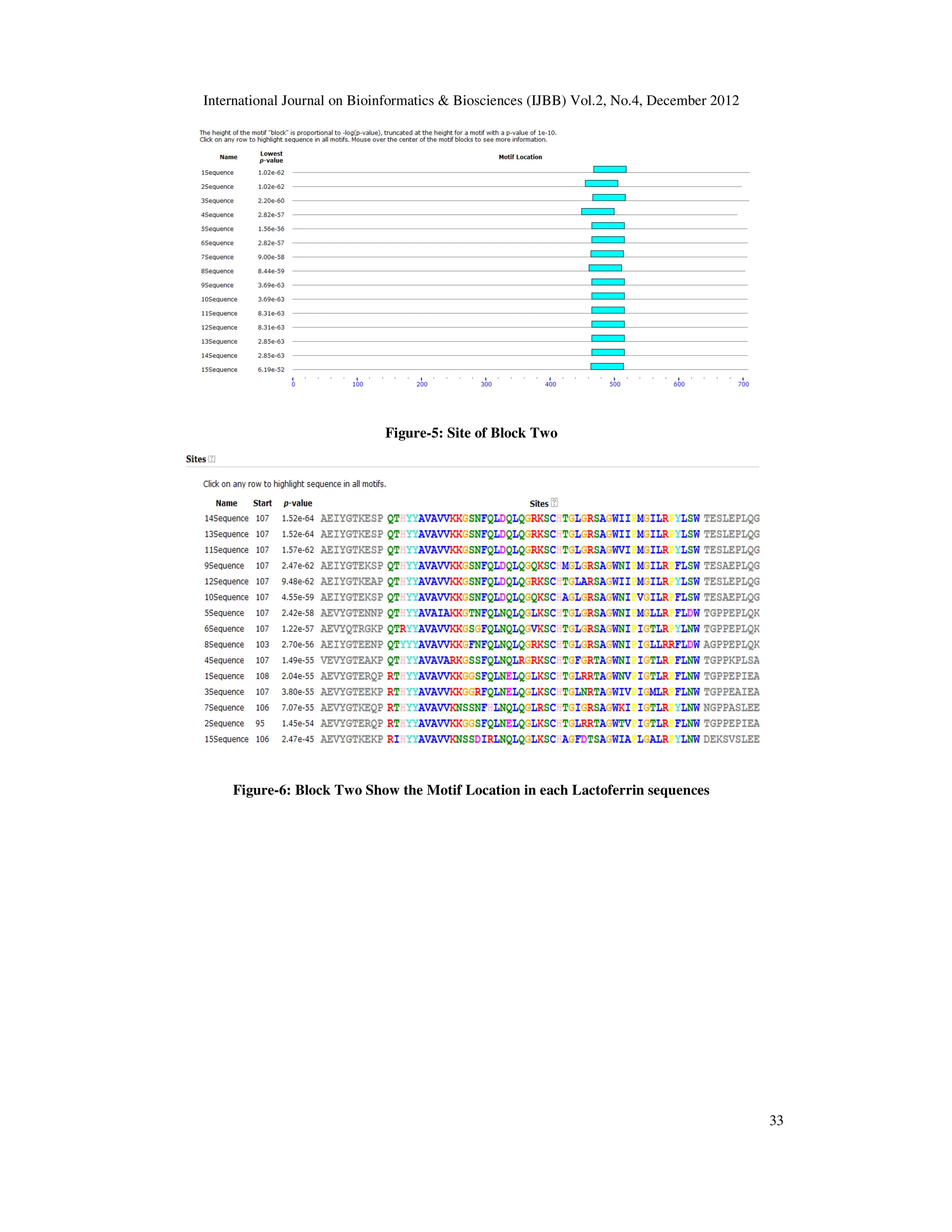
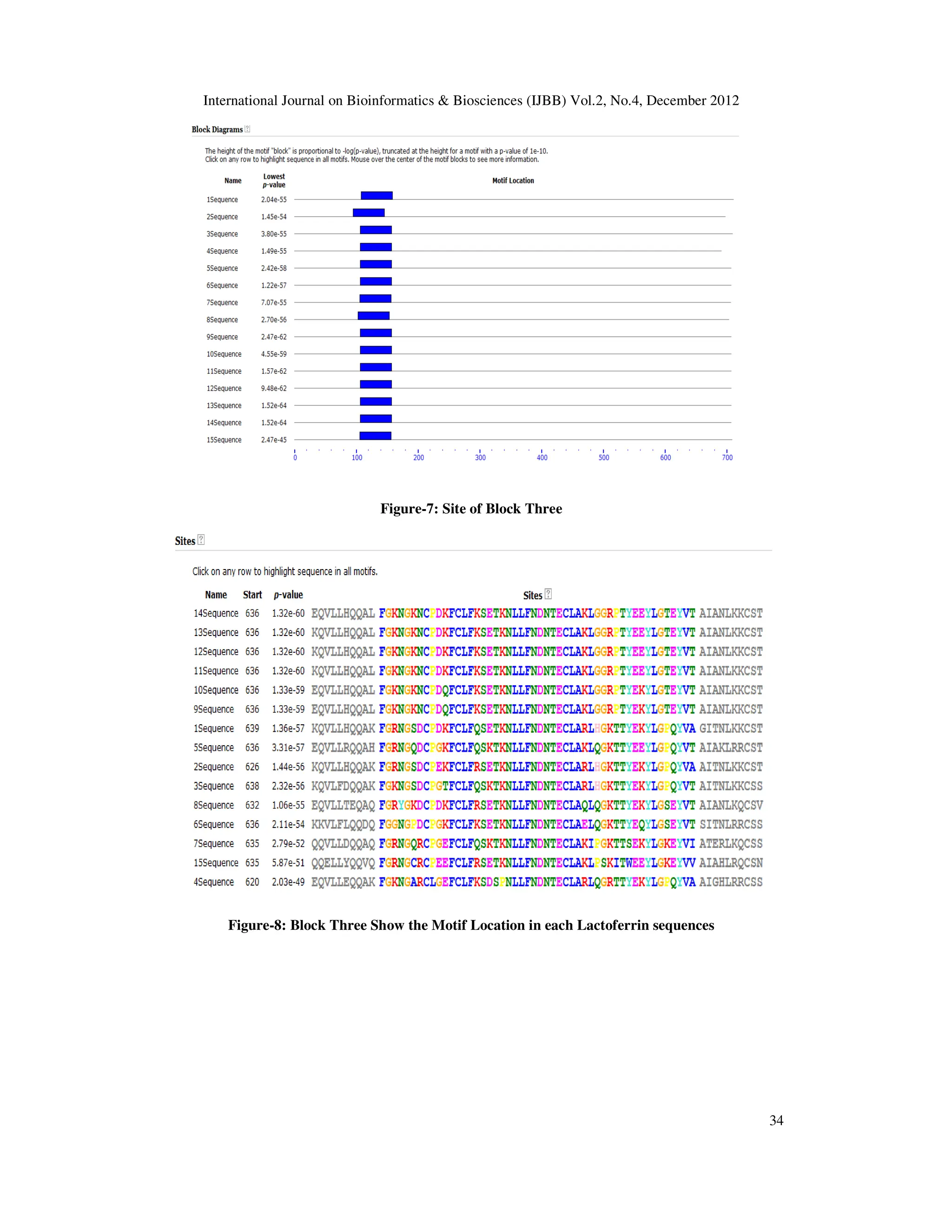
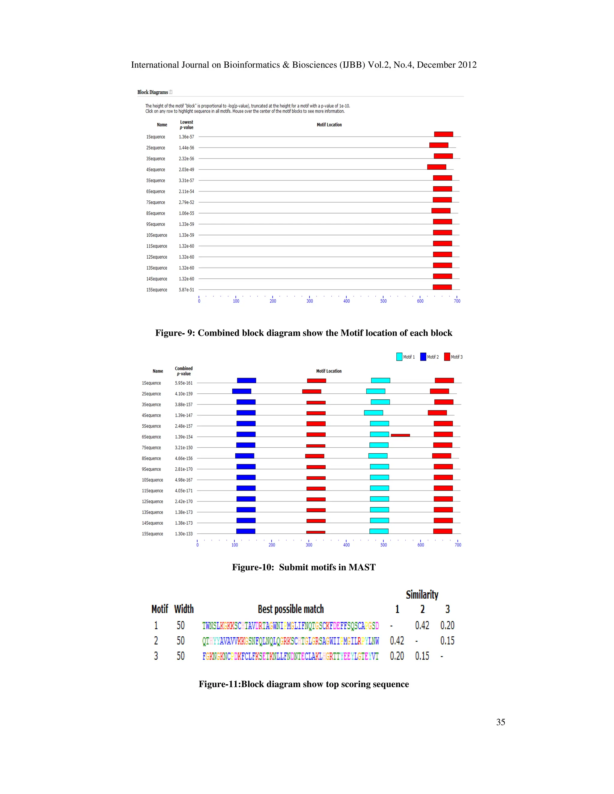
sequencesfrom different resources.By analysis of these sequences, three motifs were retrieved. It is to be
noted that all fifteen sequences contains all three motifs but start points of those are different. Each motif
has 15 sites and 50 widths. On the bases of motif analysis, it is distinct that LTF retrieved from any milk
recourse, have common conserved patterns.
![International Journal on Bioinformatics & Biosciences (IJBB) Vol.2, No.4, December 2012
DOI : 10.5121/ijbb.2012.2403 29
IDENTIFICATION OF CONSERVED
FUNCTIONAL MOTIFS IN LACTOFERRIN
USING MEME
* Shashank Rana1
Shashank.bioinfo@gmail.com
Shrikant Sharma1
shribioinfo@gmail.com
1. Research Scholar, Bioinformatics Facility, Department of Immunology, College of
Biotechnology, Sardar VallabhBhai Patel University of Agriculture & Technology.
Meerut (U.P.)
Raghvendar Singh2
Raghvendar@gmail.com
2. Head of Department, Bioinformatics Facility, Department of Immunology, College of
Biotechnology, SardarVallabhBhai Patel University of Agriculture & Technology.
Meerut (U.P.)
Corresponding Author-*Shashank Rana (Shashank.bioinfo@gmail.com)
Abstract
Lactoferrin is an iron binding globular protein with antimicrobial activity was firstly isolated in bovine
milk. Lactoferrin (LF) is structurally similar to the transferrins. So it is also known as lactotransferrin
(LTF) is a globular multifunctional protein. Our work was on motif discovery by using OOPS modal of
MEME (Multiple EM for Motif Elicitation) tool. The aim of motif discovery is to detect short, highly
conserved patterns in a collection of unaligned DNA or protein sequences. We have taken fifteen LTF AA
sequencesfrom different resources.By analysis of these sequences, three motifs were retrieved. It is to be
noted that all fifteen sequences contains all three motifs but start points of those are different. Each motif
has 15 sites and 50 widths. On the bases of motif analysis, it is distinct that LTF retrieved from any milk
recourse, have common conserved patterns.
Keywords- MEME, LTF, OOPS, Conserved pattern, MAST
Introduction
Lactoferrin form bovine milk was first isolated by Sorenson and Sorenson firstly in 1939 [1]. It
is well known fact that LF have an iron binding properties and it also have similarity with
transferrin, therefore also called lactotransferrin. Lactoferrin is considered a multifunctional or
multi-tasking protein. LF has antibacterial, antiviral, antifungal, anti-inflammatory, antioxidant
and Immunomodulatory activities [2]. LF is reported to have metal transfer, antiviral, anti-
ageing agents. Javed et. al. (2001) reported lactoferrin from camel has two lobs N (Iron binding)
and C (discharge of Iron) [7]. We have taken fifteen protein sequences of LTF from different
milk producing animals. The length of shortest sequence was 692 residues and the longest
sequence 711 residues and the modal for motif generation was OOPS. The purpose of MEME
(Multiple EM for Motif Elicitation) (rhymes with ‘team’) [5] is to allow users to discover signals
(called ‘motifs’) in DNA or protein sequences. For motif study of lactoferrin, we use by default
value as described by MEME suite. The MEME Suite is well known software package with a](https://image.slidesharecdn.com/2412ijbb03-251218040042-db715038/75/IDENTIFICATION-OF-CONSERVED-FUNCTIONAL-MOTIFS-IN-LACTOFERRIN-USING-MEME-1-2048.jpg)
![International Journal on Bioinformatics & Biosciences (IJBB) Vol.2, No.4, December 2012
30
unified web server interface that enables us to perform all four types of motif analysis wiz 1)
motif discovery, 2) motif–motif database searching, 3) motif-sequence database searching and 4)
assignment of function [6].
Figure-1: MEME overview
Materials and Methods
In present study, we have selected fifteen lactoferrin sequences (amino acid) in FASTA format
retrieved from NCBI [8] (Table-1). Motif analysis in lactoferrin sequences was conducted by
using OOPS model of MEME.. The output of this modal of MEME shows color graphical
alignment as well as common regular expression of motifs. On the hand, the block represents
start and end point of the amino acid sequences with motif length. This is well known fact that
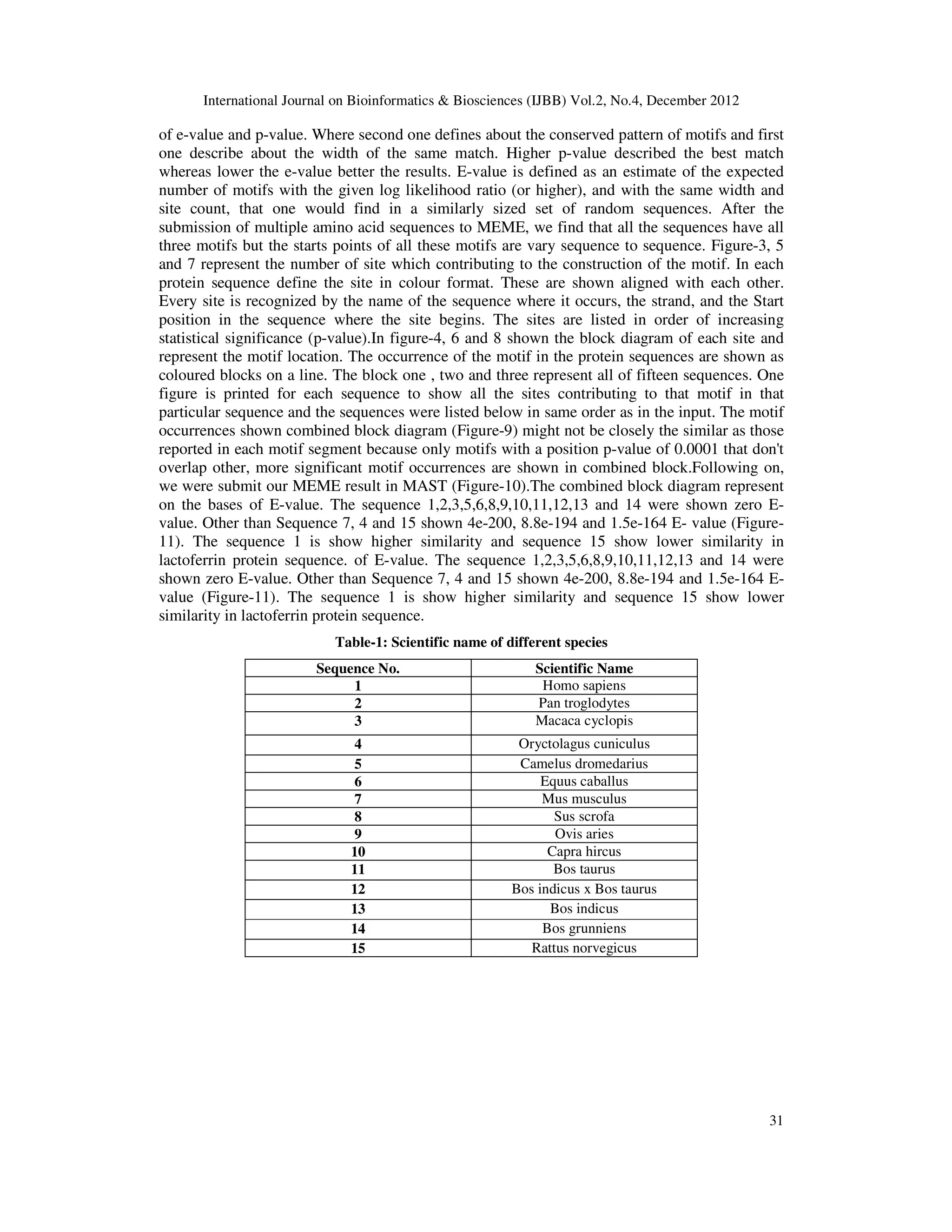
E-value describes the statistical significance of the motif. MEME [9] (ver.4.6.1) usually finds the
most statistically significant (low E-value) motifs first. The E-value is an assessment of the
probable number of motifs with the given log probability ratio (or higher) along with the same
width and site count present, that one would find in a similarly sized set of random sequences.
On the other hand, motif width defines that each motif describes a pattern of a fixed with as no
gaps are allowed in MEME motifs. In MEME package, sites define the conserved regions
present in the particular motifs. Site numbers are the important contributing factor to the
construction of the motifs. The information content of the motif (In bits), is equal to the sum of
the uncorrected information content, R (), in the columns of the LOGO as described in user
manual of MEME suite and MEME suites follows position specific probability matrices that
specify the probability of each possible letter appearing at each possible position in an
occurrence of the motif and are displayed as "sequence LOGOS", containing stacks of letters at
each position in the motif. It is to be noted that the total height of the stack is the "information
content" of that position in the motif in bits. For identification of motifs in proteins, the
categories are based on the biochemical properties of the various amino acids. Further we also
analyse motif by using Motif Alignment and Search Tool [10] (MAST ver.4.6.1). .
Results and Discussion
According to Bailey et al. (2006), by default, MEME looks for up to three motifs, each of which
may be present in some or all of the input sequences. MEME chooses the width and number of
occurrences of each motif automatically in order to minimize the ‘E-value’ of the motif—the
probability of finding an equally well-conserved pattern in random sequences. By default, only
motif widths between 6 and 50 are considered, The MEME output is HTML and shows the
motifs as local multiple alignments of (subsets of) the input sequences, as well as in several
other formats. ‘Block diagrams’ show the relative positions of the motifs in each of the input
sequences. After the submission of sequences in query box of MEME, results display in the form
of graph and seq will displays in the form of sequence logo or regular expression(Bailey et al.,
2006) (Table-2). Motif overview in figure-2 has shown 6.5e-590 E-value of motif one, 5.4e-541
E-value of motif two and 2.4e-515 E-value of motif three.In Results were analyzed on the bases](https://image.slidesharecdn.com/2412ijbb03-251218040042-db715038/75/IDENTIFICATION-OF-CONSERVED-FUNCTIONAL-MOTIFS-IN-LACTOFERRIN-USING-MEME-2-2048.jpg)
![International Journal on Bioinformatics & Biosciences (IJBB) Vol.2, No.4, December 2012
32
Table-2: motif information with sequence logo and regular expression
Figure-2: Conserved pattern of Lactoferrin
Figure-3: Site of Block one
Figure-4: Block One Show the Motif Location in each Lactoferrin sequences
Sr
.
N
o.
Mot
if
no.
Widt
h
E
Valu
e
Sit
es
Sequence logo Regular
expression
1 1 50 6.5e-
590
15 TWNS[LV][KR][GD]K
KSCHTAVDRTAG
WNIPMGL[LI][FVA]
NQTGSC[AK]FDE
[FY]FSQSCAPG[AS]D
2 2 50 5.4e-
541
15 [QR]THYYAVAVVK
KG[SG][NS]FQL[ND]
[DE]LQG[LR]KSCHT
GLGR [ST]AGW
[NI][IV]P[IM]G[IT]LR
P[FY]L[NS]W
3 3 50 2.4e-
515
15 FG[KR]NG][KS][DNR
]CP[DG][KE][FCLF]
[KQR]S[EK]TKNLLF
NDNTECLA[KR]L[G
QH]
G[KR][TP]TYE[KE]Y
LG[TP][EQ]YV[TA]](https://image.slidesharecdn.com/2412ijbb03-251218040042-db715038/75/IDENTIFICATION-OF-CONSERVED-FUNCTIONAL-MOTIFS-IN-LACTOFERRIN-USING-MEME-4-2048.jpg)