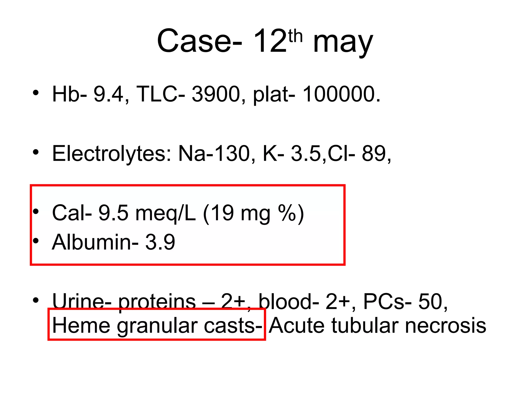
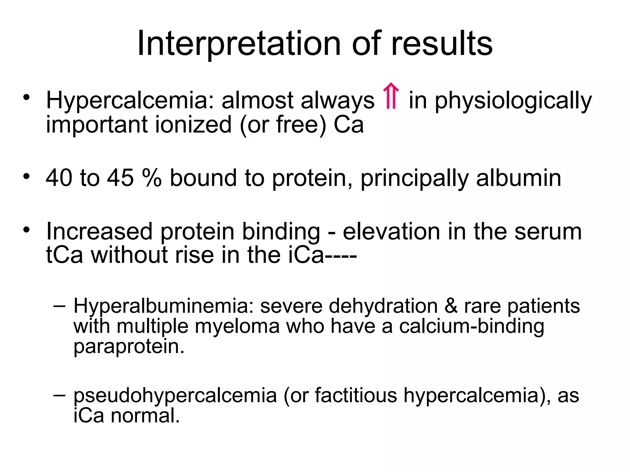
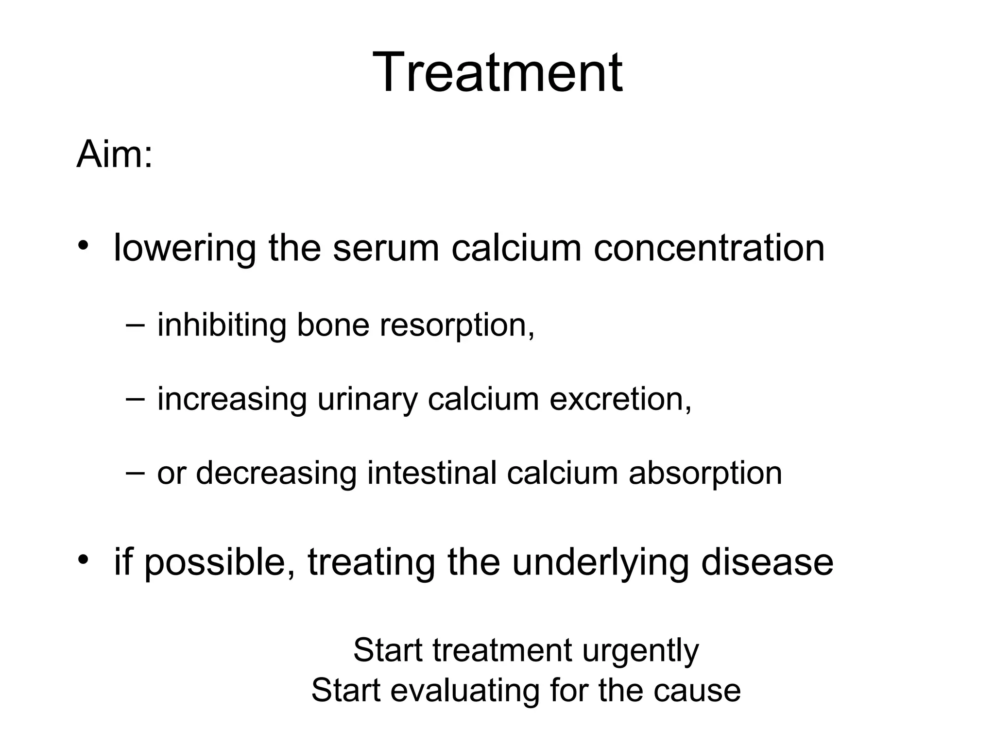
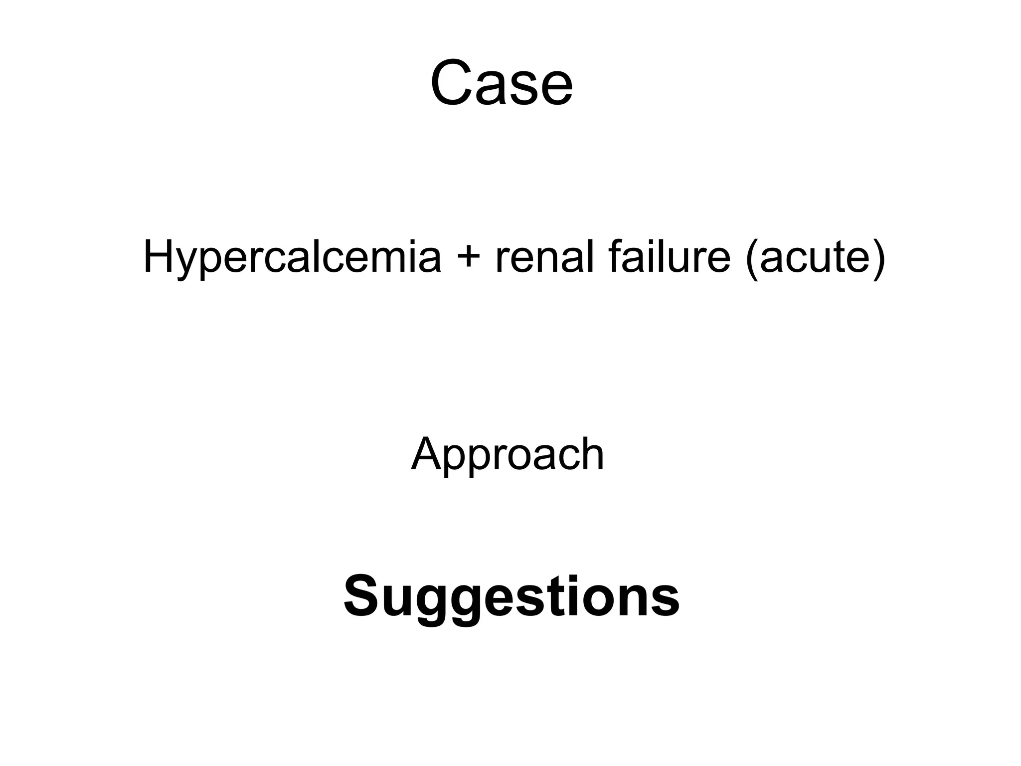
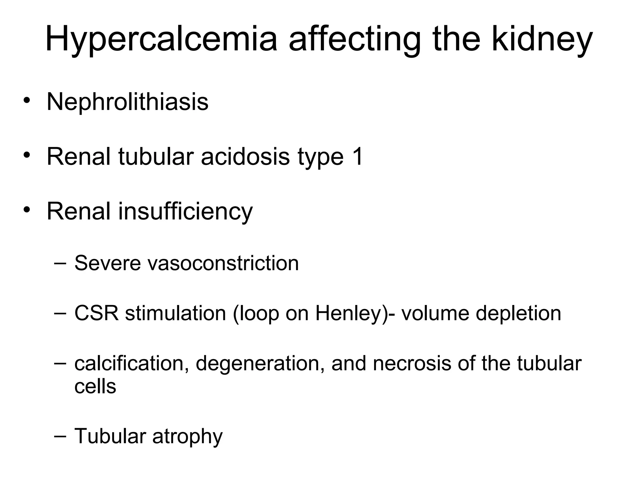
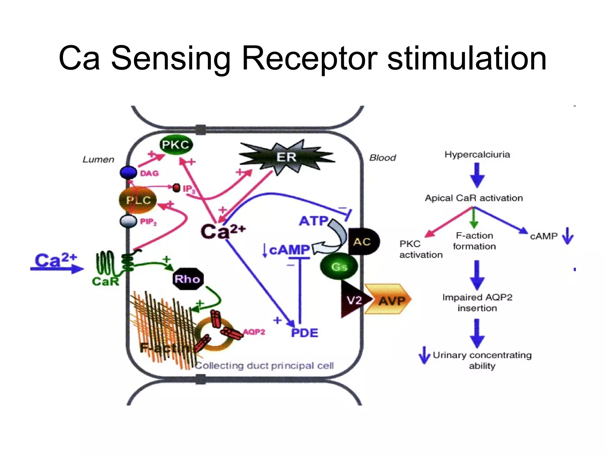
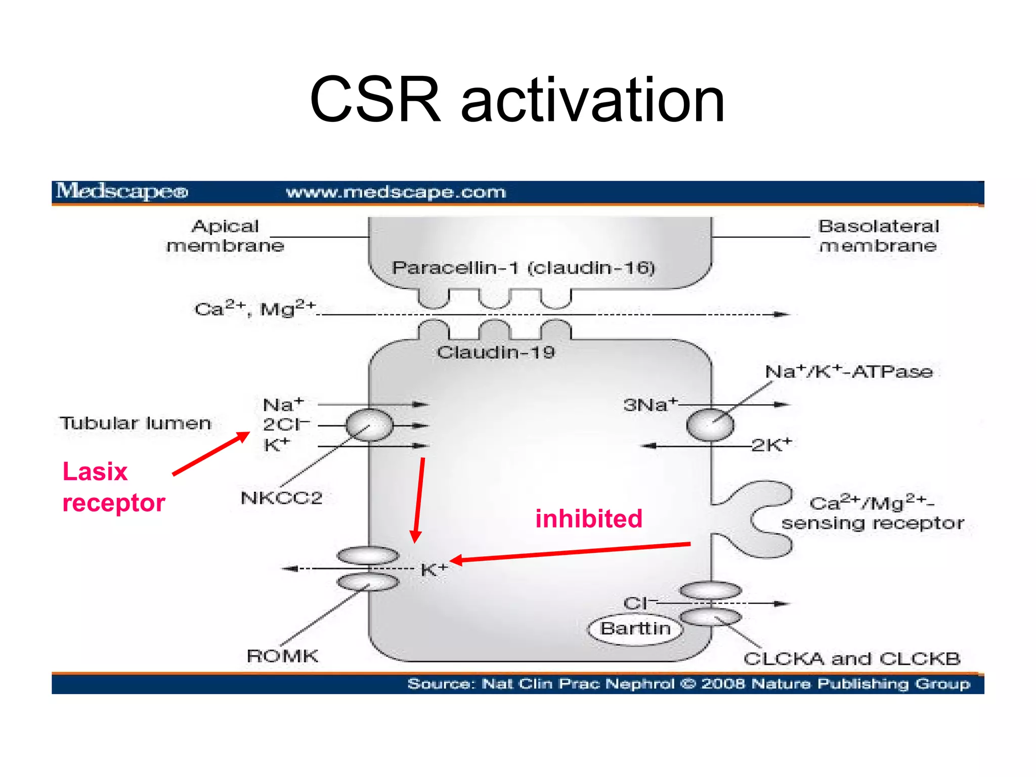
Hypercalcemia
Dr Vilas Naik,Dr Vilas Naik, DM (Nephrology)DM (Nephrology)
Fellowship in Nephrology and KidneyFellowship in Nephrology and Kidney
transplant, Toronto, Canadatransplant, Toronto, Canada
Rare cause of ARF
Case
• 65/ male, Mr SH.
• PMHx
– CAD- anterior wall thrombolysed- aug 10, s/p CABG
October 2010
– Burning feet- 3 yrs, multivitamins and sym meds
• HOPI:
– very low appetite and weight loss, nausea & vomiting-
1m.
– Admitted for evaluation of the same: 3rd
may 2011
Case: 4th
May 11
• Investigations:
– Hb- 9.8 gms, TLC- 4200, plat- 1.13
– Creatinine- 3.1 mg%
(eGFR= 20.16 ml/min)
– Electrolytes:134/5.3/97
– LFT- normal
– Total Prot/ Sr Albu- 6.9 / 3.4
Case
• UGI scopy:- grade 2 GERD
• Sr Vitamin and folate- N
• NCS- demyelinating polyneuropathy
• Progressive confusion- CT brain- WNL,
– MRI brain- N,
– CSF- WNL.
• Chest X-Ray: WNL
• USG:
– RK- 9.6 x 4.8
– LK – 9.8 x 4.2
Case- 11th
may 2011
• Progressive worsening of LOC, stuporous
• Creatinin 2.1 3.1 5.1 7 mg%
• Urine output- 1.5 to 2 L
• Referred to nephrology for potential
dialysis
Case- 12th
may
• Hb- 9.4, TLC- 3900, plat- 100000.
• Electrolytes: Na-130, K- 3.5,Cl- 89,
• Cal- 9.5 meq/L (19 mg %)
• Albumin- 3.9
• Urine- proteins – 2+, blood- 2+, PCs- 50,
Heme granular casts- Acute tubular necrosis
Interpretation of results
• Hypercalcemia: almost always ⇑ in physiologically
important ionized (or free) Ca
• 40 to 45 % bound to protein, principally albumin
• Increased protein binding - elevation in the serum
tCa without rise in the iCa----
– Hyperalbuminemia: severe dehydration & rare patients
with multiple myeloma who have a calcium-binding
paraprotein.
– pseudohypercalcemia (or factitious hypercalcemia), as
iCa normal.
Case- 12th
may
• Clinically:
– BP- 130/80, no edema, volume depleted
– Chest clear- SpO2- 98 % on room air
– No lymphadynopathy
– CNS- stuporous, no focal neurodeficit.
Treatment
Aim:
• lowering the serum calcium concentration
– inhibiting bone resorption,
– increasing urinary calcium excretion,
– or decreasing intestinal calcium absorption
• if possible, treating the underlying disease
Start treatment urgently
Start evaluating for the cause
Treatment
• Normal saline
• Furosemide: bolus f/b drip to match UO
Both increase urinary Ca excretion
• Steroids- decrease 1-25 (OH)2 D3, intestinal absorption
inhibited.
• Calcitonin - Inhibits bone reabsorption
• Biphosphonates- inhibits bone reabsorption
Treatment
• Calcimimetics: Cinacalcit- PTH receptor
antagonist
• Dialysis: Low or no calcium dialysate
– Fastest method
– Needs vascular intervention
In our patient…..
• NS bolus 2 L, then 250 ml/hr
• Lasix- bolus, drip to match urine output
• Hydrocort 100mg 6 hrly
• Biphosphonate:
– 4 mg in 100 ml NS over 30 min
Case
• Creat 7.1 6.1 4.8 3 1.8 mg%
• Parallel decrease in Sr Ca
• Clinical and biochemical improvement
• CBC- still pancytopenia
Approach
Cause of
Hypercalcemia
Case
Hypercalcemia + renal failure (acute)
Approach
Suggestions
Our patient….
• Sr electrophoresis- no M band
• Skeletal survey- no lytic lesions
• Sr immunofixation- negative
• Sr 25 hydroxy and 1-25 dihydroxy vit D3
levels- normal
• Sr iPTH (appropriately collected and sent)
Our patient….
• Sr ACEI levels- normal.
• T3/ T4/ TSH- WNL
• PSA
• CT chest (HRCT and CECT)- WNL
Case..
• BMA- in v/o persistent pancytopenia-
WNL, only mild erythroid hyperplasia
CAUSES
• PTH dependent
– Primary hyper PTH
• Familial
• Sporadic
– Tertiary hyper PTH
(Acute Renal Failure)
• PTH independent
– Hypercalcemia of malig:
• PTHrp
• Activation of extrarenal 1
alpha-hydroxylase
(increased calcitriol)
• Osteolytic bone metastases
and local cytokines
– Vitamin D intoxication
PTH independent factors
• Chronic granulomatous
disease
– Extrarenal 1 alpha-
hydroxylase (⇑ calcitriol)
• Medications:
– Thiazides
– Lithium
– Excessive Vitamin A
• Miscellaneous:
• Hyperthyroidism
• Adrenal insufficiency
• Immobilization
• Parenteral nutrition
Causes
• Primary hyperparathyroidism & malignancy: -90%
• Malignancy often evident clinically by the time it
causes hypercalcemia
• hypercalcemia of malignancy-higher calciums &
more symptomatic from than primary hyper PTH.
Approach
Hypercalcemia affecting the kidney
Mechanisms
– Nephrogenic DI:
• downregulation of aquaporin-2 water channels
• calcium deposition in the medulla with secondary
tubulointerstitial injury
• Decreased medullary osmolality and hence
decreased concentrating ability
Hypercalcemia affecting the kidney
• Nephrolithiasis
• Renal tubular acidosis type 1
• Renal insufficiency
– Severe vasoconstriction
– CSR stimulation (loop on Henley)- volume depletion
– calcification, degeneration, and necrosis of the tubular
cells
– Tubular atrophy
Ca Sensing Receptor stimulation
CSR activation
Lasix
receptor
inhibited
Follow of the patient
• Creatinine- 1.2 mg%
• Pancytopenia persistent
• Otherwise healthy
• Decided to go to Mumbai for hematology
evaluation
Follow up of the patient
• Complete hematology work up was
negative
• Follow up till 2014 november – mild
thrombocytopenia, otherwise healthy

Hypercalcemia

  • 1.
    Hypercalcemia Dr Vilas Naik,DrVilas Naik, DM (Nephrology)DM (Nephrology) Fellowship in Nephrology and KidneyFellowship in Nephrology and Kidney transplant, Toronto, Canadatransplant, Toronto, Canada
  • 2.
  • 3.
    Case • 65/ male,Mr SH. • PMHx – CAD- anterior wall thrombolysed- aug 10, s/p CABG October 2010 – Burning feet- 3 yrs, multivitamins and sym meds • HOPI: – very low appetite and weight loss, nausea & vomiting- 1m. – Admitted for evaluation of the same: 3rd may 2011
  • 4.
    Case: 4th May 11 •Investigations: – Hb- 9.8 gms, TLC- 4200, plat- 1.13 – Creatinine- 3.1 mg% (eGFR= 20.16 ml/min) – Electrolytes:134/5.3/97 – LFT- normal – Total Prot/ Sr Albu- 6.9 / 3.4
  • 5.
    Case • UGI scopy:-grade 2 GERD • Sr Vitamin and folate- N • NCS- demyelinating polyneuropathy • Progressive confusion- CT brain- WNL, – MRI brain- N, – CSF- WNL. • Chest X-Ray: WNL • USG: – RK- 9.6 x 4.8 – LK – 9.8 x 4.2
  • 6.
    Case- 11th may 2011 •Progressive worsening of LOC, stuporous • Creatinin 2.1 3.1 5.1 7 mg% • Urine output- 1.5 to 2 L • Referred to nephrology for potential dialysis
  • 7.
    Case- 12th may • Hb-9.4, TLC- 3900, plat- 100000. • Electrolytes: Na-130, K- 3.5,Cl- 89, • Cal- 9.5 meq/L (19 mg %) • Albumin- 3.9 • Urine- proteins – 2+, blood- 2+, PCs- 50, Heme granular casts- Acute tubular necrosis
  • 8.
    Interpretation of results •Hypercalcemia: almost always ⇑ in physiologically important ionized (or free) Ca • 40 to 45 % bound to protein, principally albumin • Increased protein binding - elevation in the serum tCa without rise in the iCa---- – Hyperalbuminemia: severe dehydration & rare patients with multiple myeloma who have a calcium-binding paraprotein. – pseudohypercalcemia (or factitious hypercalcemia), as iCa normal.
  • 9.
    Case- 12th may • Clinically: –BP- 130/80, no edema, volume depleted – Chest clear- SpO2- 98 % on room air – No lymphadynopathy – CNS- stuporous, no focal neurodeficit.
  • 10.
    Treatment Aim: • lowering theserum calcium concentration – inhibiting bone resorption, – increasing urinary calcium excretion, – or decreasing intestinal calcium absorption • if possible, treating the underlying disease Start treatment urgently Start evaluating for the cause
  • 11.
    Treatment • Normal saline •Furosemide: bolus f/b drip to match UO Both increase urinary Ca excretion • Steroids- decrease 1-25 (OH)2 D3, intestinal absorption inhibited. • Calcitonin - Inhibits bone reabsorption • Biphosphonates- inhibits bone reabsorption
  • 12.
    Treatment • Calcimimetics: Cinacalcit-PTH receptor antagonist • Dialysis: Low or no calcium dialysate – Fastest method – Needs vascular intervention
  • 13.
    In our patient….. •NS bolus 2 L, then 250 ml/hr • Lasix- bolus, drip to match urine output • Hydrocort 100mg 6 hrly • Biphosphonate: – 4 mg in 100 ml NS over 30 min
  • 14.
    Case • Creat 7.16.1 4.8 3 1.8 mg% • Parallel decrease in Sr Ca • Clinical and biochemical improvement • CBC- still pancytopenia
  • 15.
  • 16.
    Case Hypercalcemia + renalfailure (acute) Approach Suggestions
  • 17.
    Our patient…. • Srelectrophoresis- no M band • Skeletal survey- no lytic lesions • Sr immunofixation- negative • Sr 25 hydroxy and 1-25 dihydroxy vit D3 levels- normal • Sr iPTH (appropriately collected and sent)
  • 18.
    Our patient…. • SrACEI levels- normal. • T3/ T4/ TSH- WNL • PSA • CT chest (HRCT and CECT)- WNL
  • 19.
    Case.. • BMA- inv/o persistent pancytopenia- WNL, only mild erythroid hyperplasia
  • 20.
    CAUSES • PTH dependent –Primary hyper PTH • Familial • Sporadic – Tertiary hyper PTH (Acute Renal Failure) • PTH independent – Hypercalcemia of malig: • PTHrp • Activation of extrarenal 1 alpha-hydroxylase (increased calcitriol) • Osteolytic bone metastases and local cytokines – Vitamin D intoxication
  • 21.
    PTH independent factors •Chronic granulomatous disease – Extrarenal 1 alpha- hydroxylase (⇑ calcitriol) • Medications: – Thiazides – Lithium – Excessive Vitamin A • Miscellaneous: • Hyperthyroidism • Adrenal insufficiency • Immobilization • Parenteral nutrition
  • 22.
    Causes • Primary hyperparathyroidism& malignancy: -90% • Malignancy often evident clinically by the time it causes hypercalcemia • hypercalcemia of malignancy-higher calciums & more symptomatic from than primary hyper PTH.
  • 23.
    Approach Hypercalcemia affecting thekidney Mechanisms – Nephrogenic DI: • downregulation of aquaporin-2 water channels • calcium deposition in the medulla with secondary tubulointerstitial injury • Decreased medullary osmolality and hence decreased concentrating ability
  • 24.
    Hypercalcemia affecting thekidney • Nephrolithiasis • Renal tubular acidosis type 1 • Renal insufficiency – Severe vasoconstriction – CSR stimulation (loop on Henley)- volume depletion – calcification, degeneration, and necrosis of the tubular cells – Tubular atrophy
  • 25.
  • 26.
  • 27.
    Follow of thepatient • Creatinine- 1.2 mg% • Pancytopenia persistent • Otherwise healthy • Decided to go to Mumbai for hematology evaluation
  • 28.
    Follow up ofthe patient • Complete hematology work up was negative • Follow up till 2014 november – mild thrombocytopenia, otherwise healthy

Editor's Notes

  • #8 Even though he was malnourished, his Sr Albu was significantly preserved- at this point he was significantly volume depleted. But ATN can be a process in almost any disease, glomerulonephritis can also have a component of ATN, ischemic and toxic ATN. He had a foleys in place since- 7 days, so had PCs in urine.