The document provides an overview of the European Association for the Study of the Liver (EASL) clinical practice guidelines on managing hepatitis E infection, first presented at the International Liver Congress 2018. It covers various aspects including virology, epidemiology, key recommendations, and the management of acute and chronic infections, emphasizing the importance of evidence-based practices. The guidelines should be used as an educational resource and not as the sole basis for patient management decisions.

![• Chair
– Harry R Dalton
• Panel members
– Nassim Kamar, Sally A Baylis,
Darius Moradpour, Heiner
Wedemeyer, Francesco Negro
(EASL Governing Board
Representative)
• Reviewers
– Philippa Easterbrook, Sven
Pischke, Yury Khudyakov
Guideline panel
EASL CPG HEV. J Hepatol 2018;doi: 10.1016/j.jhep.2018.03.005 [Epub ahead of print]](https://image.slidesharecdn.com/heveasl-cpg-240603102538-5498df40/85/HEV-EASL-guidelines-2018-recent-management-guidelines-pptx-4-320.jpg)
![Outline
EASL CPG HEV. J Hepatol 2018;doi: 10.1016/j.jhep.2018.03.005 [Epub ahead of print]
• Grading evidence and recommendations
Methods
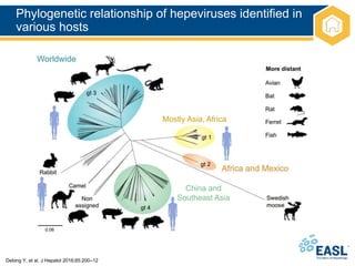
• Virology
• Epidemiology
Background
• Key recommendations
Guidelines
• Unanswered questions and perspectives
Conclusions](https://image.slidesharecdn.com/heveasl-cpg-240603102538-5498df40/85/HEV-EASL-guidelines-2018-recent-management-guidelines-pptx-5-320.jpg)
![Grading evidence and recommendations
*Level was downgraded if there was poor quality, strong bias or inconsistency between studies;
level was upgraded if there was a large effect size
1. Guyatt GH, et al. BMJ 2008:336:924–6;
2. EASL CPG HEV. J Hepatol 2018;doi: 10.1016/j.jhep.2018.03.005 [Epub ahead of print]
• Grading is adapted from the GRADE system1,2
Level of evidence* Confidence in the evidence
A Data derived from meta-analyses or
systematic reviews or from (multiple)
randomized controlled trials (RCTs) with
high quality
Further research is unlikely to change
our confidence in the estimate of
benefit and risk
B Data derived from a single RCT or multiple
non-randomized studies
Further research (if performed) is
likely to have an impact on our
confidence in the estimate of benefit
and risk and may change the
estimate
C Small studies, retrospective observational
studies, registries
Any estimate of effect is uncertain
Grade of recommendation (wording associated with the grade of recommendation)
1 Strong (“must”, “should”, or “EASL recommends”)
2 Weak (“can”, “may”, or “EASL suggests”)](https://image.slidesharecdn.com/heveasl-cpg-240603102538-5498df40/85/HEV-EASL-guidelines-2018-recent-management-guidelines-pptx-7-320.jpg)
![Virology of HEV
EASL CPG HEV. J Hepatol 2018;doi: 10.1016/j.jhep.2018.03.005 [Epub ahead of print]
Hepeviridae
Virus family
Hepeviridae viruses infect
mammals, birds and fish](https://image.slidesharecdn.com/heveasl-cpg-240603102538-5498df40/85/HEV-EASL-guidelines-2018-recent-management-guidelines-pptx-9-320.jpg)
![Virology of HEV
EASL CPG HEV. J Hepatol 2018;doi: 10.1016/j.jhep.2018.03.005 [Epub ahead of print]
Hepeviridae
Orthohepevirus
Virus family
Genus
Hepeviridae viruses infect
mammals, birds and fish
Strains infecting humans
belong to the Orthohepevirus
genus, species A
Species A D
C
B](https://image.slidesharecdn.com/heveasl-cpg-240603102538-5498df40/85/HEV-EASL-guidelines-2018-recent-management-guidelines-pptx-10-320.jpg)
![Virology of HEV
EASL CPG HEV. J Hepatol 2018;doi: 10.1016/j.jhep.2018.03.005 [Epub ahead of print]
Hepeviridae
Orthohepevirus
Virus family
Genus
Hepeviridae viruses infect
mammals, birds and fish
Strains infecting humans
belong to the Orthohepevirus
genus, species A
Species
Species A comprises
8 genotypes
A D
C
B
GT 1 GT 2 GT 3 GT 7
GT 6
GT 5
GT 4 GT 8
• Only infect humans
• Faecal–oral spread
via contaminated water
• Large outbreaks
• Brief, self-limiting
• Never chronic
• High mortality in
pregnancy (25%)
• Endemic in animal
species; eg, pigs and
wild boar
• Zoonotic infections in
humans
• High-income
countries
• China: GT 4 most
common
• S. America: GT 3 only
• Have only been
reported in wild boar
• GT 7 identified in
patient regularly
consuming camel
meat and milk
• Have since been
identified in camels](https://image.slidesharecdn.com/heveasl-cpg-240603102538-5498df40/85/HEV-EASL-guidelines-2018-recent-management-guidelines-pptx-11-320.jpg)
![Virology of HEV
EASL CPG HEV. J Hepatol 2018;doi: 10.1016/j.jhep.2018.03.005 [Epub ahead of print]
Hepeviridae
Orthohepevirus
Virus family
Genus
Hepeviridae viruses infect
mammals, birds and fish
Strains infecting humans
belong to the Orthohepevirus
genus, species A
Species
Species A comprises
8 genotypes
A D
C
B
• Only infect humans
• Faecal–oral spread
via contaminated water
• Large outbreaks
• Brief, self-limiting
• Never chronic
• High mortality in
pregnancy (25%)
• Endemic in animal
species; eg, pigs and
wild boar
• Zoonotic infections in
humans
• High-income
countries
• China: GT 4 most
common
• S. America: GT 3 only
• Have only been
reported in wild boar
• GT 7 identified in
patient regularly
consuming camel
meat and milk
• Have since been
identified in camels
Focus of this CPG
GT 1 GT 2 GT 3 GT 7
GT 6
GT 5
GT 4 GT 8](https://image.slidesharecdn.com/heveasl-cpg-240603102538-5498df40/85/HEV-EASL-guidelines-2018-recent-management-guidelines-pptx-12-320.jpg)
![HEV GT 1 and 2 in brief
*Data from 2005
EASL CPG HEV. J Hepatol 2018;doi: 10.1016/j.jhep.2018.03.005 [Epub ahead of print]
• ~20 million infections worldwide
– 3 million symptomatic cases and 70,000 deaths/year*
– WHO guidelines should be consulted for management of outbreaks
of acute HEV in resource-limited settings
• Brief, self-limiting, usually in young adults
• Never chronic
– Acute-on-chronic liver failure possible
• High mortality in pregnancy (25%)
Recommendations
• Travellers with hepatitis returning from areas endemic for HEV
GT 1 or 2 should be tested for HEV
A 1
• Pregnant women with HEV GT 1 or 2 should be cared for in a
high-dependency setting, and transferred to a liver transplant
unit if liver failure occurs
A 1
Level of evidence Grade of recommendation](https://image.slidesharecdn.com/heveasl-cpg-240603102538-5498df40/85/HEV-EASL-guidelines-2018-recent-management-guidelines-pptx-14-320.jpg)
![HEV GT 3 and 4: epidemiology
1. Dalton HR, et al. Eur J Gastroenterol Hepatol 2008;20:784–90;
EASL CPG HEV. J Hepatol 2018;doi: 10.1016/j.jhep.2018.03.005 [Epub ahead of print]
• Endemic in some developing countries, as well as most high-income
countries
• Most common cause of acute viral hepatitis in many European
countries
• Estimated that ≥2 million locally acquired HEV infections/year
– Most as a result of zoonotic infection
• Primary hosts are pigs
• HEV GT 3 and 4 tend to affect older males
– In an English study, male:female ratio was 3:1; median age, 63 years1
• Incidence varies between and within countries, and over time
– Multiple ‘hotspots’ of HEV infection in Europe](https://image.slidesharecdn.com/heveasl-cpg-240603102538-5498df40/85/HEV-EASL-guidelines-2018-recent-management-guidelines-pptx-15-320.jpg)
![HEV ‘hot spots’ in Europe
1. Thom K, et al. Euro Surveill 2018;doi: 10.2807/1560-7917.ES.2018.23.12.17-00174 [Epub ahead of print];
2. Mansuy JM, et al. Hepatology 2016;63:1145–54; 3. Zaaijer HL. Hepatology 2015;62:654;
4. Müller B, et al. Transfus Med Hemother 2015;42:1–66; 5. Adlhoch C, et al. J Clin Virol 2016;82:9–16;
6. Lucarelli C, et al. Euro Surveill 2016;21; 7. Bura M, et al, Int J Infect Dis. 2017; 61:20–2.
EASL CPG HEV. J Hepatol 2018;doi: 10.1016/j.jhep.2018.03.005 [Epub ahead of print]
Scotland, 2016: 1:2,481 donors viraemic1
SW France, 2016: incidence 34%2
The Netherlands, 2014: 1:600 donors viraemic3
Western Germany, 2015: 1:616 donors viraemic4
Czech Republic, 2015: 400 lab-confirmed cases5
Abruzzo, Italy, 2016: seroprevalence 49%6
Western/Central Poland, 2017: seroprevalence 50%7](https://image.slidesharecdn.com/heveasl-cpg-240603102538-5498df40/85/HEV-EASL-guidelines-2018-recent-management-guidelines-pptx-16-320.jpg)
![HEV ‘hot spots’ in Europe
Worldwide HEV viraemia in blood donors
Australia, 2015: 1:14,799–74,1318,9
Canada, 2017: 1:13,99310
China, 2016: 1:1,00011
Japan, 2011: 1:8,17315,07512,13
USA, 2016: 1:9,50014
References included in slide notes.
EASL CPG HEV. J Hepatol 2018;doi: 10.1016/j.jhep.2018.03.005 (and supplementary materials) [Epub ahead of print]
Scotland, 2016: 1:2,481 donors viraemic1
SW France, 2016: incidence 34%2
The Netherlands, 2014: 1:600 donors viraemic3
Western Germany, 2015: 1:616 donors viraemic4
Czech Republic, 2015: 400 lab-confirmed cases5
Abruzzo, Italy, 2016: seroprevalence 49%6
Western/Central Poland, 2018: seroprevalence 50%7](https://image.slidesharecdn.com/heveasl-cpg-240603102538-5498df40/85/HEV-EASL-guidelines-2018-recent-management-guidelines-pptx-17-320.jpg)
![Topics
EASL CPG HEV. J Hepatol 2018;doi: 10.1016/j.jhep.2018.03.005 [Epub ahead of print]
1. Clinical aspects: acute infection
2. Clinical aspects: chronic infection
3. Extrahepatic manifestations
4. Laboratory diagnosis of HEV infection
5. Differential diagnosis
6. HEV and the blood supply
7. Treatment of acute HEV infection
8. Management of patients not clearing HEV infection
9. Prevention of HEV infection
10. Conclusions
Click on a topic to skip
to that section](https://image.slidesharecdn.com/heveasl-cpg-240603102538-5498df40/85/HEV-EASL-guidelines-2018-recent-management-guidelines-pptx-19-320.jpg)
![Clinical aspects: acute infection
*Fatigue, itching and nausea
EASL CPG HEV. J Hepatol 2018;doi: 10.1016/j.jhep.2018.03.005 [Epub ahead of print]
• Acute HEV GT 3 infection is clinically silent in most patients
– <5% may develop symptoms of acute hepatitis
• Elevated liver enzymes, jaundice and non-specific symptoms*
• Immunocompetent patients clear the infection spontaneously
– Progression to ALF is rare with HEV GT 3
– ACLF occurs occasionally
• Non-sterilizing immunity develops after infection has cleared
– Re-infection possible, but with lower risk of symptomatic hepatitis
Recommendations
Should test for HEV in:
• All patients with symptoms consistent with acute hepatitis
A 1
Suggest testing for HEV in:
• Patients with unexplained flares of chronic liver disease
C 2
Level of evidence Grade of recommendation](https://image.slidesharecdn.com/heveasl-cpg-240603102538-5498df40/85/HEV-EASL-guidelines-2018-recent-management-guidelines-pptx-20-320.jpg)
![Clinical aspects: chronic infection
*Persistence of HEV replication for 3 months. In rare cases, spontaneous clearance has been observed between 3 and 6 months
EASL CPG HEV. J Hepatol 2018;doi: 10.1016/j.jhep.2018.03.005 [Epub ahead of print]
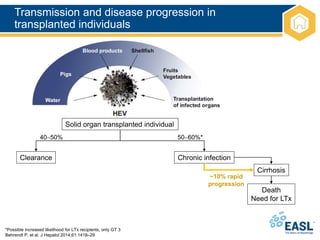
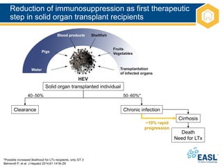
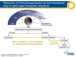
• Immunosuppressed patients can fail to clear HEV infection
– Progression to chronic hepatitis*
• Immunosuppressed groups include:
– Solid organ transplant recipients
• ~50–66% of HEV-infected organ transplant recipients develop chronic hepatitis
– Patients with haematological disorders
– Individuals living with HIV
– Patients with rheumatic disorders receiving heavy immunosuppression
• Most patients are asymptomatic and present with mild and persistent
LFT abnormalities
Recommendations
Should test for HEV in:
• All immunosuppressed patients with unexplained abnormal LFTs
A 1
Grade of evidence Grade of recommendation](https://image.slidesharecdn.com/heveasl-cpg-240603102538-5498df40/85/HEV-EASL-guidelines-2018-recent-management-guidelines-pptx-21-320.jpg)
![Clinical aspects: chronic infection
*Persistence of HEV replication for 3 months. In rare cases, spontaneous clearance has been observed between 3 and 6 months
EASL CPG HEV. J Hepatol 2018;doi: 10.1016/j.jhep.2018.03.005 [Epub ahead of print]
• Immunosuppressed patients can fail to clear HEV infection
– Progression to chronic hepatitis*
• Immunosuppressed groups include:
– Solid organ transplant recipients
• ~50–66% of HEV-infected organ transplant recipients develop chronic hepatitis
– Patients with haematological disorders
– Individuals living with HIV
– Patients with rheumatic disorders receiving heavy immunosuppression
• Most patients are asymptomatic and present with mild and persistent
LFT abnormalities
Recommendations
Should test for HEV in:
• All immunosuppressed patients with unexplained abnormal LFTs
A 1
Grade of evidence Grade of recommendation
Chronic HEV has mainly been described
in the solid organ transplant setting](https://image.slidesharecdn.com/heveasl-cpg-240603102538-5498df40/85/HEV-EASL-guidelines-2018-recent-management-guidelines-pptx-22-320.jpg)
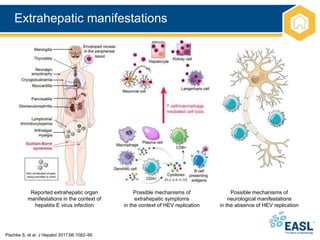
![Extrahepatic manifestations
*There is good evidence to support a causal role for HEV and these associated conditions. For the other extrahepatic
manifestations, causality remains to be established; †Case reports only
EASL CPG HEV. J Hepatol 2018;doi: 10.1016/j.jhep.2018.03.005 [Epub ahead of print]
• Extrahepatic manifestations of HEV are increasingly recognized
Organ system Clinical syndrome Notes
Neurological Neuralgic amyotrophy*
Guillain–Barré syndrome*
Meningoencephalitis*
Mononeuritis multiplex
Myositis
Bell’s palsy, vestibular neuritis,
and peripheral neuropathy
~150 cases of neurological injury (in HEV GT 3);
mainly Europe
Most (>90%) cases in the immunocompetent
Renal* Membranoproliferative and
membranous
glomerulonephritis
IgA nephropathy
Mainly immunosuppressed GT 3-infected
patients
Renal function improves and proteinuria levels
decrease following HEV clearance
Haematological Thrombocytopenia
Monoclonal immunoglobulin
Cryoglobulinaemia
Aplastic anaemia†
Haemolytic anaemia†
Mild thrombocytopenia is common; occasionally
severe
Reported in 25% of cases of acute HEV in UK
study
Occurs mainly in association with renal disease
Other Acute pancreatitis
Arthritis†
Myocarditis†
Autoimmune thyroiditis†
55 cases worldwide. HEV GT 1 only; usually mild](https://image.slidesharecdn.com/heveasl-cpg-240603102538-5498df40/85/HEV-EASL-guidelines-2018-recent-management-guidelines-pptx-24-320.jpg)
![Organ system Clinical syndrome Notes
Neurological Neuralgic amyotrophy*
Guillain–Barré syndrome*
Meningoencephalitis*
Mononeuritis multiplex
Myositis
Bell’s palsy, vestibular neuritis
and peripheral neuropathy
~150 cases of neurological injury (in HEV GT 3);
mainly Europe
Most (>90%) cases in the immunocompetent
Renal* Membranoproliferative and
membranous
glomerulonephritis
IgA nephropathy
Mainly immunosuppressed GT 3-infected
patients
Renal function improves and proteinuria levels
decrease following HEV clearance
Haematological Thrombocytopenia
Monoclonal immunoglobulin
Cryoglobulinaemia
Aplastic anaemia†
Haemolytic anaemia†
Mild thrombocytopenia is common; occasionally
severe
Reported in 25% of cases of acute HEV in UK
study
Occurs mainly in association with renal disease
Other Acute pancreatitis
Arthritis†
Myocarditis†
Autoimmune thyroiditis†
55 cases worldwide. HEV GT 1 only; usually mild
Extrahepatic manifestations
*There is good evidence to support a causal role for HEV and these associated conditions. For the other extrahepatic
manifestations, causality remains to be established; †Case reports only
EASL CPG HEV. J Hepatol 2018;doi: 10.1016/j.jhep.2018.03.005 [Epub ahead of print]
• Extrahepatic manifestations of HEV are increasingly recognized
Most important](https://image.slidesharecdn.com/heveasl-cpg-240603102538-5498df40/85/HEV-EASL-guidelines-2018-recent-management-guidelines-pptx-25-320.jpg)
![Extrahepatic manifestations
*Irrespective of LFT results
EASL CPG HEV. J Hepatol 2018;doi: 10.1016/j.jhep.2018.03.005 [Epub ahead of print]
• Existence of extrahepatic manifestations of HEV means that
testing is warranted in a number of patient populations
Recommendations
Testing for HEV recommended in:*
• Patients with neuralgic amyotrophy B 1
• Patients with Guillain–Barré syndrome B 1
Testing for HEV suggested in:
• Patients with encephalitis/myelitis
C 2
Testing for proteinuria suggested in:
• HEV-infected patients
C 2
• Patients with acute or chronic HEV infection who develop
new-onset proteinuria may be considered for a renal biopsy
C 2
Treatment
• Antiviral treatment suggested for patients with chronic HEV
infection and associated glomerular disease
C 2
Grade of evidence Grade of recommendation](https://image.slidesharecdn.com/heveasl-cpg-240603102538-5498df40/85/HEV-EASL-guidelines-2018-recent-management-guidelines-pptx-27-320.jpg)
![Laboratory diagnosis of HEV infection
EASL CPG HEV. J Hepatol 2018;doi: 10.1016/j.jhep.2018.03.005 [Epub ahead of print]
• Incubation period for HEV is ~15–60 days
– HEV RNA is detected ~3 weeks post-infection in blood and stool
• Shortly before onset of symptoms
• At clinical onset biochemical markers become elevated
– First IgM followed by IgG](https://image.slidesharecdn.com/heveasl-cpg-240603102538-5498df40/85/HEV-EASL-guidelines-2018-recent-management-guidelines-pptx-28-320.jpg)
![Laboratory diagnosis of HEV infection
*Patients with re-infection are typically anti-HEV IgM negative, but IgG and PCR positive
EASL CPG HEV. J Hepatol 2018;doi: 10.1016/j.jhep.2018.03.005 [Epub ahead of print]
• Acute HEV infection can be diagnosed by detection of anti-HEV
antibodies
– IgM, IgG or both by enzyme immunoassays in combination with HEV NAT
• Serological testing relies upon detection of anti-IgM and (rising) IgG
Infection status Positive markers
Current infection – acute HEV RNA
HEV RNA + anti-HEV IgM
HEV RNA + anti-HEV IgG*
HEV RNA + anti-HEV IgM + anti-HEV IgG
Anti-HEV IgM + anti-HEV IgG (rising)
HEV antigen
Current infection – chronic HEV RNA (± anti-HEV) ≥3 months
HEV antigen
Past infection Anti-HEV IgG](https://image.slidesharecdn.com/heveasl-cpg-240603102538-5498df40/85/HEV-EASL-guidelines-2018-recent-management-guidelines-pptx-29-320.jpg)
![Molecular analysis of HEV
EASL CPG HEV. J Hepatol 2018;doi: 10.1016/j.jhep.2018.03.005 [Epub ahead of print]
• Detection of HEV RNA in blood or stool is indicative of HEV
infection
• In immunosuppressed patients with chronic HEV, anti-HEV antibodies
are often undetectable
– NATs are the only reliable means of diagnosis
• In chronic cases, viral load testing should be used
– To evaluate patient response to treatment
– To identify relapsing infections
Recommendations
• A combination of serology and NAT testing should be used to
diagnose HEV infection
A 1
• NAT testing should be used to diagnose chronic HEV infection A 1
Grade of evidence Grade of recommendation](https://image.slidesharecdn.com/heveasl-cpg-240603102538-5498df40/85/HEV-EASL-guidelines-2018-recent-management-guidelines-pptx-30-320.jpg)
![Diagnostic algorithm for HEV infection
Serology and NAT testing are best used in combination, as a negative PCR does not exclude acute infection;
serology is sometimes negative in immunosuppressed patients with chronic infection
EASL CPG HEV. J Hepatol 2018;doi: 10.1016/j.jhep.2018.03.005 [Epub ahead of print]
Anti-HEV-IgM (and IgG)
and HEV RNA
Positive
Acute hepatitis E
Immunocompetent
Extrahepatic
manifestation?
Immunocompromised
HEV RNA
± serology
Chronic hepatitis E
Elevated liver enzymes
Pre-existing
chronic liver disease?
Acute-on-chronic
liver failure?
Transplant-centre?
Ribavirin?
Positive
HEV-infection
HEV RNA positive
>3 months?](https://image.slidesharecdn.com/heveasl-cpg-240603102538-5498df40/85/HEV-EASL-guidelines-2018-recent-management-guidelines-pptx-31-320.jpg)
![Differential diagnosis
*The differential diagnosis is in order of frequency of each condition seen at a rapid-access jaundice clinic in Southwest England
EASL CPG HEV. J Hepatol 2018;doi: 10.1016/j.jhep.2018.03.005 [Epub ahead of print]
• Diagnosis of HEV in some instances can problematic
Infection status Differential diagnosis
Acute infection* Drug-induced liver injury (DILI)
Autoimmune hepatitis (AIH)
Acute hepatitis E
Seronegative hepatitis
EBV hepatitis
Acute hepatitis B
Acute hepatitis A
Acute hepatitis C
CMV hepatitis
Chronic infection in
immunosuppressed
individuals
Graft rejection
Drug-induced liver injury
Recurrence of primary liver pathology in LTx recipients
Graft vs. host disease
Intercurrent infections; e.g. sepsis
Chronic hepatitis E
EBV and CMV reactivation](https://image.slidesharecdn.com/heveasl-cpg-240603102538-5498df40/85/HEV-EASL-guidelines-2018-recent-management-guidelines-pptx-32-320.jpg)
![Differential diagnosis
*The differential diagnosis is in order of frequency of each condition seen at a rapid-access jaundice clinic in Southwest England
EASL CPG HEV. J Hepatol 2018;doi: 10.1016/j.jhep.2018.03.005 [Epub ahead of print]
• Diagnosis of HEV in some instances can problematic
Infection status Differential diagnosis
Acute infection* Drug-induced liver injury (DILI)
Autoimmune hepatitis (AIH)
Acute hepatitis E
Seronegative hepatitis
EBV hepatitis
Acute hepatitis B
Acute hepatitis A
Acute hepatitis C
CMV hepatitis
Chronic infection in
immunosuppressed
individuals
Graft rejection
Drug-induced liver injury
Recurrence of primary liver pathology in LTx recipients
Graft vs. host disease
Intercurrent infections; e.g. sepsis
Chronic hepatitis E
EBV and CMV reactivation
In elderly patients, many of whom will
be on multiple medications, HEV
infection is often misdiagnosed as DILI](https://image.slidesharecdn.com/heveasl-cpg-240603102538-5498df40/85/HEV-EASL-guidelines-2018-recent-management-guidelines-pptx-33-320.jpg)
![Infection status Differential diagnosis
Acute infection* Drug-induced liver injury (DILI)
Autoimmune hepatitis (AIH)
Acute hepatitis E
Seronegative hepatitis
EBV hepatitis
Acute hepatitis B
Acute hepatitis A
Acute hepatitis C
CMV hepatitis
Chronic infection in
immunosuppressed
individuals
Graft rejection
Drug-induced liver injury
Recurrence of primary liver pathology in LTx recipients
Graft vs. host disease
Intercurrent infections; e.g. sepsis
Chronic hepatitis E
EBV and CMV reactivation
Differential diagnosis
*The differential diagnosis is in order of frequency of each condition seen at a rapid-access jaundice clinic in Southwest England.
EASL CPG HEV. J Hepatol 2018;doi: 10.1016/j.jhep.2018.03.005 [Epub ahead of print]
• Diagnosis of HEV in some instances can problematic
In elderly patients, many of whom will
be on multiple medications, HEV
infection is often misdiagnosed as DILI
It is also difficult to distinguish AIH
and acute HEV; AIH autoantibodies can
produce false-positive HEV results](https://image.slidesharecdn.com/heveasl-cpg-240603102538-5498df40/85/HEV-EASL-guidelines-2018-recent-management-guidelines-pptx-34-320.jpg)
![Broadening testing for HEV
*Grade of evidence A, Grade of recommendation 1; †Testing should be done at disease onset, irrespective of ALT results;
‡Testing should be done at disease onset if ALT is abnormal; §If ALT is above the limit of normal on more than one occasion
EASL CPG HEV. J Hepatol 2018;doi: 10.1016/j.jhep.2018.03.005 [Epub ahead of print]
• Previously, only patients travelling to areas in Africa and Africa
hyperendemic for HEV GT 1 or 2 were considered for testing
– Now know that most HEV infection is locally acquired
• All patients presenting with hepatitis should be tested*
– Irrespective of travel history
Immunological status Patients who should be tested for HEV
Immunocompetent Any patient with biochemical evidence of hepatitis*
Suspected drug-induced liver injury*
Decompensated chronic liver disease†
Neuralgic amyotrophy†
Guillain–Barré syndrome†
Encephalitis†
Patients with unexplained acute neurology and raised ALT‡
Immunocompromised
(developed countries)
As above
Persistently abnormal ALT§](https://image.slidesharecdn.com/heveasl-cpg-240603102538-5498df40/85/HEV-EASL-guidelines-2018-recent-management-guidelines-pptx-35-320.jpg)
![HEV and the blood supply
EASL CPG HEV. J Hepatol 2018;doi: 10.1016/j.jhep.2018.03.005 [Epub ahead of print]
• HEV can also be transmitted iatrogenically
– Through infected blood and blood products
• Universal, targeted or partial screening for HEV in donors:
– Ireland, the UK, the Netherlands, and Japan
– Germany: voluntary HEV screening by some blood transfusion companies
Recommendations
• Patients with abnormal LFTs after receiving blood
products should be tested for HEV
A 1
Blood donor screening
• Blood donor services should screen blood donors for
HEV by NAT, informed by local risk assessment and
cost-effectiveness studies
A 1
Grade of evidence Grade of recommendation](https://image.slidesharecdn.com/heveasl-cpg-240603102538-5498df40/85/HEV-EASL-guidelines-2018-recent-management-guidelines-pptx-36-320.jpg)
![Treatment of acute HEV infection
*Grade of evidence A
EASL CPG HEV. J Hepatol 2018;doi: 10.1016/j.jhep.2018.03.005 [Epub ahead of print]
• Acute HEV infection does not usually require antiviral therapy*
• Most cases of HEV infection are spontaneously cleared
– Some patients may progress to liver failure
– Ribavirin
• Early therapy of acute HEV may shorten course of disease and reduce
overall morbidity
Recommendation
• Ribavirin treatment may be considered in cases of
severe acute hepatitis or acute-on-chronic liver failure
C 2
Grade of evidence Grade of recommendation](https://image.slidesharecdn.com/heveasl-cpg-240603102538-5498df40/85/HEV-EASL-guidelines-2018-recent-management-guidelines-pptx-37-320.jpg)
![Management of patients not clearing
HEV infection
EASL CPG HEV. J Hepatol 2018;doi: 10.1016/j.jhep.2018.03.005 [Epub ahead of print]
HEV clearance
Relapse after
ceasing ribavirin
No HEV clearance
3-month course of
ribavirin monotherapy
Chronic HEV infection
Serum and stool
HEV RNA negative
No response to
ribavirin or intolerant
6-month course of
ribavirin monotherapy
Persistent HEV replication
in serum or HEV relapse
Pegylated interferon for 3 months in LTx patients
No alternative available therapy in other transplant patients
Reduction of immunosuppression](https://image.slidesharecdn.com/heveasl-cpg-240603102538-5498df40/85/HEV-EASL-guidelines-2018-recent-management-guidelines-pptx-40-320.jpg)
![Management of HEV infection
EASL CPG HEV. J Hepatol 2018;doi: 10.1016/j.jhep.2018.03.005 [Epub ahead of print]
Recommendations
• Decrease immunosuppression at diagnosis of chronic
HEV infection in solid organ transplant recipients, if
possible
B 1
• Give ribavirin for 12 weeks in patients with persisting
HEV replication 3 months after detection of HEV RNA
B 1
Monitoring of HEV RNA
• Assess in serum and stool at the end of scheduled
period of ribavirin therapy
• Stop ribavirin if undetectable in both serum and stool
B
C
1
2
Grade of evidence Grade of recommendation](https://image.slidesharecdn.com/heveasl-cpg-240603102538-5498df40/85/HEV-EASL-guidelines-2018-recent-management-guidelines-pptx-41-320.jpg)
![Management of HEV infection
*Grade of evidence C
EASL CPG HEV. J Hepatol 2018;doi: 10.1016/j.jhep.2018.03.005 [Epub ahead of print]
• Optimal treatment duration in patients who test HEV RNA positive
after 4 or 8 weeks of therapy and who are HEV RNA negative after
12 weeks of therapy is unknown*
• Optimal therapeutic approach unknown in patients who show no
response to ribavirin and/or who relapse after retreatment*
Recommendation
• If HEV RNA is still detectable in serum and/or stool after
12 weeks, ribavirin monotherapy may be continued for
an additional 3 months (6 months therapy overall)
C 2
• Liver transplant recipients who show no response to
ribavirin can be considered for treatment with pegylated
interferon-α
C 2
Grade of evidence Grade of recommendation](https://image.slidesharecdn.com/heveasl-cpg-240603102538-5498df40/85/HEV-EASL-guidelines-2018-recent-management-guidelines-pptx-42-320.jpg)
![Prevention of HEV infection
EASL CPG HEV. J Hepatol 2018;doi: 10.1016/j.jhep.2018.03.005 [Epub ahead of print]
• Consumption of undercooked meat from pigs, wild boar, and deer
is a clear risk factor for HEV infection in Europe
– In vitro food preparation data inconclusive
• Risk of patient-to-patient transmission is poorly defined
– Sexual transmission has been described in MSM
– Stool contains high amounts of infectious HEV particles
• Strict hygiene is required
• A vaccine has been developed but is only licensed in China
Recommendations
• Immunocompromised individuals and those with chronic
liver diseases should avoid consumption of undercooked
meat (pork, wild boar and venison) and shellfish
B 1
• Suggested that immunocompromised patients consume
meat only if it has been thoroughly cooked to ≥70°C
B 2
Grade of evidence Grade of recommendation](https://image.slidesharecdn.com/heveasl-cpg-240603102538-5498df40/85/HEV-EASL-guidelines-2018-recent-management-guidelines-pptx-43-320.jpg)
![Conclusions
EASL CPG HEV. J Hepatol 2018;doi: 10.1016/j.jhep.2018.03.005 [Epub ahead of print]
• Understanding of HEV infection has changed dramatically in
the last decade
• Infection with HEV represents an important global public health
problem and is a cause of significant morbidity and mortality worldwide
• There are still many knowledge gaps
• CPGs will require amendment in a few years’ time with further
research and evolving evidence](https://image.slidesharecdn.com/heveasl-cpg-240603102538-5498df40/85/HEV-EASL-guidelines-2018-recent-management-guidelines-pptx-45-320.jpg)


