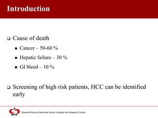
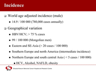
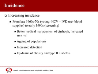
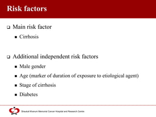
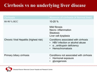
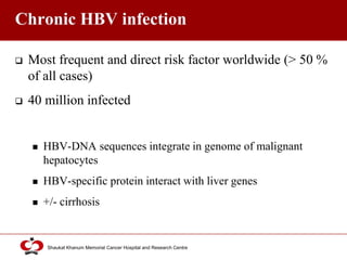
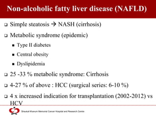
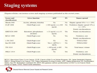
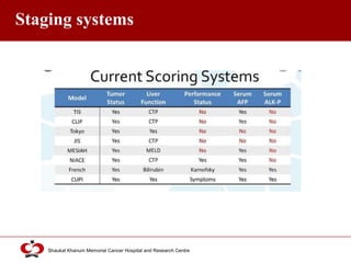
Call Girls Jp Nagar Just Call 7001305949 Top Class Call Girl Service Available
Hepatocellular carcinoma
1. Shaukat Khanum Memorial Cancer Hospital and Research Centre
Jibran Mohsin
Hepatocellular Carcinoma
Fellow Surgical Oncology
2. Click to edit Master title style
Shaukat Khanum Memorial Cancer Hospital and Research Centre
Outline
Introduction Fibrolamellar carcinoma (FLC)
Incidence Screening
Risk factors Staging systems
Pathology Treatment options
Clinical presentation Defining treatment strategy
Investigations Summary
Diagnosis
3. Click to edit Master title style
Shaukat Khanum Memorial Cancer Hospital and Research Centre
Introduction
90 % of primary liver malignancy
6th most common neoplasm (> 5% of all cancers)
5th most common malignancy (men)
9th most common malignancy (women)
2nd most common cause of cancer-related deaths
745 000 deaths worldwide (9.1 % of total; 2012)
4. Click to edit Master title style
Shaukat Khanum Memorial Cancer Hospital and Research Centre
Introduction
Cause of death
Cancer – 50-60 %
Hepatic failure – 30 %
GI bleed – 10 %
Screening of high risk patients, HCC can be identified
early
5. Click to edit Master title style
Shaukat Khanum Memorial Cancer Hospital and Research Centre
Incidence
World age adjusted incidence (male)
14.9 / 100 000 (780,000 cases annually)
Geographical variation
HBV/HCV; > 75 % cases
99 / 100 000 (Mongolian men)
Eastern and SE-Asia (> 20 cases / 100 000)
Southern Europe and north America (intermediate incidence)
Northern Europe and south central Asia ( < 5 cases / 100 000)
HCV, Alcohol, NAFLD, obesity
6. Click to edit Master title style
Shaukat Khanum Memorial Cancer Hospital and Research Centre
Incidence
Increasing incidence
From late 1960s-70s (young- HCV – IVD use- blood
supplies) to early 1990s (screening)
Better medical management of cirrhosis, increased
survival
Ageing of populations
Increased detection
Epidemic of obesity and type II diabetes
7. Click to edit Master title style
Shaukat Khanum Memorial Cancer Hospital and Research Centre
Risk factors
Main risk factor
Cirrhosis
Additional independent risk factors
Male gender
Age (marker of duration of exposure to etiological agent)
Stage of cirrhosis
Diabetes
8. Click to edit Master title style
Shaukat Khanum Memorial Cancer Hospital and Research Centre
Cirrhosis vs no underlying liver disease
Cirrhosis Absence of cirrhosis (≠ Normal liver)
80-90 % HCC 10-20 %
Mild fibrosis
Necro- inflammation
Steatosis
Liver cell dysplasia
Chronic Viral Hepatitis (highest risk) Conditions associated with cirrhosis
• HBV infection or alcohol abuse
• α 1 antitrypsin deficiency
• Hemochromatosis
Primary biliary cirrhosis Conditions not associated with cirrhosis
• Hormonal exposure
• glycogenosis
9. Click to edit Master title style
Shaukat Khanum Memorial Cancer Hospital and Research Centre
Chronic HBV infection
Most frequent and direct risk factor worldwide (> 50 %
of all cases)
40 million infected
HBV-DNA sequences integrate in genome of malignant
hepatocytes
HBV-specific protein interact with liver genes
+/- cirrhosis
10. Click to edit Master title style
Shaukat Khanum Memorial Cancer Hospital and Research Centre
Chronic HBV infection
Risk of HBV-associated HCC
Severity of underlying hepatitis
Age at infection (hence duration of infection)
Level of viral replication
10-fold risk HBsAg vs 60 fold risk HBeAg
HBV-DNA copies/mL > 104 (2.3 risk) vs > 106 (6.1 risk)
11. Click to edit Master title style
Shaukat Khanum Memorial Cancer Hospital and Research Centre
Chronic HBV infection
Cumulative risk (5 year)
10 % (western) vs 17 % (Asian)
Vertical transmission > horizontal (sexual or parenteral)
Longer duration
Additional environmental factors
12. Click to edit Master title style
Shaukat Khanum Memorial Cancer Hospital and Research Centre
HCV infection
Accounts for major proportion of increase in HCC over
last 10 years
> 70% HCC (west) : anti-HCV antibodies
Mean time around 30 years
Most frequent and 1st complication in HCV
compensated cirrhosis
0-2 % (chronic hepatitis) vs 1-4 % (7 % Japan)
(compensated cirrhosis)
13. Click to edit Master title style
Shaukat Khanum Memorial Cancer Hospital and Research Centre
Additional Risk/co- factors
Chronic HBV infection HCV infection NAFLD HH
Male gender (3-6 times
higher)
Male gender (2-3 X) Male gender Male gender
Age > 40 years Age > 55 years (2-4 x) Increasing age Age > 55 years ( 13.3 fold)
Concurrent HCV infection
(2 x)
HBV coinfection (2-6 x) HBV infection (4.9 fold)
HDV coinfection (3x) HIV-HCV ( 15-20 years
earlier)
Heavy alcohol use (2-3 x) Alcohol > 60-80 g/day (2-4
x)
Alcohol abuse (2.3 fold)
Aflatoxins (endemic
regions)
Iron deposition
Diabetes ( 2 x) diabetes
Obesity (NAFLD)
≠ HAV/ HEV ≠ viral genotype / viral
concentration
Severity of liver disease
14. Click to edit Master title style
Shaukat Khanum Memorial Cancer Hospital and Research Centre
Future
HBV HCV NAFLD
Prevention of
HBV infection
Prevention of HCV infection Growing epidemics worldwide
Immunization Prevention of progression of HCV
chronic infection to cirrhosis
• Direct acting anti-virals
(90% cure)
• Sustained virological
response
15. Click to edit Master title style
Shaukat Khanum Memorial Cancer Hospital and Research Centre
HIV infection
Due to higher prevalence of associated well-known risk
factors
Co-infection HBV/HCV
Alcohol abuse
NASH
Diabetes
25 % o all liver-related deaths
Earlier and more aggressive course (HIV-HCV vs HCV
alone)
16. Click to edit Master title style
Shaukat Khanum Memorial Cancer Hospital and Research Centre
Alcohol
Heavy (> 50-70 g/day) and prolonged use
Classical risk factor
Annual Incidence
1.7 % alcohol cirrhosis
2.2 % HBV cirrhosis
3.7 % HCV cirrhosis
Additional risk factor in HBV/HCV cirrhosis/
metabolic syndrome related chronic liver disease
17. Click to edit Master title style
Shaukat Khanum Memorial Cancer Hospital and Research Centre
Non-alcoholic fatty liver disease (NAFLD)
Simple steatosis NASH (cirrhosis)
Metabolic syndrome (epidemic)
Type II diabetes
Central obesity
Dyslipidemia
25 -33 % metabolic syndrome: Cirrhosis
4-27 % of above : HCC (surgical series: 6-10 %)
4 x increased indication for transplantation (2002-2012) vs
HCV
18. Click to edit Master title style
Shaukat Khanum Memorial Cancer Hospital and Research Centre
Non-alcoholic fatty liver disease (NAFLD)
Obesity:
Increase mortality from liver cancer far more than any other
cancer
Diabetes
Increase risk of HCC +/- acute and chronic liver disease
19. Click to edit Master title style
Shaukat Khanum Memorial Cancer Hospital and Research Centre
Non-alcoholic fatty liver disease (NAFLD)
Male gender
Increasing age
60 % cases > 50 years with DM/obesity
NASH/advance liver fibrosis
Sinusoidal iron deposition
Severity of underlying liver disease
Synergistic effect with chronic HCV infection or
alcoholic consumption
20. Click to edit Master title style
Shaukat Khanum Memorial Cancer Hospital and Research Centre
Hereditary hemochromatosis (HH)
ART
Homozygosity of C282Y mutation in hemochromatosis
gene
Excessive GI iron absorption
Others iron overload conditions
Homozygous beta thalassemia (African overload syndrome)
21. Click to edit Master title style
Shaukat Khanum Memorial Cancer Hospital and Research Centre
Cirrhosis of other etiologies
Primary biliary cirrhosis (PBC)
Low risk factor (rare incidence, M:F 1:9)
Secondary biliary cirrhosis (exceptionally rare)
Autoimmune hepatitis (low risk: female predominance,
delayed development of cirrhosis due to steroids)
Cirrhosis at time presentation
HCV may induce autoantibodies
22. Click to edit Master title style
Shaukat Khanum Memorial Cancer Hospital and Research Centre
Aflatoxin B1
Asia and sub-Saharan Africa
Contamination of imperfectly stored staple crops by
Aspergillus flavus
Tumor suppressor gene p53 mutation
Independent risk factor vs co-carcinogen (HBV)
Frequently non-cirrhotic liver
23. Click to edit Master title style
Shaukat Khanum Memorial Cancer Hospital and Research Centre
Metabolic liver disease
Increased risk Low risk
Alpha 1 anti trypsin deficiency Glycogenosis type IV
Porphyria cutanea tarda Hereditary fructose intolerance
Tyrosinemia Wilson disease
(oxygen/nitrogen species and
unsaturated aldehydes ; p53 mutation)
Hypercitrullinemia
24. Click to edit Master title style
Shaukat Khanum Memorial Cancer Hospital and Research Centre
Hepatocellular Adenoma (HCA)
Malignant transformation via Hepatocyte dysplasia
Adenomatosis: not increased risk
Female Male
≥ 4 cm As small as 1 cm
Child bearing age with prolonged use OCP/estrogens Make with metabolic syndrome
4.2 % risk 50 % risk (10 x)
Discontinuation doesn’t completely avoid risk
Resection if > 4 cm Resection /ablation irrespective of size
25. Click to edit Master title style
Shaukat Khanum Memorial Cancer Hospital and Research Centre
Hepatocellular Adenoma (HCA)
Risk factors
Phenotype: Telangiectatic or atypical > Steatotic
Genotype: β catenin (10-15 % HCAs)
Type I glycogenosis
Use of anabolic steroids (recreational) or androgens
Fanconi syndrone
26. Click to edit Master title style
Shaukat Khanum Memorial Cancer Hospital and Research Centre
Pathology (Preneoplastic lesions)
(Microscopic)
dysplastic foci
Macroscopic dysplastic nodules (DNs)
Dysplastic hepatocytes
(< 1mm size)
Nodular region < 2 cm (diameter) with dysplasia (but without definite
histological criteria of malignancy)
Chronic liver disease
(cirrhosis)
Degree of cytological or architectural atypia
Low grade DNs High grade DNs (less common)
(33 % malignant transformation)
~ 1 cm Up to 2 cm
Slightly yellowish Increased cell density
Very low chance of malignancy Irregular thin-trabecular pattern
Unpaired arteries
Distinct foci of well differentiated
HCC
Difficult to differentiate vs well
differentiated HCC
27. Click to edit Master title style
Shaukat Khanum Memorial Cancer Hospital and Research Centre
Pathology (HCC)
Subdivisions
Gross morphology
Degree of differentiation
Vascularity
Capsule
Vascular invasion
28. Click to edit Master title style
Shaukat Khanum Memorial Cancer Hospital and Research Centre
Pathology
Gross Morphology
Solitary
Multinodular Multicentric Synchronous discrete lesions in different segments
Intrahepatic
metastases
1 dominant mass and number of ‘daughter‘ nodules
in adjacent segments
Diffuse Relatively rare
Poorly defined widely infiltrative masses
Diagnostic challenges on imaging
Infiltrating Less differentiated
Ill defined margins
Fibrolamellar Young, without cirrhosis, better prognosis, cure by
resection
29. Click to edit Master title style
Shaukat Khanum Memorial Cancer Hospital and Research Centre
Pathology
Degree of Differentiation
Decreases with increase in diameter
Grade 1: resemble normal hepatocytes, near normal lobar
architecture
Imunomarker (50-73 %): Glypican 3 (GPC3), Heat Shock
Protein 70 (HSP70) and Glutamine Synthetase (GS)
100 % specificity on resected specimens
Edmondson grade (Histologic Grade (G))
Grade can’t be assessed GX
Well differentiated G1
Moderately differentiated G2
Poorly differentiated G3
Undifferentiated G4
30. Click to edit Master title style
Shaukat Khanum Memorial Cancer Hospital and Research Centre
Pathology
Molecular Pathogenesis
TERT promoter mutations (most frequent i.e. 60 %)
Subsequent increase in telomerase expression
Development/transformation and progression
Prognosis/select treatment/assess response to chemotherapy
31. Click to edit Master title style
Shaukat Khanum Memorial Cancer Hospital and Research Centre
Pathology
Vascularity
Macroregenerative nodule low grade DN High grade
DN Frank HCC
Loss of visualization of portal tracts
Development of new non-tiadal arterial vessels (arterial
neoangiogenesis)
Dominant blood supply of overt HCC lesion
Landmark of HCC diagnosis
Rationale for chemoembolization and anti-angiogenic
treatment
32. Click to edit Master title style
Shaukat Khanum Memorial Cancer Hospital and Research Centre
Pathology
Distinct fibrous capsule
80 % resected HCCs
Variable thickness
May not be complete
Frequently infiltrated by tumor cells
Microscopic capsular invasion:
33 % cases in tumor < 2 cm diameter
66 % cases in larger tunors
33. Click to edit Master title style
Shaukat Khanum Memorial Cancer Hospital and Research Centre
Pathology
Vascular invasion
Portal invasion (most important predictive factor of recurrence)
Expansive type
Poorly differentiated
Large size
Microscopic vascular invasion
20 % (< 2cm)
30-60 % (2-5 cm)
60-90 % ( > 5 cm)
34. Click to edit Master title style
Shaukat Khanum Memorial Cancer Hospital and Research Centre
Pathology
Vascular (Portal) invasion
Tumor thrombus has its own arterial supply
Mainly from site of original venous invasion
Grows rapidly in both direction (in particular towards main portal vein –
high risk of complete thrombosis and increased portal HTN –
fatal esophageal varices rupture/ liver failure i.e.
ascites/jaundice/encephalopathy
Tumor fragments spread throughout liver as thrombus crosses
segmental branches
35. Click to edit Master title style
Shaukat Khanum Memorial Cancer Hospital and Research Centre
Pathology
Vascular invasion
Hepatic veins
Possible although less frequent
Extends into supra hepatic vena cava or right atrium
Lung metastases
Biliary tract invasion (rare)
Jaundice or hemobilia
36. Click to edit Master title style
Shaukat Khanum Memorial Cancer Hospital and Research Centre
Pathology
HCC induced biliary obstruction
Intraductal tumor extension
Obstruction by fragment of necrotic tumor debris
Hemorrhage of tumor (Hemobilia)
Metastatic lymph node compression of major bile ducts in porta hepatis.
Rate of invasion
Portal vein invasion 15 % (1 in 3 cases will develop PV thrombosis)
Hepatic vein invasion 5 %
Bile duct invasion 3 %
37. Click to edit Master title style
Shaukat Khanum Memorial Cancer Hospital and Research Centre
Pathology
Metastases
Sites (descending order) Lungs (most frequent)
Adrenal glands
Bones
Lymph nodes
Meninges
Pancreas
Brain
Kidney
Risk factors Tumor size
Bilobar disease
Poor differentiation
38. Click to edit Master title style
Shaukat Khanum Memorial Cancer Hospital and Research Centre
Clinical Presentation
Rare before 40 years, peak at 70 years
M:F (2-4:1)
most pronounced in medium risk south European populations
and premenopausal women
Difference in exposure to risk factors
Higher BMI
Higher levels of androgenic hormones
39. Click to edit Master title style
Shaukat Khanum Memorial Cancer Hospital and Research Centre
Clinical Presentation
Circumstances of diagnosis
Incidental finding During routine screening (Industrialized countries)
During assessment of patients with deranged LFTs or of another
pathological condition
Symptomatic
(severity)
Liver-related (functional status of non-tumorous liver)
• Sudden onset or worsening ascites or liver decompensation
may be 1st evidence of HCC
Cancer-related ( stage of tumor)
• Abdominal pain
• Malaise
• Weight loss
• Asthenia
• Anorexia
• Fever
• Acute (tumor extension or complication- next slide)
40. Click to edit Master title style
Shaukat Khanum Memorial Cancer Hospital and Research Centre
Clinical Presentation
Spontaneous Rupture • 5-15 % cases
• Superficial or protruding tumors
• Suspect:
• Known HCC/cirrhosis + acute epigastric pain or
• Asian/African men with acute abdomen
• Minor rupture:
• Abdominal pain
• Hemorrhagic ascites
• Hypovolemic shock (50 % cases)
PV invasion Upper GI bleed
Acute ascites
Hepatic vein or IVC
invasion
PE
Sudden death
Biliary invasion/ Hemobilia
(2 %)
2 % cases
Paraneoplastic syndromes Polyglobulia, Hypercalcemia, Hypoglycemia
41. Click to edit Master title style
Shaukat Khanum Memorial Cancer Hospital and Research Centre
42. Click to edit Master title style
Shaukat Khanum Memorial Cancer Hospital and Research Centre
Clinical Presentation (examination)
Large or superficial tumors
Clinical signs of cirrhosis
Ascites
Collateral circulation
Umbilical hernia
Hepatosplenomegaly
43. Click to edit Master title style
Shaukat Khanum Memorial Cancer Hospital and Research Centre
Investigations
Hematological Liver function tests
Serum tumor markers Alpha-fetoprotein (AFP)
DCP
PIVKA-II
AFP-L3
Radiological Ultrasound
CT scan
MRI
Contrast enhanced US
Angiography
PET
Pathological Cytology
Histology
44. Click to edit Master title style
Shaukat Khanum Memorial Cancer Hospital and Research Centre
Liver function tests (LFTs)
Non-specific
Reflects underlying pathology or presence of SOL
Normal LFTs exceptional;
HCC in background of cirrhosis
HCC in normal liver are large
Jaundice most frequently result of liver decompensation
45. Click to edit Master title style
Shaukat Khanum Memorial Cancer Hospital and Research Centre
Serum Alpha fetoprotein (AFP)
Most widely recognized serum marker of HCC
Normal: 0-20 ng/mL
May increase in HCC
> 400 ng/mL (diagnostic of HCC ; 95 % accuracy)
> 10 000 ng/mL (5-10 % cases)
Poor differentiation
Tumor aggressiveness
Vascular invasion
46. Click to edit Master title style
Shaukat Khanum Memorial Cancer Hospital and Research Centre
Serum Alpha fetoprotein (AFP)
Sensitivity
> 20 ng/mL (60 %)
> 200 ng/mL (78%)
Others
Non –tumor: 30 % chronic active hepatitis without HCC
Tumor:
Non-seminal germinal tumors, Hepatoid gastric
tumors, NET
47. Click to edit Master title style
Shaukat Khanum Memorial Cancer Hospital and Research Centre
Other serum tumor markers (Japan)
Des-γ-carboxy prothrombin (DCP)
Sensitivity (early detection)
61% (AFP alone)
74 % (DCP alone)
91% (AFP + DCP)
Prothrombin induced by vitamin K absence (PIVKA-II)
> 40 mAU/mL
AFP-L3 (> 15 %)
48. Click to edit Master title style
Shaukat Khanum Memorial Cancer Hospital and Research Centre
Radiological
Objective
Screen high risk patients for development of HCC (US)
Differentiate potential HCC from other tumors/SOL
Hyper vascularity during arterial phase
‘Washout’ during portal or late phase (Hypo vascular
compared to adjacent parenchyma)
Select appropriate treatment
Number, size, extent, daughter nodules, vascular invasion,
extrahepatic spread and underlying liver disease.
49. Click to edit Master title style
Shaukat Khanum Memorial Cancer Hospital and Research Centre
Ultrasound
1st line investigation for screening
Low cost, availability, sensitivity
Accuracy
3-5 cm: 85-95 %
1 cm: 60-80 %
steatosis rates and heterogeneity of underlying liver disease
Dimension and location
Operator experience
Echogenicity
50. Click to edit Master title style
Shaukat Khanum Memorial Cancer Hospital and Research Centre
Ultrasound
Echogenicity
Small HCC: hypoechoic and homogenous (regenerating/DNs)
Large HCC: Hypo/hyperechoic but heterogeneous
Capsule: Hypoechoic peripheral rim
51. Click to edit Master title style
Shaukat Khanum Memorial Cancer Hospital and Research Centre
Ultrasound (Demerits)
Infiltrative type difficult: Grossly heterogeneous
cirrhotic liver
1 cm tumor: deep vs surface
Upper segments or edge of left lateral segments
Obesity (abdominal wall and steatotic liver)
52. Click to edit Master title style
Shaukat Khanum Memorial Cancer Hospital and Research Centre
Ultrasound
Doppler US: feeding artery +/- draining vein
Vascular invasion or biliary invasion
Indirect evidence of cirrhosis
Segmental atrophy, splenomegaly, ascites, collateral veins
53. Click to edit Master title style
Shaukat Khanum Memorial Cancer Hospital and Research Centre
Helical / Multislice spiral / multiphasic CT
More accurate than US
Multiphasic
Without contrast
With Contrast:
Arterial (25-50s)
Portal (60-65s)
Equilibrium (130-180s)
54. Click to edit Master title style
Shaukat Khanum Memorial Cancer Hospital and Research Centre
CT scan: Role
HCCs identification and distribution (lobar/segmental)
Features of underlying cirrhosis
Liver/tumor volumes
Extrahepatic tumor spread
Vascular invasion of segmental branches
55. Click to edit Master title style
Shaukat Khanum Memorial Cancer Hospital and Research Centre
CT scan: features
Usually hypodense
Spontaneous hyperdense (2-20%): iron overload or
fatty infiltration
Early uptake of contrast with mosaic shape pattern
Sharply diminished density in portal phase
Washout during late phase
56. Click to edit Master title style
Shaukat Khanum Memorial Cancer Hospital and Research Centre
57. Click to edit Master title style
Shaukat Khanum Memorial Cancer Hospital and Research Centre
CT scan: features
Variable vascularity depending on tumor grade
Capsule: Peripheral enhanced thickening in portal or
late phase
Intratumoral arterioportal fistula
Early enhancement of portal branches
Triangular area distal to tumor with contrast enhancement
different from adjacent parenchyma
58. Click to edit Master title style
Shaukat Khanum Memorial Cancer Hospital and Research Centre
MRI
More accurate than other imaging techniques
HCC vs other liver tumors (> 2cm)
T1- and T2-weighted images
Early, intermediate and late phases (gadolinium)
Features:
Mosaic shape structure
Capsule
59. Click to edit Master title style
Shaukat Khanum Memorial Cancer Hospital and Research Centre
MRI
Early uptake and late washout
Gd-EOB-DTPA
liver specific MR contrast medium
Accumulates in Kupffer cells (phagocytosis) or hepatic cells
Increased MRI accuracy
T1 weighted T2 weighted
Hypodense Hyperdense 54 % cases
Hypodense Hypodense 16 % cases
Hyperdense Fatty/copper/glycogen infiltration of tumor
60. Click to edit Master title style
Shaukat Khanum Memorial Cancer Hospital and Research Centre
61. Click to edit Master title style
Shaukat Khanum Memorial Cancer Hospital and Research Centre
Contrast-enhanced US (CEUS)
Most recent technique to assess vascularization
Contrast agent (stabilized microbubbles) : IV bolus
injection followed by saline flush
Arterial (10-20s post injection)
Portal venous (30-80s)
Late (120-360s)
62. Click to edit Master title style
Shaukat Khanum Memorial Cancer Hospital and Research Centre
63. Click to edit Master title style
Shaukat Khanum Memorial Cancer Hospital and Research Centre
Contrast-enhanced US (CEUS)
Merits
Greater sensitivity to detect arterial enhancement
microbubbles confined to vascular spaces
Continuous monitoring of images
Well differentiated vs poorly differentiated: slower wash out
Demerits
Same as other US modes
64. Click to edit Master title style
Shaukat Khanum Memorial Cancer Hospital and Research Centre
Contrast-enhanced US (CEUS)
Sonazoid-CEUS (Japan 2007; 2nd generation US
contrast agent) vs Sono Vue
Taken up by Kupffer cells in postvascular/Kupffer
phase (10 mins post injection)
Extremely stable Kupffer images suitable for repeated
scanning 10-120 mins after injection
65. Click to edit Master title style
Shaukat Khanum Memorial Cancer Hospital and Research Centre
Angiography
As part of arterial chemoembolization
Arteriography: early vascular uptake (blush)
Lipiodol (radiodense) injection
Retained: HCC, focal nodular hyperplasia, adenoma,
angioma, metastases
False-negative: Avascular, necrotic or fibrotic HCC
66. Click to edit Master title style
Shaukat Khanum Memorial Cancer Hospital and Research Centre
18F-FDG-PET
Limited role
low sensitivity (60 %)
Can detect poorly differentiated, but not well differentiated
Similar metabolism
18F-fluorocholine (FCH), PET tracer of lipid metabolism
High sensitivity and more suitable than CT or bone scan
for bone metastases in metastatic HCC
67. Click to edit Master title style
Shaukat Khanum Memorial Cancer Hospital and Research Centre
Accuracy
Contrast CT +/- MRI
Highest diagnostic accuracy (> 80 %)
Nodules < 1cm
3 monthly surveillance for at least 2 years
Nodules > 1 cm
Suspicious
Typical HCC features: no further investigations
Not typical: Percutaneous FN biopsy
68. Click to edit Master title style
Shaukat Khanum Memorial Cancer Hospital and Research Centre
Pathological (cytology/histology)
Increased accuracy: if non-tumorous tissue is available
Demerits
Hemorrhage
Pain
Needle tract neoplastic seeding (1-5 %)
Limited to subcutaneous tissue
Slow progression
Local excision without impact on survival
Vascular spread
69. Click to edit Master title style
Shaukat Khanum Memorial Cancer Hospital and Research Centre
Pathological (cytology/histology)
Risk of needle tract seeding vs risk of aggressive
treatment (resection/transplantation) in cases without
malignancy
Avoid direct puncture of nodule
Through thick area of normal liver
GPC3, HSP70, GS staining (small lesions not clearly
HCC)
70. Click to edit Master title style
Shaukat Khanum Memorial Cancer Hospital and Research Centre
Pathological (cytology/histology)
Low false-positive rate
High false-negative rate
Small lesions
Lesions difficult to access
Lesions in multinodular parenchyma
71. Click to edit Master title style
Shaukat Khanum Memorial Cancer Hospital and Research Centre
Diagnosis
Standard for diagnosis: Histology
Tumors < 3 cm
Active treatment is required
Non-invasive diagnosis (radiological imaging alone)
Rigorous technique and interpretation
72. Click to edit Master title style
Shaukat Khanum Memorial Cancer Hospital and Research Centre
Diagnostic criteria
European Association for the Study of the Liver
(EASL)
2000 (1st attempt), 2011 (updated)
AFP: lack adequate sensitivity and specificity
Based on imaging +/- biopsy
73. Click to edit Master title style
Shaukat Khanum Memorial Cancer Hospital and Research Centre
Diagnostic criteria
Nodule size
(screening US)
< 1 cm • Repeat US at 3-6 monthly (until lesion
disappeared/enlarged/HCC features) for at least 2 years
• Other imaging techniques unlikely to reliably confirm
diagnosis
• Accuracy of liver biopsy and likelihood of HCC is low
• Than routine surveillance
>1 cm with
cirrhosis
Early uptake and delayed wash out on a single dynamic imaging
study (triphasic CT or MR with gadolinium)= characteristic of HCC
(Sensitivity 71 %, PPV 100 %, Specificity 100 %)
> 1 cm without
cirrhosis Diagnostic biopsy (negative result doesn’t rule out malignancy)
Vascular features
not typical
74. Click to edit Master title style
Shaukat Khanum Memorial Cancer Hospital and Research Centre
Diagnostic criteria
Biopsy of small lesions
Expert pathologists
Not clearly HCC: CD34, CK7, GPC3, HSP-70, GS
Negative biopsy
Imaging 3-6 monthly
If enlarges but atypical of HCC: repeat biopsy
75. Click to edit Master title style
Shaukat Khanum Memorial Cancer Hospital and Research Centre
Diagnostic criteria
CEUS
False-positive HCC diagnosis
HCC vs intrahepatic cholangiocarcinoma
Withdrawn from diagnostic algorithm (American association
for the study of Liver Disease – AASLD)
76. Click to edit Master title style
Shaukat Khanum Memorial Cancer Hospital and Research Centre
Fibrolamellar carcinoma (FLC)
Prognosis better than overall HCC
5 year SR (50-75%) following resection
Resection preferred over transplantation
Imaging
Large solitary hyper vascular heterogeneous liver mass
Central hypo dense region/scar (central necrosis/fibrosis)
Low attenuation on MRI T2 images vs high attenuation of central scar
of focal nodular hyperplasia
Well defined margins and calcification (68 %)
77. Click to edit Master title style
Shaukat Khanum Memorial Cancer Hospital and Research Centre
Fibrolamellar carcinoma (FLC)
High AFP: < 10 % cases
Lymphadenectomy recommended if resection considered
Rationale: LN invasion within hepatic pedicle (60%)
Significant risk of recurrence
Liver, LN or distant
Close long-tern follow up mandatory
Recurrence /death beyond 5 years common
Repeat resection reasonable options: young population, indolent course, relative inefficacy
of non surgical treatments
78. Click to edit Master title style
Shaukat Khanum Memorial Cancer Hospital and Research Centre
Fibrolamellar carcinoma (FLC)
True FLC vs mixed FLC-HCC
“Conventional HCC displaying some distinct area with FLC features”
Transplantation: alternate option in selected cases
Survival rate 48 %
79. Click to edit Master title style
Shaukat Khanum Memorial Cancer Hospital and Research Centre
Screening
Routinely done in countries where effective therapeutic interventions are
available
Fulfill most of criteria for surveillance /screening program
Common in highly endemic areas (increasing incidence in others)
High mortality
Extremely poor survival by time patients present with cancer-related symptoms
Population at risk is clearly defined (male, > 60 years, HBV/HCV)
Effective treatment available in selected patients
Acceptable screening tests:
low morbidity and high efficacy
Allow tumor detection in latent/early stage
80. Click to edit Master title style
Shaukat Khanum Memorial Cancer Hospital and Research Centre
Screening
Diagnosis at early stage required with preserved liver function
At presentation
Symptomatic Median survival (6 months)
Asymptomatic untreated (not end-stage) 3 year survival (50%)
81. Click to edit Master title style
Shaukat Khanum Memorial Cancer Hospital and Research Centre
Screening
AFP
Little value in screening alone
Normal baseline AFP increased AFP and normal US CT/MRI
Ultrasound
Difficult in obese patients with fatty liver disease and cirrhosis
Optimal interval for periodic screening (6 monthly; 3-4 months in Japan) in high risk
patients
Tumor doubling time vary widely (average 200 days)
Undetectable to 2 cm (4-12 months)
Rapidly growing HCC to reach 3 cm ( 5 months)
Treatment most effective for tumors < 3 cm
82. Click to edit Master title style
Shaukat Khanum Memorial Cancer Hospital and Research Centre
Screening
Impact on prognosis
China on HBV carriers
Cost effective if expected HCC risk exceeds
0.2 % per year in HBV patients
1.5 % per year in HCV patients
83. Click to edit Master title style
Shaukat Khanum Memorial Cancer Hospital and Research Centre
Screening
Limitations
20-50 % HCC at presentation had previously undiagnosed cirrhosis
(escape surveillance)
Access to medical care
Compliance: 50 % drop out in 5 years (alcoholic cirrhosis)
US: operator dependent
CT/MR:
High cost/invasiveness, unsuitable for regular screening
Suitable in irregular liver parenchyma or obesity
Decision to screen cirrhotic patient: consider comorbid disease, severity of
liver disease and available treatment options (e.g. no screening in CP C if
not candidate for LT)
84. Click to edit Master title style
Shaukat Khanum Memorial Cancer Hospital and Research Centre
Screening (Summary)
US recommended as screening tool, whereas CT/MRI most useful in
confirming diagnosis
High risk groups
Established cirrhosis (HBV/HCV/Hemochromatosis)
CLD due to NASH
Male, alcohol related cirrhosis abstaining from alcohol or likely to comply
with treatment
Surveillance of at-risk patients at 6 monthly interval with US to detect HCC a
early stage
85. Click to edit Master title style
Shaukat Khanum Memorial Cancer Hospital and Research Centre
Staging systems
Objective
Predict outcome / prognosis / survival
Select treatment plan
86. Click to edit Master title style
Shaukat Khanum Memorial Cancer Hospital and Research Centre
Staging systems
Parameters affecting survival
Morphological spread of tumor • Number of tumor nodules
• vascular invasion (radiological or
microscopically)
• Tumor size (dominant nodule)
• Limitation: based on pathological findings;
applicable on resected specimens accurately
only
Presence and severity of cancer –
related symptoms
• WHO performance status or Karnofsky index
• Pain (poor outcome indicator)
Severity and evolution of
underlying cirrhosis
Child-Pugh score
• Functional reserve of cirrhotic patients
underlying portocaval shunt surgery
• Not entirely appropriate for HCC (resection/
transplantation)
MELD
87. Click to edit Master title style
Shaukat Khanum Memorial Cancer Hospital and Research Centre
Staging systems
88. Click to edit Master title style
Shaukat Khanum Memorial Cancer Hospital and Research Centre
Staging systems (Tumor spread)
T staging (Liver Cancer Study Group of Japan; LCSGJ)
Size < 2cm > 2cm
Number 1 >1 1 >1
Vascular
Invasion
- + - + - + - +
T1 T2 T2 T3 T2 T3 T3 T4
89. Click to edit Master title style
Shaukat Khanum Memorial Cancer Hospital and Research Centre
Staging systems (Tumor spread)
Tx: Primary can’t be assessed
T0: No evidence of primary tumor
T4: Main branch of portal or hepatic vein (previously T3B)
T4: direct invasion of adjacent organs (except gallbladder) or perforation of visceral
peritoneum
T staging (American Joint Committee on Cancer (AJCC) / International Union Against
Cancer (UICC)) (8th edition; 2017)
Number 1 >1
Size ≤ 2 cm > 2 cm None > 5 cm Any > 5cm
Vascular Invasion - + - +
T1a T1a T1b T2 T2 T3
Previously T1 T2 T1 T2 T2 T3A
90. Click to edit Master title style
Shaukat Khanum Memorial Cancer Hospital and Research Centre
Staging systems (Tumor spread)
Regional Lymph Nodes (N)
Nx Can’t be assessed
N0 No regional LN metastasis
N1 Regional LN metastasis
Distant Metastasis (M)
Mx Can’t be assessed
M0 -
M1 +
91. Click to edit Master title style
Shaukat Khanum Memorial Cancer Hospital and Research Centre
Staging systems (Tumor spread)
Stage T N M
IA 1a 0 0
IB 1b
II 2 0 0
IIIA 3 0 0
IIIB 4
IVA Any 1 0
IVB Any Any 1
92. Click to edit Master title style
Shaukat Khanum Memorial Cancer Hospital and Research Centre
Staging systems (Tumor spread)
Edmondson grade (Histologic Grade (G))
Grade can’t be assessed GX
Well differentiated G1
Moderately differentiated G2
Poorly differentiated G3
Undifferentiated G4
93. Click to edit Master title style
Shaukat Khanum Memorial Cancer Hospital and Research Centre
Staging systems (Tumor spread)
Fibrosis score (F)
F0 Fibrosis score 0-4 (no to moderate fibrosis)
F1 Fibrosis score 5-6 (severe fibrosis or cirrhosis)
Stage 5 year survival rate
F0 F1
I 64% 49%
II 46% 30%
III 17% 9%
AJCC manual recommends notation of fibrosis but hasn’t yet formally incorporated F classification into staging system
94. Click to edit Master title style
Shaukat Khanum Memorial Cancer Hospital and Research Centre
Staging systems (Cancer-related symptoms / patient)
Eastern Cooperative Oncology Group (ECOG) Performance status (1982) *
(ak.a. WHO performance status score / Zubrod score)
0 Fully active, able to carry on all pre-disease performance without restriction Asymptomatic
1 Restricted in physically strenuous activity but ambulatory and able to carry out work of light
or sedentary nature .e. light house work or office work
Symptomatic but
completely ambulatory
2 Ambulatory and capable of all self care but unable to carry out any work activities. Up and
about > 50 % of waking hours
Symptomatic , < 50 % in
bed during day
3 Capable of only limited self care, confined to bed/chair > 50 % of waking hours Symptomatic , > 50 % in
bed, but not bed bound
4 Completely disabled. Can’t carry on any self care. Totally confined to bed/chair Bedbound
5 Dead Death
* Oken M, Creech R, Tormey D, et al. Toxicity and response criteria of the Eastern Cooperative Oncology Group.Am
J Clin Oncol. 1982;5:649-655.
95. Click to edit Master title style
Shaukat Khanum Memorial Cancer Hospital and Research Centre
Staging systems (Cancer-related symptoms / patient)
*Karnofsky D, Burchenal J, The clinical evaluation of chemotherapeutic agents in cancer. In: MacLeod C, ed.
Evaluation of Chemotherapeutic Agents. New York, NY: Columbia University Press; 1949:191–205
96. Click to edit Master title style
Shaukat Khanum Memorial Cancer Hospital and Research Centre
Staging systems (Liver function/damage)
97. Click to edit Master title style
Shaukat Khanum Memorial Cancer Hospital and Research Centre
Staging systems (Liver function/damage)
C(T)P score
Rough estimate of gross synthetic capacity of liver
validated as predictor of survival after surgery in cirrhotic liver
A: major liver resection
B: Minor liver resection
C: ‘significant risk from anesthesia and laparotomy
Only candidate for OLT (orthotopic liver transplantation)
98. Click to edit Master title style
Shaukat Khanum Memorial Cancer Hospital and Research Centre
Staging systems (Liver function/damage)
Model for end-stage liver disease (MELD) score
• Objective measure of extent of liver disease, relying only on quantifiable lab values vs subjective physical
findings such as ascites or encephalopathy
• Most helpful in determining allocation for liver transplant
99. Click to edit Master title style
Shaukat Khanum Memorial Cancer Hospital and Research Centre
Staging systems
Italy CLIP Cancer of Liver Italian Program 1998
France GRETCH
/French
Groupe d'Etude et de Traitement du Carcinome
Hépatocellulaire
1999
Spain BCLC Barcelona Clinic Liver Cancer 1999
Hong Kong CUPI Chinese University Prognostic Index 2002
HKLC Hong Kong Liver Cancer 2014
Japan Okuda staging 1985
JIS /JSS Japan Integrated Staging (c-JIS, bm-JIS) 2003/2008
Tokyo system
Taiwan TIS Taipei Integrated Scoring System 2010
NATURE scoring system
NIACE (Number, Infiltration, AFP, CP, ECOG) score
MES(IA)H Model for Estimated Survival (in Ambulatory) HCC
100. Click to edit Master title style
Shaukat Khanum Memorial Cancer Hospital and Research Centre
Staging systems
Okuda K, Ohtsuki T, Obata H, et al. Natural history of hepatocellular carcinoma and prognosis in relation to
treatment. Study of 850 patients. Cancer 1985;56:918-28.
Stage Median Survival
I 11.5 months
II 3 months
III 0.9 months
101. Click to edit Master title style
Shaukat Khanum Memorial Cancer Hospital and Research Centre
Staging systems
A new prognostic system for hepatocellular carcinoma: a retrospective study of 435 patients: the Cancer of the Liver
Italian Program (CLIP) investigators. Hepatology 1998;28:751-5.
CLIP score Median survival
0 36-42 months
1 22-32 months
2 8-16 months
3 4-7 months
4 - 6 1-3 months
102. Click to edit Master title style
Shaukat Khanum Memorial Cancer Hospital and Research Centre
Staging systems
Chevret S, Trinchet JC, Mathieu D, et al. A new prognostic classification for predicting survival in patients with
hepatocellular carcinoma. Groupe d'Etude et de Traitement du Carcinome Hépatocellulaire. J Hepatol 1999;31:133-41.
Score Range: 0-11
103. Click to edit Master title style
Shaukat Khanum Memorial Cancer Hospital and Research Centre
Staging systems
Leung TW, Tang AM, Zee B, et al. Construction of the Chinese University Prognostic Index for hepatocellular carcinoma
and comparison with the TNM staging system, the Okuda staging system, and the Cancer of the Liver Italian Program
staging system: a study based on 926 patients. Cancer 2002;94:1760-9.
Scorerange:-7to12
104. Click to edit Master title style
Shaukat Khanum Memorial Cancer Hospital and Research Centre
Staging systems
Kudo M, Chung H, Osaki Y. Prognostic staging system for hepatocellular carcinoma (CLIP score): its value and
limitations, and a proposal for a new staging system, the Japan Integrated Staging Score (JIS score). J Gastroenterol
2003;38:207-15.
105. Click to edit Master title style
Shaukat Khanum Memorial Cancer Hospital and Research Centre
Staging systems
106. Click to edit Master title style
Shaukat Khanum Memorial Cancer Hospital and Research Centre
Staging systems (BCLC)
107. Click to edit Master title style
Shaukat Khanum Memorial Cancer Hospital and Research Centre
Staging systems (BCLC)
Merits Demerits
Externally validated Not validated in eastern countries
Each stage combined with treatment
algorithm
Advanced stage can be further divided
into 2 groups
• Locally advanced (PV invasion)
• Advanced (extrahepatic metastasis)
Supported by AASLD and EASL
108. Click to edit Master title style
Shaukat Khanum Memorial Cancer Hospital and Research Centre
Staging systems
109. Click to edit Master title style
Shaukat Khanum Memorial Cancer Hospital and Research Centre
Staging systems
110. Click to edit Master title style
Shaukat Khanum Memorial Cancer Hospital and Research Centre
Treatment
Wide range of treatment options
Curative (transplantation, resection, ablation)
Chemoembolization, systemic therapy
Multidisciplinary team meeting
Liver surgeon
Interventional radiologist
Oncologist
Hepatologist
111. Click to edit Master title style
Shaukat Khanum Memorial Cancer Hospital and Research Centre
Treatment
In case of underlying liver disease(cirrhosis)
Only transplantation is curative
Recurrence constant with all other options
112. Click to edit Master title style
Shaukat Khanum Memorial Cancer Hospital and Research Centre
Treatment
Without cirrhosis (small group)
Resection: ideal treatment
With cirrhosis
Challenging: Patient , liver (FLR) and tumor factors need to
be considered
HCC identified by surveillance
Usually smaller; hence curative treatment
Access to curative treatment ? (22.8 %)
113. Click to edit Master title style
Shaukat Khanum Memorial Cancer Hospital and Research Centre
Treatment
HCC in normal liver (no or minimal coexisting
fibrosis)
Partial liver resection (treatment of choice)
Non-tumourous liver has high regenerating capacity
Perioperative mortality < 1 % ( especially in metabolic
syndrome)
Perioperative morbidity < 15 %
5 year survival rate > 50 %
Lymphadenectomy recommended (15 % risk vs < 5 % in
cirrhotic)
114. Click to edit Master title style
Shaukat Khanum Memorial Cancer Hospital and Research Centre
Treatment
HCC in normal liver (no or minimal coexisting
fibrosis)
Adjuvant chemotherapy not recommended
Regular follow up with CT-chest/abdomen 6 monthly
Early detection and treatment of recurrence may improve
survival
Percutaneous ablation
No role (large size tumor at diagnosis)
115. Click to edit Master title style
Shaukat Khanum Memorial Cancer Hospital and Research Centre
Treatment
HCC in normal liver (no or minimal coexisting
fibrosis)
Transplantation
10 % perioperative mortality
Long term immunosuppression
Long term results similar to resection
116. Click to edit Master title style
Shaukat Khanum Memorial Cancer Hospital and Research Centre
Treatment
HCC in normal liver (no or minimal coexisting
fibrosis)
Transplantation (indications) (5 year SR: 59 %)
Primary treatment
Partial resection preclude by anatomical factors
Need to preserve sufficient volume of liver remnant
Rescue treatment
Intrahepatic tumor recurrence not amenable to repeat
resection
117. Click to edit Master title style
Shaukat Khanum Memorial Cancer Hospital and Research Centre
Treatment
HCC in cirrhotic patients
Liver resection
Limitations
20-60 % multifocal at time of diagnosis (ideally 1)
Cirrhosis (post operative complications)
High risk of recurrence (cirrhosis persists)
Oncological resections (wide margins) vs liver
disease (parenchymal sparing)
118. Click to edit Master title style
Shaukat Khanum Memorial Cancer Hospital and Research Centre
Treatment
HCC in cirrhotic patients
Liver resection (risk of surgery)
Morbidity
coagulation defects (increased PT ; peak on day 1)
portal HTN,
liver failure (raised bilirubin; peak on day 3-5)
PT/bilirubin normalize in 5-7 days but delayed or absent in cirrhosis)
impaired regeneration PT< 50 % normal and bilirubin > 50umol/L on day 5;
post operative mortality 50 %)
Mortality (10 % or more; 1990’s; now decreased to (0%) 4-6 %)
Patient selection,
operative technique and
perioperative management
119. Click to edit Master title style
Shaukat Khanum Memorial Cancer Hospital and Research Centre
Treatment
HCC in cirrhotic patients
Liver resection (selection)
Child-Pugh A
Without clinically significant portal HTN
Hepatic venous pressure gradient > 10 mmHg
Presence of esophageal varices
Splenomegaly (> 12 cm)
Thrombocytopenia < 100 000/uL
Portosystemic shunts (patent umbilical vein)
Ascites
Unifocal resectable
120. Click to edit Master title style
Shaukat Khanum Memorial Cancer Hospital and Research Centre
Treatment
HCC in cirrhotic patients
Liver resection
CP - B/C: prohibitive risk of early liver failure even after
minor resection or mere laparotomy
CP – A: still increased risk of liver failure after major
liver resection (impaired regeneration)
Correlates with fibrosis grade (F) (F1> F0)
Additional selection criteria: Indocyanine green
(ICG)
121. Click to edit Master title style
Shaukat Khanum Memorial Cancer Hospital and Research Centre
Treatment
Indocyanine green (ICG; Japan)
0.5 mg ICG/Kg
Retention after 15 mins (ICG-R15) in peripheral blood
Normal 10 %
Minor resection: 22 % or less
Major resection: 14-17 %
USA/Europe
Absence of significant portal HTN or cytolysis
122. Click to edit Master title style
Shaukat Khanum Memorial Cancer Hospital and Research Centre
Treatment
HCC in cirrhotic patients
Liver resection (prognostic factors/indicators)
Normal bilirubin
Absence of clinically significant portal HTN
MELD score
Thrombocytopenia
Irrespective of CP grade and tumor features
123. Click to edit Master title style
Shaukat Khanum Memorial Cancer Hospital and Research Centre
Treatment
In cirrhotic patients , who have single resectable lesion,
hepatectomy should only be performed if they have well-
preserved liver function, with normal bilirubin and heaptic
venous pressure gradient < 10 mmHg (so called BCLC
stage A)
124. Click to edit Master title style
Shaukat Khanum Memorial Cancer Hospital and Research Centre
Treatment
Optimal management of remnant liver
More selective use of inflow occlusion
Avoiding excessive mobilization of liver
Measuring future remnant liver volume (RLV) using CT
reconstruction. (40 % of total liver volume in CLD required
for major hepatectomy)
Preoperative PV embolization (PVE)
Test ability of liver to regenerate
Ethanol/glue: atrophy of right lobe in 2-6 weeks and
compensatory hypertrophy of left lobe
125. Click to edit Master title style
Shaukat Khanum Memorial Cancer Hospital and Research Centre
Treatment
HCC in cirrhotic patients
Liver resection
PVE +/- ablation: routine before right hepatectomy in
cirrhotic liver
Parenchymal size vs functional evaluation of remnant
liver volume
126. Click to edit Master title style
Shaukat Khanum Memorial Cancer Hospital and Research Centre
Treatment
HCC in cirrhotic patients
Liver resection (Technique)
Anatomical Resections (remove both the tumor and
adjacent segments that have same portal tributaries) vs
limted resections / tumorectomies
Wide margins > 1-2 cm to ensure potential satellite
nodules are also resected vs limited margins
Improves long term survival without increasing
perioperative risk
127. Click to edit Master title style
Shaukat Khanum Memorial Cancer Hospital and Research Centre
Treatment
HCC in cirrhotic patients
Liver resection (Technique)
Rationale: tumor spread via microvascular invasion
Tumor diameter
Poor differentiation
Survival Benefit
Anatomical: 20 % improved
Margins: 75 % 5-yr SR (2 cm) vs 49 % (1 cm)
128. Click to edit Master title style
Shaukat Khanum Memorial Cancer Hospital and Research Centre
Treatment
HCC in cirrhotic patients
Liver resection (Technique)
Laparoscopic:
Less intraoperative bleeding
Fewer postoperative complications
Less analgesia, short stay
Low risk of postoperative ascites
Facilitates subsequent transplantation (less adhesions)
129. Click to edit Master title style
Shaukat Khanum Memorial Cancer Hospital and Research Centre
Treatment
HCC in cirrhotic patients
Liver resection (Outcome)
5 year SR: 44 % (as high as 68 % CP grade A, well
encapsulated , < 2 cm)
10 year SR: 29 %
Factors
Age, degree of liver damage, AFP level, tumor diameter,
number of nodules, vascular invasion an surgical margins
130. Click to edit Master title style
Shaukat Khanum Memorial Cancer Hospital and Research Centre
Treatment
HCC in cirrhotic patients
Liver resection (Treatment of recurrence)
Major cause of death after resection (persist precursor)
40 %(1 year), 60 %( 3 years), 80 % (5 years)
True recurrence (60-70%) vs de novo tumors (30-40%)
Within 2 years (vascular invasion, poor grade,
satellites, number of nodules)
> 2 years (main risk factors same as primary HCC)
Multifocal (50 %), distant metastasis (15 %; lungs,
adrenal, bones) (distant recurrence alone infrequent)
131. Click to edit Master title style
Shaukat Khanum Memorial Cancer Hospital and Research Centre
Treatment
HCC in cirrhotic patients
Liver resection (Treatment of recurrence)
Neoadjuvant/adjuvant treatments not recommended for recurrence
reduction
Preoperative chemoembolization
Neoadjuvant/adjuvant chemotherapy
Internal radiation with 131I-labelled lipiodol (adjuvant; improved
survival till 7th year post operatively; 88 % HBV -HCC)
Adoptive imnnuotherapy
Retinoic acid or interferon (low quality studies) or anti angiogenic
therapy (under evaluation)
132. Click to edit Master title style
Shaukat Khanum Memorial Cancer Hospital and Research Centre
Treatment
HCC in cirrhotic patients
Liver resection (Treatment of recurrence)
Most effective to prevent recurrence: Transplantation in
selected patients
Others:
Management of underlying cirrhosis/CLD (prognosis
and recurrence)
Active surveillance post surgery and active treat
recurrences if confined to liver (surgery or
transplantation) (survival benefit ?)
133. Click to edit Master title style
Shaukat Khanum Memorial Cancer Hospital and Research Centre
Treatment
HCC in cirrhotic patients
Liver transplantation; LT (rationale)
Only tumor for which transplant plays significant role
Most attractive therapy:
removes detectable and undetectable tumor
Removes all pre neoplastic nodules
treats underlying cirrhosis
Prevent development of postoperative or distant
complications associated with portal HTN and liver
failure
134. Click to edit Master title style
Shaukat Khanum Memorial Cancer Hospital and Research Centre
Treatment
HCC in cirrhotic patients
Liver transplantation; LT (selection)
Limitations
Availability in most endemic areas of HCC
Donor shortage
Only in fraction of HCC cases (< 5 %)
Potential candidate if survival is around same as those transplanted
for other indications
Confined to liver (i.e. no extrahepatic disease including LNs)
Without vascular extension
Limited tumor burden
135. Click to edit Master title style
Shaukat Khanum Memorial Cancer Hospital and Research Centre
Treatment
HCC in cirrhotic patients
Liver transplantation; LT (selection; definition of tumor burden)
Milan criteria
Single tumor < 5 cm
2 or 3 tumors < 3 cm
5 year SR: 60 %-75 %
Limitations: Too restrictive (need for expanded criteria)
UCSF criteria
Single tumor < 6.5 cm
3 or fewer, largest < 4.5 cm, sum of diameters < 8 cm
Others: Poor differentiation, high/increasing AFP
2012 consensus: limited expansion of standard Milan criteria
Future: Molecular profiling rather than tumor morphology
136. Click to edit Master title style
Shaukat Khanum Memorial Cancer Hospital and Research Centre
Treatment
HCC in cirrhotic patients
Liver transplantation; LT (treatment on waiting list)
Average time (listing transplant): > 12 months
25 % excluded due to disease progression
3 approaches
Living-donor transplant (LDT)
Risks: inherent risk for donor, small-for-size graft (25-30 % have
potential donor)
Merits: perform rapidly, avoid drop-out on waiting list
MELD organ allocation policy (2002; USA) priority to HCC within
Milan criteria
Resection/ablation/chemoembolization during waiting period to
prevent progression beyond criteria.
137. Click to edit Master title style
Shaukat Khanum Memorial Cancer Hospital and Research Centre
Treatment
HCC in cirrhotic patients
Trans arterial chemoembolization (TACE)
Combination of intra arterially infused chemotherapy and hepatic artery
occlusion
Infused into liver either before embolization or impregnated in gelatin
sponges used for embolisation
Transcatheter Arterial Embolization (TAE)
Omits doxorubicin (most common chemotherapeutic agent)
138. Click to edit Master title style
Shaukat Khanum Memorial Cancer Hospital and Research Centre
Treatment
HCC in cirrhotic patients
Trans arterial chemoembolization (TACE) (Technique)
HCC (vs normal liver) receives 100% blood supply from artery
Obstruction of feeding artery; ischemia; necrosis
Supra selective embolization: accessory arteries like diaphragmatic or
mammary should also be embolised
Iodized oil (Lipiodol):
Improve efficacy, hyperdense on CT , cleared from normal liver,
retained in malignant cells (several weeks-year)
Used for targeting cytotoxic drugs and increasing their concentration
in tumor (DC-Beads ; drug eluting beads loaded with doxorubicin)
TACE and antiangiogenic combination (under evaluation)
139. Click to edit Master title style
Shaukat Khanum Memorial Cancer Hospital and Research Centre
Treatment
HCC in cirrhotic patients
Trans arterial chemoembolization (TACE) (Contraindications)
Liver decompensation
Biliary obstruction
Bilio-enteric anastomosis
Impaired kidney function
PV thrombosis (unless limited to section of liver only and TACE an be
performed in highly selective manner on limited tumor volume)
140. Click to edit Master title style
Shaukat Khanum Memorial Cancer Hospital and Research Centre
Treatment
HCC in cirrhotic patients
Trans arterial chemoembolization (TACE) (M&M)
Mortality < 1 %
Post-embolization syndrome (> 75 %)
Fever, abdominal pain, nausea, high ALT/AST
Not prevented by antibiotics/NSAID
Self limiting (< 1 week)
Others (< 5 %): Cholecystitis, GB infarction, gastric or duodenal wall
necrosis, acute pancreatitis (less if supraselective TACE)
Hepatic abscess (0.3 %), high mortality
Risk factors: bilioenteric anastomosis, large tumor, PVT
141. Click to edit Master title style
Shaukat Khanum Memorial Cancer Hospital and Research Centre
Treatment
HCC in cirrhotic patients
Trans arterial chemoembolization (TACE) (Monitoring)
CT at 1 month
Disappearance of arterial vascular supply to tumor
Decrease in diameter
Persistent vascularization with decrease size (residual tumor)
Compact lipiodol uptake without residual vascularization
(complete necrosis without size reduction)
142. Click to edit Master title style
Shaukat Khanum Memorial Cancer Hospital and Research Centre
Treatment
HCC in cirrhotic patients
Trans arterial chemoembolization (TACE) (Efficacy)
Improves survival
Median survival 34 months
1- year (82 %), 3 – year (47%), 5-year (26 %) and 7-year
(16 %) survival
Prognostic factors (decreasing order)
Degree of liver damage, PV invasion, maximum tumor
size, number of lesions, AFP levels
143. Click to edit Master title style
Shaukat Khanum Memorial Cancer Hospital and Research Centre
Treatment
HCC in cirrhotic patients
Trans arterial chemoembolization (TACE) (Efficacy)
One of 2 non curative treatment options (besides sorafenib)
that can improve survival (unresectable HCC)
Recommended as 1st line palliative treatment for
non-surgical patients with compensated CP A and
large or multifocal HCC, without PVT or extrahepatic
metastasis (BCLC stage B)
144. Click to edit Master title style
Shaukat Khanum Memorial Cancer Hospital and Research Centre
Treatment
HCC in cirrhotic patients
Trans arterial chemoembolization (TACE) (Efficacy)
Surgical resection preferred over TACE
Seuqential TACE and RFA in multiple tumors
145. Click to edit Master title style
Shaukat Khanum Memorial Cancer Hospital and Research Centre
Treatment
HCC in cirrhotic patients
Percutaneous locoregional ablative therapy (Technique)
Damaging agents: ethanol or acetic acid
Energy sources:
High temperature: RFA(most effective), microwave
or interstitial laser photocoagulation
Low temperature: cryoablation
Non-thermal: irreversible electroporation – high
voltage DC, nanopores in cell membrane, apoptosis
146. Click to edit Master title style
Shaukat Khanum Memorial Cancer Hospital and Research Centre
Treatment
HCC in cirrhotic patients
RFA (Technique)
US or CT guidance
High frequency AC
Approach: Percutaneous/laparoscopic/open
147. Click to edit Master title style
Shaukat Khanum Memorial Cancer Hospital and Research Centre
Treatment
HCC in cirrhotic patients
RFA (Technique)
Conversion of electromagnetic (RF) energy into heat via
needle electrode (15-18G) inserted in tumor ionic
agitation and frictional heat coagulative necrosis ->
death
Patient is made into electric circuit by grounding pads
applied to their thighs
Maintain temperature of 55 -100 degree Celsius
throughout the entire volume for sufficient time
148. Click to edit Master title style
Shaukat Khanum Memorial Cancer Hospital and Research Centre
Treatment
HCC in cirrhotic patients
RFA (Technique)
Impedance monitoring: excessive heating tissue
charring , increased tissue impedance and less energy
absorption
1st line ablation technique.
PEI vs RFA
Low local recurrence
Fewer sessions
149. Click to edit Master title style
Shaukat Khanum Memorial Cancer Hospital and Research Centre
Treatment
HCC in cirrhotic patients
RFA (Advantages)
Minimally invasive
Preserve uninvolved liver parenchyma
No systemic side effects
Avoid M&M of major liver surgery
150. Click to edit Master title style
Shaukat Khanum Memorial Cancer Hospital and Research Centre
Treatment
HCC in cirrhotic patients
RFA (Drawbacks)
Size: < 5 cm (smaller size, greater complete local control)
Number: > 3
repeated punctures
Multifocal carcinogenesis vs vascular extension (local therapy
ineffective)
Isoechoic HCC or tumor in segment 4,7 and 8 or left lateral section
extending beyond spleen (not clearly visible on US)
Superficial or protruding tumors (no intervening hepatic parenchyma –
intraperitoneal bleed or seedling) (absence of safe path)
151. Click to edit Master title style
Shaukat Khanum Memorial Cancer Hospital and Research Centre
Treatment
HCC in cirrhotic patients
RFA (Contraindications)
Gross ascites (intraperitoneal bleed)
Uncorrected coagulopathy
Bilio-enteric anastomosis or endoscopic sphincterotomy (bile bacterial
contamination , risk of abscess formation)
Proximity to colon, duodenum, stomach, biliary confluence
(injured/perforated by heating)
Pacemaker(RFA, not microwave)
Proximity of vascular pedicle (cooling effect) (vs MWA)
Costly, prolonged (20-90 mins) and painful, under G/A (vs short, very
cheap under light sedation; PEI) (MWA: quick, G/A)
152. Click to edit Master title style
Shaukat Khanum Memorial Cancer Hospital and Research Centre
Treatment
HCC in cirrhotic patients
RFA (Complications)
Mortality < 1%
Morbidity < 10 %
Most frequent (no /limited impact): Pleural effusion and segmental
intrahepatic dilation
Severe complications: abscess formation, perforation of adjacent organs
and intraperitoneal bleed
Tumor seedling (< 5 %)
Risk factors: subscapular location, poor grade
Coagulating needle tract may reduce risk
153. Click to edit Master title style
Shaukat Khanum Memorial Cancer Hospital and Research Centre
Treatment
HCC in cirrhotic patients
Percutaneous ablation (Methods and margins)
Tumor and margin control
Safety margin (control satellite nodules)
At least 5mm
3 cm tumor: ablation 4 cm (thermal > chemical)
Mutlipolar ablation (several probes around tumor)
Combining ablation with TACE
Monitoring: CT/MRI at least 1 month
RFA: hypervascular rim of fibrotic tissue at periphery on late phase CT/MRI
Follow up scan : 3 monthly for recurrence
154. Click to edit Master title style
Shaukat Khanum Memorial Cancer Hospital and Research Centre
Treatment
HCC in cirrhotic patients
Percutaneous ablation (Indications)
Unsuitable for resection surgery as per AASLD / EASL
Neoadjuvant in liver transplant candidates
Recurrence after liver resection
Alternate or 1st line in selected cases
< 2 cm (sustained CR 97 %, 5 yr-SR 68 %)
Meta analyses still favor surgery to ablation in terms of local control and 3
year SR
US/Iltaly: trend of ablation with low survival – need for evaluation of
expansion of indications
155. Click to edit Master title style
Shaukat Khanum Memorial Cancer Hospital and Research Centre
Treatment
HCC in cirrhotic patients
Conventional systemic chemotherapy
No rationale for using chemotherapy in unresectable HCC outside
clinical trials.
Single agent chemotherapy (5-FU, Doxorubicin, Cisplatin, Vinblastine,
etoposide, mitoxantrone): transient 15-20 % response
Combination chemotherapy: no benefit
Most active agent: Doxorubicin (overall response 19 %)
156. Click to edit Master title style
Shaukat Khanum Memorial Cancer Hospital and Research Centre
Treatment
HCC in cirrhotic patients
Anti-angiogenic targeted therapies
Sorafenib: tyrosine kinases VEGFR 2 and 3, PDGFRβ.
Median overall survival - CP A cirrhotic advanced HCC (10.7
months vs 7.9 months) (SHARP trail)
Median time to tumor progression (24 vs 12 weeks) in advanced
(unresectable or metastatic)
157. Click to edit Master title style
Shaukat Khanum Memorial Cancer Hospital and Research Centre
Treatment
HCC in cirrhotic patients
Anti-angiogenic targeted therapies
Side effects: Diarrhea (39 %), hand-foot syndrome (21%),
anorexia (14 %), alopecia (14%)
Safety profile, anti-tumor effect and PK similar for CP A
and B.
1st line palliative option patients not eligible for resection,
transplantation, ablation or TACE, (Advanced HCC) if
they still have preserved liver function (CP A (or B))
Others: bevacizumab, sunitinib
158. Click to edit Master title style
Shaukat Khanum Memorial Cancer Hospital and Research Centre
Treatment
HCC in cirrhotic patients
Anti-angiogenic targeted therapies
Anti-EGFR agents
Elotinib
Cetuximab
Contraindications
CAD, Cardiac failure, systemic HTN, CP B or C
cirrhosis
159. Click to edit Master title style
Shaukat Khanum Memorial Cancer Hospital and Research Centre
Treatment
HCC in cirrhotic patients
Radiotherapy
External beam radiotherapy: limited role as normal liver is very
radiosensitive
Safe dose: 30 Gy. Higher dose (Radiation hepatitis and liver failure)
Indications: Unresectable HCC in hilum or adjacent to main PV or
IVC (local ablation not possible) (avoids subsequent liver failure)
Modified stereotactic radiation techniques
Hypofractionation
High ablative dose to center of tumor
Controlling for respiratory motion
160. Click to edit Master title style
Shaukat Khanum Memorial Cancer Hospital and Research Centre
Treatment
HCC in cirrhotic patients
Radioembolisation
Radioisotopes (131iodine-iodised oil or 90Yttrium (90Y)-
labelled microspheres directly into hepatic artery
Less toxicity and increased delivery to tumor
Iodised oil: comparable efficacy to chemoembolization in
HCC not complicated by PVT and superior if PVT
25 um spheres lodge at distal arteriolar level, emitted
radiation penetrates 10 mm, delivering dose of 150 Gy
161. Click to edit Master title style
Shaukat Khanum Memorial Cancer Hospital and Research Centre
Treatment
HCC in cirrhotic patients
Radioembolisation (vs TACE in BCLC stage A and B)
90Y: safe and effective and alternate to TACE in PVT
Prolongs time of progression (> 26 months vs 6.8 months)
Impact on survival unknown
Indications
Not candidate for ablation/transplantation/resection
Bridge/downstage advanced HCC
162. Click to edit Master title style
Shaukat Khanum Memorial Cancer Hospital and Research Centre
Treatment
HCC in cirrhotic patients
High dose, hypofractionated proton beam therapy (Phase II trail)
Local control for unresectable HCC (CP A or B)
Other treatments (ineffective)
Antiandrogenic
Antiestrogenic
Somatostatin analogues
163. Click to edit Master title style
Shaukat Khanum Memorial Cancer Hospital and Research Centre
Defining a treatment strategy
Uncomplicated HCC with CLD
Transplantation (if available, 1st choice)
Extent of liver disease, age, associated conditions
> 6 months waiting time: resection/ablation/TACE
Resection (if transplantation is not available or not indicated)
Limitation: number (ideally 1), severity (CP A and neither
cytolysis, portal HTN nor impaired ICG test)
Right hepatectomy: PVE +/- TACE before resection
164. Click to edit Master title style
Shaukat Khanum Memorial Cancer Hospital and Research Centre
Defining a treatment strategy
Uncomplicated HCC with CLD
RFA
If no resection due to severity of liver disease and nodule is single (or
less than 3) and size < 5 cm
1st line treatment for single < 2cm tumor (alternative to resection)
TACE
If neither resection nor RFA indicated
Pre requisite : no ascites or liver failure (bilirubin < 50umol/L) and
tumor burden not too extensive (no vascular invasion or extrahepatic
metastases)
165. Click to edit Master title style
Shaukat Khanum Memorial Cancer Hospital and Research Centre
Defining a treatment strategy
Uncomplicated HCC with CLD
Anti-angiogenic treatments
Remaining cases
Neither liver failure nor vascular disease
Transplantation (< 5 %)
Resection (10-15 %)
Ablation (15-20 %)
TACE (30-40%)
166. Click to edit Master title style
Shaukat Khanum Memorial Cancer Hospital and Research Centre
Defining a treatment strategy
Complicated HCC (with macroscopic PV invasion)
Contraindication for transplantation and ablation
TACE
Traditionally contraindicated
Today occasionally performed if thrombus limited to section of liver
(highly selective embolization with reduced doses and partial arterial
occlusion as endpoint)
Surgical resection: if main PV not involved
Otherwise:
Radio embolization
Anti angiogenic therapy
167. Click to edit Master title style
Shaukat Khanum Memorial Cancer Hospital and Research Centre
Defining a treatment strategy
Complicated HCC (with macroscopic hepatic vein invasion)
Confined to hepatic vein: resection if possible can be proposed
High risk of pulmonary metastases within 6-12 months post
surgery
Extension in IVC or right atrium: beyond any treatment
168. Click to edit Master title style
Shaukat Khanum Memorial Cancer Hospital and Research Centre
Defining a treatment strategy
Complicated HCC (ruptured)
Actively treated unless terminal presentation (multiple tumors, PVT and
end stage liver disease)
Primary aim of treatment: Stop bleeding (arterial embolization)
Subsequent hepatectomy (long term survival)
Source of bleed:
Tumor rupture
Rupture of artery at junction of tumor and adjacent parenchyma
Rupture not always associated with peritoneal seeding of tumor cells
169. Click to edit Master title style
Shaukat Khanum Memorial Cancer Hospital and Research Centre
THANK YOU
surgicalpresentations