Call Girls In Andheri East Call 9920874524 Book Hot And Sexy Girls
Cáncer de hígado
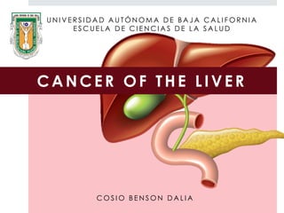
1. U N I V E R S I D A D A U T Ó N O M A D E B A J A C A L I F O R N I A
E S C U E L A D E C I E N C I A S D E L A S A L U D
CANCER OF THE L IVER
C O S I O B E N S O N D A L I A
2. I N T R O D U C T I O N
Primary cancer of the liver:
Represent de fith most common malignancy worldwide and the
Second most common cause of death from cancer
Dismal prognosis: 5-year survival rate below 10%
Effective treatment options
Lack of a true breakthrough in screening and early detection,
and the ontinued rising incidence of HCC globally.
DeVita et al. CANCER principles and practicesof oncology. 10 ed. 2015
HCC HBV HCV
3. E P I D E M I O L O G Y
ANUAL NUMER OF WORLDWIDE LIVER
CANCER CASES: 748,300
CLOSELY RESEMBLES THE NUMBER OF
DEATHS 695,900
LONG-TERM SURVIVAL RATE: 3-5%
HCC 2.3 MOST COMMON IN MEN THAN
IN WOMEN.
USA INCREASE IN THE INCIDENCE OF
HCC DURING THE PAST 25 YEARS.
DeVita et al. CANCER principles and practicesof oncology. 10 ed. 2015
>
4. E P I D E M I O L O G Y
I n c i d e n c e a n d m o r t a l i t y
M E N W O M E N
5O
9O
G L O B O C A N 2 0 1 2
( I A R C )
2O
5O
5. E P I D E M I O L O G Y
Rate incidence and mortality
7o
G L O B O C A N 2 0 1 2
4o
B O T H G E N D E R S
Incidence 7o
Mortality 4o
MÉXICO
6. HCC represent the 85% of primary cancer of the liver.
The incidence of HCC increase with age:
E P I D E M I O L O G Y
It is endemic in regions
where HBV is endemic
The varying geographic of HCC varies significantly
In Western countries HCV
infection and alcoholic
cirrhosis are major risk
factors for HCC
5ta y 6ta
decade
7. VIRAL HEPATITIS AND HEPATOCELULLAR CARCINOMA
E T I O L O G I C F A C T O R S
-Strong association between
chronic hepatitis B and
increased incidence og HCC-
8. The exact mechanism by wich HBV causes HCC is not known.
The effect of HBV on hepatic carcinogenesis is indirect
E T I O L O G Y F A C T O R S
• Inflamation
• Regeneration
• Fibrosis associated with chronic
hepatitis
CIRROSIS
9. Direct viral effect
E T I O L O G Y F A C T O R S
HBV DNA may become integrated
within the chromosomes of
infected hepatocytes, this
integration may occur in critical
location.
HBV
- Retinoic acid
receptor alpha gene
-Human cyclin A
gene
HCV RNA virus, cannot integrate
into hepatocyte DNA
Integration appears to
be random:
hepatitis B x gene (HBx)
transcripcional
activator associated
with growth control.
Antigene Bx (HBxAg) has been found to interact with p53, interfering with
its funtion as a tumor suppressor.
10. Comparison between HVB- HBC
E T I O L O G Y F A C T O R S
Factor VHB VHC
Middle ages 52-56 (20-80) 62
Increased incidence 400 millons, Asia and África
170 millons infected worldwide
Cause: 50% of HCC in Japón,
EU and Europa occidental
Cirrosis 25-50% >75%
Morphology Solitary lesions
Multifocal lesions , more
severe inflammation
HCC Progression 10-30 years >30 years
HCC growth (%) 4% per year 1-7% per year
Phan A. Gastric and esophageal cancer. En: The MD Anderson
Manual of Medical Oncology. 2da edición. Mc Graw-Hill: 2011.
11. E T I O L O G Y F A C T O R S
Major risk factors
Chronic HBV infection
Chronic HCV infection
Cirrhosis
Exhibition aflatoxin B1 in the diet
Others conditions
Α1 -antitrypsin deficiency
Hemochromatosis
Membranous obstruction of the inferior
vena cava
Storage disease type 1 glycogen and 2
Hereditary tyrosinemia type 1
Wilson disease
Hereditary conditions not
associated with liver disease
Ataxia-telangectasia
Hipercitrulinemia
Others factors
Smoking
Diabetes mellitus
Oral contraceptives
12. Chronic viral
hepatitis
77% of HCC cases worldwide are
attributable to viral hepatitis
Hepatitis B Virus
Hepatitis C Virus
It is the most important risk
factor for the
development HCC
HCV alone causes about 40 % of HCC cases in the
US
HCV carriers and chronic HBV usually take 10 to 20
years to develop cirrhosis and 30 to 40 years to
develop HCC
E T I O L O G Y F A C T O R S
13. Alcohol and cirrhosis
Excessive alcohol
consumption
Risk of
HCC
Autopsies of patients with
alcoholic cirrhosis have
reported more than 10% of
HCC undiagnosed
E T I O L O G Y F A C T O R S
Reactive oxygen species
Acetaldehyde (Protein /
DNA)
Cell damage
14. Nonalcoholic fatty liver
disease
Metabolic
syndrome
Risk of
HCC appers to
be less than chronic
hepatitis C
It is present in 30% of the
general adult of population.
90% in morbidly obese adults.
E T I O L O G Y F A C T O R S
Independent risk
factors
15. Aflatoxin B1
Food contaminated with
aflatoxin
A mycotoxin found in grains
Aspergillus flavus
Aspergillus parasiticus
It is an important factor for HCC in
parts of Asia and Africa
Inactivating mutation third base
of codon 249 of tumor suppressor
gene P53
E T I O L O G Y F A C T O R S
16. The use of oral contraceptives (OC) significantly increases the incidence
of benign hepatic adenomas.
There is some evidence that OC also increase the risk of developing HCC.
Others environmental factors
Smoking Occupational exposure to
arsenic or vinyl chloride
CHC
Risk of
developing liver
angiosarcoma
Oral contraceptives
E T I O L O G Y F A C T O R S
17. HCC develops in more than 45% of patients with
hemochromatosis.
Patients with Wilson's disease occasionally
develop HCC, but only with the presence of
cirrhosis
Others liver conditions
Malignant transformation occurred earlier
in patients with cirrhosis (for
hemochromatosis) , but this complication
has also been reported in patients without
cirrhosis
Carcinogenic
Generation of mutations by
reactive oxygen species
E T I O L O G Y F A C T O R S
Excessive
iron in tissues
18. C L I N I C A L P R E S E N T A T I O N
Most cases of HCC are incidentally identified by scanning programs in
high-risk individuals (people with cirrhosis, chronic hepatitis C ,
hemochromatosis , etc. )
It is common that patients are asymptomatic until the disease is well
advanced.
<30% of patients are candidates
for surgery or other direct therapy at the
time of presentation
Many patients have symptoms of
advanced liver cirrhosis and HCC
dysfunction
HCC commonly coexists with
cirrhosis
Abdominal pain in the
right upper quadrant
The most common initial
symptom is
19. #1 Right upper
quadrant pain or
epigastrium
#2 Anorexia or early
satiety with weight
loss
Symptom
Frequency
(%)
Sign Frequency (%)
Abdominal Pain 59-95 Hepatomegaly 54-98
Weight loss 34-71 Ascites 35-61
Weakness 22-53 Fever 11-54
Increased abdominal size 28-43 Splenomegaly 27-42
Unspecific symptoms 25-28 Jaundice 4-35
Jaundice 5-26 Hepatic vascular mumur 6-25
C L I N I C A L P R E S E N T A T I O N
20. HCC may present with various paraneoplastic symptoms by secretion of many hormones ,
their presentation is rare and unusual.
One of the most important is type B hypoglycemia , which occurs in less than 5% of patients,
manifested by severe hypoglycemia in the early course of the disease.
Type A hypoglycemia manifests as a lower grade Glucopenia occurs in the terminal stages
of HCC (and other malignant tumors of the liver).
Paraneoplastic Syndromes
It is believed to be a defect in
processing of the precursor growth
factor similar to insulin II ( pre- IGFII ) by
malignant hepatocytes
Disability liver ( and widely infiltrated
and commonly cirrhotic ) to meet the
demands of glucose
Type B hypoglycemia Type A hypoglycemia
A large tumor with rapid growth and for other
body tissues
C L I N I C A L P R E S E N T A T I O N
21. Policitemia (eritrocitosis)
It occurs in less than 10% of patients with HCC.
It seems to be caused by the synthesis of erythropoietin or a substance similar to
erythropoietin produced by malignant hepatocytes
Paraneoplastic syndrome
Other paraneoplastic syndrome associated with
HCC
carcinoid syndrome
hypercalcemia
hypertrophic osteoarthropathy
Neuropathy
Osteoporosis
polymyositis
porphyria
Sexual changes (gynecomastia , feminization )
hypertension
Tirocoxicosis
thrombophlebitis migrans
Watery diarrhea syndrome
C L I N I C A L P R E S E N T A T I O N
22. P A T H O L O G Y
Based on the pattern of growth, CHC can be classified into 4 general anatomical
types
Disfuse infiltrative Multifocal Encapsulated
Encapsulated
combined
pattern
Normal liver
parenchyma
23. P A T H O L O G Y
Difuse
inffiltrative
type
Growth with nodular ,
pseudolobular or invasive pattern
with poorly defined margins
Occur at the beginning
of liver cirrhosis , and
corresponds to 50 % of
cases in the US
Multifocal
type
It has multiple tumors of similar size
that makes it difficult to
determine whether the lesions are
intrahepatic or secondary to a
primary tumor mestástasis
Encapsulated
type
It presents an expansive growth ,
which compresses and distorts the
surrounding tissue
24. hepatocellular carcinoma
Neoplasia massive unifocal
encapsulated in which most or all
of the right hepatic lobe is
replaced.
It is well circumscribed and shows
numerous small foci bleeding .
P A T H O L O G Y
25. D I A G N O S I S
Complete history and physical examination
BHC electrolytes
Liver function tests ( LFTs)
Albumin
Prothrombin time
Serology for HCV and HBV
Tumor markers ( AFP )
Preferred
treatment
26. Clinic history
Investigate potential risk factors for HCC
Transfusions
Tattoos
Intravenous drug abuse
High-risk sexual practices
familial syndromes
Use of oral contraceptive or hormone replacement
Steroids androgens
Exposure to chemicalsndrógenos
D I A G N O S I S
27. TUMOR MARKERS
Tumor serum markers alone do not usually make the diagnosis of HCC but may be
useful in conjunction with imaging findings for diagnosis.
They can make us suspect a CHC , which would make us more sensitive imaging
studies to look for any abnormalities in the liver. The most useful marker is alpha-
fetoprotein ( AFP )
AFP is a
α1 - globulin
Normally present in high
concentrations in fetal serum but
only in small amounts thereafter .
The reappearance of high serum levels of
AFP strongly suggest the presence of CHC
(or hepatoblastoma )
Especially in populations where HCC is most
prevalent :
The vast majority of Chinese and black African
patients have an elevated AFP (> 10 ng / mL )
and approximately 75% have diagnostic levels (>
500 ng / mL ) .
D I A G N O S I S
28. levels greater than 500 ng / mL usually indicate hepatocellular carcinoma.
It can be seen sometimes in patients with active viral hepatitis.
Diagnosis can be considered a higher concentration of AFP 200 ng / mL in
patients with cirrhosis and liver injury greater than 2 cm in diameter .
The mean values of AFP in patients in regions with a high incidence of HCC is
60,000 to 80,000 ng / mL compared to approximately 8,000 ng / mL in regions
with low or intermediate incidence
D I A G N Ó S T I C O
AFP
D I A G N O S I S
29. Alpha Fetoprotein fucosylated
Des - γ - Carboxy Prothrombin glypican 3
Golgi protein 73
Hepatocyte growth
Factor Insulin-like growth factor 1
Transforming growth factor β -1
Mass spectrometry " flight time " desorption - ionization by laser surface (
SELDI-TOF- MS )
Others tumor markers
D I A G N Ó S T I C OD I A G N O S I S
30. The diagnosis of HCC generally requires evidence of a focal lesion images ,
although large infiltrative lesions may also be diagnostic.
A blood hyperenhancement , particularly in contrast images , observed by the
formation of new blood vessels to supply the tumor ( neoangiogenesis ).
D I A G N Ó S T I C O
Imaging studies
US
Detects most but not distinguish HCC
tumor lesions other forms of liver solid
TC
Multiphase or dynamic is the imaging
technique of choice for the diagnosis of
HCC
Laparoscopy
It can be used to detect peritoneal spread or
other extrahepatic lesions
Biopsies can be obtained under direct vision of
the lesion
D I A G N O S I S
32. S T A G I N G
Classification TNM
Tumor primario (T)
TX No se puede evaluar el tumor primario
T0 No hay evidencia de tumor primario
T1 Tumor solitario sin invasión vascular
T2 Tumor solitario con invasión vascular o múltiples tumores no > 5 cm
T3a Tumores múltiples > 5 cm
T3b Tumor único o múltiples tumores de cualquier tamaño involucrando a la
rama principal de la vena porta hepática
T4 Tumor(s)con invasión directa a órganos adyacentes, además de la
vesícula biliar y peritoneo visceral
Nódulos linfáticos regionales (N)
NX No se puede evaluar nódulos linfáticos regionales
N0 No hay nódulos linfáticos regionales
N1 Nódulos linfáticos regionales
Metástasis a distancia (M)
M0 No hay metástasis a distancia
M1 Hay metástasis a distancia
33. E S T A D I F I C A C I Ó N
AJCC classification system combined - TNM
AJCC: American Joint Committe on Cancer
Estadio
Grupo
TNM Descripción
Estadio I T1, N0, M0 Tumor único <2 cm sin invasión vascular
Estadio II T2, N0, M0
Tumor único <2 cm con invasión vascular o múltiples tumores < 2 cm
en un lóbulo o tumor único de 2 cm sin invasión vascular
Estadio IIIA T3, N0, M0
Tumores múltiples en un lóbulo ± invasión vascular o cualquier tumor
>5 cm o tumor único >2 cm con invasión vascular
Estadio IIIB T1-3, N1. M0 Nódulos linfáticos regionales positivos
Estadio IVA T4, N0, M0
Tumores múltiples en 2+ lóbulos o tumores que involucran la vena
portal hepática
Estadio IVB T1-4, N0-1, M1 Metástasis remota
Puntaje de
fibrosis
F0
F1
0-4 ninguna o moderada
5-6 fibrosis severa/cirrosis
S T A G I N G
34. E S T A D I F I C A C I Ó N
Pons F, Varela M, Llovet J. Staging systmes in
hepatocellular carcinoma. HPB (Oxford). 2005;
7(1): 35-41.
S T A G I N G
35. T R E A T M E N T
BCLC: Algoritmo for staging and treatment of HCC (2014)
Forner, A. et al. Treatment of intermediate-stage hepatocellular carcinoma
Nat. Rev. Clin. Oncol. doi:10.1038/nrclinonc.2014.122
36. T R E A T M E N T
BCLC: Algoritmo for staging and treatment of HCC ( 2014 )
(2009)
37. T R E A T M E N T
Child-Pugh Grading of Cirrhosis
38. B I B L I O G R A P H Y
GLOBOCAN 2012 (IARS). Organización Mundial de la Salud.
Bartlett D, Di Bisceglie A, Dawson L. Cancer of the Liver . En: De Vita, Hellman, Rosenberg.
Cancer, principles & practice of oncology. 10th edición. Philadelphia: Lippincot Williams;
2012. p. 1129-1156.
Phan A. Hepatobiliary malignancies. En: The MD Anderson Manual of Medical Oncology. 2da
ed. Mc Graw-Hill: 2011.
Ferlman M, Friedman L, Brandt L. Sleisengen and Fordtran’s- Garstrointestinal and liver disease:
pathophysiology/diagnosis/management. 9na ed. Philadelphia: Sounders Elservier: 2010.
Pons F, Varela M, Llovet J. Staging systmes in hepatocellular carcinoma. HPB (Oxford). 2005;
7(1): 35-41.