More Related Content
Similar to hemorragia postparto desde el sangrado vaginal (20)
hemorragia postparto desde el sangrado vaginal
- 1. The new engl and jour nal of medicine
n engl j med 384;17 nejm.org April 29, 2021 1635
Review Article
From the Division of Maternal-Fetal
Medicine, Department of Gynecology and
Obstetrics, Johns Hopkins University
School of Medicine, Baltimore. Address
reprint requests to Dr. Bienstock at the
Division of Maternal-Fetal Medicine, De-
partment of Gynecology and Obstetrics,
Johns Hopkins University School of Med-
icine, 600 N. Wolfe St., Baltimore, MD
21287, or at jbienst@jhmi.edu.
N Engl J Med 2021;384:1635-45.
DOI: 10.1056/NEJMra1513247
Copyright © 2021 Massachusetts Medical Society.
P
ostpartum hemorrhage continues to be the leading prevent-
able cause of maternal illness and death globally.1,2
Worldwide, postpartum
hemorrhage accounts for 8% of maternal deaths in developed regions of the
world and 20% of maternal deaths in developing regions.2
The United States has
one of the highest maternal mortality rates among developed countries, with ap-
proximately 11% of all maternal deaths associated with postpartum hemorrhage.3
During the period from 1993 through 2014, the rate of postpartum hemorrhage
(which was defined as blood loss >1000 ml after vaginal or cesarean delivery) re-
quiring a blood transfusion4
increased from approximately 8 cases per 10,000
deliveries to 40 per 10,000 deliveries in the United States.5
With the increasing prevalence of postpartum hemorrhage, well-designed cohort
studies and randomized clinical trials that evaluate interventions that are critical
for predicting, preventing, and managing postpartum hemorrhage remain a high
priority.6
However, as a result of challenges in the quantification of blood loss,
various definitions of postpartum hemorrhage, and differences in outcome report-
ing, data from relevant randomized clinical trials are difficult to interpret and
compare across studies.6
In addition, guidelines for preventing and managing post-
partum hemorrhage vary substantially among major national obstetrics and gyne-
cology organizations, including the American College of Obstetricians and Gyne-
cologists,7
the Society of Obstetricians and Gynaecologists of Canada, the French
College of Gynecologists and Obstetricians, the Royal College of Obstetricians and
Gynaecologists (United Kingdom), and the Royal Australian and New Zealand
College of Obstetricians and Gynaecologists.8
This review discusses the causes,
identification, management, prevention, and prediction of postpartum hemorrhage.
Definition, Causes, and Risk Factors
The normal rate of blood flow to the uterus at full term is approximately 600 ml
per minute, in contrast to approximately 60 ml per minute in the nonpregnant
state.9
The control of postpartum blood loss depends primarily on uterine contrac-
tions and, to a lesser degree, on activation of the coagulation cascade.
The traditional definition of postpartum hemorrhage is blood loss of more than
500 ml after a vaginal delivery or more than 1000 ml after a cesarean delivery.10
More recently, postpartum hemorrhage has been redefined as a cumulative blood
loss of 1000 ml or more or blood loss associated with signs or symptoms of hypo-
volemia, irrespective of the route of delivery.10
Typical clinical signs and symptoms
of hypovolemia (e.g., hypotension and tachycardia) due to postpartum hemorrhage
may not appear until blood loss exceeds 25% of total blood volume (>1500 ml during
late pregnancy).11
Postpartum hemorrhage is considered to be primary when it occurs within the
first 24 hours after delivery and secondary when it occurs between 24 hours and up
to 12 weeks after delivery.10,12
The causes of postpartum hemorrhage can be sum-
marized by the four “T’s”: tone (uterine atony), trauma (lacerations or uterine rupture),
Dan L. Longo, M.D., Editor
Postpartum Hemorrhage
Jessica L. Bienstock, M.D., M.P.H., Ahizechukwu C. Eke, M.D., Ph.D.,
and Nancy A. Hueppchen, M.D.
The New England Journal of Medicine
Downloaded from nejm.org by Damian Absalon Herrera Gonzales on May 11, 2021. For personal use only. No other uses without permission.
Copyright © 2021 Massachusetts Medical Society. All rights reserved.
- 2. n engl j med 384;17 nejm.org April 29, 2021
1636
The new engl and jour nal of medicine
tissue (retained placenta or clots), and thrombin
(clotting-factor deficiency).10
The most common
cause is uterine atony (accounting for approxi-
mately 70% of cases), followed by obstetrical lac-
erations (approximately 20%), retained placen-
tal tissue (approximately 10%), and clotting-factor
deficiencies (<1%).10
Postpartum hemorrhage can
lead to severe anemia requiring blood transfu-
sion, disseminated intravascular coagulopathy, hys-
terectomy, multisystem organ failure, and death.10
Postpartum hemorrhage due to uterine atony is
often preceded by chorioamnionitis, therapeutic
use of magnesium sulfate, prolonged labor or
precipitous delivery, labor induction or augmen-
tation, uterine fibroids, or uterine overdistention
as a result of multiple gestation, fetal macrosomia,
or polyhydramnios. Cesarean delivery is associat-
ed with a higher risk of postpartum hemorrhage
than vaginal delivery. Advanced maternal age and
extremes of parity (0 and >4) are additional risk
factors.
Other risk factors for postpartum hemorrhage
are closely linked to the type of hemorrhage that
develops. For example, obstetrical lacerations can
be caused by operative vaginal delivery, precipitous
delivery, or episiotomy, whereas retained placen-
tal tissue can be caused by placenta accreta
spectrum (PAS; a spectrum of abnormal placenta-
tion disorders, including placenta accreta, placenta
increta, and placenta percreta), which is associ-
ated with prior uterine surgery. Retained placen-
tal tissue can also be the result of incomplete
delivery of the placental tissue and membranes.
Maternal coagulopathy that leads to postpartum
hemorrhage can be a complication of severe pre-
eclampsia and eclampsia, HELLP (hemolysis, ele-
vated liver-enzyme level, and low platelet count)
syndrome, intrauterine fetal death, placental ab-
ruption, or a coagulation disorder that is acquired
(e.g., amniotic fluid embolism) or inherited.
Despite efforts to identify patients who are at
increased risk for postpartum hemorrhage, this
life-threatening complication often occurs in
women who have no identifiable risk factors.10
Therefore, vigilance is crucial after all deliveries.
Management of Postpartum
Hemorrhage
General Approach
Management of postpartum hemorrhage neces-
sitates a coordinated multidisciplinary approach,
which involves good communication, accurate
assessment of blood loss, monitoring of mater-
nal vital signs and symptoms, fluid replacement,
and arrest of the source of hemorrhage, all oc-
curring concurrently (Fig. 1).10,13
Assessment of
ongoing blood loss is a critical step in the man-
agement of postpartum hemorrhage. Blood loss
can be assessed on the basis of visual estimation
or weighing of materials, including blood and
amniotic fluid–soaked surgical sponges and
drapes.7
Although there is no strong evidence
that one method of assessing blood loss is better
than the other, quantification provides a more
accurate estimation of blood loss, as compared
with subjective assessment.7,14
Morbidity among
women with severe postpartum hemorrhage may
be reduced when quantitative estimation of blood
loss is used as a component of maternal safety
protocols,7
but this method has not consistently
been found to improve clinical outcomes. A re-
cent Cochrane review and meta-analysis showed
no evidence that quantitative estimation of blood
loss reduces the need for uterotonic agents, blood
transfusion, or volume expanders during post-
partum hemorrhage.15
Despite these limitations,
some obstetrics and gynecology societies7
favor
quantification of blood loss by weighing of blood-
soaked materials (lap sponges and pads), moni-
toring of fluid used during irrigation, and use of
underbuttock, graduated cylinder drapes during
postpartum hemorrhage. More recently, the use
of colorimetric techniques, which involve elec-
tronic artificial intelligence (e.g., smartphone
applications), to estimate blood loss in real time
seems encouraging.16
Once a woman is admitted for delivery, if
there is a high index of suspicion for the devel-
opment of postpartum hemorrhage (e.g., placenta
previa, PAS, or active vaginal bleeding), two large-
bore intravenous cannulas should be inserted, a
complete blood count should be obtained, and
a specimen should be sent to the blood bank.
The blood bank should be notified, and at least
2 units of blood should be typed and cross-
matched for the patient. Additional maternal
monitoring should be tailored to the cause and
degree of increased risk of postpartum hemor-
rhage and can include the use of continuous
pulse oximetry, assessment of urine output with
the use of an indwelling bladder catheter, con-
tinuous cardiac monitoring, assessment of coagu-
lation status (on the basis of prothrombin time,
fibrinogen level, and activated thromboplastin
time), and a comprehensive metabolic panel.10
If
The New England Journal of Medicine
Downloaded from nejm.org by Damian Absalon Herrera Gonzales on May 11, 2021. For personal use only. No other uses without permission.
Copyright © 2021 Massachusetts Medical Society. All rights reserved.
- 3. n engl j med 384;17 nejm.org April 29, 2021 1637
Postpartum Hemorrhage
the patient is at very high risk for postpartum
hemorrhage, central venous and arterial cathe-
ters should be placed. A heating–cooling circu-
lating water pad or a forced-air warming system
may be used to help mitigate the hypothermia
that is often associated with massive fluid resus-
citation and prolonged surgery. Although both
crystalloids and colloids can be used as intrave-
nous fluids,17
crystalloids are slightly favored
over colloids.18
Management of Retained Placental Tissue
Inspection of the placenta after delivery is im-
portant to rule out retained placental tissue or a
retained succenturiate lobe of the placenta (an
abnormality in the placental structure in which
one or more accessory lobes is connected to the
main part of the placenta by blood vessels).
When retained placental tissue is suspected,
evacuation with manual exploration or a banjo
(blunt) curette under ultrasonographic guidance
Figure 1. Postpartum Hemorrhage (PPH) Screening, Evaluation, and Management.
The abbreviation aPTT denotes activated partial-thromboplastin time, BMP basic metabolic panel, CBC complete blood count, FFP
fresh-frozen plasma, IV intravenous, PT prothrombin time, and T and S type and screen.
Estimated Blood Loss of 1000–1500 ml Estimated Blood Loss of >3000 ml
Estimated Blood Loss of >1500–3000 ml
During Pregnancy
Screen all women for risk factors for PPH
Identify women at risk for PPH
Optimize hemoglobin levels before delivery
Seek multidisciplinary consultation (e.g., for inherited hematologic disorders)
On Admission to Labor and Delivery
Assess patients for risk factors for PPH
Categorize patients into risk strata (low, medium, and high risk for PPH)
Create IV access with a large-bore cannula (2 IV cannulas for patients at increased risk)
Obtain baseline laboratory values (CBC, BMP, T and S, fibrinogen, PT, aPTT)
Notify anesthesia and other consultation services (e.g., hematology, gynecologic oncology)
Document pertinent contraindications to specific pharmacotherapies (e.g., asthma,
hypertension)
Call for help from nursing, additional obstetrical
providers
Notify anesthesia, blood bank, interventional
radiology suite, operating room
Place additional IV catheter
Deliver placenta, membranes, and any retained
products of conception
Place urinary catheter to monitor urine output
and assist in uterine contraction
Perform bimanual uterine massage and manual
uterine exploration and evacuate retained
tissue or clots
Administer IV fluid to replace blood loss
(crystalloids, colloids)
Administer pharmacotherapy (oxytocin,
methylergonovine, carboprost, misoprostol)
Examine and repair any genital tract lacerations
Monitor laboratory values (CBC, BMP, T and S,
fibrinogen, PT, aPTT, lactate)
Monitor for coagulation (thromboelastography
or rotational thromboelastometry)
Place arterial catheter, central venous catheters
Administer blood and blood products (especially
if patient is hemodynamically unstable)
Continue to administer pharmacotherapy to
maximum tolerated doses
Place uterine tamponade device (e.g., Bakri or
Rusch balloon tamponade)
Administer tranexamic acid
Continue transfusion of red cells, FFP, and
platelets according to local protocols
(e.g., 1:1:1 ratio)
Continue to monitor laboratory values (CBC,
BMP, T and S, fibrinogen, PT, aPTT, lactate)
Continue to monitor for coagulation (thrombo-
elastography or rotational thromboelastometry)
Consider readministration of tranexamic acid
Consider recombinant factor VIIa therapy
(in hemophilia A or B)
Consider uterine artery embolization (if patient
is stable) with interventional radiology
Perform laparotomy
Repair any uterine lacerations or uterine rupture
Place uterine compression sutures (e.g.,
B-Lynch, Hayman, Cho, Esike methods)
Perform stepwise suture ligation
Perform bilateral uterine artery ligation
(O’Leary sutures)
Perform bilateral utero-ovarian artery ligation
Perform internal iliac artery ligation
Perform hysterectomy (total or supracervical —
may be appropriate earlier in management,
particularly if future childbearing is not
desired)
Development of Postpartum Hemorrhage
The New England Journal of Medicine
Downloaded from nejm.org by Damian Absalon Herrera Gonzales on May 11, 2021. For personal use only. No other uses without permission.
Copyright © 2021 Massachusetts Medical Society. All rights reserved.
- 4. n engl j med 384;17 nejm.org April 29, 2021
1638
The new engl and jour nal of medicine
is recommended; the positive and negative pre-
dictive values of ultrasonography in detecting
retained placental tissue are approximately 58%
and 87%, respectively.19
Abnormal uterine bleed-
ing that warrants manual removal of the pla-
centa increases the likelihood that the bleeding
is due to a PAS disorder.20
Management of Genital Tract Lacerations
Careful inspection of the lower genital tract for
cervical, vaginal, perineal, or rectovaginal lac-
erations is important. Lacerations should be
promptly repaired with absorbable sutures. There
is insufficient evidence in support of routine
antibiotic prophylaxis after uterine evacuation
for postpartum hemorrhage or repair of a peri-
neal laceration.21
Management of Uterine Atony
Bimanual uterine massage is usually the first step
in the management of postpartum hemorrhage
due to uterine atony. Massage is performed in an
attempt to induce uterine contractions by stimu-
lating endogenous prostaglandins.10,22
Oxytocin
(administered intravenously or intramuscularly)
is the mainstay of treatment for controlling
postpartum hemorrhage due to uterine atony;
administration of oxytocin is usually begun simul-
taneously with uterine massage, if the agent has
not already been administered prophylactically.
The uterine response after the administration of
intravenous oxytocin is usually immediate (oxy-
tocin half-life, 1 to 6 minutes in plasma).23
Additional agents (e.g., methylergonovine
maleate, a semisynthetic ergot alkaloid) and
intramuscular prostaglandins (e.g., carboprost
tromethamine, a 15-methyl analogue of prosta-
glandin F2α
) can be used as second-line pharma-
cotherapy to control postpartum hemorrhage. A
Cochrane review and meta-analysis have ques-
tioned the usefulness of misoprostol, a prosta-
glandin E1
analogue.24,25
Although oxytocin causes
rhythmic contractions of the uterus, methylergo-
novine maleate stimulates uterine smooth mus-
cle and uterine vascular α1
-adrenergic receptors
in a sustained manner, causing vasoconstriction
and cessation of bleeding. Methylergonovine
maleate is often considered the next agent to be
administered after oxytocin.23
The indications
and contraindications for pharmacotherapy in
postpartum hemorrhage are listed in Table 1.
If pharmacotherapy fails in the management
of uterine atony, mechanical methods, including
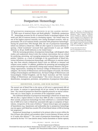
balloon tamponade (Fig. 2A) and uterine com-
pression sutures (Fig. 2B), can be lifesaving.
Balloon tamponade systems, such as the Bakri
balloon, first described in 2001,26
involve instill-
ing fluid (to a maximum volume of approxi-
mately 500 ml) into an intrauterine balloon,
with removal of the balloon up to 24 hours after
insertion; the tamponade effect of the filled bal-
loon is intended to stop or reduce intrauterine
bleeding.27
A 2020 systematic review and meta-
analysis concluded that uterine balloon tampon-
ade systems appear to be safe,28
with a success
rate of more than 85% in the management of
postpartum hemorrhage.
Uterine compression sutures, also known as
“brace sutures,” were first described in 1997 by
B-Lynch and colleagues and were shown to be
highly effective in controlling postpartum hem-
orrhage.29
Since 1997, several other compression
suture techniques have been described.30-36
Some
techniques involve sutures that enter the uterine
cavity34
and abut the anterior and posterior walls
of the uterus, with a potential to increase the
risk of uterine synechiae, but other techniques
do not.29,36
Several systematic reviews of case
series have shown a combined success rate of
more than 90% with the use of brace sutures in
managing postpartum hemorrhage.37,38
Uterine
necrosis and intrauterine synechiae are possible
complications of uterine compression proce-
dures.39,40
The frequency of successful pregnancy
after management of postpartum hemorrhage
with uterine compression sutures has ranged
from 11 to 75%.37
Uterine and vaginal packing has been used
successfully in cases of postpartum hemor-
rhage, but it is not routinely recommended be-
cause of the potential for intrauterine infection.41
Although a positive tamponade test (decreased
bleeding with bimanual uterine compression
involving a hand on the maternal abdomen to
compress the uterine fundus from above and a
hand in the vagina to compress from below) has
not been rigorously studied in postpartum hem-
orrhage trials, it is a reasonable pretest to con-
sider using before choosing uterine balloon place-
ment or compression sutures.42
In severe cases of postpartum hemorrhage,
when pharmacologic therapy, uterine compres-
sion or tamponade, and other conservative mea-
sures have failed to control bleeding, surgical
The New England Journal of Medicine
Downloaded from nejm.org by Damian Absalon Herrera Gonzales on May 11, 2021. For personal use only. No other uses without permission.
Copyright © 2021 Massachusetts Medical Society. All rights reserved.
- 5. n engl j med 384;17 nejm.org April 29, 2021 1639
Postpartum Hemorrhage
Table
1.
Medical
Therapy
for
Postpartum
Hemorrhage.*
Medication
Mechanism
of
Action
Route
of
Administration
and
Dose
Concerns
and
Contraindications
Adverse
Effects
First-line
therapy:
oxytocin
Stimulates
oxytocin
receptors
in
the
uterus
IV
route,
10–40
IU/500–1000
ml
of
lactated
Ringer’s
solution;
IM
or
IMM
route,
5–10
IU
for
up
to
4
doses
SIADH,
hypotension
Rapid
bolus
administration
may
cause
hyponatre-
mia,
hypotension,
tachy-
cardia,
and
arrhythmia
Second-line
therapy
Methylergonovine
maleate
(ergot
alkaloid)
Partial
agonist
or
antagonist
at
serotoninergic,
dopaminergic,
α
1
-adrenergic
receptors
in
the
uterus
IM
or
IMM
route,
0.2
mg
every
2–4
hr,
for
a
maximum
of
5
doses;
oral
route,
0.2
mg
every
6–8
hr
for
2–7
days
Hypertension,
cardiovascular
disease
(stroke,
Renaud’s
disease)
Elevated
blood
pressure,
nausea,
vomiting,
myo-
cardial
infarction
Carboprost
tromethamine
(PGF
2α
)
PGF
2α
agonist
in
uterine
myometrium
IM
or
IMM
route,
250
μg
every
15–90
min
for
a
maximum
of
8
doses
Asthma,
cardiovascular
disease,
hepatic
disease,
renal
disease
Nausea,
vomiting,
and
diarrhea
Adjunctive
agents
Tranexamic
acid
Diminishes
the
dissolution
of
hemostatic
fibrin
by
plasmin,
stabilizing
clot
in
uterine
vessels
IV
route,
1
g
(100
mg/ml)
over
a
10-min
period;
if
bleeding
persists
after
30
min
or
stops
and
restarts
within
24
hr
after
the
first
dose,
a
second
dose
may
be
administered
Contraindicated
if
known
hypersensitivity
to
tranexamic
acid,
thromboembolic
event
during
pregnancy,
history
of
hypercoagulopathy
Headache,
musculoskeletal
pain,
nausea,
diarrhea
Recombinant
factor
VIIa
Activates
clotting
cascade
by
cleaving
factor
IX
and
factor
X,
which
activates
these
fac-
tors
and
leads
to
activation
of
thrombin
and
fibrin
IV
route,
50–100
μg/kg
(single
dose)
Severe
anemia,
severe
thrombocytope-
nia,
hyperfibrinogenemia,
allergy
to
mouse,
hamster,
or
bovine
proteins
Thromboembolic
events,
cerebrovascular
infarcts,
myocardial
infarction
Treatment
of
uncertain
useful-
ness:
misoprostol
PGE
1
agonist
in
the
uterine
myometrium
Sublingual,
oral,
or
rectal
route
(sublingual
route
preferred),
600–1000
μg
in
single
dose;
repeat
doses
not
recommended
Sepsis,
allergy
to
misoprostol,
concurrent
anticoagulant
therapy,
cardiovascular
disease;
efficacy
is
disputed
Nausea,
vomiting,
fever,
diarrhea
*
I
M
denotes
intramuscular,
IMM
intramyometrial,
IV
intravascular,
PGE
1
prostaglandin
E
1
,
PGF
2α
15-methyl
prostaglandin
F
2α
,
and
SIADH
syndrome
of
inappropriate
antidiuretic
hor-
mone
secretion.
The New England Journal of Medicine
Downloaded from nejm.org by Damian Absalon Herrera Gonzales on May 11, 2021. For personal use only. No other uses without permission.
Copyright © 2021 Massachusetts Medical Society. All rights reserved.
- 6. n engl j med 384;17 nejm.org April 29, 2021
1640
The new engl and jour nal of medicine
A Bakri Balloon Tamponade
B Uterine Compression B-Lynch Suture
C
Ovarian
ligament
Fallopian
tube
Round
ligament
Ovarian
artery
Ligated ascending
uterine artery for
hemorrhage control
Ligature
Ovarian suspensory
ligament
Uterus
Vagina
Cervix
Ovary
Uterus
Ureter
Section
of uterine wall
showing suture path
Internal iliac
artery
Uterine
artery
Round
Round
Round
Round
Round
Suture
ligament
Inflated
intrauterine
balloon providing
compression and
hemorrhage
control
Suture providing
uterine compression
and hemorrhage
control
Uterine Artery Ligation
Syringe with sterile liquid used
for balloon inflation
5
0
6
0
1
2 3
4
5
5
5
6
1
2
3
4
4
5
6
Broad
ligament
Myometrium
Ligature
The New England Journal of Medicine
Downloaded from nejm.org by Damian Absalon Herrera Gonzales on May 11, 2021. For personal use only. No other uses without permission.
Copyright © 2021 Massachusetts Medical Society. All rights reserved.
- 7. n engl j med 384;17 nejm.org April 29, 2021 1641
Postpartum Hemorrhage
methods can be lifesaving. Bilateral uterine ar-
tery ligation (Fig. 2C) is an appropriate next step
at the time of laparotomy. Described by Waters
in 195243
and by O’Leary and O’Leary in 1966,44
this surgical technique involves suture ligation
of the uterine vessels on the lateral aspect of the
lower uterine segment. If bilateral uterine artery
ligation fails, the vessels of the utero-ovarian
pedicle can be suture-ligated in a stepwise fash-
ion (bilateral utero-ovarian artery ligation). Inter-
nal iliac artery ligation, initially described in
1964 by Burchell et al. for controlling postpar-
tum hemorrhage,45
is usually a suture ligation
procedure of last resort, with a 50 to 60% suc-
cess rate, but it has largely fallen out of favor
because of the extent of surgical dissection that
is necessary.45
Hysterectomy (total or supracervi-
cal) for the control of postpartum hemorrhage
can be a lifesaving procedure.10
Use of Blood Products
Although there are no strict criteria for initiating
blood transfusion in cases of postpartum hem-
orrhage, transfusion is typically begun when the
estimated blood loss exceeds 1500 ml or when
hemodynamic changes become apparent.10,46
If
the need arises, massive blood transfusion (de-
fined as infusion of ≥10 units of packed red cells
in a 24-hour period or ≥4 units of packed red
cells within 1 hour)47
is initiated. No data from
randomized clinical trials provide the ratio for
transfusing blood products in obstetrics10
; the
obstetrical protocols for transfusion of packed
red cells, fresh-frozen plasma, and platelets in a
ratio of 6:4:1, 4:4:1, or 1:1:1 were derived from
the trauma literature.48,49
Treatment goals are to
maintain the hemoglobin level at more than 8 g
per deciliter, the fibrinogen level at more than
2 g per liter, the platelet count at more than
50,000 per microliter, and the activated partial-
thromboplastin and prothrombin times at less
than 1.5 times the normal values, on the basis
of practical guidelines such as those established
by the British Committee for Standards in Hae-
matology.50
In an observational study, thrombo-
elastography or rotational thromboelastometry
was recommended for maintaining adequate
coagulation while managing severe postpartum
hemorrhage.51
Blood-product replacement ther-
apy for the management of postpartum hemor-
rhage, when to administer it, and dosage recom-
mendations are listed in Table 2.
Placenta Accreta Spectrum Disorders
The frequency of peripartum hysterectomy per-
formed for the management of postpartum
hemorrhage due to PAS disorders continues to
rise with increased cesarean delivery rates.52
There is insufficient evidence to determine the
optimal time of delivery; however, the American
College of Obstetricians and Gynecologists rec-
ommends planned cesarean delivery, with or
without hysterectomy, at 34 weeks to 35 weeks
6 days of gestation in cases of PAS disorders,
whereas the Royal College of Obstetricians and
Gynaecologists53
recommends delivery between
35 weeks and 36 weeks 6 days of gestation.
Cesarean hysterectomy in women with PAS
disorders is a complex procedure. When per-
formed by obstetricians and gynecologists with
expertise in complex pelvic surgery, working in
collaboration with other surgical specialties, such
as vascular surgery, interventional radiology,
urology, and hematology, cesarean hysterectomy
has the potential to reduce maternal morbidity
and mortality.54
Both ureters may be stented
before the procedure to facilitate their identifica-
tion and reduce the risk of injury during the
procedure, especially if extensive pelvic dissec-
tion is anticipated.55
In women thought to be at
very high risk for placenta percreta, placement of
balloon catheters in the internal iliac arteries
immediately before the procedure, with inflation
immediately after delivery of the fetus, may re-
duce intraoperative bleeding.55
Although defini-
tive management of PAS disorders involves im-
mediate hysterectomy with the placenta left in
situ, some experts recommend expectant man-
agement and delayed hysterectomy in selected
cases in order to minimize hemorrhage and the
need for massive blood transfusion.56
In planned
cases of cesarean hysterectomy, a midline verti-
cal incision should ideally be used, since it mini-
mizes dissection of tissue planes that may bleed
if coagulopathy develops and provides good visu-
alization of the abdomen, uterine pedicles, and
pelvis.
Figure 2 (facing page). Mechanical Methods for Managing
Uterine Atony.
Panel A shows balloon tamponade (with a Bakri balloon),
Panel B uterine compression sutures (B-Lynch sutures,
placed according to the numbers, from 1 to 6), and
Panel C uterine artery ligation.
The New England Journal of Medicine
Downloaded from nejm.org by Damian Absalon Herrera Gonzales on May 11, 2021. For personal use only. No other uses without permission.
Copyright © 2021 Massachusetts Medical Society. All rights reserved.
- 8. n engl j med 384;17 nejm.org April 29, 2021
1642
The new engl and jour nal of medicine
Management of Uterine Inversion
Uterine inversion, a protrusion of the uterus
through the vaginal orifice at the time of deliv-
ery, can cause postpartum hemorrhage and hypo-
tension, which may be disproportionate to blood
loss. The first step in management is immediate
manual replacement of the uterus (with the pla-
centa still in place). If this attempt is unsuccess-
ful, relaxing the uterus with tocolytic agents
(nitroglycerin, terbutaline, magnesium sulfate, or
halothane) is an appropriate next step.57
If replac-
ing the uterus is still unsuccessful, laparotomy
can be performed, followed by one of several
techniques: reduction of the uterine inversion
back into the abdomen by gentle upward trac-
tion with Allis clamps placed at both uterine
cornua (Huntington’s method)58
; posterior longi-
tudinal incision of the cervical ring, followed by
gentle upward traction of the uterus with Allis
clamps placed at both cornua (Haultain’s meth-
od)59
; or placement of a Silastic cup of a vacuum
extractor on the fundus from above and use of
negative pressure to restore the uterus back to
its normal position (Antonelli’s method).60
Once
the uterus is replaced, uterotonic agents are ad-
ministered to aid uterine contraction, and man-
ual extraction of the placenta may be performed.
Other Management Approaches
A nonpneumatic antishock garment has been
used in the treatment of hypovolemic shock from
postpartum hemorrhage. The garment is worn
to decrease blood flow in the aorta and increase
venous return from the inferior vena cava,61
making it an invaluable device in cases of hypo-
volemic shock to temporarily maintain blood
pressure while awaiting definitive management.62
If the patient’s condition is stable enough for the
patient to be transported to the radiology suite
and preservation of fertility is desired, uterine
artery embolization (often as a supplement to
intrauterine balloon tamponade) can be consid-
ered. The uterine artery embolization procedure
involves injection of gelatin or polyvinyl alcohol
particles into the uterine artery or the anterior
division of the internal iliac arteries through the
femoral arteries with the use of the Seldinger
technique under fluoroscopic and ultrasonograph-
ic guidance.63
Success rates in the control of
postpartum hemorrhage range from 75 to 100%,64
and pregnancy after uterine artery embolization
has been reported in 43 to 48% of women.65,66
Secondary Postpartum
Hemorrhage
Secondary postpartum hemorrhage accounts for
approximately 1 to 2% of cases.10
The causes
include uterine subinvolution, retained products
of conception, endomyometritis, uterine vascular
disorders such as arteriovenous malformations,
and coagulopathies such as von Willebrand dis-
ease.10
Management of secondary postpartum
hemorrhage is directed at correcting the sus-
pected cause of the hemorrhage.10
Complications of Postpartum
Hemorrhage
In the immediate postpartum period, complica-
tions of postpartum hemorrhage include hypo-
volemic shock from massive blood loss, dissemi-
nated intravascular coagulopathy, acute renal
failure, hepatic failure, and complications of
blood transfusion, including transfusion-related
acute lung injury, acute respiratory distress syn-
Table 2. Blood-Product Replacement Therapy for Postpartum Hemorrhage.
Blood Product Component Therapy Dose When to Administer
Packed red cells Red cells 1 Unit is 450 ml in volume and is
expected to increase the maternal
hemoglobin level by 1 g/dl
If hemoglobin 7 or 8 g/dl (depending on
local protocols and coexisting maternal
conditions)
Fresh-frozen plasma Plasma proteins, clotting
factors (except platelets),
fibrinogen, anticoagulants
(proteins C and S)
1 Unit is approximately 250 ml in
volume; a dose of 10–20 ml/kg
will increase clotting factors by
10–20%
After every 1, 4, or 6 units of packed red
cells (depending on local protocols) or
if prothrombin time is prolonged (INR
or aPTT 1.5 times the normal value)*
Platelet concentrate Platelets 1 Pack of pooled platelets If platelet count 75,000/μl or after every 1,
4, or 6 units of packed red cells
Cryoprecipitate Factor VIII, fibrinogen 2 Pools of cryoprecipitate If fibrinogen 1 or 2 g/liter
*
The abbreviation aPTT denotes activated partial-thromboplastin time, and INR international normalized ratio.
The New England Journal of Medicine
Downloaded from nejm.org by Damian Absalon Herrera Gonzales on May 11, 2021. For personal use only. No other uses without permission.
Copyright © 2021 Massachusetts Medical Society. All rights reserved.
- 9. n engl j med 384;17 nejm.org April 29, 2021 1643
Postpartum Hemorrhage
drome, transfusion-associated circulatory over-
load, and death.10,67
Late complications such as
Sheehan’s syndrome (pituitary necrosis and pan-
hypopituitarism) and infertility may also occur.10,67
It is critical to manage postpartum hemorrhage
promptly and adequately in order to minimize
the risk of these complications.
Prevention of Postpartum
Hemorrhage
Preventive measures for postpartum hemorrhage
should be undertaken when possible, ideally
beginning before conception, with identification
of women at high risk and interventions to in-
crease iron stores and hemoglobin levels when
necessary. Screening women during pregnancy
and labor for risk factors for postpartum hemor-
rhage can be useful in the preparation for deliv-
ery, including identifying an appropriate loca-
tion for delivery (Table 3). Blood typing and
screening are important for women at moderate
risk for postpartum hemorrhage, whereas those
at high risk should undergo blood typing and
cross-matching of at least 2 units of packed red
cells in anticipation of possible postpartum
hemorrhage.
Active management of the third stage of
labor, including prophylactic use of uterotonic
agents and controlled umbilical cord traction,
has been shown to reduce blood loss during this
stage68
and to reduce the risk of postpartum
hemorrhage by approximately 66%, as compared
with expectant management.68
However, con-
trolled umbilical cord traction has limited bene-
fits in cases of severe postpartum hemorrhage
and may lead to uterine inversion if the manage-
ment team is inexperienced.69
Another method,
early cord clamping, can result in decreased neo-
natal iron stores and an increased risk of infant
anemia70
and therefore is no longer recom-
mended as a component of active management
of the third stage. Uterine massage, although a
mainstay of management, has not been consis-
tently shown to be beneficial in the prevention
of postpartum hemorrhage.22
Prediction of Postpartum
Hemorrhage
Identification of patients at risk for postpartum
hemorrhage, early intervention with the use of
standardized protocols, and a coordinated, team-
based approach once hemorrhage occurs have
been shown to decrease maternal morbidity and
mortality.7,71
Prenatal diagnosis of PAS disorders
in women who have undergone prior uterine
surgery is invaluable for surgical planning.72
Al-
though obstetrical ultrasonography (color Dop-
pler or three-dimensional power Doppler) and
magnetic resonance imaging (MRI) have similar
diagnostic accuracy in detecting PAS disorders
(sensitivity of approximately 94% and specificity
of approximately 84%),73
MRI can complement
ultrasonography in assessing the depth of uter-
ine muscular and parametrial invasion.72
Catego-
rization of patients on admission to labor and
delivery into risk strata (low, medium, or high
risk) (Table 3) may identify up to 85% of preg-
nant women at risk for postpartum hemor-
rhage,74
with negative predictive values of more
than 98%.71,75
In a case–control study, Nyfløt et al.
showed that prolonged active labor (duration
12 hr) is associated with an increased risk of
severe postpartum hemorrhage.76
Risk stratifica-
tion can help the multidisciplinary team to be
alert to a patient’s risk and make informed
choices about the need for and availability of
intravenous access, uterotonic medications, blood
products, and additional personnel.
Table 3. Classification of Postpartum Hemorrhage Risk and Potential Need
for Transfusion.
Risk Level (Preparation
for Transfusion) Defining Factors
Low risk (having blood speci-
men available in case
blood products become
needed)
No previous uterine incision
Singleton pregnancy
≤4 Previous vaginal deliveries
No known bleeding disorders
No history of postpartum hemorrhage
Medium risk (blood typing
and screening)
Prior cesarean delivery or uterine surgery
Multiple gestation
4 Previous vaginal deliveries
Chorioamnionitis
History of postpartum hemorrhage
Large uterine fibroids
Fetal death
Estimated fetal weight 4000 g
Morbid obesity (body-mass index 40)*
High risk (blood typing and
cross-matching of at least
2 units of packed red
cells)
Placenta previa or low-lying placenta
Suspected placenta accreta spectrum
Hemoglobin 10 mg/dl and other risk factors
Platelet count 100,000/μl
Active bleeding on admission
Known coagulopathy
*
The body-mass index is the weight in kilograms divided by the square of the
height in meters.
The New England Journal of Medicine
Downloaded from nejm.org by Damian Absalon Herrera Gonzales on May 11, 2021. For personal use only. No other uses without permission.
Copyright © 2021 Massachusetts Medical Society. All rights reserved.
- 10. n engl j med 384;17 nejm.org April 29, 2021
1644
The new engl and jour nal of medicine
Conclusions
Postpartum hemorrhage remains a clinically
significant cause of maternal complications
and death; worldwide, one woman dies from
postpartum hemorrhage every 7 minutes. There-
fore, prompt identification of patients who are
at risk for postpartum hemorrhage, routine ac-
tive management of the third stage of labor,
expeditious assessment of blood loss, appropri-
ate patient monitoring, and management of
postpartum hemorrhage are important.77
No potential conflict of interest relevant to this article was
reported.
Disclosure forms provided by the authors are available with
the full text of this article at NEJM.org.
References
1. Making pregnancy safer. Geneva:
World Health Organization, 2007 (https://
www.who.int/maternal_child_adolescent/
documents/newsletter/mps_newsletter_
issue4.pdf).
2. Say L, Chou D, Gemmill A, et al.
Global causes of maternal death: a WHO
systematic analysis. Lancet Glob Health
2014;2(6):e323-e333.
3. Centers for Disease Control and Preven-
tion. Pregnancy Mortality Surveillance Sys-
tem. Trends in pregnancy-related mortality
in the United States: 1987-2017 (graph)
(https://www.cdc.gov/reproductivehealth/
maternal-mortality/pregnancy-mortality
-surveillance-system.htm).
4. Borovac-Pinheiro A, Pacagnella RC,
Cecatti JG, et al. Postpartum hemorrhage:
new insights for definition and diagnosis.
Am J Obstet Gynecol 2018;
219:
162-8.
5. Centers for Disease Control and Pre-
vention. Postpartum hemorrhage, 1993-2014
(graph) (https://www.cdc.gov/reproductive
health/maternalinfanthealth/pregnancy
-complications-data.htm#post).
6. Meher S. How should we diagnose
and assess the severity of PPH in clinical
trials? Best Pract Res Clin Obstet Gynae-
col 2019;61:41-54.
7. Quantitative blood loss in obstetric
hemorrhage: ACOG committee opinion,
number 794. Obstet Gynecol 2019;
134(6):
e150-e156.
8. Dahlke JD, Mendez-Figueroa H, Mag-
gio L, et al. Prevention and management
of postpartum hemorrhage: a comparison
of 4 national guidelines. Am J Obstet Gy-
necol 2015;213(1):76.e1-76.e10.
9. Dobiesz VA, Robinson DW. Trauma in
pregnancy. In:Walls RM, Hockberger R,
Gausche-Hill M, eds. Rosen’s emergency
medicine: concepts and clinical practice.
9th ed. Philadelphia:Elsevier, 2017:
2314-
22.
10. Committee on Practice Bulletins-
Obstetrics. Practice bulletin no. 183: post-
partum hemorrhage. Obstet Gynecol 2017;
130(4):e168-e186.
11. Pacagnella RC, Souza JP, Durocher J,
et al. A systematic review of the relation-
ship between blood loss and clinical signs.
PLoS One 2013;8(3):e57594.
12. Prevention and management of post-
partum haemorrhage: Green-top Guide-
line no. 52. BJOG 2017;
124(5):
e106-e149.
13. Cho HY, Na S, Kim MD, et al. Imple-
mentation of a multidisciplinary clinical
pathway for the management of postpar-
tum hemorrhage: a retrospective study.
Int J Qual Health Care 2015;
27:
459-65.
14. Bose P, Regan F, Paterson-Brown S.
Improving the accuracy of estimated
blood loss at obstetric haemorrhage using
clinical reconstructions. BJOG 2006;
113:
919-24.
15. Diaz V, Abalos E, Carroli G. Methods
for blood loss estimation after vaginal
birth. Cochrane Database Syst Rev 2018;
9:CD010980.
16. Gerdessen L, Meybohm P, Choorapoi-
kayil S, et al. Comparison of common
perioperative blood loss estimation tech-
niques: a systematic review and meta-
analysis. J Clin Monit Comput 2020 Au-
gust 19 (Epub ahead of print).
17. Alderson P, Schierhout G, Roberts I,
Bunn F. Colloids versus crystalloids for
fluid resuscitation in critically ill pa-
tients. Cochrane Database Syst Rev 2000;
2:
CD000567.
18. WHO guidelines for the management
of postpartum haemorrhage and retained
placenta. Geneva:World Health Organi-
zation, 2009.
19. Carlan SJ, Scott WT, Pollack R, Harris
K. Appearance of the uterus by ultra-
sound immediately after placental deliv-
ery with pathologic correlation. J Clin
Ultrasound 1997;25:301-8.
20. Porreco RP, Stettler RW. Surgical rem-
edies for postpartum hemorrhage. Clin
Obstet Gynecol 2010;53:182-95.
21. ACOG practice bulletin no. 120: use
of prophylactic antibiotics in labor and
delivery. Obstet Gynecol 2011;
117:
1472-
83.
22. Hofmeyr GJ, Abdel-Aleem H, Abdel-
Aleem MA. Uterine massage for prevent-
ing postpartum haemorrhage. Cochrane
Database Syst Rev 2013;
7:
CD006431.
23. Butwick AJ, Carvalho B, Blumenfeld
YJ, El-Sayed YY, Nelson LM, Bateman BT.
Second-line uterotonics and the risk of
hemorrhage-related morbidity. Am J Ob-
stet Gynecol 2015;212(5):642.e1-642.e7.
24. Tunçalp Ö, Hofmeyr GJ, Gülmezoglu
AM. Prostaglandins for preventing post-
partum haemorrhage. Cochrane Database
Syst Rev 2012;8:CD000494.
25. Gallos ID, Papadopoulou A, Man R,
et al. Uterotonic agents for preventing post-
partum haemorrhage: a network meta-
analysis. Cochrane Database Syst Rev
2018;12:CD011689.
26. Bakri YN, Amri A, Abdul Jabbar F.
Tamponade-balloon for obstetrical bleed-
ing. Int J Gynaecol Obstet 2001;
74:
139-42.
27. Vintejoux E, Ulrich D, Mousty E, et al.
Success factors for Bakri balloon usage
secondary to uterine atony: a retrospec-
tive, multicentre study. Aust N Z J Obstet
Gynaecol 2015;55:572-7.
28. Suarez S, Conde-Agudelo A, Borovac-
Pinheiro A, et al. Uterine balloon tam-
ponade for the treatment of postpartum
hemorrhage: a systematic review and meta-
analysis. Am J Obstet Gynecol 2020;
222:
(4)293.e1-293.e52.
29. B-Lynch C, Coker A, Lawal AH, Abu J,
Cowen MJ. The B-Lynch surgical tech-
nique for the control of massive postpar-
tum haemorrhage: an alternative to hys-
terectomy? Five cases reported. Br J Obstet
Gynaecol 1997;104:372-5.
30. Ouahba J, Piketty M, Huel C, et al.
Uterine compression sutures for postpar-
tum bleeding with uterine atony. BJOG
2007;114:619-22.
31. Zheng J, Xiong X, Ma Q, Zhang X, Li
M. A new uterine compression suture for
postpartum haemorrhage with atony. BJOG
2011;118:370-4.
32. Marasinghe JP, Condous G, Senevi-
ratne HR, Marasinghe U. Modified an-
chored B-Lynch uterine compression su-
ture for post partum bleeding with
uterine atony. Acta Obstet Gynecol Scand
2011;90:280-3.
33. Pereira A, Nunes F, Pedroso S, Saraiva
J, Retto H, Meirinho M. Compressive uter-
ine sutures to treat postpartum bleeding
secondary to uterine atony. Obstet Gyne-
col 2005;106:569-72.
34. Cho JH, Jun HS, Lee CN. Hemostatic
suturing technique for uterine bleeding
during cesarean delivery. Obstet Gynecol
2000;96:129-31.
35. Hayman RG, Arulkumaran S, Steer
PJ. Uterine compression sutures: surgical
management of postpartum hemorrhage.
Obstet Gynecol 2002;99:502-6.
36. Esike COU. A uterus-preserving treat-
ment for uncontrollable postpartum hem-
orrhage: Esike’s technique. Obstet Gyne-
col 2020;136:466-9.
37. Matsubara S, Yano H, Ohkuchi A, Ku-
wata T, Usui R, Suzuki M. Uterine com-
pression sutures for postpartum hemor-
The New England Journal of Medicine
Downloaded from nejm.org by Damian Absalon Herrera Gonzales on May 11, 2021. For personal use only. No other uses without permission.
Copyright © 2021 Massachusetts Medical Society. All rights reserved.
- 11. n engl j med 384;17 nejm.org April 29, 2021 1645
Postpartum Hemorrhage
rhage: an overview. Acta Obstet Gynecol
Scand 2013;92:378-85.
38. Mallappa Saroja CS, Nankani A, El-
Hamamy E. Uterine compression sutures,
an update: review of efficacy, safety and
complications of B-Lynch suture and other
uterine compression techniques for post-
partum haemorrhage. Arch Gynecol Ob-
stet 2010;281:581-8.
39. B-Lynch C. Partial ischemic necrosis of
the uterus following a uterine brace com-
pression suture. BJOG 2005;
112:
126-7.
40. Joshi VM, Shrivastava M. Partial ische
mic necrosis of the uterus following a
uterine brace compression suture. BJOG
2004;111:279-80.
41. Dildy GA III. Postpartum hemor-
rhage: new management options. Clin
Obstet Gynecol 2002;45:330-44.
42. Sebghati M, Chandraharan E. An up-
date on the risk factors for and manage-
ment of obstetric haemorrhage. Womens
Health (Lond) 2017;13:34-40.
43. Waters EG. Surgical management of
postpartum hemorrhage with particular
reference to ligation of uterine arteries.
Am J Obstet Gynecol 1952;
64:
1143-8.
44. O’Leary JL, O’Leary JA. Uterine artery
ligation in the control of intractable post-
partum hemorrhage. Am J Obstet Gyne-
col 1966;94:920-4.
45. Joshi VM, Otiv SR, Majumder R, Nikam
YA, Shrivastava M. Internal iliac artery li-
gation for arresting postpartum haemor-
rhage. BJOG 2007;114:356-61.
46. Shields LE, Wiesner S, Fulton J, Pelle-
treau B. Comprehensive maternal hemor-
rhage protocols reduce the use of blood
products and improve patient safety. Am J
Obstet Gynecol 2015;212:272-80.
47. Malone DL, Hess JR, Fingerhut A.
Massive transfusion practices around the
globe and a suggestion for a common
massive transfusion protocol. J Trauma
2006;60:Suppl:S91-S96.
48. Young PP, Cotton BA, Goodnough LT.
Massive transfusion protocols for patients
with substantial hemorrhage. Transfus
Med Rev 2011;25:293-303.
49. Holcomb JB, Tilley BC, Baraniuk S,
et al. Transfusion of plasma, platelets, and
red blood cells in a 1:1:1 vs a 1:1:2 ratio
and mortality in patients with severe
trauma: the PROPPR randomized clinical
trial. JAMA 2015;313:471-82.
50. Hunt BJ, Allard S, Keeling D, Norfolk
D, Stanworth SJ, Pendry K. A practical
guideline for the haematological manage-
ment of major haemorrhage. Br J Haema-
tol 2015;170:788-803.
51. Toffaletti JG, Buckner KA. Use of
earlier-reported rotational thromboelas-
tometry parameters to evaluate clotting
status, fibrinogen, and platelet activities
in postpartum hemorrhage compared to
surgery and intensive care patients. Anesth
Analg 2019;128:414-23.
52. Piñas Carrillo A, Chandraharan E. Pla-
centa accreta spectrum: risk factors, diag-
nosis and management with special refer-
ence to the Triple P procedure. Womens
Health (Lond) 2019;15:1745506519878081.
53. Jauniaux E, Alfirevic Z, Bhide AG, et al.
Placenta praevia and placenta accreta: di-
agnosis and management: Green-top
Guideline no. 27a. BJOG 2019;
126(1):
e1-
e48.
54. Oyelese Y, Scorza WE, Mastrolia R,
Smulian JC. Postpartum hemorrhage. Ob-
stet Gynecol Clin North Am 2007;
34:
421-
41.
55. Collins SL, Alemdar B, van Beekhui-
zen HJ, et al. Evidence-based guidelines
for the management of abnormally inva-
sive placenta: recommendations from the
International Society for Abnormally In-
vasive Placenta. Am J Obstet Gynecol
2019;220:511-26.
56. Zuckerwise LC, Craig AM, Newton
JM, Zhao S, Bennett KA, Crispens MA.
Outcomes following a clinical algorithm
allowing for delayed hysterectomy in the
management of severe placenta accreta
spectrum. Am J Obstet Gynecol 2020;
222(2):179.e1-179.e9.
57. Vijayaraghavan R, Sujatha Y. Acute
postpartum uterine inversion with haemor-
rhagic shock: laparoscopic reduction: a new
method of management? BJOG 2006;
113:
1100-2.
58. Huntington JL, Irving FC, Kellogg FS,
Mass B. Abdominal reposition in acute
inversion of the puerperal uterus. Am J
Obstet Gynecol 1928;15:34-8.
59. Haultain FW. The treatment of chron-
ic uterine inversion by abdominal hyster-
ectomy, with a successful case. Br Med J
(Clin Res Ed) 1901;
2:
974.
60. Antonelli E, Irion O, Tolck P, Morales
M. Subacute uterine inversion: descrip-
tion of a novel replacement technique us-
ing the obstetric ventouse. BJOG 2006;
113:846-7.
61. Lester F, Stenson A, Meyer C, Morris J,
Vargas J, Miller S. Impact of the non-
pneumatic antishock garment on pelvic
blood flow in healthy postpartum women.
Am J Obstet Gynecol 2011;
204(5):
409.e1-
409.e5.
62. Turan J, Ojengbede O, Fathalla M, et al.
Positive effects of the non-pneumatic anti-
shock garment on delays in accessing care
for postpartum and postabortion hemor-
rhage in Egypt and Nigeria. J Womens
Health (Larchmt) 2011;20:91-8.
63. Gipson MG, Smith MT. Endovascular
therapies for primary postpartum hemor-
rhage: techniques and outcomes. Semin
Intervent Radiol 2013;30:333-9.
64. Ruiz Labarta FJ, Pintado Recarte MP,
Alvarez Luque A, et al. Outcomes of pelvic
arterial embolization in the management
of postpartum haemorrhage: a case series
study and systematic review. Eur J Obstet
Gynecol Reprod Biol 2016;
206:
12-21.
65. McLucas B, Voorhees WD III, Elliott
S. Fertility after uterine artery emboliza-
tion: a review. Minim Invasive Ther Allied
Technol 2016;25:1-7.
66. Likis FE, Sathe NA, Morgans AK, et al.
Management of postpartum hemorrhage.
AHRQ comparative effectiveness reviews
no. 151. Rockville, MD:Agency for Health-
care Research and Quality, 2015.
67. Lu MC, Korst LM, Fridman M, Muth-
engi E, Gregory KD. Identifying women
most likely to benefit from prevention
strategies for postpartum hemorrhage.
J Perinatol 2009;29:422-7.
68. Begley CM, Gyte GM, Devane D, Mc-
Guire W, Weeks A, Biesty LM. Active ver-
sus expectant management for women in
the third stage of labour. Cochrane Data-
base Syst Rev 2019;
2:
CD007412.
69. Hofmeyr GJ, Mshweshwe NT, Gül-
mezoglu AM. Controlled cord traction for
the third stage of labour. Cochrane Data-
base Syst Rev 2015;
1:
CD008020.
70. McDonald SJ, Middleton P, Dowswell
T, Morris PS. Effect of timing of umbilical
cord clamping of term infants on mater-
nal and neonatal outcomes. Cochrane
Database Syst Rev 2013;
7:
CD004074.
71. Doomah YH, Xu S-Y, Cao L-X, Liang
S-L, Nuer-Allornuvor GF, Ying X-Y. A fuzzy
expert system to predict the risk of post-
partum hemorrhage. Acta Inform Med
2019;27:318-26.
72. Jauniaux E, Bhide A, Kennedy A,
Woodward P, Hubinont C, Collins S. FIGO
consensus guidelines on placenta accreta
spectrum disorders: prenatal diagnosis
and screening. Int J Gynaecol Obstet 2018;
140:274-80.
73. D’Antonio F, Iacovella C, Palacios-
Jaraquemada J, Bruno CH, Manzoli L,
Bhide A. Prenatal identification of inva-
sive placentation using magnetic reso-
nance imaging: systematic review and
meta-analysis. Ultrasound Obstet Gyne-
col 2014;44:8-16.
74. Dilla AJ, Waters JH, Yazer MH. Clini-
cal validation of risk stratification criteria
for peripartum hemorrhage. Obstet Gyne-
col 2013;122:120-6.
75. Hussain SA, Guarini CB, Blosser C,
Poole AT. Obstetric hemorrhage outcomes
by intrapartum risk stratification at a
single tertiary care center. Cureus 2019;
11(12):e6456.
76. Nyfløt LT, Stray-Pedersen B, Forsén L,
Vangen S. Duration of labor and the risk
of severe postpartum hemorrhage: a case-
control study. PLoS One 2017;
12(4):
e0175306.
77. Hu K, Lapinski MM, Mischler G, Allen
RH, Manbachi A, Chan Seay R. Improved
treatment of postpartum hemorrhage: de-
sign, development, and bench-top valida-
tion of a reusable intrauterine tamponade
device for low-resource settings. J Med
Devices 2020;14:014503 (https://doi.org/
10.1115/1.4045965).
Copyright © 2021 Massachusetts Medical Society.
The New England Journal of Medicine
Downloaded from nejm.org by Damian Absalon Herrera Gonzales on May 11, 2021. For personal use only. No other uses without permission.
Copyright © 2021 Massachusetts Medical Society. All rights reserved.