1. The study examines the gene and protein expression of HDAC3 and GDF11 in endometriosis.
2. Results show that HDAC3 is upregulated in endometriotic tissues and cell lines compared to normal endometrium. HDAC3 expression is also differentially regulated by steroid hormones in endometrial cells.
3. GDF11 is downregulated in endometriosis lesions and its protein expression is negatively correlated with HDAC3 protein expression in tissue samples. This suggests HDACs may regulate GDF11 expression in endometriosis.
Chromosomal Abnormalities in a Male Partner Who was a Candidate for Assisted ...Apollo Hospitals
Cytogenetic analysis was performed according to standard methods on cultured cells obtained from the patient with low sperm quality in respect of morphology and motility. The patients were interviewed about their histories and their reproductive problems, family background, and possible consanguinity.
Level of 17-β Estradiol in follicular fluid for patient undergoes IVF as corr...iosrjce
IOSR Journal of Dental and Medical Sciences is one of the speciality Journal in Dental Science and Medical Science published by International Organization of Scientific Research (IOSR). The Journal publishes papers of the highest scientific merit and widest possible scope work in all areas related to medical and dental science. The Journal welcome review articles, leading medical and clinical research articles, technical notes, case reports and others.
The two main sex hormones — estrogen and testosterone — have wide-ranging effects in the body. Produced primarily by the ovaries (estrogen) and testes (testosterone), these hormones affect not just your sexual function but also your bones, brain, and blood vessels.
Adrenocorticoids sex hormones & insulin.Lecturer notes-Dr.Jibachha SahDr. Jibachha Sah
Adrenocorticoids sex hormones & insulin is lecturer notes on pharmacology for B.V.Sc & A.H. sixth semester students of Nepal Polytechnic Institute,Chitwan,Nepal.This lecturer notes will beneficial other veterinary college students. I would like to comment and suggest from concerning specialist.Email:jibachhashah@gmail.com;mobile.00977-9845024121
Reproductive Genetics: Introduction to Genetic Testing Optionskanew396
GenomeSmart can help you navigate the different reproductive genetic testing options to allow you to make informed decisions for the health of yourself and your family.
American Urological Association (AUA) Lecture given at the American Society of Andrology (ASA) 40th annual conference, April 18 – 21, 2015 in Salt Lake City, Utah.
IOSR Journal of Pharmacy and Biological Sciences(IOSR-JPBS) is a double blind peer reviewed International Journal that provides rapid publication (within a month) of articles in all areas of Pharmacy and Biological Science. The journal welcomes publications of high quality papers on theoretical developments and practical applications in Pharmacy and Biological Science. Original research papers, state-of-the-art reviews, and high quality technical notes are invited for publications.
IOSR Journal of Pharmacy and Biological Sciences(IOSR-JPBS) is an open access international journal that provides rapid publication (within a month) of articles in all areas of Pharmacy and Biological Science. The journal welcomes publications of high quality papers on theoretical developments and practical applications in Pharmacy and Biological Science. Original research papers, state-of-the-art reviews, and high quality technical notes are invited for publications.
Identification of a Novel Mutation (p.G328W) in the NR5A1 Gene in a Boy with ...CrimsonPublishersGJEM
The gene NR5A1 (nuclear receptor subfamily 5 group A member 1, OMIM +184757) encodes for the Steroidogenic factor1 (SF1), a key regulator of the adrenal/gonadal development and function. Disorders/Differences of Sex Development (DSD) are defined as conditions where the chromosomal/anatomical, phenotypic sex is atypical. Heterozygous mutations of NR5A1 gene are associated, in 46, XY DSD patients, with a wide phenotypic spectrum of the external genitalia, with or without adrenal failure. Here we report a 46, XY newborn carrying a novel heterozygous NR5A1 mutation that presents with ambiguous genitalia without adrenal failure.
E-Mail [email protected]Clinical Genetic Aspects of Consangu.docxsagarlesley
E-Mail [email protected]
Clinical Genetic Aspects of Consanguinity
Hum Hered 2014;77:108–117
DOI: 10.1159/000360763
Consanguinity and Disorders of
Sex Development
Anu Bashamboo Ken McElreavey
Human Developmental Genetics, Institut Pasteur, Paris , France
Introduction
Disorders of sex development (DSD) are defined as
‘congenital conditions in which the development of chro-
mosomal, gonadal, or anatomical sex is atypical’ [1] . DSD
has recently been coined to encompass terms such as in-
tersex, pseudohermaphroditism, hermaphroditism and
sex-reversal, which can be confusing to clinicians, pa-
tients and parents. The definition provides a rational ba-
sis for designating each phenotype. 46,XY DSD includes
errors of testis determination or undermasculinization of
an XY male due to errors in either androgen synthesis or
androgen action. 46,XY gonadal dysgenesis is an error of
testis determination that is either complete (complete go-
nadal dysgenesis; CGD) or partial (partial gonadal dys-
genesis; PGD). 46,XY CGD is characterized by complete-
ly female external genitalia, well-developed Müllerian
structures and a gonad composed of a streak of fibrous
tissue. On the other hand, 46,XY PGD is characterized by
partial testis formation, usually a mixture of Wolffian and
Müllerian ducts and varying degrees of masculinization
of the external genitalia. 46,XX DSD includes overviril-
ization or masculinization of an XX individual due to an-
drogen excess, and the vast majority of cases of 46,XX
DSD are associated with congenital adrenal hyperplasia
(CAH) [2] . The much rarer 46,XX testicular DSD refers
to a male with testes and a normal male habitus, whereas
46,XX ovotesticular DSD refers to individuals that have
Key Words
Consanguinity · Disorders of sex development
Abstract
Disorders of sex development (DSD) are defined as ‘congen-
ital conditions in which the development of chromosomal,
gonadal, or anatomical sex is atypical’ [Lee et al., Pediatrics
2006; 118:e488–e500]. Studies conducted in Western coun-
tries, with low rates of consanguinity, show that truly am-
biguous genitalia have an estimated incidence of 1: 5,000
births. There are indications that the prevalence of DSD is
higher in endogamous communities. The incidence of am-
biguous genitalia in Saudi Arabia has been estimated at
1: 2,500 live births; whilst in Egypt, it has been estimated at
1: 3,000 live births. This may be due in part to an increase in
disorders of androgen synthesis associated with 46,XX DSD.
There is clearly a need for further studies to address the fre-
quency of DSD in communities with high levels of consan-
guinity. This will be challenging, as an accurate diagnosis is
difficult and expensive even in specialized centres. In devel-
oping countries with high levels of consanguinity, these lim-
itations can be compounded by cultural, social and religious
factors. Overall there is an indication that consan ...
Prenatal Diagnosis of Denys-Drash Syndromenavasreni
Denys-Drash syndrome consists of the triad of progressive nephropathy characterised by diffuse
mesangial sclerosis (DMS), genital abnormalities, and Wilms tumour. Nephropathy may range
from early onset proteinuria to nephrotic syndrome to end stage renal failure. Genital malformations affects both external and internal genitalia. It may range from penoscrotal hypospadias,
bilateral cryptorchidism to an enlarged clitoris with fused labia and a urogenital sinus to atrophic
uterus to streak ovaries or dysgenetic testes. The risk of developing Wilms’ tumor may be as high
as 50%.
Denys-Drash syndrome consists of the triad of progressive nephropathy characterised by diffuse mesangial sclerosis (DMS), genital abnormalities, and Wilms tumour. Nephropathy may range from early onset proteinuria to nephrotic syndrome to end stage renal failure. Genital malformations affects both external and internal genitalia...
Prenatal Diagnosis of Denys-Drash Syndromepateldrona
Denys-Drash syndrome consists of the triad of progressive nephropathy characterised by diffuse mesangial sclerosis (DMS), genital abnormalities, and Wilms tumour. Nephropathy may range from early onset proteinuria to nephrotic syndrome to end stage renal failure. Genital malformations affects both external and internal genitalia...
Prenatal Diagnosis of Denys-Drash Syndromeeshaasini
Denys-Drash syndrome consists of the triad of progressive nephropathy characterised by diffuse
mesangial sclerosis (DMS), genital abnormalities, and Wilms tumour. Nephropathy may range
from early onset proteinuria to nephrotic syndrome to end stage renal failure. Genital malformations affects both external and internal genitalia. It may range from penoscrotal hypospadias,
bilateral cryptorchidism to an enlarged clitoris with fused labia and a urogenital sinus to atrophic
uterus to streak ovaries or dysgenetic testes. The risk of developing Wilms’ tumor may be as high
as 50%.
Prenatal Diagnosis of Denys-Drash Syndromegeorgemarini
Denys-Drash syndrome consists of the triad of progressive nephropathy characterised by diffuse mesangial sclerosis (DMS), genital abnormalities, and Wilms tumour. Nephropathy may range from early onset proteinuria to nephrotic syndrome to end stage renal failure. Genital malformations affects both external and internal genitalia...
Prenatal Diagnosis of Denys-Drash SyndromeSarkarRenon
Denys-Drash syndrome consists of the triad of progressive nephropathy characterised by diffuse mesangial sclerosis (DMS), genital abnormalities, and Wilms tumour. Nephropathy may range from early onset proteinuria to nephrotic syndrome to end stage renal failure.
Prenatal Diagnosis of Denys-Drash SyndromeAnonIshanvi
Denys-Drash syndrome consists of the triad of progressive nephropathy characterised by diffuse mesangial sclerosis (DMS), genital abnormalities, and Wilms tumour. Nephropathy may range from early onset proteinuria to nephrotic syndrome to end stage renal failure. Genital malformations affects both external and internal genitalia...
Prenatal Diagnosis of Denys-Drash Syndromekomalicarol
Denys-Drash syndrome consists of the triad of progressive nephropathy characterised by diffuse
mesangial sclerosis (DMS), genital abnormalities, and Wilms tumour. Nephropathy may range
from early onset proteinuria to nephrotic syndrome to end stage renal failure. Genital malformations affects both external and internal genitalia. It may range from penoscrotal hypospadias,
bilateral cryptorchidism to an enlarged clitoris with fused labia and a urogenital sinus to atrophic
uterus to streak ovaries or dysgenetic testes. The risk of developing Wilms’ tumor may be as high
as 50%.
Management of poor ovarian reserve- Dr Parul KatiyarDr Parul Katiyar
Premature ovarian aging or ovarian failure is a major cause of female factor infertility. Dr Parul explains the mechanism of premature ovarian failure and discusses some simple measures to preserve/ regain fertility among women.
Differences in the endometrial transcript profile during the receptive period between women who were refractory to implantation and those who achieved pregnancy.
By Luis Alberto Velásquez Cumplido
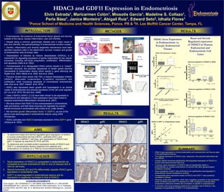
1. Elvin Estrada1, Maricarmen Colón1, Miosotis García1, Madeline S. Collazo1,
Perla Báez1, Janice Monteiro1, Abigail Ruiz1, Edward Seto2, Idhaliz Flores1
1Ponce School of Medicine and Health Sciences, Ponce, PR & 2H. Lee Moffitt Cancer Center, Tampa, FL
• Endometriosis, the presence of endometrial tissue (glands and stroma)
outside of the uterus, causes inflammation, pain and infertility.
• Despite being a common gynecologic disease and a major cause of
pain and infertility, the pathophysiology of endometriosis remains unclear.
• Genetic, inflammatory and recently epigenetic mechanisms have been
ascribed important roles in the attachment, survival, invasion and growth
of endometriotic cells at ectopic sites.
• Deacetylation of histones by histone deacetylases (HDACs), is a
mechanism for regulating expression of genes responsible for key cellular
processes including cell-cycle progression, proliferation, differentiation,
and apoptosis (Kelly et al, 2002).
• Overexpression of or dysregulated HDAC activity results in a “closed”
chromatin structure; the promoter sequences of certain genes become
inaccessible to transcription factors which results in gene silencing (de
Ruijter et al, 2003, Marks et al, 2005, Arts et al, 2003).
• Previous studies have shown that TSA, a histone deacetylase inhibitor
(HDACi), attenuates invasiveness, E-cadherin expression and
proliferation while increasing apoptosis of immortalized endometriotic
cells (Guo et al, 2007; Imesch et al, 2010).
• HDACi also decreased lesion growth and hyperalgesia in an animal
model of endometriosis and induced expression of the cell cycle regulator
p21 (Lu et al, 2010; Wu and Guo, 2008).
• Class I HDACs (HDAC1,2,3,8) have been shown to be expressed in
endometrium and alterations in their expression have been linked to
impaired endometrial differentiation (Krusche et al, 2007).
• We have shown that HDAC1/2 are overexpressed in endometriotic
cells and lesions, and that these enzymes are regulated by ovarian
steroid hormones in endometrial cells (Colón et al, under review).
• We identified growth differentiation factor 11 (GDF11), as one of the
genes most downregulated in endometriotic lesions using cDNA
microarrays.
• HDACi activates and HDAC3 represses expression of the GDF11 gene
(Zhang et al, 2004).
HDAC3 and GDF11 Expression in Endometriosis
ACKNOWLEDGEMENTS
Grant support : R01-HD050559 (I.F.); NIH-MBRS S06-GM08239 (I.F.); NIH-NIGMS
1R25GM082406 (M.C. and A.R.); U56CA126379 (TMA construction). Dr. A. Fazleabas
provided HESC and EEC cells. Dr. A. Mendoza from Southern Pathology Inc. archived
tissues.
RESULTS
1. To determine basal and steroid regulated gene expression of HDAC3
in human endometrial stromal (HESC), epithelial (EEC) and
endometriotic (Hs832cT) cell lines, and expression of HDAC3 and
gdf11 in tissues by real-time RT-PCR (qPCR).
2. To determine and correlate protein expression levels of HDAC3 and
GDF11 in endometriotic lesions obtained from patients and
endometrium of controls on a TMA by Immunohistochemistry (IHC).
INTRODUCTION
• HDAC3 was expressed in normal endometrial and endometriotic cell lines as seen
in the pathological pictures.
• The highest expression of HDAC3 was observed in the stromal endometrial cell
line (HESC).
• Ovarian steroid hormones significantly downregulated expression of HDAC3 in
human endometrial stromal cells only.
• In tissues, gene expression of HDAC3 was not significantly higher in
endometriosis vs. eutopic endometrium of women with other gynaecological
conditions (controls).
• Gene expression of gdf11 was confirmed to be significantly downregulated in
endometriotic lesions compared to controls.
• Unexpectedly gene expression levels of HDAC3 and GDF11 were positively
correlated in tissues.
• However, at the level of protein, there was a negative correlation in the expression
levels of HDAC3 and GDF11 in tissues on a TMA. Analysis is currently being
conducted to determine whether this correlation is specific to endometriosis.
• In summary, this study presents evidence for a possible role of HDACs in the
regulation of expression of GDF11 in endometriosis.
SUMMARY
METHODS
1. Gene expression of HDAC3 is upregulated in endometriotic as
compared to normal endometrium both in vivo (in tissues) and in
vitro (in cell lines).
2. Steroid hormones (E2 and P4) differentially regulate HDAC3 gene
expression in endometrial cells.
3. GDF11 is downregulated in endometriosis lesions and its
expression is negatively correlated to that of HDAC3.
AIMS
HYPOTHESES
RESULTS
RNeasy
kit (Qiagen)
HESC, EEC and Hs832cT
E2 +/- P4
24 hrs
TaqMan® Gene Expression
assays (ABI)
RNA
Endometriotic
Cells (Hs578cT)
Endometrial
Epithelial Cells
(EEC)
Endometrial
Stromal Cells (HESC)
Endometriosis Tissue Microarray
Immunohistochemistry
Archived
Human
Tissue
Samples
FFPE
Pathology
Confirma>on
TMA
construc>on
Substrate
Signal
HRP
QuickTime™anda
decompressor
areneededtoseethispicture.
Tissue Type Localization n
Proliferative
Endometrium
from Controls
(PE-Control)
Eutopic 14
Secretory
Endometrium
from Controls
(SE-Control)
Eutopic 38
Proliferative
Endometrium
from
Endometriosis
Patients
(PE-Endo)
Eutopic 22
Endometriosis
Ovary 29
Fallopian Tube 16
Peritoneum 34
Skin 4
Gastrointestinal
(GI)
7
Total 164
HDAC Gene Expression
in Endometriotic vs.
Eutopic Endometrial
Tissues
Basal and Steroid
Regulated expression
of HDAC3 in Human
Endometrial and
Endometriotic Cell
Lines