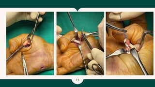
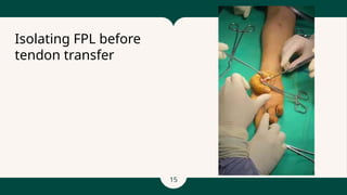
27 nov 2024
HAND UNIT CENSUS
2
53yo Chinese Lady
Right handed
Works in HR for a private firm
Underlying:
1. Migraine
2. Rheumatoid arthritis
3. Bronchial asthma
4. Thyroid nodule
History of right carpal tunnel release and right pronator release on 2/8/24
Madam TSM
3
- Numbness -right middle, ring and
little fingers - 6 months
- surgery on August 2024, numbness
over the right thumb and index
fingers improved
- Inability to flex right thumb at IP joint
- Inability to perform opposition of
thumb
- pain -medial aspect right elbow -
radiates ring and little fingers
- Pain over volar aspect of the proximal
forearm resolved
HISTORY
4
- scars over the volar aspect of proximal
forearm and at the wrist
- muscle wasting thenar eminence
- Reduced sensation right middle, ring and
little fingers
- Tinel’s sign positive over the right cubital
tunnel
- Limitation of flexion over IP joint of right
thumb
- Unable to perform opposition of thumb to
fingers
- Radial pulse palpable
- CRT all fingers < 2 secs
Physical examination
of the right upper
limb
5
X Rays
6
X rays
8
Diagnosis:
Right cubital tunnel syndrome
Surgery performed:
1. Right cubital tunnel release
2. Extensor Indicis Proprius (EIP)
to Flexor Pollicis Longus (FPL)
transfer
Operation done on 22
November 2024
9
2. EIP to FPL tendon transfer
Extensor indicis propius (EIP) tendon was
idenfied and cut at
level of extensor zone 5
Proximal stump of EIP was tunnel
subcutaneous from dorsal to
volar at hypothenar region and brought to
MCPJ of thumb
FPL tendon was identified and imbrication
was done
EIP tendon was sutured to Flexor pollicis
longus (FPL) tendon at
level of MCPJ using Pulvetaft technique
Radial digital nerve noted, close to
imbrication and tendon
transfer site, nerve was transferred more
radially
Post tendon transfer, position of thumb in
flexion at IP joint
Intraoperative
findings
1. Cubital tunnel release:
- Cubital tunnel decompression was done -
Osborne ligament + 2 heads of FCU was
release
- Ulnar nerve appears pale and constricted
at area of compression at Osborne
ligament +2 heads of FCU
- Ulnar nerve was decompressed from
proximal to distal up to 7cm below elbow
joint
- Post release, ulnar nerve vaso nervorum seen
10
Intraop pictures
11
Intraop pictures
12
13
14
Tunneling of EIP to FPL
15
Isolating FPL before
tendon transfer
16
- Tendon of a functioning muscle is detached
from its insertion & reattached to another
tendon or bone to replace the function of a
paralysed muscle or injured tendon.
- The transferred tendon remains attached to
its parent muscle with an intact
neurovascular pedicle.
- “Use of the power of a functioning muscle
unit to activate a non functioning nerve/
muscle/ tendon unit”
- Tendon transfers could be used to correct
instability, imbalance, lack of coordination
and restore function by redistributing
remaining muscular forces
What is a tendon
transfer?
17
â—Ź Paralysed muscle due to:
Nerve injury – peripheral or brachial plexus
High cervical quadriplegia (needs some input to brachial plexus/hand)
Neurological disease
Nerve repair with early transfer as internal splint
â—Ź Injured (ruptured or avulsed) tendon or muscle
Considerations
- Graft vs. transfer (adhesions more likely in graft – 2 anastomoses)
- Quality of available donors
- Length of time since injury
- Nature of tendon bed
â—Ź Balancing deformed hand e.g. cerebral palsy or rheumatoid arthritis
â—Ź Some congenital abnormalities
Indications of tendon transfers
18
1. Choose a tendon donor for transfer that minimizes functional loss. The donor tendon must be
expendable.
2. The muscle strength of the donor's tendon must be near normal as it will lose a function grade
with the transfer (a tendon with 5/5 strength will decrease to 4/5 strength after transfer).
3. Excursion of donor's tendon should be similar to an excursion of recipient's tendon, wrist
extension and flexion tendons have 33 mm excursion, finger extensors 50 mm of excursion, and
finger flexors 70 mm of excursion (Smith 3-5-7 rule). Utilizing the tenodesis effect of the wrist
can compensate for an additional 20 mm to 30 mm of finger tendon excursion.
4. The donor tendon should be routed in the direction of pull that is line with the recipient's
tendon.
5. A single tendon transfer should aim to restore one function.
6. Soft tissue adjacent to transfer site should be stable and pliable to allow for tendon gliding.
7. The full passive range of motion of the joint controlled by the transferred tendon should be
achieved before surgery.
8. Donor tendons should be in the same phase as recipient's tendons if possible (finger extensor
act in phase with wrist flexors and finger flexors act in phase with wrist extensors).
Principles of tendon transfers
20
Can adjust with pulley or tenodesis
effect
Smith 3-5-7 rule
3 cm excursion - wrist flexors, wrist
extensors
5 cm excursion - EDC, FPL, EPL
7 cm excursion - FDS, FDP
Appropriate
excursion
21
Examples of tendon
transfers
22
- Improper initial graft tensioning
- Repair site rupture or loosening
as a result of slit propagation or
knot failure
- Alternative to pulvertaft weave
techniques are such as spiral
linking and loop-tendon suture
can improve biomechanical
strength of repair but with trade-
off of increased bulk of the
repair.
- Adhesions requiring aggressive
physiotherapy or secondary
tenolysis
Complications of
tendon transfers
23
•Described by Toth 1986
1. Protective phase
•Begins at surgery and lasts 3 – 5 weeks
•Objectives:-
–Protective splinting
–Oedema control
–Mobilise uninvolved joints
2. Mobilisation phase
•Begins when tendon healing is adequate for activation (usually 3
– 5 weeks post op)
•Objectives
–Mobilise tendon transfer
–Immobilise soft tissue
–Continue immobilisation of uninvolved joints to prevent joint stiffness
from disuse
–Reinforce preoperative teaching and patient education
–Continue oedema control and protective splinting
–Begin home rehabilitation program
•Usually day time dynamic splinting with nightly static splinting
Basic Principles of Post Operative Rehabilitation
3. Intermediate phase
•Begins 5 – 8 weeks post operatively
•Gradually increases hand activity and passive range of motion
exercises
•Limited functional movements permitted
4. Resistive phase
•Beginning at 8 – 12 weeks
•Tendon junctions are strong enough to withstand increasing
resistance
•Therapeutic objective is to increase endurance and strength of
transferred muscles
•Work related simulated tasks are begun to patient tolerance
Thank You

hand census microsurgery 27 Nov 24.pptx

  • 1.
    27 nov 2024 HANDUNIT CENSUS
  • 2.
    2 53yo Chinese Lady Righthanded Works in HR for a private firm Underlying: 1. Migraine 2. Rheumatoid arthritis 3. Bronchial asthma 4. Thyroid nodule History of right carpal tunnel release and right pronator release on 2/8/24 Madam TSM
  • 3.
    3 - Numbness -rightmiddle, ring and little fingers - 6 months - surgery on August 2024, numbness over the right thumb and index fingers improved - Inability to flex right thumb at IP joint - Inability to perform opposition of thumb - pain -medial aspect right elbow - radiates ring and little fingers - Pain over volar aspect of the proximal forearm resolved HISTORY
  • 4.
    4 - scars overthe volar aspect of proximal forearm and at the wrist - muscle wasting thenar eminence - Reduced sensation right middle, ring and little fingers - Tinel’s sign positive over the right cubital tunnel - Limitation of flexion over IP joint of right thumb - Unable to perform opposition of thumb to fingers - Radial pulse palpable - CRT all fingers < 2 secs Physical examination of the right upper limb
  • 5.
  • 6.
  • 7.
    8 Diagnosis: Right cubital tunnelsyndrome Surgery performed: 1. Right cubital tunnel release 2. Extensor Indicis Proprius (EIP) to Flexor Pollicis Longus (FPL) transfer Operation done on 22 November 2024
  • 8.
    9 2. EIP toFPL tendon transfer Extensor indicis propius (EIP) tendon was idenfied and cut at level of extensor zone 5 Proximal stump of EIP was tunnel subcutaneous from dorsal to volar at hypothenar region and brought to MCPJ of thumb FPL tendon was identified and imbrication was done EIP tendon was sutured to Flexor pollicis longus (FPL) tendon at level of MCPJ using Pulvetaft technique Radial digital nerve noted, close to imbrication and tendon transfer site, nerve was transferred more radially Post tendon transfer, position of thumb in flexion at IP joint Intraoperative findings 1. Cubital tunnel release: - Cubital tunnel decompression was done - Osborne ligament + 2 heads of FCU was release - Ulnar nerve appears pale and constricted at area of compression at Osborne ligament +2 heads of FCU - Ulnar nerve was decompressed from proximal to distal up to 7cm below elbow joint - Post release, ulnar nerve vaso nervorum seen
  • 9.
  • 10.
  • 11.
  • 12.
  • 13.
  • 14.
  • 15.
    16 - Tendon ofa functioning muscle is detached from its insertion & reattached to another tendon or bone to replace the function of a paralysed muscle or injured tendon. - The transferred tendon remains attached to its parent muscle with an intact neurovascular pedicle. - “Use of the power of a functioning muscle unit to activate a non functioning nerve/ muscle/ tendon unit” - Tendon transfers could be used to correct instability, imbalance, lack of coordination and restore function by redistributing remaining muscular forces What is a tendon transfer?
  • 16.
    17 ● Paralysed muscledue to: Nerve injury – peripheral or brachial plexus High cervical quadriplegia (needs some input to brachial plexus/hand) Neurological disease Nerve repair with early transfer as internal splint ● Injured (ruptured or avulsed) tendon or muscle Considerations - Graft vs. transfer (adhesions more likely in graft – 2 anastomoses) - Quality of available donors - Length of time since injury - Nature of tendon bed ● Balancing deformed hand e.g. cerebral palsy or rheumatoid arthritis ● Some congenital abnormalities Indications of tendon transfers
  • 17.
    18 1. Choose atendon donor for transfer that minimizes functional loss. The donor tendon must be expendable. 2. The muscle strength of the donor's tendon must be near normal as it will lose a function grade with the transfer (a tendon with 5/5 strength will decrease to 4/5 strength after transfer). 3. Excursion of donor's tendon should be similar to an excursion of recipient's tendon, wrist extension and flexion tendons have 33 mm excursion, finger extensors 50 mm of excursion, and finger flexors 70 mm of excursion (Smith 3-5-7 rule). Utilizing the tenodesis effect of the wrist can compensate for an additional 20 mm to 30 mm of finger tendon excursion. 4. The donor tendon should be routed in the direction of pull that is line with the recipient's tendon. 5. A single tendon transfer should aim to restore one function. 6. Soft tissue adjacent to transfer site should be stable and pliable to allow for tendon gliding. 7. The full passive range of motion of the joint controlled by the transferred tendon should be achieved before surgery. 8. Donor tendons should be in the same phase as recipient's tendons if possible (finger extensor act in phase with wrist flexors and finger flexors act in phase with wrist extensors). Principles of tendon transfers
  • 18.
    20 Can adjust withpulley or tenodesis effect Smith 3-5-7 rule 3 cm excursion - wrist flexors, wrist extensors 5 cm excursion - EDC, FPL, EPL 7 cm excursion - FDS, FDP Appropriate excursion
  • 19.
  • 20.
    22 - Improper initialgraft tensioning - Repair site rupture or loosening as a result of slit propagation or knot failure - Alternative to pulvertaft weave techniques are such as spiral linking and loop-tendon suture can improve biomechanical strength of repair but with trade- off of increased bulk of the repair. - Adhesions requiring aggressive physiotherapy or secondary tenolysis Complications of tendon transfers
  • 21.
    23 •Described by Toth1986 1. Protective phase •Begins at surgery and lasts 3 – 5 weeks •Objectives:- –Protective splinting –Oedema control –Mobilise uninvolved joints 2. Mobilisation phase •Begins when tendon healing is adequate for activation (usually 3 – 5 weeks post op) •Objectives –Mobilise tendon transfer –Immobilise soft tissue –Continue immobilisation of uninvolved joints to prevent joint stiffness from disuse –Reinforce preoperative teaching and patient education –Continue oedema control and protective splinting –Begin home rehabilitation program •Usually day time dynamic splinting with nightly static splinting Basic Principles of Post Operative Rehabilitation 3. Intermediate phase •Begins 5 – 8 weeks post operatively •Gradually increases hand activity and passive range of motion exercises •Limited functional movements permitted 4. Resistive phase •Beginning at 8 – 12 weeks •Tendon junctions are strong enough to withstand increasing resistance •Therapeutic objective is to increase endurance and strength of transferred muscles •Work related simulated tasks are begun to patient tolerance
  • 22.