SlideShare a Scribd company logo
1 of 10
Download to read offline
Hyaluronic Acid-Binding Scaffold for Articular Cartilage Repair
Shimon A. Unterman, Ph.D.,1,2
Matthew Gibson, B.S.,1,2
Janice H. Lee, Ph.D.,1,2
Joshua Crist, M.S.,1,2
Thanissara Chansakul, B.S.,1,2
Elaine C. Yang, B.S.,1,2
and Jennifer H. Elisseeff, Ph.D.1,2
Hyaluronic acid (HA) is an extracellular matrix molecule with multiple physical and biological functions found
in many tissues, including cartilage. HA has been incorporated in a number of biomaterial and scaffold systems.
However, HA in the material may be difficult to control if it is not chemically modified and chemical modifi-
cation of HA may negatively impact biological function. In this study, we developed a poly(ethylene glycol)
hydrogel with noncovalent HA-binding capabilities and evaluated its ability to support cartilage formation
in vitro and in an articular defect model. Chondrogenic differentiation of mesenchymal stem cells encapsulated
in the HA-interactive scaffolds containing various amounts of exogenous HA was evaluated. The HA-binding
hydrogel without exogenous HA produced the best cartilage as determined by biochemical content (glysoca-
minoglycan and collagen), histology (Safranin O and type II collagen staining), and gene expression analysis for
aggrecan, type I collagen, type II collagen, and sox-9. This HA-binding formulation was then translated to an
osteochondral defect model in the rat knee. After 6 weeks, histological analysis demonstrated improved cartilage
tissue production in defects treated with the HA-interactive hydrogel compared to noninteractive control
scaffolds and untreated defects. In addition to the tissue repair in the defect space, the Safranin O staining in
cartilage tissue surrounding the defect was greater in treatment groups where the HA-binding scaffold was
applied. In sum, incorporation of a noncovalent HA-binding functionality into biomaterials provides an ability
to interact with local or exogenous HA, which can then impact tissue remodeling and ultimately new tissue
production.
Introduction
Hyaluronic acid (HA) is used extensively in tissue
engineering scaffolds due to its important structural
and signaling roles in a variety of tissues, including the joint.
It is a nonsulfated glysocaminoglycan (GAG) composed of
repeating disaccharide units of glucuronic acid and N-
acetylglucosamine. The carboxylate group of glucuronic acid
allows for relatively facile crosslinking and chemical modi-
fication of HA to form hydrogels or sponges, which has led
to its evaluation as a scaffold material for a variety of tis-
sues.1–7
However, the resultant HA-based scaffolds exhibit
little similarity with the natural structure and presentation of
HA found in the body. The bioactivity of HA is highly de-
pendent on the molecular weight of the polymer and its
associations with other proteins and extracellular matrix
(ECM) components, and it is unclear how crosslinked HA
scaffolds would affect cellular behavior compared to its
natural presentation. Furthermore, the covalent modification
of the HA backbone itself may significantly change its bio-
logical activity in unanticipated ways. A more natural, bio-
logically relevant presentation of the HA may yield greater
insight into the effects of HA-based scaffolds for tissue en-
gineering, and may better potentiate tissue repair.
Cartilage tissue engineering aims to develop an effective
therapy to repair articular cartilage lost due to trauma or
disease. Cartilage has poor endogenous repair capacity, and
currently available therapies are largely ineffective at pro-
ducing a robust, healthy repair tissue. Given the aging
population and increasing incidence of cartilage damage and
osteoarthritis, there is significant interest in cartilage repair
and restoring joint function. Biomaterials play an important
role in serving as a scaffold to direct tissue repair. Tissue
engineering scaffolds normally are composed of combina-
tions of biological and synthetic polymer systems. While
biological polymer systems often exhibit good bioactivity
and regeneration potential, they frequently are mechanically
weak, and difficult to control and purify. Attempts to
chemically modify biological polymers to increase scaffold
strength and control are often challenging and may cause a
1
Translational Tissue Engineering Center, Wilmer Eye Institute, Johns Hopkins University, Baltimore, Maryland.
2
Department of Biomedical Engineering, Johns Hopkins University, Baltimore, Maryland.
TISSUE ENGINEERING: Part A
Volume 18, Numbers 23 and 24, 2012
ª Mary Ann Liebert, Inc.
DOI: 10.1089/ten.tea.2011.0711
2497
loss of biological activity.8
In contrast, synthetic systems
boast a high degree of control over physical properties, but
exhibit little to no biological activity.9,10
Of recent interest is
the combination of synthetic materials with biologically ac-
tive molecules to form biosynthetic composite materials that
share the high degree of control found in synthetic materi-
als with the biological functionalities found in biological
polymers. These composite biomaterials include synthetic
polymers modified with bioactive proteins or peptides to
introduce specific biological functionalities such as cell
adhesion, growth factor activity, or cell-mediated degrada-
tion.11
Short, synthetic peptides are easy to synthesize, pu-
rify, and modify, yet still exhibit significant biological
activity. IKVAV, YRGDS, collagen mimetic peptides, and
matrix metalloproteinase-sensitive peptides are all examples
of peptides that have been incorporated into biomaterial
systems to introduce biological functionalities.11–14
More re-
cently, scaffolds that were engineered with fibronectin do-
mains designed to simultaneously interact with growth
factors as well as integrins were shown to enhance growth
factor-driven wound healing through local presentation of
growth factors to cells.15
These techniques allow for the
controlled introduction of specific biological functionalities
to a broader polymer system to tailor the cellular microen-
vironment for the desired task.
To achieve a more natural presentation of HA, we de-
signed an HA-interacting hydrogel scaffold that non-
covalently binds HA. Mummert et al. discovered an
HA-binding peptide (HABPep) through phage display that
specifically binds HA that was applied to inhibiting HA-
mediated leukocyte trafficking. Moreover, a fluorescent-
labeled derivative of HABPep can efficiently and specifically
label HA in tissues.16,17
In this study, we conjugated HAB-
Pep to a synthetic hydrogel scaffold based on poly(ethylene
glycol) diacrylate (PEGDA) and investigated the resulting
biomaterial’s ability to interact with HA using an in vitro
model system. This scaffold can interact with HA in the local
ECM environment, including cell-secreted HA and ex-
ogenously supplied HA. We hypothesized that this HA-
interacting hydrogel would improve chondrogenesis of bone
marrow-derived mesenchymal stem cells (MSCs) in an
in vitro culture system, since HA is a key molecule in carti-
lage matrix. To extend this to a clinically relevant model, we
implanted the HABPep-functionalized hydrogels in a rat
osteochondral defect model to determine their ability to
potentiate cartilage repair in vivo.
Materials and Methods
Synthesis of HA binding-hydrogels
HA-binding peptide (HABPep; sequence GAHWQF-
NALTVR) and sequence-scrambled HABPep controls
(sHABPep; WRHGFALTAVNQ) were synthesized using
standard Fmoc-mediated solid-phase peptide synthesis on a
Symphony Quartet peptide synthesizer (Protein Technolo-
gies). Following synthesis, peptides were cleaved using a so-
lution of trifluoroacetic acid, triisopropylsilane, and water in a
95:2.5:2.5 ratio. The crude product was purified using reverse-
phase high-performance liquid chromatography (HPLC, C18
Grace-Vydac column) on a water/acetonitrile gradient.
Purified peptides were frozen and lyophilized; identity of
purified peptides was confirmed using matrix-assisted laser-
desorption ionization time of flight (MALDI-TOF) mass
spectroscopy (Voyager DE-STR; Applied Biosystems).
Peptides were conjugated to acryl-PEG-N-hydroxysucci-
nimide (Acryl-PEG-NHS; 3.4 kD, Laysan Bio) as previously
described.11
Briefly, peptides were reacted with a 1.2-fold
molar excess of PEG in 50 mM sodium bicarbonate at pH 8.0
for 2 h at room temperature. The resultant acryl-PEG-peptides
were lyophilized and stored at -20°C.
HA-interacting scaffolds were prepared by dissolving 10%
(w/v) polyethylene glycol diacrylate (PEGDA; 3.4 kD, Sunbio)
with 2% (w/v) acryl-PEG-HABPep and 0.05% photoinitiator
(Irgacure 2959; Ciba) in phosphate-buffered saline (PBS; In-
vitrogen). Controls without HA-binding functionality were
substituted with either sequence-scrambled 2% acryl-PEG-
sHABPep or 2% PEG monoacrylate (PEGMA; 5 kD, Laysan
Bio in place of HABPep). Macromers were combined with HA
solutions as indicated (HA; 980 kD, Lifecore). Cylindrical
polypropylene molds (*5.5-mm diameter) were filled with
100mL of macromer solution. The solution was polymerized by
exposure to ultraviolet light at 365 nm (5 mW/cm2
) for 5 min.
Quantification of HA release from hydrogels
Release of HA from hydrogels was determined as a
function of HA loading and presence of HABPep. Control
sHABPep hydrogels were prepared with 0, 1, 5, 10, and
20 mg/mL HA. HABPep scaffolds were prepared with
5 mg/mL HA, based on preliminary studies indicating that
concentration had little nonspecific interactions between the
HA and the PEG network. Polymerized constructs were
immersed in PBS or 50 U/mL hyaluronidase solution (Sig-
ma), which were collected at various time points and assayed
for the presence of HA using a carbazole assay as previously
shown.18
Briefly, 3 mL sodium tetraborohydrate solution
(9.5 mg/mL in sulfuric acid, Sigma) was placed in test tubes
and cooled to 4°C. Glucuronic acid standards or samples
(0.5 mL) were carefully layered over the sodium tetra-
borohydrate. Tubes were then heated for 10 min in a boiling
water bath and cooled to room temperature. Carbazole so-
lution (0.1/mL, 12.5/mg in 9.9875/g of ethanol, Sigma) was
added to the tubes and shaken. The test tubes were heated in
a boiling water bath for 15 min and cooled to room tem-
perature. Absorption of the samples was measured at 530 nm
against water blanks and compared to a glucuronic acid
standard curve. All experiments were performed in triplicate.
Chondrogenic differentiation of goat MSCs
Goat bone marrow-derived MSCs were isolated and ex-
panded as previously described.19
After three or four pas-
sages, MSCs were trypsinized, centrifuged, and resuspended
in a macromer solution containing 10% PEGDA and 2% ac-
ryl-PEG-peptide or PEGMA as well as varying HA concen-
trations (0, 0.5, 2.5, 5 mg/mL). Cells were suspended at 20
million/mL and hydrogels polymerized in 100-mL cylindrical
molds as described above. Hydrogel constructs were trans-
ferred to 24-well plates containing the chondrogenic differ-
entiation medium containing 100 nM dexamethasone
(Sigma), 40 mg/L Proline (Sigma), 50 mg/L ascorbic acid-2-
phosphate (Sigma), 100 mg/L sodium pyruvate (Invitrogen),
50 mg/mL ITS Premix (insulin, transferring, selenous acid;
BD Biosciences), 1% penicillin/streptomycin (Invitrogen),
and 10 ng/mL transforming growth factor b (TGFb-1).
2498 UNTERMAN ET AL.
Constructs were cultured for up to 6 weeks, after which they
were evaluated on the basis of biochemical content, chon-
drogenic gene expression, and histological analysis.
Biochemical characterization
of in vitro chondrogenesis
Hydrogel constructs were harvested at time points up to 6
weeks for biochemical analysis as previously described.19
Constructs were weighed, lyophilized, and weighed again to
obtain a dry weight and a swelling ratio. Dried hydrogels
were homogenized with pellet pestles and digested over-
night in papain (Worthington Biochemical). DNA content was
assayed using Hoescht 33258 dye (Molecular Probes) and a
DynaQuant 200 fluorometer (Hoefer) against a calf thymus
DNA standard curve. Glycosaminoglycan (GAG) content was
assayed by measuring absorbance at 525nm with di-
methylmethylene blue dye against a standard curve using
chondrotin sulfate C (Sigma). A hydroxyproline assay was
used to determine collagen content by hydrolysis overnight in
hydrochloric acid followed by reaction with p-dimethylami-
nobenzaldehyde (Sigma) and chloramine T (Sigma). Absor-
bance was read on a spectrophotometer at 563 nm and
compared to hydroxyproline standards (Sigma). Biochemical
content was normalized to DNA content and dry weight to
account for variations in the construct size and cellularity. All
biochemical data had a sample size of 4.
Histological characterization of in vitro chondrogenesis
Hydrogel constructs were fixed in 4% paraformaldehyde
(Sigma) and stored in 70% ethanol. Constructs were dehy-
drated, embedded in paraffin, and sectioned into 5-mm sec-
tions using a microtome (Leica). Sections were stained with
Safranin O to assess GAG content. Immunohistochemistry
was performed using rabbit polyclonal antibodies against type
I and type II collagen followed by visualization with horse-
radish peroxidase using the Histostain SP kit (Invitrogen).
Images were captured using a Zeiss Axiovert microscope.
Real-time polymerase chain reaction analysis
of in vitro chondrogenesis
Constructs were homogenized with pellet pestles, and
RNA was isolated from three separate constructs using Trizol
(Invitrogen) following standard protocols. RNA concentra-
tions were obtained using a Nanodrop 2000 spectrophotom-
eter. One mg of RNA was reverse-transcribed to cDNA using
the Superscript First Strand Synthesis kit (Invitrogen). Real-
time polymerase chain reaction (PCR) was performed on the
cDNA using a Step One Plus system (Applied Biosystems) and
the SYBR Green master mix (Applied Biosystems) using
primers shown in Supplementary Table S1 (Supplementary
Data are available online at www.liebertpub.com/tea). Re-
lative expression levels compared to b-actin were determined
using the 2- DDCt
method. The reference condition chosen was
PEGMA scaffolds containing no encapsulated HA at 4 days;
all data were normalized to this condition.
In vivo osteochondral defect model
A rat osteochondral defect model was used to assess the
potential of HA binding hydrogels to effect in vivo repair. All
animal procedures were approved by the Johns Hopkins
Animal Care and Use Committee (protocol #RA08A450).
Male Sprague-Dawley rats (8 weeks) were anesthetized with
2%–3% Isoflurane using a tabletop anesthesia system (Vet-
Equip). Hind limbs prepared using standard aseptic tech-
niques, and an incision was made medial to the patellar
tendon. The patella was displaced laterally to expose the ar-
ticular surface of the femur. Round, 1-mm osteochondral de-
fects were made in the patellar groove of the femur
approximately 3 mm anterior to the ACL insertion point up to
a depth of 1 mm. Defect depth was designed to approximate
the depth used for microfracture procedures. Cartilage defect
size was standardized through the use of a constant diameter
drill bit to control defect diameter at 1 mm. Defect depth was
controlled by drilling to a previously marked point on the bit.
Following defect creation, macromer solutions containing
10% PEGDA, 2% acryl-PEG-HABPep (or sHABPep), and
0.05% photoinitiator in PBS were placed into the defect site.
Polymer solutions were photopolymerized by exposure to
ultraviolet light for 5 min (365 nm, 5 mW/cm2
, Acticure 4000).
During polymerization, hydrogels were partially mixed with
blood and bone marrow that were present during the defect
creation. Controls included scrambled peptide hydrogels and
untreated defects. Incisions were closed and animals were
allowed unrestricted movement for the duration of the study.
A sample size of 6 knees was used for each material condition
at each time point (4 days, 3 weeks, 6 weeks).
Histological evaluation of in vivo repair
At each time point, knees were dissected and excised.
Implant areas were grossly imaged using a Zeiss Axiovert
dissection microscope. Knees were decalcified and fixed for
approximately a week in a solution of 10% formalin and 10%
formic acid. Solution changes were performed every other
day, at which point solutions were qualitatively assayed for
calcium content using an oxalate precipitation test. Following
a negative test, samples were immerse in increasing concen-
trations of a sucrose solution (up to 20% w/v) as a cryopro-
tectant, taking care to give adequate time for full tissue
penetration. Then, samples were immersed in graded solu-
tions of 20% sucrose and optimal cutting temperature (OCT)
solution (Tissue-Tek), embedded in OCT, and frozen. Knees
were cryosectioned at -20°C using a cryostat microtome
(Leica) to section thicknesses of 7–10 mm. Sections were stained
with Safranin-O and immunostained with type II collagen to
visualize tissue morphology and repair.
Statistical analysis
Quantitative biochemical data were evaluated using
multifactor analysis of variance to determine the significance
of main factor effects to a significance level of 0.05. Multiple
comparisons of individual condition means were carried out
using the Tukey’s honestly significant difference test. Statis-
tical analysis was carried out in MATLAB (Mathworks).
Results
Incorporation of HA-binding elements into PEG
hydrogels increases HA retention
HA release from PEG hydrogels depended on the initial
HA loading dose. HA release from standard PEG hydrogels
(without the HA interaction) was determined as a function of
HA-INTERACTING SCAFFOLDS FOR CARTILAGE REPAIR 2499
initial HA loading. Various concentrations of HA (from 0–
20 mg/mL) were loaded into control hydrogels (with no
specific HA interaction) before polymerization, and the HA
release profiles were measured by the carbazole assay to
determine baseline HA release and any effects of nonspecific
entanglement of HA with the crosslinked PEG gels (Fig. 1A).
When high concentrations of HA were encapsulated in the
PEG hydrogels, minimal HA was released, likely due to en-
tanglement with the PEG network. When lower concentra-
tions of HA (1 and 5 mg/mL) were encapsulated into the PEG
hydrogels, the HA was quickly released. Percent HA release
was significantly dependent on both HA loading and time
( p < 0.05, Supplementary Fig. S1). Control hydrogels without
HA did not yield any detectable levels of uronic acid using the
carbazole assay. No evidence was found for covalent inter-
actions between HA and the PEG network. Based on other
studies with similar materials, it was assumed that high con-
centrations of HA can drive phase-separation processes that
may create small pockets of higher concentration HA sur-
rounded by pockets of higher concentration PEG, resulting in
different diffusion kinetics.20
Incorporation of HA-binding peptides into PEG hydrogels
modulated interactions, diffusion, and ultimately release of
HA. PEG hydrogels conjugated with HA-binding or scrambled
peptides were loaded with 5 mg/mL HA, the dose in which
fast release was observed, and incubated in the presence and
absence of hyaluronidase (Fig. 1B). The 5 mg/mL concentra-
tion of HA was selected for these release studies, since the HA
is normally quickly released from PEG hydrogels. HA-inter-
acting hydrogels released significantly less HA at equilibrium
( p <0.05) compared to hydrogels modified with scrambled
peptide controls. With the addition of hyaluronidase, HA re-
lease did exhibit a small, but significant increase compared to
incubation in PBS ( p <0.05, Supplementary Fig. S2).
Chondrogenic differentiation of MSCs in HA-binding
PEG hydrogels
Cartilage formation by MSCs improved in HA-interacting
scaffolds as evaluated by biochemical content, gene expres-
sion, and histological analysis. MSCs were encapsulated in
HA-interacting scaffolds containing varying concentrations
of HA and incubated in the chondrogenic medium for 6
weeks. The physical properties of these hydrogels varied
depending on initial HA content and changed over the
course of chondrogenic differentiation (Fig. 2A). At 4 days,
the swelling ratio of HA-interacting and control hydrogels
increased in an HA dose-dependent manner. However, as
tissue developed over the culture period, the water content
in the hydrogels varied. The swelling ratio depended sig-
nificantly ( p < 0.05) as a function of HA loading, and showed
significant interactions between HA loading and time as well
as hydrogel choice and time. Swelling did not directly de-
pend on time or hydrogel choice (Supplementary Fig. S3).
HA-binding hydrogels increased cartilage production as
determined by ECM production, cell number, and gene ex-
pression analysis. DNA content or cell number, in the control
PEG hydrogels, decreased in an HA dose-dependent manner
at all time points, but increased in HA-binding hydrogels
containing increasing levels of exogenous HA at 4 days and 3
weeks. However, at later time points (6 weeks), DNA levels
in the HA-interacting hydrogels decreased (Fig. 2B). Overall,
DNA content depended significantly ( p < 0.05) as a function
of time and hydrogel choice. While HA loading was not a
significant main effect, it demonstrated significant interac-
tions with both time and hydrogel choice (Supplementary
Fig. S4). GAG deposition in the hydrogels increased over
time in all conditions (Fig. 2C, E). However, GAG levels
decreased with higher exogenous HA loading in a dose-de-
pendent manner for both control and HA-interacting hy-
drogels. HA-binding hydrogels produced significantly
greater GAG levels than PEG and sHABPep controls, and
hydrogels without any exogenous HA produced the greatest
GAG matrix levels compared to all groups. GAG content
depended significantly on all main factor effects ( p < 0.05),
and HA loading demonstrated significant interactions with
both time and hydrogel choice (Supplementary Fig. S5).
Total collagen deposition, represented by hydroxyproline
content, in both HA-interacting and control hydrogels also
increased over 6 weeks of chondrogenic culture (Fig. 2D, F).
FIG. 1. Hyaluronic acid (HA)-interacting hydrogels increase retention of HA. (A) Release of HA from control noninteracting
hydrogels was assessed at various HA loadings to determine the effects of nonspecific interactions between HA and
poly(ethylene glycol) diacrylate. At steady state, HA loading concentrations of 1 mg/mL and 5 mg/mL were fully released
from the hydrogel. (B) Specific interactions between HA and HABPep-functionalized hydrogels were assessed at 5 mg/mL
HA loading. HABPep was shown to significantly decrease HA release at steady state (*denotes significance between HABPep
and scrambled controls at each time point, p < 0.05). Addition of hyaluronidase increased the kinetics of release, but not the
equilibrium behavior. HABPep, HA-binding peptide.
2500 UNTERMAN ET AL.
Hydroxyproline content depended significantly on hydrogel
choice and time ( p < 0.05), and HA loading interacted sig-
nificantly with time (Supplementary Fig. S6). Overall, the
ECM analysis in the hydrogels suggests that the HA-inter-
acting hydrogels with little to no exogenous HA loading
produced the greatest levels of new cartilage production.
Gene expression analysis of cartilage-related markers
supported the chondrogenic differentiation of MSCs in the
hydrogels (Fig. 3). Expression of aggrecan, an important
protein in GAG structure and assembly, was largely un-
changed at 4 days between HA-interacting and control
hydrogels, though decreased with increasing HA loading.
At 3 weeks, HA-interacting hydrogels demonstrated dra-
matically higher aggrecan expression than control hydro-
gels, peaking at 2.5 mg/mL exogenous HA loading.
However, at 6 weeks, the control hydrogels expressed
higher levels of aggrecan compared to HA-binding hydro-
gels, with exogenous HA producing a dose-dependent de-
crease in expression. In the case of type II collagen, no
significant differences were observed between HA-binding
hydrogels and controls at 4 days and 3 weeks, while in-
termediate HA loading of HA-binding hydrogels exhibited
a significant upregulation at 6 weeks. Levels of type I
collagen expression were also upregulated in HA-binding
hydrogels, but to a much lesser degree than the upregula-
tion of type II collagen and aggrecan.
FIG. 2. HA-interacting hydrogels increased cartilage production by mesenchymal stem cells (MSCs). (A) Physical prop-
erties of PEG, HA-interacting (HAB), and scrambled peptide control (sHAB) hydrogels containing encapsulated MSCs varied
with initial HA loading and culture time. Swelling was significantly dependent on HA loading ( p < 0.05, see Fig. S3). (B) Cell
number, as measured by DNA content, was initially highly dependent on scaffold type and HA loading, but differences
decreased as scaffolds matured at 6 weeks. DNA was significantly dependent on hydrogel type and time, but not HA loading
( p < 0.05, Fig. S4). (C) Glycosaminoglycan (GAG) content, normalized to DNA, increased with time for all scaffolds, with
strong HA dose dependence at later weeks. GAG levels were significantly dependent on hydrogel type, HA loading, and
time ( p < 0.05). (D) Overall collagen production, as measured by hydroxyproline content normalized to DNA, increased over
time for all scaffolds, but showed no specific trend across HA concentrations. Collagen content was significantly dependent
on hydrogel type and time, but not HA loading ( p < 0.05). GAG content (E) and collagen content (F) were plotted for
representative HAB and PEG conditions over time. PEG, poly(ethylene glycol).
HA-INTERACTING SCAFFOLDS FOR CARTILAGE REPAIR 2501
Histological analysis supports the biochemical results that
the HA-interactive scaffolds produce greater levels of carti-
lage tissue components after 6 weeks. Safranin O staining of
harvested constructs (Fig. 4) exhibited a substantial increase
in GAG deposition in HA-interacting hydrogels at 3 weeks
compared to controls. In addition to the concentrated GAG
staining in the pericellular region, HA-binding hydrogels
contained higher staining in the intercellular regions of the
hydrogel material, suggesting the scaffold has retained cell-
secreted proteoglycans by binding to the HA core. The
differences in staining were less pronounced at 6 weeks be-
tween the groups, though HA-interactive scaffolds still had
more intense staining. Safranin O staining was also a func-
tion of initial HA loading, with more intense staining ob-
served for lower loading for both control and HA-binding
scaffolds, similar to the quantified ECM results. Type II
collagen immunostaining of control hydrogels at 3 weeks
was slightly more intense than HA-interactive scaffolds.
However, after 6 weeks, HA-binding PEG hydrogels pro-
duced significantly greater Type II collagen staining com-
pared to PEG controls.
Repair of osteochondral defects in vivo with hydrogels
Implantation of HA-interactive PEG hydrogels increased
cartilage tissue production in osteochondral defects created
on the rat femoral condyle compared to control hydrogels
and untreated defects. Acellular scaffolds were implanted in
the defects to avoid the challenge of delivering exogenous
cells. The implanted scaffolds were able to integrate with the
surrounding tissue, and gross images of harvested knees
after 4 days following implantation demonstrated that hy-
drogels remained in the defects and achieved good material-
tissue integration (Supplementary Fig. S7).
Implantation of HA-interactive hydrogels in osteochon-
dral defects resulted in a more robust cartilage tissue repair
compared to control hydrogels and untreated defects (Fig. 5).
After 4 days, discrete hydrogel material was clearly visible in
FIG. 3. Gene expression demonstrates chondrogenic differentiation of MSCs in HA-interacting hydrogels. Sox-9, aggrecan,
type II collagen, and type I collagen expression were analyzed for all hydrogel conditions. All expression levels were normalized
to individual b-actin levels and observed expression for PEG scaffolds containing no HA at 4 days. HA-interacting hydrogels
increased aggrecan production at 3 weeks and type II collagen production at 6 weeks.
2502 UNTERMAN ET AL.
the HA-interactive and scrambled peptide control condi-
tions, while the untreated defects were filled with a clot,
cells, and tissue debris. Both HA-binding and control hy-
drogels appeared to be well-integrated into the surrounding
tissue with cell infiltration present at the margins of the
implants. Scrambled peptide controls exhibited a signifi-
cantly stronger tissue response with more cells at the implant
edge. After 3 weeks, the implant material was still evident
for both HA-binding and control hydrogels, though the
surrounding tissue response had mostly subsided and the
underlying subchondral bone was undergoing repair.
Although there was as yet no clear, differentiated carti-
lage-like repair tissue visible, there were some early signs of
repair at the margins of the defect that did not stain positive
FIG. 4. HA-interactive
scaffolds accumulate more
cartilage matrix components
at 6 weeks. Safranin O
staining of HA-interactive
scaffolds and PEG scaffolds
across 6 weeks demonstrates
increased GAG deposition
in HA-interactive scaffolds.
Staining is highest for
low HA loadings.
Immunohistochemical
staining for Type II collagen
indicated initially higher
staining for PEG controls at
3 weeks, but substantially
higher staining for HA-
interactive scaffolds at 6
weeks. Bar = 100 mm.
FIG. 5. HA-interactive scaffolds assist in the repair of rat osteochondral defects in vivo. Safranin O staining of osteochondral
defects in representative rat knees demonstrated that HA-interactive hydrogels produced more GAG-positive repair tissue
than scrambled peptide controls (sHABPep) or untreated defects. In addition, tissue adjacent to the defects treated with HA-
interactive scaffolds contained more GAGs than controls. Representative Safranin O sections are shown for two separate
animals at 3 and 6 weeks postimplantation for comparison. Type II collagen immunostaining did not show substantial
differences in collagen content. Bar = 200 mm.
HA-INTERACTING SCAFFOLDS FOR CARTILAGE REPAIR 2503
for Safranin O. Untreated defect controls exhibited a smaller
defect depth, but no visible Safranin O-positive cartilage re-
pair. At 6 weeks, implant material was entirely absent
and replaced with varying levels of repair tissue. Neither
HA-binding nor scrambled peptide control hydrogels caused
a complete regeneration of healthy cartilage at this time
point, but HA binding hydrogels produced a greater volume
of repair tissue with stronger Safranin O staining compared
to controls, though still weaker than healthy tissue. Un-
treated defects showed a well-integrated repair tissue with
minimal Safranin O staining, indicative of the expected fi-
brocartilagenous repair.
The presence of the HA-interactive hydrogels in the os-
teochondral defects also had an impact on the ECM of the
cartilage surrounding the defect. At 3 weeks, all knees ex-
hibited reduced Safranin O staining for GAGs on articular
cartilage surrounding the defect and in the joint space com-
pared to day 4 staining and untreated controls. At 6 weeks,
Safranin O staining of cartilage outside the defect area
was further reduced, suggesting the presence of a discrete
osteochondral defect also caused degenerative joint changes.
However, joints containing defects treated with HA-inter-
active scaffolds exhibited greater proteoglycan staining, in-
dicating reduced cartilage degeneration. Overall, these results
indicate that HA-binding hydrogels produced an improve-
ment in the repair of osteochondral defects and the mainte-
nance of cartilage tissue surrounding the defect.
Discussion
The goal of this research was to develop a novel HA-
binding hydrogel that would interact noncovalently with
HA. While the HA-interacting scaffold was able to modulate
the release of HA doped into the hydrogel, the potential
scaffold interaction with cell-secreted or local ECM-derived
HA may also influence tissue remodeling and regeneration.
Noncovalent binding of a critical matrix building block may
allow a more efficient matrix assembly when required during
the tissue development process. Since HA is a critical matrix
component or the building block of the ECM in many tissues,
providing a scaffold that can reversibly bind the molecule
could have widespread application in regeneration.
The role of HA in the development of cartilage has been
well studied, but its application to tissue engineering has
yielded varying results. In development, HA plays a critical
role during mesenchymal condensation that leads to carti-
lage formation. Early in this process, the limb mesenchyme is
composed of dispersed cells throughout an ECM that con-
tains significant quantities of HA. Upon initiation of con-
densation, mesenchymal cells aggregate, hyaluronidases are
upregulated, and HA concentrations drop. It is believed that
HA helps mediate cell aggregation as an intercell bridge
through multivalent binding to HA receptors, notably CD44.
High concentrations of HA can saturate the cell receptors
without the attendant bridging effects, and thus inhibit or
slow mesenchymal condensation. After condensation, HA
synthesis is again upregulated as the cells differentiate to-
ward a functional cartilage tissue.21
This is supported by
research showing that addition of HA inhibits chick limb
bud morphogenesis and later cartilage formation, and that
HAase activity is spatially and temporally regulated in a
very precise manner during limb bud development.22–24
The connection between embryological roles of HA and its
uses in tissue engineering are less understood. Previous
studies in our laboratory show agreement that HA encap-
sulation in synthetic hydrogels does not produce a strong
chondrogenic response in vitro, but may in fact help osteo-
genesis.7
Other studies from Burdick and coworkers, how-
ever, who have used an HA-based hydrogel with a variety of
cell types, indicate that while modified HA hydrogels are
supportive of embryonic stem cell self-renewal, they are
supportive of differentiation and tissue formation when used
with MSCs and chondrocytes. These results suggest that less
differentiated cell types respond to HA as a proliferative and
self-renewal signal, while partially differentiated cells re-
spond to HA by enhancing chondrogenesis.6,25,26
In the
present studies, HA-interactive scaffolds (with little exoge-
nous HA) significantly improved cartilage tissue formation
in vitro. However, hydrogels containing exogenous HA
increased cell number at early time points, evidencing either
an increase in proliferation or an improvement in cell via-
bility. We speculate that the observed high levels of matrix
synthesis in HA-interactive scaffolds at later time points
may act as a signal that there are too many matrix-producing
cells in a tissue, resulting in a lower cell number at 6 weeks.
These results of HA improving cell survival or proliferation
at early time points, yet inhibiting matrix production at later
time points, highlight the importance of controlling the
temporal presentation of extracellular signals during cell
differentiation.
In addition to the overall improvement in chondrogenesis
observed with the HA-interactive scaffolds, the temporal
dynamics of tissue formation more closely resembled the
embryologic development of cartilage tissue. Histological
staining and gene expression both confirmed early increases
in GAG deposition and aggrecan synthesis, with lower Type
II collagen production compared to controls. However by 6
weeks, Type II collagen expression and deposition signifi-
cantly increased compared to controls. These data correlate
with observations during limb embryogenesis, where GAG
deposition precedes Type II collagen synthesis.27
Previous
studies have suggested that pericellular GAG molecules can
aid in the organization and deposition of Type II collagen
during chondrogenesis.28
This may explain the marked in-
crease in Type II collagen deposition by 6 weeks compared to
control materials. The cell-secreted proteoglycans, bound to
the HA-interactive scaffold through their HA cores, may aid
in the deposition and organization of Type II collagen.
After in vitro testing, the biomaterial formulation that
produced the greatest cartilage, the HA-binding hydrogels
with no HA loading, was selected for translation to an in vivo
study to evaluate repair of osteochondral defects. The goal of
the in vivo studies was to provide a material that would
direct the differentiation of endogenous repair cells. While
cells can be added to the biomaterial before implantation,
clinical translation potential is increased if an off-the-shelf
material is available to combine with surgical procedures.
Defects treated with the HA-interactive scaffolds produced
more organized repair tissue that stained more strongly for
Safranin O compared to untreated defects, though still below
that of healthy cartilage. Repair tissue was still clearly dis-
tinguishable from the surrounding cartilage after 6 weeks,
and in some cases active remodeling was still present.
Additional time, up to 9 or 12 weeks, may provide the
2504 UNTERMAN ET AL.
opportunity for more remodeling and ultimately more
complete repair.
An unexpected observation in joint studies was the impact
of the HA-interactive scaffolds on reducing degeneration of
tissue surrounding the defects. The cartilage tissue surround-
ing the osteochondral defects demonstrated degenerative
changes, such as reduced proteoglycan content, over the
course of the experiment. The potential of cartilage defects to
lead to generalized degenerative changes in the joint is a well-
known phenomenon that is the basis of the clinical desire to
treat cartilage defects to prevent post-traumatic osteoarthritis.
Implanting defects with the HA-interactive scaffolds reduced
the decrease in Safranin O staining in the surrounding tissues.
There are a number of potential physical and biological
mechanisms for this observation. HA bound to the surface of
the hydrogel may enhance lubrication, reducing friction at the
defect and surrounding cartilage surface. HA also has anti-
inflammatory properties, which may improve overall joint
homeostasis as the defect is undergoing repair.
In conclusion, HA-interacting hydrogels can improve
cartilage tissue formation in vitro and in vivo. The results
indicated that early presentation of HA to MSCs results in a
more proliferative phenotype, while later presentation of
cell-secreted HA results in a more chondrogenic phenotype
than controls. The use of a smart, matrix-interacting material
allowed for the recapitulation of events during limb bud
development with temporal increases in proteoglycan fol-
lowed by Type II collagen production. The HA-binding hy-
drogels improved repair of osteochondral defects, and were
able to reduce degeneration in cartilage surrounding the
defects. Matrix-interactive materials are a promising candi-
date as biomaterials for applications, including repair and
regeneration due to their high degree of control, the natural
presentation of native matrix molecules, and their dynamic,
cell-directed presentation of ECM components.
Acknowledgments
This investigation was supported by the National Institutes
of Health under Ruth L. Kirschstein National Research Service
Award #AG328232. We gratefully acknowledge Johns Hop-
kins A.B. Mass Spectrometry/Proteomic Facility for providing
the MALDI-TOF spectrometer and the Johns Hopkins De-
partment of Chemistry Instrumentation Facility for providing
the peptide synthesizer.
Disclosure Statement
No competing financial interests exist.
References
1. Volpi, N., Schiller, J., Stern, R., and Soltes, L. Role, metabo-
lism, chemical modifications and applications of hyalur-
onan. Curr Med Chem 16, 1718, 2009.
2. Chung, C., Beecham, M., Mauck, R.L., and Burdick, J.A. The
influence of degradation characteristics of hyaluronic acid
hydrogels on in vitro neocartilage formation by mesenchy-
mal stem cells. Biomaterials 30, 4287, 2009.
3. Shu, X.Z., Ahmad, S., Liu, Y., and Prestwich, G.D. Synthesis
and evaluation of injectable, in situ crosslinkable synthetic
extracellular matrices for tissue engineering. J Biomed Mater
Res Part A 79, 902, 2006.
4. Park, Y.D., Tirelli, N., and Hubbell, J.A. Photopolymerized
hyaluronic acid-based hydrogels and interpenetrating net-
works. Biomaterials 24, 893, 2003.
5. Liu, Y., Shu, X.Z., and Prestwich, G.D. Osteochondral defect
repair with autologous bone marrow-derived mesenchymal
stem cells in an injectable, in situ, cross-linked synthetic ex-
tracellular matrix. Tissue Eng 12, 3405, 2006.
6. Gerecht, S., Burdick, J.A., Ferreira, L.S., Townsend, S.A.,
Langer, R., and Vunjak-Novakovic, G. Hyaluronic acid
hydrogel for controlled self-renewal and differentiation of
human embryonic stem cells. Proc Natl Acad Sci U S A 104,
11298, 2007.
7. Hwang, N.S., Varghese, S., Li, H., and Elisseeff, J. Regulation
of osteogenic and chondrogenic differentiation of mesen-
chymal stem cells in PEG-ECM hydrogels. Cell Tissue Res
344, 499, 2011.
8. Mano, J.F., Silva, G.A., Azevedo, H.S., Malafaya, P.B., Sousa,
R.A., Silva, S.S., et al. Natural origin biodegradable systems in
tissue engineering and regenerative medicine: present status
and some moving trends. J R Soc Interface 4, 999, 2007.
9. Gunatillake, P., Mayadunne, R., and Adhikari, R. Recent
developments in biodegradable synthetic polymers. Bio-
technol Annu Rev 12, 301, 2006.
10. Ifkovits, J.L., and Burdick, J.A. Review: photopolymerizable
and degradable biomaterials for tissue engineering applica-
tions. Tissue Eng 13, 2369, 2007.
11. Hern, D.L., and Hubbell, J.A. Incorporation of adhesion
peptides into nonadhesive hydrogels useful for tissue res-
urfacing. J Biomed Mater Res 39, 266, 1998.
12. Zustiak, S.P., Durbal, R., and Leach, J.B. Influence of cell-
adhesive peptide ligands on poly(ethylene glycol) hydrogel
physical, mechanical and transport properties. Acta Bioma-
terialia 6, 3404, 2010.
13. Lee, H.J., Yu, C., Chansakul, T., Hwang, N.S., Varghese, S.,
Yu, S.M., et al. Enhanced chondrogenesis of mesenchymal
stem cells in collagen mimetic peptide-mediated microen-
vironment. Tissue Eng Part A 14, 1843, 2008.
14. Salinas, C.N., and Anseth, K.S. The enhancement of chon-
drogenic differentiation of human mesenchymal stem cells
by enzymatically regulated RGD functionalities. Biomater-
ials 29, 2370, 2008.
15. Martino, M.M., Tortelli, F., Mochizuki, M., Traub, S.,
Ben-David, D., Kuhn, G.A., et al. Engineering the growth
factor microenvironment with fibronectin domains to pro-
mote wound and bone tissue healing. Sci Transl Med 3,
100ra89. 2011.
16. Mummert, M.E., Mohamadzadeh, M., Mummert, D.I., Mi-
zumoto, N., and Takashima, A. Development of a peptide
inhibitor of hyaluronan-mediated leukocyte trafficking. J
Exp Med 192, 769, 2000.
17. Zmolik, J.M., and Mummert, M.E. Pep-1 as a novel probe for
the in situ detection of hyaluronan. J Histochem Cytochem
53, 745, 2005.
18. Bitter, T., and Muir, H.M. A modified uronic acid carbazole
reaction. Anal Biochem 4, 330, 1962.
19. Williams, C.G., Kim, T.K., Taboas, A., Malik, A., Manson, P.,
and Elisseeff, J. In vitro chondrogenesis of bone marrow-
derived mesenchymal stem cells in a photopolymerizing
hydrogel. Tissue Eng 9, 679, 2003.
20. Dong, Y., Hassan, W., Zheng, Y., Saeed, A.O., Cao, H., Tai,
H., et al. Thermoresponsive hyperbranched copolymer with
multi acrylate functionality for in situ cross-linkable hya-
luronic acid composite semi-IPN hydrogel. J Mater Sci Mater
Med 23, 25, 2012.
HA-INTERACTING SCAFFOLDS FOR CARTILAGE REPAIR 2505
21. Maleski, M.P., and Knudson, C.B. Hyaluronan-mediated
aggregation of limb bud mesenchyme and mesenchymal
condensation during chondrogenesis. Exp Cell Res 225,
55, 1996.
22. Toole, B.P., Jackson, G., and Gross, J. Hyaluronate in mor-
phogenesis: inhibition of chondrogenesis in vitro. Proc Natl
Acad Sci U S A 69, 1384, 1972.
23. Kulyk, W.M., and Kosher, R.A. Temporal and spatial anal-
ysis of hyaluronidase activity during development of the
embryonic chick limb bud. Dev Biol 120, 535, 1987.
24. Li, Y., Toole, B.P., Dealy, C.N., and Kosher, R.A. Hyaluronan
in limb morphogenesis. Dev Biol 305, 411, 2007.
25. Chung, C., and Burdick, J.A. Influence of three-dimensional
hyaluronic acid microenvironments on mesenchymal stem
cell chondrogenesis. Tissue Eng Part A 15, 243, 2009.
26. Chung, C., Mesa, J., Randolph, M.A., Yaremchuk, M., and
Burdick, J.A. Influence of gel properties on neocartilage
formation by auricular chondrocytes photoencapsulated in
hyaluronic acid networks. J Biomed Mater Res Part A 77,
518, 2006.
27. Mo´dis, L. Organization of the Extracellular Matrix: A Po-
larization Microscopic Approach. Boca Raton, FL: CRC
Press, 1990.
28. Graff, R.D., Kelley, S.S., and Lee, G.M. Role of pericellular
matrix in development of a mechanically functional neo-
cartilage. Biotechnol Bioeng 82, 457, 2003.
Address correspondence to:
Jennifer H. Elisseeff, Ph.D.
Department of Biomedical Engineering
Johns Hopkins University
Smith Building Rm. 5033
400 N. Broadway
Baltimore, MD 21231
E-mail: jhe@jhu.edu
Received: December 15, 2011
Accepted: June 22, 2012
Online Publication Date: August 14, 2012
2506 UNTERMAN ET AL.

More Related Content

What's hot

Enzyme Immobilization_ 2
Enzyme Immobilization_ 2Enzyme Immobilization_ 2
Enzyme Immobilization_ 2Esam Yahya
 
Enzyme immobilization
Enzyme immobilizationEnzyme immobilization
Enzyme immobilizationsiva38
 
IMMOBILIZED ENZYME SYSTEMS
IMMOBILIZED ENZYME SYSTEMSIMMOBILIZED ENZYME SYSTEMS
IMMOBILIZED ENZYME SYSTEMSRashmi .
 
Enzyme of immobilization by kk sahu
Enzyme of immobilization by kk sahuEnzyme of immobilization by kk sahu
Enzyme of immobilization by kk sahuKAUSHAL SAHU
 
Stability Of Peptides And Proteins
Stability Of Peptides And ProteinsStability Of Peptides And Proteins
Stability Of Peptides And ProteinsThilak Chandra
 
Enzyme immobilization ,Methods ,advantages and disadvantages and applications
Enzyme immobilization ,Methods ,advantages and disadvantages and applicationsEnzyme immobilization ,Methods ,advantages and disadvantages and applications
Enzyme immobilization ,Methods ,advantages and disadvantages and applicationsTaufica Nusrat
 
Stability studies of proteins and peptides.
Stability studies of proteins and peptides.Stability studies of proteins and peptides.
Stability studies of proteins and peptides.SULABH910
 
Immobilization Of Enzymes
Immobilization Of EnzymesImmobilization Of Enzymes
Immobilization Of EnzymesSuraj Choudhary
 
Immobilization of enzymes
Immobilization of enzymesImmobilization of enzymes
Immobilization of enzymesAlen Shaji
 
Enzyme Immobilization
Enzyme ImmobilizationEnzyme Immobilization
Enzyme Immobilizationkanwer iman
 
Stability of protein drugs
Stability of protein drugsStability of protein drugs
Stability of protein drugsZahir Khan
 
Methods of enzyme immobilization and its effect on mass transfer
Methods of enzyme immobilization and its effect on mass transfer Methods of enzyme immobilization and its effect on mass transfer
Methods of enzyme immobilization and its effect on mass transfer AysvaryaGGanesan
 
Enzyme immobilization 1
Enzyme immobilization 1Enzyme immobilization 1
Enzyme immobilization 1Esam Yahya
 
BMCL_2006_LSmithII
BMCL_2006_LSmithIIBMCL_2006_LSmithII
BMCL_2006_LSmithIILeon Smith
 

What's hot (20)

Immobilized enzymes
Immobilized enzymesImmobilized enzymes
Immobilized enzymes
 
Enzyme Immobilization_ 2
Enzyme Immobilization_ 2Enzyme Immobilization_ 2
Enzyme Immobilization_ 2
 
Enzyme immobilization
Enzyme immobilizationEnzyme immobilization
Enzyme immobilization
 
IMMOBILIZED ENZYME SYSTEMS
IMMOBILIZED ENZYME SYSTEMSIMMOBILIZED ENZYME SYSTEMS
IMMOBILIZED ENZYME SYSTEMS
 
Enzyme of immobilization by kk sahu
Enzyme of immobilization by kk sahuEnzyme of immobilization by kk sahu
Enzyme of immobilization by kk sahu
 
Stability Of Peptides And Proteins
Stability Of Peptides And ProteinsStability Of Peptides And Proteins
Stability Of Peptides And Proteins
 
Peptidomimetics
PeptidomimeticsPeptidomimetics
Peptidomimetics
 
Immobilization techniques
Immobilization techniquesImmobilization techniques
Immobilization techniques
 
Enzyme immobilization ,Methods ,advantages and disadvantages and applications
Enzyme immobilization ,Methods ,advantages and disadvantages and applicationsEnzyme immobilization ,Methods ,advantages and disadvantages and applications
Enzyme immobilization ,Methods ,advantages and disadvantages and applications
 
Stability studies of proteins and peptides.
Stability studies of proteins and peptides.Stability studies of proteins and peptides.
Stability studies of proteins and peptides.
 
Immobilization Of Enzymes
Immobilization Of EnzymesImmobilization Of Enzymes
Immobilization Of Enzymes
 
enzyme immobilization
enzyme immobilizationenzyme immobilization
enzyme immobilization
 
ENZYME IMMOBILIZATION
ENZYME IMMOBILIZATIONENZYME IMMOBILIZATION
ENZYME IMMOBILIZATION
 
Immobilization of enzymes
Immobilization of enzymesImmobilization of enzymes
Immobilization of enzymes
 
Enzyme Immobilization
Enzyme ImmobilizationEnzyme Immobilization
Enzyme Immobilization
 
Stability of protein drugs
Stability of protein drugsStability of protein drugs
Stability of protein drugs
 
Methods of enzyme immobilization and its effect on mass transfer
Methods of enzyme immobilization and its effect on mass transfer Methods of enzyme immobilization and its effect on mass transfer
Methods of enzyme immobilization and its effect on mass transfer
 
Enzyme immobilization and applications
Enzyme immobilization and applicationsEnzyme immobilization and applications
Enzyme immobilization and applications
 
Enzyme immobilization 1
Enzyme immobilization 1Enzyme immobilization 1
Enzyme immobilization 1
 
BMCL_2006_LSmithII
BMCL_2006_LSmithIIBMCL_2006_LSmithII
BMCL_2006_LSmithII
 

Viewers also liked

La economía de la educación.para lecturas y exposición
La economía de la educación.para lecturas y exposiciónLa economía de la educación.para lecturas y exposición
La economía de la educación.para lecturas y exposiciónJoel Jesús Espinoza Alayo
 
Proyecto mediterránea-mente
Proyecto mediterránea-menteProyecto mediterránea-mente
Proyecto mediterránea-menteaciencia2013
 
EL COMUNAL - NÚMERO 0
EL COMUNAL - NÚMERO 0EL COMUNAL - NÚMERO 0
EL COMUNAL - NÚMERO 0Hebe Sosa
 
Mit Wikipedia Informationskompetenz fördern
Mit Wikipedia Informationskompetenz fördernMit Wikipedia Informationskompetenz fördern
Mit Wikipedia Informationskompetenz fördernWolf-Rüdiger Wagner
 
Presentación Arkade IngenieríA Ld
Presentación Arkade IngenieríA LdPresentación Arkade IngenieríA Ld
Presentación Arkade IngenieríA Lddiegosmedina
 
Terms and conditions may apply finale version
Terms and conditions may apply   finale versionTerms and conditions may apply   finale version
Terms and conditions may apply finale versionMarcel Engelmann
 
DAMAGE SURVEY EXPERTS TO OCEAN SHIPPING ACCIDENTS AND INCIDENTS - 3.PDF
DAMAGE SURVEY EXPERTS TO OCEAN SHIPPING ACCIDENTS AND INCIDENTS - 3.PDFDAMAGE SURVEY EXPERTS TO OCEAN SHIPPING ACCIDENTS AND INCIDENTS - 3.PDF
DAMAGE SURVEY EXPERTS TO OCEAN SHIPPING ACCIDENTS AND INCIDENTS - 3.PDFDAMAGE SURVEY WITNESS EXPERT
 
Qubit Bright Sparks NYC 2014 - Edmund Optics
Qubit Bright Sparks NYC 2014 - Edmund OpticsQubit Bright Sparks NYC 2014 - Edmund Optics
Qubit Bright Sparks NYC 2014 - Edmund OpticsQubit
 
Blogging
BloggingBlogging
BloggingWs M
 
Objektbeskrivning släntstigen 6
Objektbeskrivning släntstigen 6Objektbeskrivning släntstigen 6
Objektbeskrivning släntstigen 6Malin Johansson
 
Úlceras, heridas y calidad asistencial : Modelos estrategias basadas en el pa...
Úlceras, heridas y calidad asistencial : Modelos estrategias basadas en el pa...Úlceras, heridas y calidad asistencial : Modelos estrategias basadas en el pa...
Úlceras, heridas y calidad asistencial : Modelos estrategias basadas en el pa...GNEAUPP.
 

Viewers also liked (20)

Blue Planet Foundation Motion
Blue Planet Foundation MotionBlue Planet Foundation Motion
Blue Planet Foundation Motion
 
La economía de la educación.para lecturas y exposición
La economía de la educación.para lecturas y exposiciónLa economía de la educación.para lecturas y exposición
La economía de la educación.para lecturas y exposición
 
Proyecto mediterránea-mente
Proyecto mediterránea-menteProyecto mediterránea-mente
Proyecto mediterránea-mente
 
RETO SEMARNAT
RETO SEMARNATRETO SEMARNAT
RETO SEMARNAT
 
EL COMUNAL - NÚMERO 0
EL COMUNAL - NÚMERO 0EL COMUNAL - NÚMERO 0
EL COMUNAL - NÚMERO 0
 
Mit Wikipedia Informationskompetenz fördern
Mit Wikipedia Informationskompetenz fördernMit Wikipedia Informationskompetenz fördern
Mit Wikipedia Informationskompetenz fördern
 
Naranjas de la china
Naranjas de la chinaNaranjas de la china
Naranjas de la china
 
Presentación Arkade IngenieríA Ld
Presentación Arkade IngenieríA LdPresentación Arkade IngenieríA Ld
Presentación Arkade IngenieríA Ld
 
Terms and conditions may apply finale version
Terms and conditions may apply   finale versionTerms and conditions may apply   finale version
Terms and conditions may apply finale version
 
DAMAGE SURVEY EXPERTS TO OCEAN SHIPPING ACCIDENTS AND INCIDENTS - 3.PDF
DAMAGE SURVEY EXPERTS TO OCEAN SHIPPING ACCIDENTS AND INCIDENTS - 3.PDFDAMAGE SURVEY EXPERTS TO OCEAN SHIPPING ACCIDENTS AND INCIDENTS - 3.PDF
DAMAGE SURVEY EXPERTS TO OCEAN SHIPPING ACCIDENTS AND INCIDENTS - 3.PDF
 
Qubit Bright Sparks NYC 2014 - Edmund Optics
Qubit Bright Sparks NYC 2014 - Edmund OpticsQubit Bright Sparks NYC 2014 - Edmund Optics
Qubit Bright Sparks NYC 2014 - Edmund Optics
 
OMNISOFT
OMNISOFTOMNISOFT
OMNISOFT
 
Dezembro 2009
Dezembro 2009Dezembro 2009
Dezembro 2009
 
Blogging
BloggingBlogging
Blogging
 
Objektbeskrivning släntstigen 6
Objektbeskrivning släntstigen 6Objektbeskrivning släntstigen 6
Objektbeskrivning släntstigen 6
 
Memoria de actividades COEPA 2010
Memoria de actividades COEPA 2010Memoria de actividades COEPA 2010
Memoria de actividades COEPA 2010
 
Channel Crossing
Channel CrossingChannel Crossing
Channel Crossing
 
Chuchelandia
ChuchelandiaChuchelandia
Chuchelandia
 
SISTEMA OPERATIVO GNU LINUX
SISTEMA OPERATIVO GNU LINUXSISTEMA OPERATIVO GNU LINUX
SISTEMA OPERATIVO GNU LINUX
 
Úlceras, heridas y calidad asistencial : Modelos estrategias basadas en el pa...
Úlceras, heridas y calidad asistencial : Modelos estrategias basadas en el pa...Úlceras, heridas y calidad asistencial : Modelos estrategias basadas en el pa...
Úlceras, heridas y calidad asistencial : Modelos estrategias basadas en el pa...
 

Similar to Ha based scaffold

JBEI Research Highlights - October 2018
JBEI Research Highlights - October 2018 JBEI Research Highlights - October 2018
JBEI Research Highlights - October 2018 Irina Silva
 
JBEI Research Highlights - February 2018
JBEI Research Highlights - February 2018JBEI Research Highlights - February 2018
JBEI Research Highlights - February 2018Irina Silva
 
Proteoimic presentation
Proteoimic presentationProteoimic presentation
Proteoimic presentationSham Sadiq
 
JBEI Research Highlights November 2016
JBEI Research Highlights November 2016JBEI Research Highlights November 2016
JBEI Research Highlights November 2016Irina Silva
 
Design, Synthesis, and 3D Fabrication of Novel Nutraceutical Hydrogels to Rep...
Design, Synthesis, and 3D Fabrication of Novel Nutraceutical Hydrogels to Rep...Design, Synthesis, and 3D Fabrication of Novel Nutraceutical Hydrogels to Rep...
Design, Synthesis, and 3D Fabrication of Novel Nutraceutical Hydrogels to Rep...Charles Malcolm Roberson
 
2012 J Biomed Mater Res B 1-8
2012 J Biomed Mater Res B 1-82012 J Biomed Mater Res B 1-8
2012 J Biomed Mater Res B 1-8Helen Cox
 
Eckenhoff et al 2001
Eckenhoff et al 2001Eckenhoff et al 2001
Eckenhoff et al 2001Kin Chan
 
Pharma supplements recombinant proteins as excipients
Pharma supplements recombinant proteins as excipients Pharma supplements recombinant proteins as excipients
Pharma supplements recombinant proteins as excipients Stephen Berezenko
 
Comparative evaluation of hyaluronic acid production by Streptococcus thermop...
Comparative evaluation of hyaluronic acid production by Streptococcus thermop...Comparative evaluation of hyaluronic acid production by Streptococcus thermop...
Comparative evaluation of hyaluronic acid production by Streptococcus thermop...Journal of Research in Biology
 
In vitro and in vivo evaluation of positively charged liposaccharide derivati...
In vitro and in vivo evaluation of positively charged liposaccharide derivati...In vitro and in vivo evaluation of positively charged liposaccharide derivati...
In vitro and in vivo evaluation of positively charged liposaccharide derivati...Adel Abdelrahim, PhD
 
Layer by-layer decorated herbal cell compatible scaffolds for bone tissue eng...
Layer by-layer decorated herbal cell compatible scaffolds for bone tissue eng...Layer by-layer decorated herbal cell compatible scaffolds for bone tissue eng...
Layer by-layer decorated herbal cell compatible scaffolds for bone tissue eng...Advancells Stem Cell Therapies
 
Biotechnology and gene expression profiling for mechanistic understanding of ...
Biotechnology and gene expression profiling for mechanistic understanding of ...Biotechnology and gene expression profiling for mechanistic understanding of ...
Biotechnology and gene expression profiling for mechanistic understanding of ...Hana Fayed
 

Similar to Ha based scaffold (20)

JBEI Research Highlights - October 2018
JBEI Research Highlights - October 2018 JBEI Research Highlights - October 2018
JBEI Research Highlights - October 2018
 
thesis
thesisthesis
thesis
 
JBEI Research Highlights - February 2018
JBEI Research Highlights - February 2018JBEI Research Highlights - February 2018
JBEI Research Highlights - February 2018
 
AMI_ Rahul
AMI_ RahulAMI_ Rahul
AMI_ Rahul
 
Proteoimic presentation
Proteoimic presentationProteoimic presentation
Proteoimic presentation
 
analog design.pptx
analog design.pptxanalog design.pptx
analog design.pptx
 
JBEI Research Highlights November 2016
JBEI Research Highlights November 2016JBEI Research Highlights November 2016
JBEI Research Highlights November 2016
 
Design, Synthesis, and 3D Fabrication of Novel Nutraceutical Hydrogels to Rep...
Design, Synthesis, and 3D Fabrication of Novel Nutraceutical Hydrogels to Rep...Design, Synthesis, and 3D Fabrication of Novel Nutraceutical Hydrogels to Rep...
Design, Synthesis, and 3D Fabrication of Novel Nutraceutical Hydrogels to Rep...
 
2046-1682-6-1
2046-1682-6-12046-1682-6-1
2046-1682-6-1
 
PhD Thesis
PhD ThesisPhD Thesis
PhD Thesis
 
2012 J Biomed Mater Res B 1-8
2012 J Biomed Mater Res B 1-82012 J Biomed Mater Res B 1-8
2012 J Biomed Mater Res B 1-8
 
Eckenhoff et al 2001
Eckenhoff et al 2001Eckenhoff et al 2001
Eckenhoff et al 2001
 
Heat shock proteins
Heat shock proteinsHeat shock proteins
Heat shock proteins
 
Pharma supplements recombinant proteins as excipients
Pharma supplements recombinant proteins as excipients Pharma supplements recombinant proteins as excipients
Pharma supplements recombinant proteins as excipients
 
Comparative evaluation of hyaluronic acid production by Streptococcus thermop...
Comparative evaluation of hyaluronic acid production by Streptococcus thermop...Comparative evaluation of hyaluronic acid production by Streptococcus thermop...
Comparative evaluation of hyaluronic acid production by Streptococcus thermop...
 
Biotransformation
BiotransformationBiotransformation
Biotransformation
 
Heme binding to GAPDH_Biochemistry 2012
Heme binding to GAPDH_Biochemistry 2012Heme binding to GAPDH_Biochemistry 2012
Heme binding to GAPDH_Biochemistry 2012
 
In vitro and in vivo evaluation of positively charged liposaccharide derivati...
In vitro and in vivo evaluation of positively charged liposaccharide derivati...In vitro and in vivo evaluation of positively charged liposaccharide derivati...
In vitro and in vivo evaluation of positively charged liposaccharide derivati...
 
Layer by-layer decorated herbal cell compatible scaffolds for bone tissue eng...
Layer by-layer decorated herbal cell compatible scaffolds for bone tissue eng...Layer by-layer decorated herbal cell compatible scaffolds for bone tissue eng...
Layer by-layer decorated herbal cell compatible scaffolds for bone tissue eng...
 
Biotechnology and gene expression profiling for mechanistic understanding of ...
Biotechnology and gene expression profiling for mechanistic understanding of ...Biotechnology and gene expression profiling for mechanistic understanding of ...
Biotechnology and gene expression profiling for mechanistic understanding of ...
 

Recently uploaded

CALL ON ➥9907093804 🔝 Call Girls Baramati ( Pune) Girls Service
CALL ON ➥9907093804 🔝 Call Girls Baramati ( Pune)  Girls ServiceCALL ON ➥9907093804 🔝 Call Girls Baramati ( Pune)  Girls Service
CALL ON ➥9907093804 🔝 Call Girls Baramati ( Pune) Girls ServiceMiss joya
 
Call Girls Service Surat Samaira ❤️🍑 8250192130 👄 Independent Escort Service ...
Call Girls Service Surat Samaira ❤️🍑 8250192130 👄 Independent Escort Service ...Call Girls Service Surat Samaira ❤️🍑 8250192130 👄 Independent Escort Service ...
Call Girls Service Surat Samaira ❤️🍑 8250192130 👄 Independent Escort Service ...CALL GIRLS
 
Russian Call Girls in Chennai Pallavi 9907093804 Independent Call Girls Servi...
Russian Call Girls in Chennai Pallavi 9907093804 Independent Call Girls Servi...Russian Call Girls in Chennai Pallavi 9907093804 Independent Call Girls Servi...
Russian Call Girls in Chennai Pallavi 9907093804 Independent Call Girls Servi...Nehru place Escorts
 
Low Rate Call Girls Ambattur Anika 8250192130 Independent Escort Service Amba...
Low Rate Call Girls Ambattur Anika 8250192130 Independent Escort Service Amba...Low Rate Call Girls Ambattur Anika 8250192130 Independent Escort Service Amba...
Low Rate Call Girls Ambattur Anika 8250192130 Independent Escort Service Amba...narwatsonia7
 
Low Rate Call Girls Pune Esha 9907093804 Short 1500 Night 6000 Best call girl...
Low Rate Call Girls Pune Esha 9907093804 Short 1500 Night 6000 Best call girl...Low Rate Call Girls Pune Esha 9907093804 Short 1500 Night 6000 Best call girl...
Low Rate Call Girls Pune Esha 9907093804 Short 1500 Night 6000 Best call girl...Miss joya
 
Russian Call Girls in Bangalore Manisha 7001305949 Independent Escort Service...
Russian Call Girls in Bangalore Manisha 7001305949 Independent Escort Service...Russian Call Girls in Bangalore Manisha 7001305949 Independent Escort Service...
Russian Call Girls in Bangalore Manisha 7001305949 Independent Escort Service...narwatsonia7
 
VIP Call Girls Indore Kirti 💚😋 9256729539 🚀 Indore Escorts
VIP Call Girls Indore Kirti 💚😋  9256729539 🚀 Indore EscortsVIP Call Girls Indore Kirti 💚😋  9256729539 🚀 Indore Escorts
VIP Call Girls Indore Kirti 💚😋 9256729539 🚀 Indore Escortsaditipandeya
 
High Profile Call Girls Jaipur Vani 8445551418 Independent Escort Service Jaipur
High Profile Call Girls Jaipur Vani 8445551418 Independent Escort Service JaipurHigh Profile Call Girls Jaipur Vani 8445551418 Independent Escort Service Jaipur
High Profile Call Girls Jaipur Vani 8445551418 Independent Escort Service Jaipurparulsinha
 
Call Girl Service Bidadi - For 7001305949 Cheap & Best with original Photos
Call Girl Service Bidadi - For 7001305949 Cheap & Best with original PhotosCall Girl Service Bidadi - For 7001305949 Cheap & Best with original Photos
Call Girl Service Bidadi - For 7001305949 Cheap & Best with original Photosnarwatsonia7
 
Russian Call Girls in Pune Tanvi 9907093804 Short 1500 Night 6000 Best call g...
Russian Call Girls in Pune Tanvi 9907093804 Short 1500 Night 6000 Best call g...Russian Call Girls in Pune Tanvi 9907093804 Short 1500 Night 6000 Best call g...
Russian Call Girls in Pune Tanvi 9907093804 Short 1500 Night 6000 Best call g...Miss joya
 
VIP Call Girls Tirunelveli Aaradhya 8250192130 Independent Escort Service Tir...
VIP Call Girls Tirunelveli Aaradhya 8250192130 Independent Escort Service Tir...VIP Call Girls Tirunelveli Aaradhya 8250192130 Independent Escort Service Tir...
VIP Call Girls Tirunelveli Aaradhya 8250192130 Independent Escort Service Tir...narwatsonia7
 
Russian Call Girls in Pune Riya 9907093804 Short 1500 Night 6000 Best call gi...
Russian Call Girls in Pune Riya 9907093804 Short 1500 Night 6000 Best call gi...Russian Call Girls in Pune Riya 9907093804 Short 1500 Night 6000 Best call gi...
Russian Call Girls in Pune Riya 9907093804 Short 1500 Night 6000 Best call gi...Miss joya
 
Call Girls Service Jaipur Grishma WhatsApp ❤8445551418 VIP Call Girls Jaipur
Call Girls Service Jaipur Grishma WhatsApp ❤8445551418 VIP Call Girls JaipurCall Girls Service Jaipur Grishma WhatsApp ❤8445551418 VIP Call Girls Jaipur
Call Girls Service Jaipur Grishma WhatsApp ❤8445551418 VIP Call Girls Jaipurparulsinha
 
Call Girls Service Chennai Jiya 7001305949 Independent Escort Service Chennai
Call Girls Service Chennai Jiya 7001305949 Independent Escort Service ChennaiCall Girls Service Chennai Jiya 7001305949 Independent Escort Service Chennai
Call Girls Service Chennai Jiya 7001305949 Independent Escort Service ChennaiNehru place Escorts
 
Russian Call Girls Chickpet - 7001305949 Booking and charges genuine rate for...
Russian Call Girls Chickpet - 7001305949 Booking and charges genuine rate for...Russian Call Girls Chickpet - 7001305949 Booking and charges genuine rate for...
Russian Call Girls Chickpet - 7001305949 Booking and charges genuine rate for...narwatsonia7
 
CALL ON ➥9907093804 🔝 Call Girls Hadapsar ( Pune) Girls Service
CALL ON ➥9907093804 🔝 Call Girls Hadapsar ( Pune)  Girls ServiceCALL ON ➥9907093804 🔝 Call Girls Hadapsar ( Pune)  Girls Service
CALL ON ➥9907093804 🔝 Call Girls Hadapsar ( Pune) Girls ServiceMiss joya
 
Call Girls Yelahanka Just Call 7001305949 Top Class Call Girl Service Available
Call Girls Yelahanka Just Call 7001305949 Top Class Call Girl Service AvailableCall Girls Yelahanka Just Call 7001305949 Top Class Call Girl Service Available
Call Girls Yelahanka Just Call 7001305949 Top Class Call Girl Service Availablenarwatsonia7
 
Kesar Bagh Call Girl Price 9548273370 , Lucknow Call Girls Service
Kesar Bagh Call Girl Price 9548273370 , Lucknow Call Girls ServiceKesar Bagh Call Girl Price 9548273370 , Lucknow Call Girls Service
Kesar Bagh Call Girl Price 9548273370 , Lucknow Call Girls Servicemakika9823
 
College Call Girls Pune Mira 9907093804 Short 1500 Night 6000 Best call girls...
College Call Girls Pune Mira 9907093804 Short 1500 Night 6000 Best call girls...College Call Girls Pune Mira 9907093804 Short 1500 Night 6000 Best call girls...
College Call Girls Pune Mira 9907093804 Short 1500 Night 6000 Best call girls...Miss joya
 
Call Girls Horamavu WhatsApp Number 7001035870 Meeting With Bangalore Escorts
Call Girls Horamavu WhatsApp Number 7001035870 Meeting With Bangalore EscortsCall Girls Horamavu WhatsApp Number 7001035870 Meeting With Bangalore Escorts
Call Girls Horamavu WhatsApp Number 7001035870 Meeting With Bangalore Escortsvidya singh
 

Recently uploaded (20)

CALL ON ➥9907093804 🔝 Call Girls Baramati ( Pune) Girls Service
CALL ON ➥9907093804 🔝 Call Girls Baramati ( Pune)  Girls ServiceCALL ON ➥9907093804 🔝 Call Girls Baramati ( Pune)  Girls Service
CALL ON ➥9907093804 🔝 Call Girls Baramati ( Pune) Girls Service
 
Call Girls Service Surat Samaira ❤️🍑 8250192130 👄 Independent Escort Service ...
Call Girls Service Surat Samaira ❤️🍑 8250192130 👄 Independent Escort Service ...Call Girls Service Surat Samaira ❤️🍑 8250192130 👄 Independent Escort Service ...
Call Girls Service Surat Samaira ❤️🍑 8250192130 👄 Independent Escort Service ...
 
Russian Call Girls in Chennai Pallavi 9907093804 Independent Call Girls Servi...
Russian Call Girls in Chennai Pallavi 9907093804 Independent Call Girls Servi...Russian Call Girls in Chennai Pallavi 9907093804 Independent Call Girls Servi...
Russian Call Girls in Chennai Pallavi 9907093804 Independent Call Girls Servi...
 
Low Rate Call Girls Ambattur Anika 8250192130 Independent Escort Service Amba...
Low Rate Call Girls Ambattur Anika 8250192130 Independent Escort Service Amba...Low Rate Call Girls Ambattur Anika 8250192130 Independent Escort Service Amba...
Low Rate Call Girls Ambattur Anika 8250192130 Independent Escort Service Amba...
 
Low Rate Call Girls Pune Esha 9907093804 Short 1500 Night 6000 Best call girl...
Low Rate Call Girls Pune Esha 9907093804 Short 1500 Night 6000 Best call girl...Low Rate Call Girls Pune Esha 9907093804 Short 1500 Night 6000 Best call girl...
Low Rate Call Girls Pune Esha 9907093804 Short 1500 Night 6000 Best call girl...
 
Russian Call Girls in Bangalore Manisha 7001305949 Independent Escort Service...
Russian Call Girls in Bangalore Manisha 7001305949 Independent Escort Service...Russian Call Girls in Bangalore Manisha 7001305949 Independent Escort Service...
Russian Call Girls in Bangalore Manisha 7001305949 Independent Escort Service...
 
VIP Call Girls Indore Kirti 💚😋 9256729539 🚀 Indore Escorts
VIP Call Girls Indore Kirti 💚😋  9256729539 🚀 Indore EscortsVIP Call Girls Indore Kirti 💚😋  9256729539 🚀 Indore Escorts
VIP Call Girls Indore Kirti 💚😋 9256729539 🚀 Indore Escorts
 
High Profile Call Girls Jaipur Vani 8445551418 Independent Escort Service Jaipur
High Profile Call Girls Jaipur Vani 8445551418 Independent Escort Service JaipurHigh Profile Call Girls Jaipur Vani 8445551418 Independent Escort Service Jaipur
High Profile Call Girls Jaipur Vani 8445551418 Independent Escort Service Jaipur
 
Call Girl Service Bidadi - For 7001305949 Cheap & Best with original Photos
Call Girl Service Bidadi - For 7001305949 Cheap & Best with original PhotosCall Girl Service Bidadi - For 7001305949 Cheap & Best with original Photos
Call Girl Service Bidadi - For 7001305949 Cheap & Best with original Photos
 
Russian Call Girls in Pune Tanvi 9907093804 Short 1500 Night 6000 Best call g...
Russian Call Girls in Pune Tanvi 9907093804 Short 1500 Night 6000 Best call g...Russian Call Girls in Pune Tanvi 9907093804 Short 1500 Night 6000 Best call g...
Russian Call Girls in Pune Tanvi 9907093804 Short 1500 Night 6000 Best call g...
 
VIP Call Girls Tirunelveli Aaradhya 8250192130 Independent Escort Service Tir...
VIP Call Girls Tirunelveli Aaradhya 8250192130 Independent Escort Service Tir...VIP Call Girls Tirunelveli Aaradhya 8250192130 Independent Escort Service Tir...
VIP Call Girls Tirunelveli Aaradhya 8250192130 Independent Escort Service Tir...
 
Russian Call Girls in Pune Riya 9907093804 Short 1500 Night 6000 Best call gi...
Russian Call Girls in Pune Riya 9907093804 Short 1500 Night 6000 Best call gi...Russian Call Girls in Pune Riya 9907093804 Short 1500 Night 6000 Best call gi...
Russian Call Girls in Pune Riya 9907093804 Short 1500 Night 6000 Best call gi...
 
Call Girls Service Jaipur Grishma WhatsApp ❤8445551418 VIP Call Girls Jaipur
Call Girls Service Jaipur Grishma WhatsApp ❤8445551418 VIP Call Girls JaipurCall Girls Service Jaipur Grishma WhatsApp ❤8445551418 VIP Call Girls Jaipur
Call Girls Service Jaipur Grishma WhatsApp ❤8445551418 VIP Call Girls Jaipur
 
Call Girls Service Chennai Jiya 7001305949 Independent Escort Service Chennai
Call Girls Service Chennai Jiya 7001305949 Independent Escort Service ChennaiCall Girls Service Chennai Jiya 7001305949 Independent Escort Service Chennai
Call Girls Service Chennai Jiya 7001305949 Independent Escort Service Chennai
 
Russian Call Girls Chickpet - 7001305949 Booking and charges genuine rate for...
Russian Call Girls Chickpet - 7001305949 Booking and charges genuine rate for...Russian Call Girls Chickpet - 7001305949 Booking and charges genuine rate for...
Russian Call Girls Chickpet - 7001305949 Booking and charges genuine rate for...
 
CALL ON ➥9907093804 🔝 Call Girls Hadapsar ( Pune) Girls Service
CALL ON ➥9907093804 🔝 Call Girls Hadapsar ( Pune)  Girls ServiceCALL ON ➥9907093804 🔝 Call Girls Hadapsar ( Pune)  Girls Service
CALL ON ➥9907093804 🔝 Call Girls Hadapsar ( Pune) Girls Service
 
Call Girls Yelahanka Just Call 7001305949 Top Class Call Girl Service Available
Call Girls Yelahanka Just Call 7001305949 Top Class Call Girl Service AvailableCall Girls Yelahanka Just Call 7001305949 Top Class Call Girl Service Available
Call Girls Yelahanka Just Call 7001305949 Top Class Call Girl Service Available
 
Kesar Bagh Call Girl Price 9548273370 , Lucknow Call Girls Service
Kesar Bagh Call Girl Price 9548273370 , Lucknow Call Girls ServiceKesar Bagh Call Girl Price 9548273370 , Lucknow Call Girls Service
Kesar Bagh Call Girl Price 9548273370 , Lucknow Call Girls Service
 
College Call Girls Pune Mira 9907093804 Short 1500 Night 6000 Best call girls...
College Call Girls Pune Mira 9907093804 Short 1500 Night 6000 Best call girls...College Call Girls Pune Mira 9907093804 Short 1500 Night 6000 Best call girls...
College Call Girls Pune Mira 9907093804 Short 1500 Night 6000 Best call girls...
 
Call Girls Horamavu WhatsApp Number 7001035870 Meeting With Bangalore Escorts
Call Girls Horamavu WhatsApp Number 7001035870 Meeting With Bangalore EscortsCall Girls Horamavu WhatsApp Number 7001035870 Meeting With Bangalore Escorts
Call Girls Horamavu WhatsApp Number 7001035870 Meeting With Bangalore Escorts
 

Ha based scaffold

  • 1. Hyaluronic Acid-Binding Scaffold for Articular Cartilage Repair Shimon A. Unterman, Ph.D.,1,2 Matthew Gibson, B.S.,1,2 Janice H. Lee, Ph.D.,1,2 Joshua Crist, M.S.,1,2 Thanissara Chansakul, B.S.,1,2 Elaine C. Yang, B.S.,1,2 and Jennifer H. Elisseeff, Ph.D.1,2 Hyaluronic acid (HA) is an extracellular matrix molecule with multiple physical and biological functions found in many tissues, including cartilage. HA has been incorporated in a number of biomaterial and scaffold systems. However, HA in the material may be difficult to control if it is not chemically modified and chemical modifi- cation of HA may negatively impact biological function. In this study, we developed a poly(ethylene glycol) hydrogel with noncovalent HA-binding capabilities and evaluated its ability to support cartilage formation in vitro and in an articular defect model. Chondrogenic differentiation of mesenchymal stem cells encapsulated in the HA-interactive scaffolds containing various amounts of exogenous HA was evaluated. The HA-binding hydrogel without exogenous HA produced the best cartilage as determined by biochemical content (glysoca- minoglycan and collagen), histology (Safranin O and type II collagen staining), and gene expression analysis for aggrecan, type I collagen, type II collagen, and sox-9. This HA-binding formulation was then translated to an osteochondral defect model in the rat knee. After 6 weeks, histological analysis demonstrated improved cartilage tissue production in defects treated with the HA-interactive hydrogel compared to noninteractive control scaffolds and untreated defects. In addition to the tissue repair in the defect space, the Safranin O staining in cartilage tissue surrounding the defect was greater in treatment groups where the HA-binding scaffold was applied. In sum, incorporation of a noncovalent HA-binding functionality into biomaterials provides an ability to interact with local or exogenous HA, which can then impact tissue remodeling and ultimately new tissue production. Introduction Hyaluronic acid (HA) is used extensively in tissue engineering scaffolds due to its important structural and signaling roles in a variety of tissues, including the joint. It is a nonsulfated glysocaminoglycan (GAG) composed of repeating disaccharide units of glucuronic acid and N- acetylglucosamine. The carboxylate group of glucuronic acid allows for relatively facile crosslinking and chemical modi- fication of HA to form hydrogels or sponges, which has led to its evaluation as a scaffold material for a variety of tis- sues.1–7 However, the resultant HA-based scaffolds exhibit little similarity with the natural structure and presentation of HA found in the body. The bioactivity of HA is highly de- pendent on the molecular weight of the polymer and its associations with other proteins and extracellular matrix (ECM) components, and it is unclear how crosslinked HA scaffolds would affect cellular behavior compared to its natural presentation. Furthermore, the covalent modification of the HA backbone itself may significantly change its bio- logical activity in unanticipated ways. A more natural, bio- logically relevant presentation of the HA may yield greater insight into the effects of HA-based scaffolds for tissue en- gineering, and may better potentiate tissue repair. Cartilage tissue engineering aims to develop an effective therapy to repair articular cartilage lost due to trauma or disease. Cartilage has poor endogenous repair capacity, and currently available therapies are largely ineffective at pro- ducing a robust, healthy repair tissue. Given the aging population and increasing incidence of cartilage damage and osteoarthritis, there is significant interest in cartilage repair and restoring joint function. Biomaterials play an important role in serving as a scaffold to direct tissue repair. Tissue engineering scaffolds normally are composed of combina- tions of biological and synthetic polymer systems. While biological polymer systems often exhibit good bioactivity and regeneration potential, they frequently are mechanically weak, and difficult to control and purify. Attempts to chemically modify biological polymers to increase scaffold strength and control are often challenging and may cause a 1 Translational Tissue Engineering Center, Wilmer Eye Institute, Johns Hopkins University, Baltimore, Maryland. 2 Department of Biomedical Engineering, Johns Hopkins University, Baltimore, Maryland. TISSUE ENGINEERING: Part A Volume 18, Numbers 23 and 24, 2012 ª Mary Ann Liebert, Inc. DOI: 10.1089/ten.tea.2011.0711 2497
  • 2. loss of biological activity.8 In contrast, synthetic systems boast a high degree of control over physical properties, but exhibit little to no biological activity.9,10 Of recent interest is the combination of synthetic materials with biologically ac- tive molecules to form biosynthetic composite materials that share the high degree of control found in synthetic materi- als with the biological functionalities found in biological polymers. These composite biomaterials include synthetic polymers modified with bioactive proteins or peptides to introduce specific biological functionalities such as cell adhesion, growth factor activity, or cell-mediated degrada- tion.11 Short, synthetic peptides are easy to synthesize, pu- rify, and modify, yet still exhibit significant biological activity. IKVAV, YRGDS, collagen mimetic peptides, and matrix metalloproteinase-sensitive peptides are all examples of peptides that have been incorporated into biomaterial systems to introduce biological functionalities.11–14 More re- cently, scaffolds that were engineered with fibronectin do- mains designed to simultaneously interact with growth factors as well as integrins were shown to enhance growth factor-driven wound healing through local presentation of growth factors to cells.15 These techniques allow for the controlled introduction of specific biological functionalities to a broader polymer system to tailor the cellular microen- vironment for the desired task. To achieve a more natural presentation of HA, we de- signed an HA-interacting hydrogel scaffold that non- covalently binds HA. Mummert et al. discovered an HA-binding peptide (HABPep) through phage display that specifically binds HA that was applied to inhibiting HA- mediated leukocyte trafficking. Moreover, a fluorescent- labeled derivative of HABPep can efficiently and specifically label HA in tissues.16,17 In this study, we conjugated HAB- Pep to a synthetic hydrogel scaffold based on poly(ethylene glycol) diacrylate (PEGDA) and investigated the resulting biomaterial’s ability to interact with HA using an in vitro model system. This scaffold can interact with HA in the local ECM environment, including cell-secreted HA and ex- ogenously supplied HA. We hypothesized that this HA- interacting hydrogel would improve chondrogenesis of bone marrow-derived mesenchymal stem cells (MSCs) in an in vitro culture system, since HA is a key molecule in carti- lage matrix. To extend this to a clinically relevant model, we implanted the HABPep-functionalized hydrogels in a rat osteochondral defect model to determine their ability to potentiate cartilage repair in vivo. Materials and Methods Synthesis of HA binding-hydrogels HA-binding peptide (HABPep; sequence GAHWQF- NALTVR) and sequence-scrambled HABPep controls (sHABPep; WRHGFALTAVNQ) were synthesized using standard Fmoc-mediated solid-phase peptide synthesis on a Symphony Quartet peptide synthesizer (Protein Technolo- gies). Following synthesis, peptides were cleaved using a so- lution of trifluoroacetic acid, triisopropylsilane, and water in a 95:2.5:2.5 ratio. The crude product was purified using reverse- phase high-performance liquid chromatography (HPLC, C18 Grace-Vydac column) on a water/acetonitrile gradient. Purified peptides were frozen and lyophilized; identity of purified peptides was confirmed using matrix-assisted laser- desorption ionization time of flight (MALDI-TOF) mass spectroscopy (Voyager DE-STR; Applied Biosystems). Peptides were conjugated to acryl-PEG-N-hydroxysucci- nimide (Acryl-PEG-NHS; 3.4 kD, Laysan Bio) as previously described.11 Briefly, peptides were reacted with a 1.2-fold molar excess of PEG in 50 mM sodium bicarbonate at pH 8.0 for 2 h at room temperature. The resultant acryl-PEG-peptides were lyophilized and stored at -20°C. HA-interacting scaffolds were prepared by dissolving 10% (w/v) polyethylene glycol diacrylate (PEGDA; 3.4 kD, Sunbio) with 2% (w/v) acryl-PEG-HABPep and 0.05% photoinitiator (Irgacure 2959; Ciba) in phosphate-buffered saline (PBS; In- vitrogen). Controls without HA-binding functionality were substituted with either sequence-scrambled 2% acryl-PEG- sHABPep or 2% PEG monoacrylate (PEGMA; 5 kD, Laysan Bio in place of HABPep). Macromers were combined with HA solutions as indicated (HA; 980 kD, Lifecore). Cylindrical polypropylene molds (*5.5-mm diameter) were filled with 100mL of macromer solution. The solution was polymerized by exposure to ultraviolet light at 365 nm (5 mW/cm2 ) for 5 min. Quantification of HA release from hydrogels Release of HA from hydrogels was determined as a function of HA loading and presence of HABPep. Control sHABPep hydrogels were prepared with 0, 1, 5, 10, and 20 mg/mL HA. HABPep scaffolds were prepared with 5 mg/mL HA, based on preliminary studies indicating that concentration had little nonspecific interactions between the HA and the PEG network. Polymerized constructs were immersed in PBS or 50 U/mL hyaluronidase solution (Sig- ma), which were collected at various time points and assayed for the presence of HA using a carbazole assay as previously shown.18 Briefly, 3 mL sodium tetraborohydrate solution (9.5 mg/mL in sulfuric acid, Sigma) was placed in test tubes and cooled to 4°C. Glucuronic acid standards or samples (0.5 mL) were carefully layered over the sodium tetra- borohydrate. Tubes were then heated for 10 min in a boiling water bath and cooled to room temperature. Carbazole so- lution (0.1/mL, 12.5/mg in 9.9875/g of ethanol, Sigma) was added to the tubes and shaken. The test tubes were heated in a boiling water bath for 15 min and cooled to room tem- perature. Absorption of the samples was measured at 530 nm against water blanks and compared to a glucuronic acid standard curve. All experiments were performed in triplicate. Chondrogenic differentiation of goat MSCs Goat bone marrow-derived MSCs were isolated and ex- panded as previously described.19 After three or four pas- sages, MSCs were trypsinized, centrifuged, and resuspended in a macromer solution containing 10% PEGDA and 2% ac- ryl-PEG-peptide or PEGMA as well as varying HA concen- trations (0, 0.5, 2.5, 5 mg/mL). Cells were suspended at 20 million/mL and hydrogels polymerized in 100-mL cylindrical molds as described above. Hydrogel constructs were trans- ferred to 24-well plates containing the chondrogenic differ- entiation medium containing 100 nM dexamethasone (Sigma), 40 mg/L Proline (Sigma), 50 mg/L ascorbic acid-2- phosphate (Sigma), 100 mg/L sodium pyruvate (Invitrogen), 50 mg/mL ITS Premix (insulin, transferring, selenous acid; BD Biosciences), 1% penicillin/streptomycin (Invitrogen), and 10 ng/mL transforming growth factor b (TGFb-1). 2498 UNTERMAN ET AL.
  • 3. Constructs were cultured for up to 6 weeks, after which they were evaluated on the basis of biochemical content, chon- drogenic gene expression, and histological analysis. Biochemical characterization of in vitro chondrogenesis Hydrogel constructs were harvested at time points up to 6 weeks for biochemical analysis as previously described.19 Constructs were weighed, lyophilized, and weighed again to obtain a dry weight and a swelling ratio. Dried hydrogels were homogenized with pellet pestles and digested over- night in papain (Worthington Biochemical). DNA content was assayed using Hoescht 33258 dye (Molecular Probes) and a DynaQuant 200 fluorometer (Hoefer) against a calf thymus DNA standard curve. Glycosaminoglycan (GAG) content was assayed by measuring absorbance at 525nm with di- methylmethylene blue dye against a standard curve using chondrotin sulfate C (Sigma). A hydroxyproline assay was used to determine collagen content by hydrolysis overnight in hydrochloric acid followed by reaction with p-dimethylami- nobenzaldehyde (Sigma) and chloramine T (Sigma). Absor- bance was read on a spectrophotometer at 563 nm and compared to hydroxyproline standards (Sigma). Biochemical content was normalized to DNA content and dry weight to account for variations in the construct size and cellularity. All biochemical data had a sample size of 4. Histological characterization of in vitro chondrogenesis Hydrogel constructs were fixed in 4% paraformaldehyde (Sigma) and stored in 70% ethanol. Constructs were dehy- drated, embedded in paraffin, and sectioned into 5-mm sec- tions using a microtome (Leica). Sections were stained with Safranin O to assess GAG content. Immunohistochemistry was performed using rabbit polyclonal antibodies against type I and type II collagen followed by visualization with horse- radish peroxidase using the Histostain SP kit (Invitrogen). Images were captured using a Zeiss Axiovert microscope. Real-time polymerase chain reaction analysis of in vitro chondrogenesis Constructs were homogenized with pellet pestles, and RNA was isolated from three separate constructs using Trizol (Invitrogen) following standard protocols. RNA concentra- tions were obtained using a Nanodrop 2000 spectrophotom- eter. One mg of RNA was reverse-transcribed to cDNA using the Superscript First Strand Synthesis kit (Invitrogen). Real- time polymerase chain reaction (PCR) was performed on the cDNA using a Step One Plus system (Applied Biosystems) and the SYBR Green master mix (Applied Biosystems) using primers shown in Supplementary Table S1 (Supplementary Data are available online at www.liebertpub.com/tea). Re- lative expression levels compared to b-actin were determined using the 2- DDCt method. The reference condition chosen was PEGMA scaffolds containing no encapsulated HA at 4 days; all data were normalized to this condition. In vivo osteochondral defect model A rat osteochondral defect model was used to assess the potential of HA binding hydrogels to effect in vivo repair. All animal procedures were approved by the Johns Hopkins Animal Care and Use Committee (protocol #RA08A450). Male Sprague-Dawley rats (8 weeks) were anesthetized with 2%–3% Isoflurane using a tabletop anesthesia system (Vet- Equip). Hind limbs prepared using standard aseptic tech- niques, and an incision was made medial to the patellar tendon. The patella was displaced laterally to expose the ar- ticular surface of the femur. Round, 1-mm osteochondral de- fects were made in the patellar groove of the femur approximately 3 mm anterior to the ACL insertion point up to a depth of 1 mm. Defect depth was designed to approximate the depth used for microfracture procedures. Cartilage defect size was standardized through the use of a constant diameter drill bit to control defect diameter at 1 mm. Defect depth was controlled by drilling to a previously marked point on the bit. Following defect creation, macromer solutions containing 10% PEGDA, 2% acryl-PEG-HABPep (or sHABPep), and 0.05% photoinitiator in PBS were placed into the defect site. Polymer solutions were photopolymerized by exposure to ultraviolet light for 5 min (365 nm, 5 mW/cm2 , Acticure 4000). During polymerization, hydrogels were partially mixed with blood and bone marrow that were present during the defect creation. Controls included scrambled peptide hydrogels and untreated defects. Incisions were closed and animals were allowed unrestricted movement for the duration of the study. A sample size of 6 knees was used for each material condition at each time point (4 days, 3 weeks, 6 weeks). Histological evaluation of in vivo repair At each time point, knees were dissected and excised. Implant areas were grossly imaged using a Zeiss Axiovert dissection microscope. Knees were decalcified and fixed for approximately a week in a solution of 10% formalin and 10% formic acid. Solution changes were performed every other day, at which point solutions were qualitatively assayed for calcium content using an oxalate precipitation test. Following a negative test, samples were immerse in increasing concen- trations of a sucrose solution (up to 20% w/v) as a cryopro- tectant, taking care to give adequate time for full tissue penetration. Then, samples were immersed in graded solu- tions of 20% sucrose and optimal cutting temperature (OCT) solution (Tissue-Tek), embedded in OCT, and frozen. Knees were cryosectioned at -20°C using a cryostat microtome (Leica) to section thicknesses of 7–10 mm. Sections were stained with Safranin-O and immunostained with type II collagen to visualize tissue morphology and repair. Statistical analysis Quantitative biochemical data were evaluated using multifactor analysis of variance to determine the significance of main factor effects to a significance level of 0.05. Multiple comparisons of individual condition means were carried out using the Tukey’s honestly significant difference test. Statis- tical analysis was carried out in MATLAB (Mathworks). Results Incorporation of HA-binding elements into PEG hydrogels increases HA retention HA release from PEG hydrogels depended on the initial HA loading dose. HA release from standard PEG hydrogels (without the HA interaction) was determined as a function of HA-INTERACTING SCAFFOLDS FOR CARTILAGE REPAIR 2499
  • 4. initial HA loading. Various concentrations of HA (from 0– 20 mg/mL) were loaded into control hydrogels (with no specific HA interaction) before polymerization, and the HA release profiles were measured by the carbazole assay to determine baseline HA release and any effects of nonspecific entanglement of HA with the crosslinked PEG gels (Fig. 1A). When high concentrations of HA were encapsulated in the PEG hydrogels, minimal HA was released, likely due to en- tanglement with the PEG network. When lower concentra- tions of HA (1 and 5 mg/mL) were encapsulated into the PEG hydrogels, the HA was quickly released. Percent HA release was significantly dependent on both HA loading and time ( p < 0.05, Supplementary Fig. S1). Control hydrogels without HA did not yield any detectable levels of uronic acid using the carbazole assay. No evidence was found for covalent inter- actions between HA and the PEG network. Based on other studies with similar materials, it was assumed that high con- centrations of HA can drive phase-separation processes that may create small pockets of higher concentration HA sur- rounded by pockets of higher concentration PEG, resulting in different diffusion kinetics.20 Incorporation of HA-binding peptides into PEG hydrogels modulated interactions, diffusion, and ultimately release of HA. PEG hydrogels conjugated with HA-binding or scrambled peptides were loaded with 5 mg/mL HA, the dose in which fast release was observed, and incubated in the presence and absence of hyaluronidase (Fig. 1B). The 5 mg/mL concentra- tion of HA was selected for these release studies, since the HA is normally quickly released from PEG hydrogels. HA-inter- acting hydrogels released significantly less HA at equilibrium ( p <0.05) compared to hydrogels modified with scrambled peptide controls. With the addition of hyaluronidase, HA re- lease did exhibit a small, but significant increase compared to incubation in PBS ( p <0.05, Supplementary Fig. S2). Chondrogenic differentiation of MSCs in HA-binding PEG hydrogels Cartilage formation by MSCs improved in HA-interacting scaffolds as evaluated by biochemical content, gene expres- sion, and histological analysis. MSCs were encapsulated in HA-interacting scaffolds containing varying concentrations of HA and incubated in the chondrogenic medium for 6 weeks. The physical properties of these hydrogels varied depending on initial HA content and changed over the course of chondrogenic differentiation (Fig. 2A). At 4 days, the swelling ratio of HA-interacting and control hydrogels increased in an HA dose-dependent manner. However, as tissue developed over the culture period, the water content in the hydrogels varied. The swelling ratio depended sig- nificantly ( p < 0.05) as a function of HA loading, and showed significant interactions between HA loading and time as well as hydrogel choice and time. Swelling did not directly de- pend on time or hydrogel choice (Supplementary Fig. S3). HA-binding hydrogels increased cartilage production as determined by ECM production, cell number, and gene ex- pression analysis. DNA content or cell number, in the control PEG hydrogels, decreased in an HA dose-dependent manner at all time points, but increased in HA-binding hydrogels containing increasing levels of exogenous HA at 4 days and 3 weeks. However, at later time points (6 weeks), DNA levels in the HA-interacting hydrogels decreased (Fig. 2B). Overall, DNA content depended significantly ( p < 0.05) as a function of time and hydrogel choice. While HA loading was not a significant main effect, it demonstrated significant interac- tions with both time and hydrogel choice (Supplementary Fig. S4). GAG deposition in the hydrogels increased over time in all conditions (Fig. 2C, E). However, GAG levels decreased with higher exogenous HA loading in a dose-de- pendent manner for both control and HA-interacting hy- drogels. HA-binding hydrogels produced significantly greater GAG levels than PEG and sHABPep controls, and hydrogels without any exogenous HA produced the greatest GAG matrix levels compared to all groups. GAG content depended significantly on all main factor effects ( p < 0.05), and HA loading demonstrated significant interactions with both time and hydrogel choice (Supplementary Fig. S5). Total collagen deposition, represented by hydroxyproline content, in both HA-interacting and control hydrogels also increased over 6 weeks of chondrogenic culture (Fig. 2D, F). FIG. 1. Hyaluronic acid (HA)-interacting hydrogels increase retention of HA. (A) Release of HA from control noninteracting hydrogels was assessed at various HA loadings to determine the effects of nonspecific interactions between HA and poly(ethylene glycol) diacrylate. At steady state, HA loading concentrations of 1 mg/mL and 5 mg/mL were fully released from the hydrogel. (B) Specific interactions between HA and HABPep-functionalized hydrogels were assessed at 5 mg/mL HA loading. HABPep was shown to significantly decrease HA release at steady state (*denotes significance between HABPep and scrambled controls at each time point, p < 0.05). Addition of hyaluronidase increased the kinetics of release, but not the equilibrium behavior. HABPep, HA-binding peptide. 2500 UNTERMAN ET AL.
  • 5. Hydroxyproline content depended significantly on hydrogel choice and time ( p < 0.05), and HA loading interacted sig- nificantly with time (Supplementary Fig. S6). Overall, the ECM analysis in the hydrogels suggests that the HA-inter- acting hydrogels with little to no exogenous HA loading produced the greatest levels of new cartilage production. Gene expression analysis of cartilage-related markers supported the chondrogenic differentiation of MSCs in the hydrogels (Fig. 3). Expression of aggrecan, an important protein in GAG structure and assembly, was largely un- changed at 4 days between HA-interacting and control hydrogels, though decreased with increasing HA loading. At 3 weeks, HA-interacting hydrogels demonstrated dra- matically higher aggrecan expression than control hydro- gels, peaking at 2.5 mg/mL exogenous HA loading. However, at 6 weeks, the control hydrogels expressed higher levels of aggrecan compared to HA-binding hydro- gels, with exogenous HA producing a dose-dependent de- crease in expression. In the case of type II collagen, no significant differences were observed between HA-binding hydrogels and controls at 4 days and 3 weeks, while in- termediate HA loading of HA-binding hydrogels exhibited a significant upregulation at 6 weeks. Levels of type I collagen expression were also upregulated in HA-binding hydrogels, but to a much lesser degree than the upregula- tion of type II collagen and aggrecan. FIG. 2. HA-interacting hydrogels increased cartilage production by mesenchymal stem cells (MSCs). (A) Physical prop- erties of PEG, HA-interacting (HAB), and scrambled peptide control (sHAB) hydrogels containing encapsulated MSCs varied with initial HA loading and culture time. Swelling was significantly dependent on HA loading ( p < 0.05, see Fig. S3). (B) Cell number, as measured by DNA content, was initially highly dependent on scaffold type and HA loading, but differences decreased as scaffolds matured at 6 weeks. DNA was significantly dependent on hydrogel type and time, but not HA loading ( p < 0.05, Fig. S4). (C) Glycosaminoglycan (GAG) content, normalized to DNA, increased with time for all scaffolds, with strong HA dose dependence at later weeks. GAG levels were significantly dependent on hydrogel type, HA loading, and time ( p < 0.05). (D) Overall collagen production, as measured by hydroxyproline content normalized to DNA, increased over time for all scaffolds, but showed no specific trend across HA concentrations. Collagen content was significantly dependent on hydrogel type and time, but not HA loading ( p < 0.05). GAG content (E) and collagen content (F) were plotted for representative HAB and PEG conditions over time. PEG, poly(ethylene glycol). HA-INTERACTING SCAFFOLDS FOR CARTILAGE REPAIR 2501
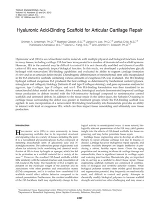
  • 6. Histological analysis supports the biochemical results that the HA-interactive scaffolds produce greater levels of carti- lage tissue components after 6 weeks. Safranin O staining of harvested constructs (Fig. 4) exhibited a substantial increase in GAG deposition in HA-interacting hydrogels at 3 weeks compared to controls. In addition to the concentrated GAG staining in the pericellular region, HA-binding hydrogels contained higher staining in the intercellular regions of the hydrogel material, suggesting the scaffold has retained cell- secreted proteoglycans by binding to the HA core. The differences in staining were less pronounced at 6 weeks be- tween the groups, though HA-interactive scaffolds still had more intense staining. Safranin O staining was also a func- tion of initial HA loading, with more intense staining ob- served for lower loading for both control and HA-binding scaffolds, similar to the quantified ECM results. Type II collagen immunostaining of control hydrogels at 3 weeks was slightly more intense than HA-interactive scaffolds. However, after 6 weeks, HA-binding PEG hydrogels pro- duced significantly greater Type II collagen staining com- pared to PEG controls. Repair of osteochondral defects in vivo with hydrogels Implantation of HA-interactive PEG hydrogels increased cartilage tissue production in osteochondral defects created on the rat femoral condyle compared to control hydrogels and untreated defects. Acellular scaffolds were implanted in the defects to avoid the challenge of delivering exogenous cells. The implanted scaffolds were able to integrate with the surrounding tissue, and gross images of harvested knees after 4 days following implantation demonstrated that hy- drogels remained in the defects and achieved good material- tissue integration (Supplementary Fig. S7). Implantation of HA-interactive hydrogels in osteochon- dral defects resulted in a more robust cartilage tissue repair compared to control hydrogels and untreated defects (Fig. 5). After 4 days, discrete hydrogel material was clearly visible in FIG. 3. Gene expression demonstrates chondrogenic differentiation of MSCs in HA-interacting hydrogels. Sox-9, aggrecan, type II collagen, and type I collagen expression were analyzed for all hydrogel conditions. All expression levels were normalized to individual b-actin levels and observed expression for PEG scaffolds containing no HA at 4 days. HA-interacting hydrogels increased aggrecan production at 3 weeks and type II collagen production at 6 weeks. 2502 UNTERMAN ET AL.
  • 7. the HA-interactive and scrambled peptide control condi- tions, while the untreated defects were filled with a clot, cells, and tissue debris. Both HA-binding and control hy- drogels appeared to be well-integrated into the surrounding tissue with cell infiltration present at the margins of the implants. Scrambled peptide controls exhibited a signifi- cantly stronger tissue response with more cells at the implant edge. After 3 weeks, the implant material was still evident for both HA-binding and control hydrogels, though the surrounding tissue response had mostly subsided and the underlying subchondral bone was undergoing repair. Although there was as yet no clear, differentiated carti- lage-like repair tissue visible, there were some early signs of repair at the margins of the defect that did not stain positive FIG. 4. HA-interactive scaffolds accumulate more cartilage matrix components at 6 weeks. Safranin O staining of HA-interactive scaffolds and PEG scaffolds across 6 weeks demonstrates increased GAG deposition in HA-interactive scaffolds. Staining is highest for low HA loadings. Immunohistochemical staining for Type II collagen indicated initially higher staining for PEG controls at 3 weeks, but substantially higher staining for HA- interactive scaffolds at 6 weeks. Bar = 100 mm. FIG. 5. HA-interactive scaffolds assist in the repair of rat osteochondral defects in vivo. Safranin O staining of osteochondral defects in representative rat knees demonstrated that HA-interactive hydrogels produced more GAG-positive repair tissue than scrambled peptide controls (sHABPep) or untreated defects. In addition, tissue adjacent to the defects treated with HA- interactive scaffolds contained more GAGs than controls. Representative Safranin O sections are shown for two separate animals at 3 and 6 weeks postimplantation for comparison. Type II collagen immunostaining did not show substantial differences in collagen content. Bar = 200 mm. HA-INTERACTING SCAFFOLDS FOR CARTILAGE REPAIR 2503
  • 8. for Safranin O. Untreated defect controls exhibited a smaller defect depth, but no visible Safranin O-positive cartilage re- pair. At 6 weeks, implant material was entirely absent and replaced with varying levels of repair tissue. Neither HA-binding nor scrambled peptide control hydrogels caused a complete regeneration of healthy cartilage at this time point, but HA binding hydrogels produced a greater volume of repair tissue with stronger Safranin O staining compared to controls, though still weaker than healthy tissue. Un- treated defects showed a well-integrated repair tissue with minimal Safranin O staining, indicative of the expected fi- brocartilagenous repair. The presence of the HA-interactive hydrogels in the os- teochondral defects also had an impact on the ECM of the cartilage surrounding the defect. At 3 weeks, all knees ex- hibited reduced Safranin O staining for GAGs on articular cartilage surrounding the defect and in the joint space com- pared to day 4 staining and untreated controls. At 6 weeks, Safranin O staining of cartilage outside the defect area was further reduced, suggesting the presence of a discrete osteochondral defect also caused degenerative joint changes. However, joints containing defects treated with HA-inter- active scaffolds exhibited greater proteoglycan staining, in- dicating reduced cartilage degeneration. Overall, these results indicate that HA-binding hydrogels produced an improve- ment in the repair of osteochondral defects and the mainte- nance of cartilage tissue surrounding the defect. Discussion The goal of this research was to develop a novel HA- binding hydrogel that would interact noncovalently with HA. While the HA-interacting scaffold was able to modulate the release of HA doped into the hydrogel, the potential scaffold interaction with cell-secreted or local ECM-derived HA may also influence tissue remodeling and regeneration. Noncovalent binding of a critical matrix building block may allow a more efficient matrix assembly when required during the tissue development process. Since HA is a critical matrix component or the building block of the ECM in many tissues, providing a scaffold that can reversibly bind the molecule could have widespread application in regeneration. The role of HA in the development of cartilage has been well studied, but its application to tissue engineering has yielded varying results. In development, HA plays a critical role during mesenchymal condensation that leads to carti- lage formation. Early in this process, the limb mesenchyme is composed of dispersed cells throughout an ECM that con- tains significant quantities of HA. Upon initiation of con- densation, mesenchymal cells aggregate, hyaluronidases are upregulated, and HA concentrations drop. It is believed that HA helps mediate cell aggregation as an intercell bridge through multivalent binding to HA receptors, notably CD44. High concentrations of HA can saturate the cell receptors without the attendant bridging effects, and thus inhibit or slow mesenchymal condensation. After condensation, HA synthesis is again upregulated as the cells differentiate to- ward a functional cartilage tissue.21 This is supported by research showing that addition of HA inhibits chick limb bud morphogenesis and later cartilage formation, and that HAase activity is spatially and temporally regulated in a very precise manner during limb bud development.22–24 The connection between embryological roles of HA and its uses in tissue engineering are less understood. Previous studies in our laboratory show agreement that HA encap- sulation in synthetic hydrogels does not produce a strong chondrogenic response in vitro, but may in fact help osteo- genesis.7 Other studies from Burdick and coworkers, how- ever, who have used an HA-based hydrogel with a variety of cell types, indicate that while modified HA hydrogels are supportive of embryonic stem cell self-renewal, they are supportive of differentiation and tissue formation when used with MSCs and chondrocytes. These results suggest that less differentiated cell types respond to HA as a proliferative and self-renewal signal, while partially differentiated cells re- spond to HA by enhancing chondrogenesis.6,25,26 In the present studies, HA-interactive scaffolds (with little exoge- nous HA) significantly improved cartilage tissue formation in vitro. However, hydrogels containing exogenous HA increased cell number at early time points, evidencing either an increase in proliferation or an improvement in cell via- bility. We speculate that the observed high levels of matrix synthesis in HA-interactive scaffolds at later time points may act as a signal that there are too many matrix-producing cells in a tissue, resulting in a lower cell number at 6 weeks. These results of HA improving cell survival or proliferation at early time points, yet inhibiting matrix production at later time points, highlight the importance of controlling the temporal presentation of extracellular signals during cell differentiation. In addition to the overall improvement in chondrogenesis observed with the HA-interactive scaffolds, the temporal dynamics of tissue formation more closely resembled the embryologic development of cartilage tissue. Histological staining and gene expression both confirmed early increases in GAG deposition and aggrecan synthesis, with lower Type II collagen production compared to controls. However by 6 weeks, Type II collagen expression and deposition signifi- cantly increased compared to controls. These data correlate with observations during limb embryogenesis, where GAG deposition precedes Type II collagen synthesis.27 Previous studies have suggested that pericellular GAG molecules can aid in the organization and deposition of Type II collagen during chondrogenesis.28 This may explain the marked in- crease in Type II collagen deposition by 6 weeks compared to control materials. The cell-secreted proteoglycans, bound to the HA-interactive scaffold through their HA cores, may aid in the deposition and organization of Type II collagen. After in vitro testing, the biomaterial formulation that produced the greatest cartilage, the HA-binding hydrogels with no HA loading, was selected for translation to an in vivo study to evaluate repair of osteochondral defects. The goal of the in vivo studies was to provide a material that would direct the differentiation of endogenous repair cells. While cells can be added to the biomaterial before implantation, clinical translation potential is increased if an off-the-shelf material is available to combine with surgical procedures. Defects treated with the HA-interactive scaffolds produced more organized repair tissue that stained more strongly for Safranin O compared to untreated defects, though still below that of healthy cartilage. Repair tissue was still clearly dis- tinguishable from the surrounding cartilage after 6 weeks, and in some cases active remodeling was still present. Additional time, up to 9 or 12 weeks, may provide the 2504 UNTERMAN ET AL.
  • 9. opportunity for more remodeling and ultimately more complete repair. An unexpected observation in joint studies was the impact of the HA-interactive scaffolds on reducing degeneration of tissue surrounding the defects. The cartilage tissue surround- ing the osteochondral defects demonstrated degenerative changes, such as reduced proteoglycan content, over the course of the experiment. The potential of cartilage defects to lead to generalized degenerative changes in the joint is a well- known phenomenon that is the basis of the clinical desire to treat cartilage defects to prevent post-traumatic osteoarthritis. Implanting defects with the HA-interactive scaffolds reduced the decrease in Safranin O staining in the surrounding tissues. There are a number of potential physical and biological mechanisms for this observation. HA bound to the surface of the hydrogel may enhance lubrication, reducing friction at the defect and surrounding cartilage surface. HA also has anti- inflammatory properties, which may improve overall joint homeostasis as the defect is undergoing repair. In conclusion, HA-interacting hydrogels can improve cartilage tissue formation in vitro and in vivo. The results indicated that early presentation of HA to MSCs results in a more proliferative phenotype, while later presentation of cell-secreted HA results in a more chondrogenic phenotype than controls. The use of a smart, matrix-interacting material allowed for the recapitulation of events during limb bud development with temporal increases in proteoglycan fol- lowed by Type II collagen production. The HA-binding hy- drogels improved repair of osteochondral defects, and were able to reduce degeneration in cartilage surrounding the defects. Matrix-interactive materials are a promising candi- date as biomaterials for applications, including repair and regeneration due to their high degree of control, the natural presentation of native matrix molecules, and their dynamic, cell-directed presentation of ECM components. Acknowledgments This investigation was supported by the National Institutes of Health under Ruth L. Kirschstein National Research Service Award #AG328232. We gratefully acknowledge Johns Hop- kins A.B. Mass Spectrometry/Proteomic Facility for providing the MALDI-TOF spectrometer and the Johns Hopkins De- partment of Chemistry Instrumentation Facility for providing the peptide synthesizer. Disclosure Statement No competing financial interests exist. References 1. Volpi, N., Schiller, J., Stern, R., and Soltes, L. Role, metabo- lism, chemical modifications and applications of hyalur- onan. Curr Med Chem 16, 1718, 2009. 2. Chung, C., Beecham, M., Mauck, R.L., and Burdick, J.A. The influence of degradation characteristics of hyaluronic acid hydrogels on in vitro neocartilage formation by mesenchy- mal stem cells. Biomaterials 30, 4287, 2009. 3. Shu, X.Z., Ahmad, S., Liu, Y., and Prestwich, G.D. Synthesis and evaluation of injectable, in situ crosslinkable synthetic extracellular matrices for tissue engineering. J Biomed Mater Res Part A 79, 902, 2006. 4. Park, Y.D., Tirelli, N., and Hubbell, J.A. Photopolymerized hyaluronic acid-based hydrogels and interpenetrating net- works. Biomaterials 24, 893, 2003. 5. Liu, Y., Shu, X.Z., and Prestwich, G.D. Osteochondral defect repair with autologous bone marrow-derived mesenchymal stem cells in an injectable, in situ, cross-linked synthetic ex- tracellular matrix. Tissue Eng 12, 3405, 2006. 6. Gerecht, S., Burdick, J.A., Ferreira, L.S., Townsend, S.A., Langer, R., and Vunjak-Novakovic, G. Hyaluronic acid hydrogel for controlled self-renewal and differentiation of human embryonic stem cells. Proc Natl Acad Sci U S A 104, 11298, 2007. 7. Hwang, N.S., Varghese, S., Li, H., and Elisseeff, J. Regulation of osteogenic and chondrogenic differentiation of mesen- chymal stem cells in PEG-ECM hydrogels. Cell Tissue Res 344, 499, 2011. 8. Mano, J.F., Silva, G.A., Azevedo, H.S., Malafaya, P.B., Sousa, R.A., Silva, S.S., et al. Natural origin biodegradable systems in tissue engineering and regenerative medicine: present status and some moving trends. J R Soc Interface 4, 999, 2007. 9. Gunatillake, P., Mayadunne, R., and Adhikari, R. Recent developments in biodegradable synthetic polymers. Bio- technol Annu Rev 12, 301, 2006. 10. Ifkovits, J.L., and Burdick, J.A. Review: photopolymerizable and degradable biomaterials for tissue engineering applica- tions. Tissue Eng 13, 2369, 2007. 11. Hern, D.L., and Hubbell, J.A. Incorporation of adhesion peptides into nonadhesive hydrogels useful for tissue res- urfacing. J Biomed Mater Res 39, 266, 1998. 12. Zustiak, S.P., Durbal, R., and Leach, J.B. Influence of cell- adhesive peptide ligands on poly(ethylene glycol) hydrogel physical, mechanical and transport properties. Acta Bioma- terialia 6, 3404, 2010. 13. Lee, H.J., Yu, C., Chansakul, T., Hwang, N.S., Varghese, S., Yu, S.M., et al. Enhanced chondrogenesis of mesenchymal stem cells in collagen mimetic peptide-mediated microen- vironment. Tissue Eng Part A 14, 1843, 2008. 14. Salinas, C.N., and Anseth, K.S. The enhancement of chon- drogenic differentiation of human mesenchymal stem cells by enzymatically regulated RGD functionalities. Biomater- ials 29, 2370, 2008. 15. Martino, M.M., Tortelli, F., Mochizuki, M., Traub, S., Ben-David, D., Kuhn, G.A., et al. Engineering the growth factor microenvironment with fibronectin domains to pro- mote wound and bone tissue healing. Sci Transl Med 3, 100ra89. 2011. 16. Mummert, M.E., Mohamadzadeh, M., Mummert, D.I., Mi- zumoto, N., and Takashima, A. Development of a peptide inhibitor of hyaluronan-mediated leukocyte trafficking. J Exp Med 192, 769, 2000. 17. Zmolik, J.M., and Mummert, M.E. Pep-1 as a novel probe for the in situ detection of hyaluronan. J Histochem Cytochem 53, 745, 2005. 18. Bitter, T., and Muir, H.M. A modified uronic acid carbazole reaction. Anal Biochem 4, 330, 1962. 19. Williams, C.G., Kim, T.K., Taboas, A., Malik, A., Manson, P., and Elisseeff, J. In vitro chondrogenesis of bone marrow- derived mesenchymal stem cells in a photopolymerizing hydrogel. Tissue Eng 9, 679, 2003. 20. Dong, Y., Hassan, W., Zheng, Y., Saeed, A.O., Cao, H., Tai, H., et al. Thermoresponsive hyperbranched copolymer with multi acrylate functionality for in situ cross-linkable hya- luronic acid composite semi-IPN hydrogel. J Mater Sci Mater Med 23, 25, 2012. HA-INTERACTING SCAFFOLDS FOR CARTILAGE REPAIR 2505
  • 10. 21. Maleski, M.P., and Knudson, C.B. Hyaluronan-mediated aggregation of limb bud mesenchyme and mesenchymal condensation during chondrogenesis. Exp Cell Res 225, 55, 1996. 22. Toole, B.P., Jackson, G., and Gross, J. Hyaluronate in mor- phogenesis: inhibition of chondrogenesis in vitro. Proc Natl Acad Sci U S A 69, 1384, 1972. 23. Kulyk, W.M., and Kosher, R.A. Temporal and spatial anal- ysis of hyaluronidase activity during development of the embryonic chick limb bud. Dev Biol 120, 535, 1987. 24. Li, Y., Toole, B.P., Dealy, C.N., and Kosher, R.A. Hyaluronan in limb morphogenesis. Dev Biol 305, 411, 2007. 25. Chung, C., and Burdick, J.A. Influence of three-dimensional hyaluronic acid microenvironments on mesenchymal stem cell chondrogenesis. Tissue Eng Part A 15, 243, 2009. 26. Chung, C., Mesa, J., Randolph, M.A., Yaremchuk, M., and Burdick, J.A. Influence of gel properties on neocartilage formation by auricular chondrocytes photoencapsulated in hyaluronic acid networks. J Biomed Mater Res Part A 77, 518, 2006. 27. Mo´dis, L. Organization of the Extracellular Matrix: A Po- larization Microscopic Approach. Boca Raton, FL: CRC Press, 1990. 28. Graff, R.D., Kelley, S.S., and Lee, G.M. Role of pericellular matrix in development of a mechanically functional neo- cartilage. Biotechnol Bioeng 82, 457, 2003. Address correspondence to: Jennifer H. Elisseeff, Ph.D. Department of Biomedical Engineering Johns Hopkins University Smith Building Rm. 5033 400 N. Broadway Baltimore, MD 21231 E-mail: jhe@jhu.edu Received: December 15, 2011 Accepted: June 22, 2012 Online Publication Date: August 14, 2012 2506 UNTERMAN ET AL.