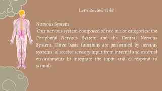
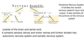
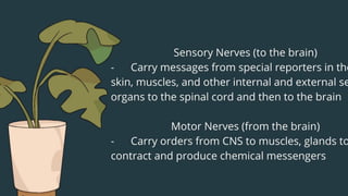
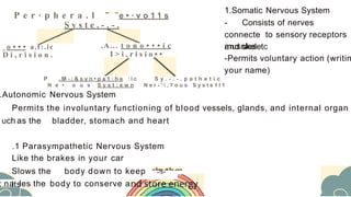
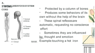
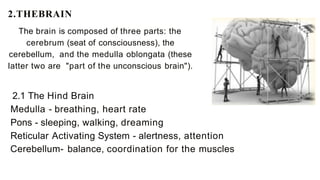
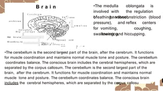
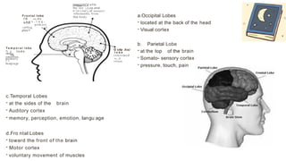
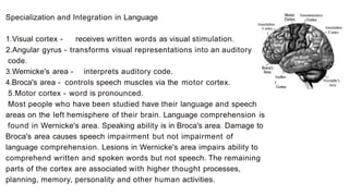
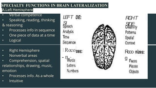
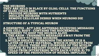
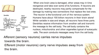
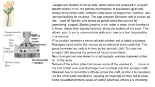
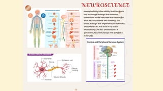
C)
N e r v o u s Syste111.
C e n t r a l ••s
S y s 1 n )
S p · o I Co:a=d
P e r · p b e 1 al ,e 1
v o u s
S . ) ' S t e D l
Peripheral Nervous System
It handles the central
nervous system's input and
output. It also contains all
the portions of the nervous
system
outside of the brain and spinal cord.
It contains sensory nerves and motor nerves and further divided into
autonomic nervous system and somatic nervous system.
P e r · p h e r a . l e • · v o 1 1 s
S y s t e . - . - .
o • • • a.t:.ic
D i , r i s i o n .
.A... t o n o • • • i c
I > i , r i s i o • •
P . M - : & s y n • p a 1 : h e : i c
N e • o u s S y s t : e w n
S y . - . - . p a t h e t i c
N e r - '  , 7 o u s S y s t e 1 l 1
.Autonomic Nervous System
Permits the involuntary functioning of blood vessels, glands, and internal organ
uch as the bladder, stomach and heart
.1 Parasympathetic Nervous System
1.Somatic Nervous System
- Consists of nerves
connecte to sensory receptors
and skeletc
muscles
-Permits voluntary action (writin
your name)
Like the brakes in your car
Slows the body down to keep
its
-!l-
: na les the body to conserve a
(
A.
CENTRALNERVOUSSYSTEM
I.SPINAL
CORD
Sacrum---,r:;j
k'« Coccyx-
---
7 Cervical
vertebrae
12 Thoracic
vertebrae
5 Lumbar
vertebrae
MAM.
- Protected by a column of bones
- Produces some behaviors of its
own without the help of the brain
- These spinal reflexesare
automatic, requesting no conscience
effort
- Sometimes they are influenced
by thought and emotion
Example:touching a hot iron
2.THEBRAIN
The brain is composed of three parts: the
cerebrum (seat of consciousness), the
cerebellum, and the medulla oblongata (these
latter two are "part of the unconscious brain").
2.1 The Hind Brain
Medulla - breathing, heart rate
Pons - sleeping, walking, dreaming
Reticular Activating System - alertness, attention
Cerebellum- balance, coordination for the muscles
B r a i n
C e r e b r u m
o r c b r r u n
a
T h ..,,,. : u n u
A n i y g d a l a .
o
H i p c a r n p u
L
Mid b r a i n _ /
y p
-H
--o m a l s n u
•The medulla oblongata is
involved with the regulation
of heartbeat,
breathing, vasoconstriction (blood
pressure), and reflex centers
for vomiting, coughing,
sneezing,
swallowing, and hiccupping.
Spinal ord
H i n d b r a . i n
[P
o
n ,J
r c b e u n ,
Mcdull:.a. o b l o n g - a t . a
w w w
__
Gt L.I C
Olr _ r _e>r
g
•The cerebellum is the second largest part of the brain, after the cerebrum. It functions
for muscle coordination and maintains normal muscle tone and posture. The cerebellum
coordinates balance. The conscious brain includes the cerebral hemispheres, which are
separated by the corpus callosum. The cerebellum is the second largest part of the
brain, after the cerebrum. It functions for muscle coordination and maintains normal
muscle tone and posture. The cerebellum coordinates balance. The conscious brain
includes the cerebral hemispheres, which are separated by the corpus callosu
2.2 The Forebrain
. .
.:---i---r1e hypothalamus regulates homeost
asis. It has regulatory areas for thirst,
hunger, body
temperature, water balance, and blood
pressure, and links the Nervous System to
the Endocrine System. The mid brain and
pons are also part of the unconscious brain.
The thalamus serves as a central relay point
for IncomIng nervous messages.
•Thalamus - Direct sensory messages to
higher centers in the brain. The sight of sunset
is directed to a visual area. The only sense
that completely bypasses the
thalamus is the sense of smell, which
has its private switching station,
the olfactory bulb.
•Hypothalamus - It is involved with drives
associated with survival such as hunger, thirst,
emotion, sex, and
1.2The Limbic System
Amygdala - Responsible for evaluating sensory
information. It determines its emotional importance. It
makes the decision to approach or to withdraw. Its
initial response may be overridden by the appraisal of
the cerebral cortex
Hippocampus - is the gate way to memory.
Cerebrum - Higher forms of thinking take place in it. It is
divided into two halves called the cerebral
hemispheres that are connected by a large band of
fibers called the corpus callosum. They have different
tasks (lateralization). The cerebrum governs
intelligence and reasoning, learning and memory.
The Cerebral Cortex -The cerebrum is covered by
several thin layers of densely packed cells known as the
cerebral cortex. On each cerebral hemisphere, deep
fissures divide the cortex into 4 lobes.
.----....
.
F r o n t a l lobe­
re -to d o
wrth
; n a x . i n g .
prob.err,
-;olYlr,g a,r,d
plan'l
T e m p o r a l l o b e­
N g t o d o
"'""th
memory.
emotloo.
L C
la.ngva.gc
conccm('d wTth
lhc rec ;,t,on and
P"'OCCSSll"g of sensor)'
rtformatJoi, from
the body
c.Temporal Lobes
- at the sides of the brain
- Auditory cortex
- memory, perception, emotion, langu age
d.Fro ntal Lobes
- toward the front of the brain
- Motor cortex
- voluntary movement of muscles
O cdp Jt:aJ
l o b e­
concemed
w , , h
vision
a.Occipital Lobes
- located at the back of the head
- Visual cortex
b. Parietal Lobe
- at the top of the brain
- Somato- sensory cortex
- pressure, touch, pain
Specialization and Integration in Language Somatoscnsory
Cortex
Association
Cortex Association
Cortex
1.Visual cortex - receives written words as visual stimulation.
2.Angular gyrus - transforms visual representations into an auditory
code.
3.Wernicke's area - interprets auditory code.
4.Broca's area - controls speech muscles via the motor cortex.
5.Motor cortex - word is pronounced.
Most people who have been studied have their language and speech
areas on the left hemisphere of their brain. Language comprehension is
found in Wernicke's area. Speaking ability is in Broca's area. Damage to
Broca's area causes speech impairment but not impairment of
language comprehension. Lesions in Wernicke's area impairs ability to
comprehend written and spoken words but not speech. The remaining
parts of the cortex are associated with higher thought processes,
planning, memory, personality and other human activities.
Auditor
y
Cortex
Wernidtc's
Arca
SPECIALTY FUNCTIONS IN BRAIN LATERALIZATION
A.Left Hemisphere
L
E
FT
SI
R
I
G
HT
SIDE:
1Roco
- n,-
Rcoo
g
...)
J
Dendrite Axon
terminal
Cell body
Node of
Ranv
Nucleus
I Schwann
cell
Myelln
sheath
When one brain area is damaged, other areas may in time
reorganize and take over some of its functions. If neurons are
destroyed, nearby neurons may partly compensate for the
damage by making new connections that replace the lost ones.
The neuron is the functional unit of the nervous system.
Humans have about 100 billion neurons in their brain alone!
While variable in size and shape, all neurons have three parts.
Dendrites receive information from another cell and transmit
the message to the cell body. The cell body contains the
nucleus, mitochondria and other organelles typical of eukaryotic
cells. The axon conducts messages away from the cell body.
Afferent (sensory neurons) carries nerve impulses
towards the brain.
Efferent (motor neurons) carry nerve impulses away from
the brain.
[ Neurotransmitte
rvesicle s
Syna
ptic
cleft
Motor
end plate
/
/
Skeletal muscle
fiber
_,
.
.,
,.
Myelin
Axon of
motor
neuron
,
Mitochondria
1
Neurotransmitte
r receptors
Ganglia are clusters of nerve cells. Some axons are wrapped in a myelin
sheath formed from the plasma membranes of specialized glial cells
known as Schwann cells. Schwann cells serve as supportive, nutritive, and
service facilities for neurons. The gap between Schwann cells is known as
the node of Ranvier, and serves as points along the neuron for
generating a signal. Signals jumping from node to node travel hundreds
of times faster than signals traveling along the surface of the axon. This
allows your brain to communicate with your toes in a few thousandths
of a second.
•The junction between a nerve cell and another cell is called a synapse.
Messages travel within the neuron as an electrical action potential. The
space between two cells is known as the synaptic cleft. To cross the
synaptic cleft requires the actions of neurotransmitters.
Neurotransmitters are stored in small synaptic vesicles clustered at the
tip of the axon.
•Arrival of the action potential causes some of the vesicles to move to
the end of the axon and discharge their contents into the synaptic cleft.
Released neurotransmitters diffuse across the cleft, and bind to receptors
on the other cell's membrane, causing ion channels on that cell to open.
Some neurotransmitters cause an action potential, others are inhibitory.
THE BRAIN IS THE ULTIMATE ORGAN OF ADAPTATION. IT TAKES IN INFORMATION
AND ORCHESTRATES COMPLEX BEHAVIORAL REPERTOIRES THAT ALLOW HUMAN
BEINGS TO ACT IN SOMETIMES MARVELOUS, SOMETIMES TERRIBLE WAYS. MOST OF
WHAT PEOPLE THINK OF AS THE ''SELF''-WHAT WE THINK, WHAT WE REMEMBER,
WHAT WE CAN DO, HOW WE FEEL-IS ACQUIRED BY THE BRAIN FROM THE
EXPERIENCES THAT OCCUR AFTER BIRTH. SOME OF THIS INFORMATION IS
ACQUIRED DURING CRITICAL OR SENSITIVE PERIODS OF DEVELOPMENT, WHEN THE
BRAIN APPEARS UNIQUELY READY TO TAKE IN CERTAIN KINDS OF INFORMATION,
WHILE OTHER INFORMATION CAN BE ACQUIRED ACROSS BROAD SWATHS OF
DEVELOPMENT THAT CAN EXTEND INTO ADULTHOOD.
THIS SPECTRUM OF POSSIBILITIES IS WELL CAPTURED BY COINCIDING EVIDENCE
OF
BOTH THE REMARKABLY RAPID BRAIN DEVELOPMENT THAT CHARACTERIZES THE
EARLY CHILDHOOD PERIOD AND THE BRAIN'S LIFELONG CAPACITY FOR
GROWTH AND CHANGE. THE BALANCE BETWEEN THE ENDURING SIGNIFICANCE
OF EARLY BRAIN DEVELOPMENT AND ITS IMPRESSIVE CONTINUING
PLASTICITY LIES AT THE HEART OF THE CURRENT CONTROVERSY ABOUT THE
EFFECTS ON THE BRAIN OF EARLY EXPERIENCE.
GROUP-3 the information in the topic three .pptx

GROUP-3 the information in the topic three .pptx

  • 3.
  • 5.
    N e rv o u s Syste111. C e n t r a l ••s S y s 1 n ) S p · o I Co:a=d P e r · p b e 1 al ,e 1 v o u s S . ) ' S t e D l Peripheral Nervous System It handles the central nervous system's input and output. It also contains all the portions of the nervous system outside of the brain and spinal cord. It contains sensory nerves and motor nerves and further divided into autonomic nervous system and somatic nervous system.
  • 7.
    P e r· p h e r a . l e • · v o 1 1 s S y s t e . - . - . o • • • a.t:.ic D i , r i s i o n . .A... t o n o • • • i c I > i , r i s i o • • P . M - : & s y n • p a 1 : h e : i c N e • o u s S y s t : e w n S y . - . - . p a t h e t i c N e r - ' , 7 o u s S y s t e 1 l 1 .Autonomic Nervous System Permits the involuntary functioning of blood vessels, glands, and internal organ uch as the bladder, stomach and heart .1 Parasympathetic Nervous System 1.Somatic Nervous System - Consists of nerves connecte to sensory receptors and skeletc muscles -Permits voluntary action (writin your name) Like the brakes in your car Slows the body down to keep its -!l- : na les the body to conserve a
  • 8.
  • 9.
    A. CENTRALNERVOUSSYSTEM I.SPINAL CORD Sacrum---,r:;j k'« Coccyx- --- 7 Cervical vertebrae 12Thoracic vertebrae 5 Lumbar vertebrae MAM. - Protected by a column of bones - Produces some behaviors of its own without the help of the brain - These spinal reflexesare automatic, requesting no conscience effort - Sometimes they are influenced by thought and emotion Example:touching a hot iron
  • 10.
    2.THEBRAIN The brain iscomposed of three parts: the cerebrum (seat of consciousness), the cerebellum, and the medulla oblongata (these latter two are "part of the unconscious brain"). 2.1 The Hind Brain Medulla - breathing, heart rate Pons - sleeping, walking, dreaming Reticular Activating System - alertness, attention Cerebellum- balance, coordination for the muscles
  • 11.
    B r ai n C e r e b r u m o r c b r r u n a T h ..,,,. : u n u A n i y g d a l a . o H i p c a r n p u L Mid b r a i n _ / y p -H --o m a l s n u •The medulla oblongata is involved with the regulation of heartbeat, breathing, vasoconstriction (blood pressure), and reflex centers for vomiting, coughing, sneezing, swallowing, and hiccupping. Spinal ord H i n d b r a . i n [P o n ,J r c b e u n , Mcdull:.a. o b l o n g - a t . a w w w __ Gt L.I C Olr _ r _e>r g •The cerebellum is the second largest part of the brain, after the cerebrum. It functions for muscle coordination and maintains normal muscle tone and posture. The cerebellum coordinates balance. The conscious brain includes the cerebral hemispheres, which are separated by the corpus callosum. The cerebellum is the second largest part of the brain, after the cerebrum. It functions for muscle coordination and maintains normal muscle tone and posture. The cerebellum coordinates balance. The conscious brain includes the cerebral hemispheres, which are separated by the corpus callosu
  • 12.
    2.2 The Forebrain .. .:---i---r1e hypothalamus regulates homeost asis. It has regulatory areas for thirst, hunger, body temperature, water balance, and blood pressure, and links the Nervous System to the Endocrine System. The mid brain and pons are also part of the unconscious brain. The thalamus serves as a central relay point for IncomIng nervous messages. •Thalamus - Direct sensory messages to higher centers in the brain. The sight of sunset is directed to a visual area. The only sense that completely bypasses the thalamus is the sense of smell, which has its private switching station, the olfactory bulb. •Hypothalamus - It is involved with drives associated with survival such as hunger, thirst, emotion, sex, and 1.2The Limbic System Amygdala - Responsible for evaluating sensory information. It determines its emotional importance. It makes the decision to approach or to withdraw. Its initial response may be overridden by the appraisal of the cerebral cortex Hippocampus - is the gate way to memory. Cerebrum - Higher forms of thinking take place in it. It is divided into two halves called the cerebral hemispheres that are connected by a large band of fibers called the corpus callosum. They have different tasks (lateralization). The cerebrum governs intelligence and reasoning, learning and memory. The Cerebral Cortex -The cerebrum is covered by several thin layers of densely packed cells known as the cerebral cortex. On each cerebral hemisphere, deep fissures divide the cortex into 4 lobes.
  • 13.
    .----.... . F r on t a l lobe­ re -to d o wrth ; n a x . i n g . prob.err, -;olYlr,g a,r,d plan'l T e m p o r a l l o b e­ N g t o d o "'""th memory. emotloo. L C la.ngva.gc conccm('d wTth lhc rec ;,t,on and P"'OCCSSll"g of sensor)' rtformatJoi, from the body c.Temporal Lobes - at the sides of the brain - Auditory cortex - memory, perception, emotion, langu age d.Fro ntal Lobes - toward the front of the brain - Motor cortex - voluntary movement of muscles O cdp Jt:aJ l o b e­ concemed w , , h vision a.Occipital Lobes - located at the back of the head - Visual cortex b. Parietal Lobe - at the top of the brain - Somato- sensory cortex - pressure, touch, pain
  • 14.
    Specialization and Integrationin Language Somatoscnsory Cortex Association Cortex Association Cortex 1.Visual cortex - receives written words as visual stimulation. 2.Angular gyrus - transforms visual representations into an auditory code. 3.Wernicke's area - interprets auditory code. 4.Broca's area - controls speech muscles via the motor cortex. 5.Motor cortex - word is pronounced. Most people who have been studied have their language and speech areas on the left hemisphere of their brain. Language comprehension is found in Wernicke's area. Speaking ability is in Broca's area. Damage to Broca's area causes speech impairment but not impairment of language comprehension. Lesions in Wernicke's area impairs ability to comprehend written and spoken words but not speech. The remaining parts of the cortex are associated with higher thought processes, planning, memory, personality and other human activities. Auditor y Cortex Wernidtc's Arca
  • 15.
    SPECIALTY FUNCTIONS INBRAIN LATERALIZATION A.Left Hemisphere L E FT SI R I G HT SIDE: 1Roco - n,- Rcoo g
  • 16.
  • 17.
    Dendrite Axon terminal Cell body Nodeof Ranv Nucleus I Schwann cell Myelln sheath When one brain area is damaged, other areas may in time reorganize and take over some of its functions. If neurons are destroyed, nearby neurons may partly compensate for the damage by making new connections that replace the lost ones. The neuron is the functional unit of the nervous system. Humans have about 100 billion neurons in their brain alone! While variable in size and shape, all neurons have three parts. Dendrites receive information from another cell and transmit the message to the cell body. The cell body contains the nucleus, mitochondria and other organelles typical of eukaryotic cells. The axon conducts messages away from the cell body. Afferent (sensory neurons) carries nerve impulses towards the brain. Efferent (motor neurons) carry nerve impulses away from the brain.
  • 18.
    [ Neurotransmitte rvesicle s Syna ptic cleft Motor endplate / / Skeletal muscle fiber _, . ., ,. Myelin Axon of motor neuron , Mitochondria 1 Neurotransmitte r receptors Ganglia are clusters of nerve cells. Some axons are wrapped in a myelin sheath formed from the plasma membranes of specialized glial cells known as Schwann cells. Schwann cells serve as supportive, nutritive, and service facilities for neurons. The gap between Schwann cells is known as the node of Ranvier, and serves as points along the neuron for generating a signal. Signals jumping from node to node travel hundreds of times faster than signals traveling along the surface of the axon. This allows your brain to communicate with your toes in a few thousandths of a second. •The junction between a nerve cell and another cell is called a synapse. Messages travel within the neuron as an electrical action potential. The space between two cells is known as the synaptic cleft. To cross the synaptic cleft requires the actions of neurotransmitters. Neurotransmitters are stored in small synaptic vesicles clustered at the tip of the axon. •Arrival of the action potential causes some of the vesicles to move to the end of the axon and discharge their contents into the synaptic cleft. Released neurotransmitters diffuse across the cleft, and bind to receptors on the other cell's membrane, causing ion channels on that cell to open. Some neurotransmitters cause an action potential, others are inhibitory.
  • 19.
    THE BRAIN ISTHE ULTIMATE ORGAN OF ADAPTATION. IT TAKES IN INFORMATION AND ORCHESTRATES COMPLEX BEHAVIORAL REPERTOIRES THAT ALLOW HUMAN BEINGS TO ACT IN SOMETIMES MARVELOUS, SOMETIMES TERRIBLE WAYS. MOST OF WHAT PEOPLE THINK OF AS THE ''SELF''-WHAT WE THINK, WHAT WE REMEMBER, WHAT WE CAN DO, HOW WE FEEL-IS ACQUIRED BY THE BRAIN FROM THE EXPERIENCES THAT OCCUR AFTER BIRTH. SOME OF THIS INFORMATION IS ACQUIRED DURING CRITICAL OR SENSITIVE PERIODS OF DEVELOPMENT, WHEN THE BRAIN APPEARS UNIQUELY READY TO TAKE IN CERTAIN KINDS OF INFORMATION, WHILE OTHER INFORMATION CAN BE ACQUIRED ACROSS BROAD SWATHS OF DEVELOPMENT THAT CAN EXTEND INTO ADULTHOOD. THIS SPECTRUM OF POSSIBILITIES IS WELL CAPTURED BY COINCIDING EVIDENCE OF BOTH THE REMARKABLY RAPID BRAIN DEVELOPMENT THAT CHARACTERIZES THE EARLY CHILDHOOD PERIOD AND THE BRAIN'S LIFELONG CAPACITY FOR GROWTH AND CHANGE. THE BALANCE BETWEEN THE ENDURING SIGNIFICANCE OF EARLY BRAIN DEVELOPMENT AND ITS IMPRESSIVE CONTINUING PLASTICITY LIES AT THE HEART OF THE CURRENT CONTROVERSY ABOUT THE EFFECTS ON THE BRAIN OF EARLY EXPERIENCE.