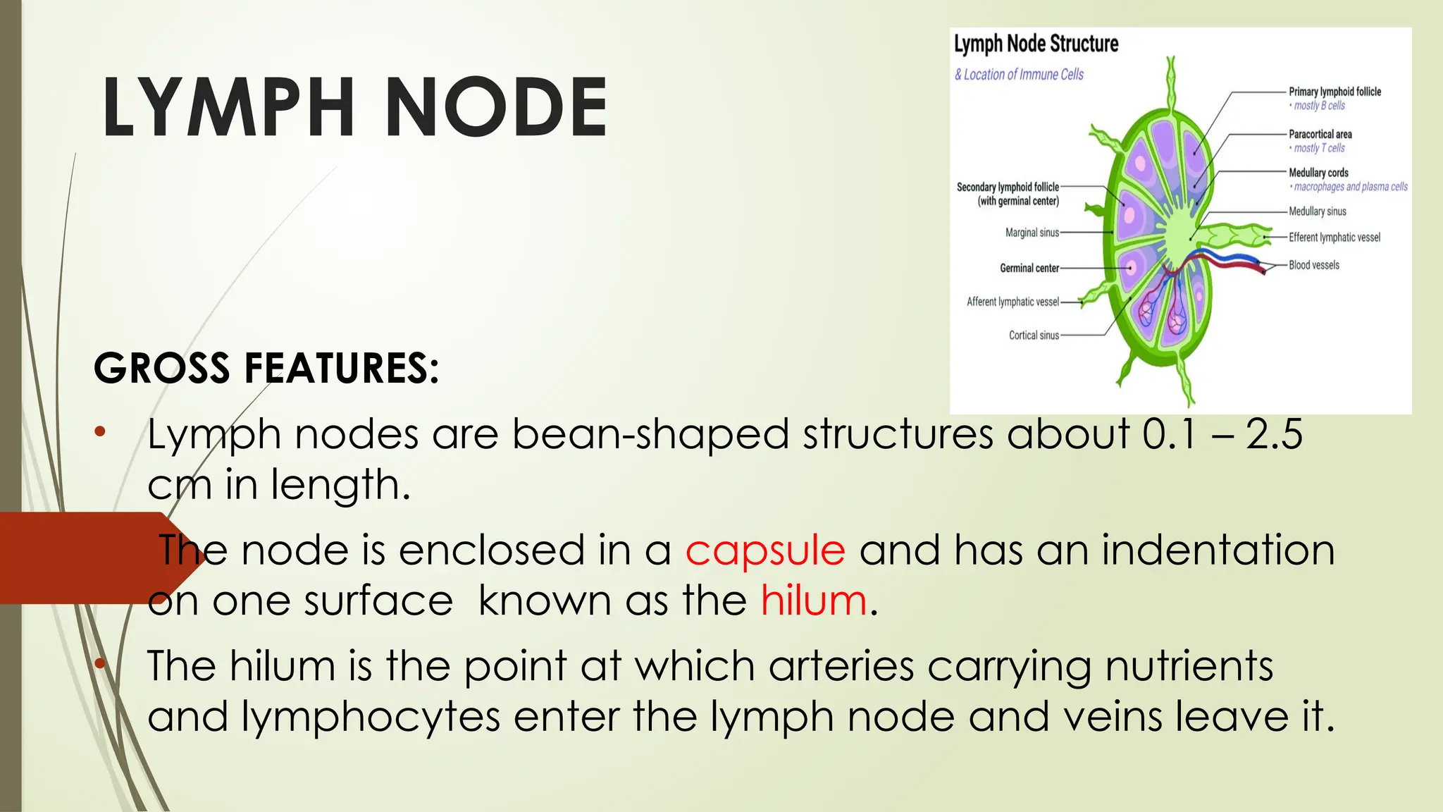
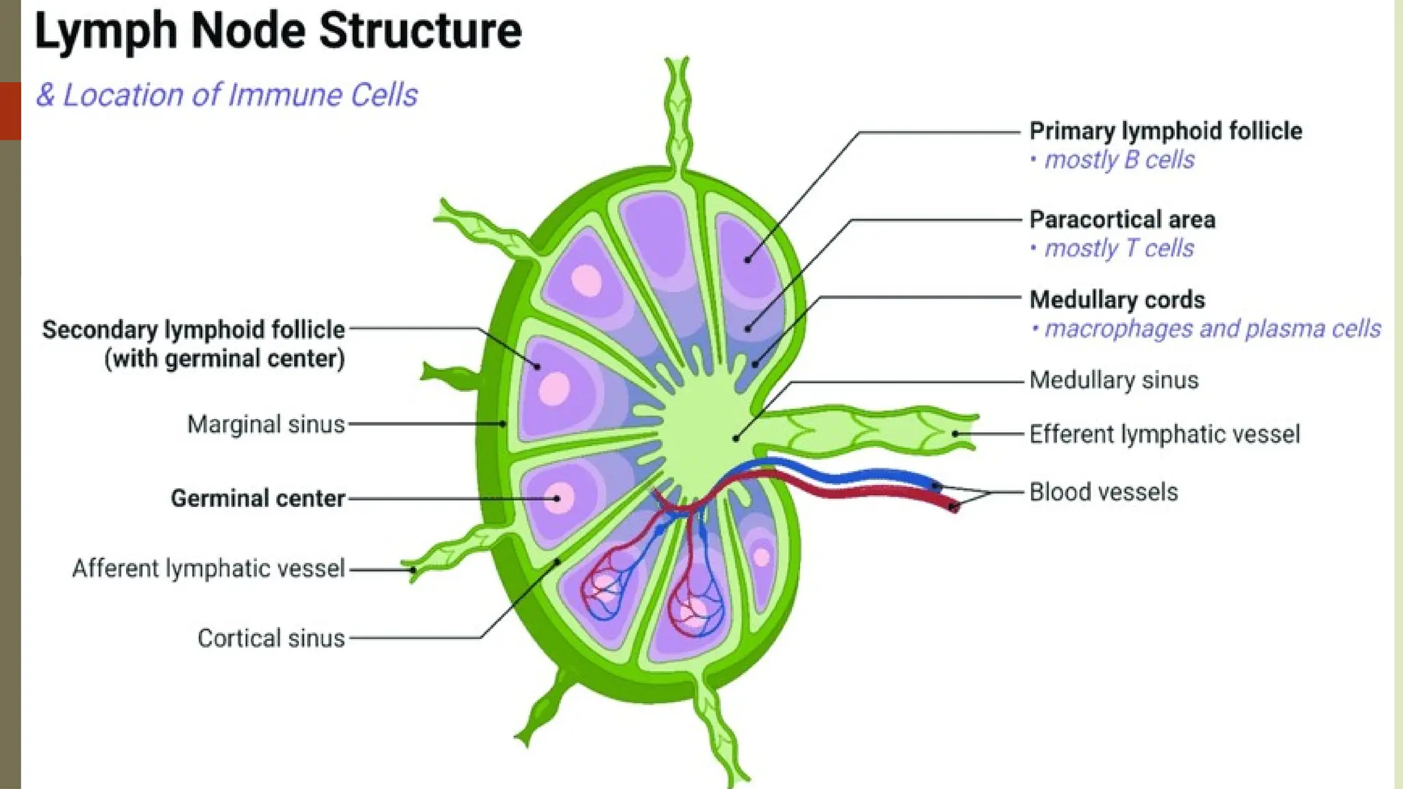
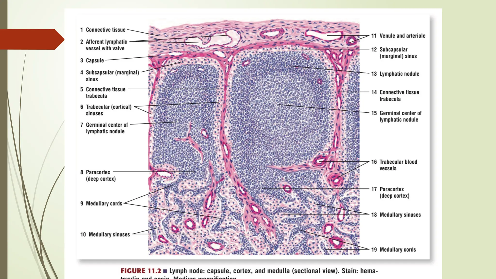
GROSS AND MICROSCOPIC
FEATURES OF LYMPH NODE
DR SURAJ PD SAH
IMMUNE SYSTEM
 Protects the organism against invading pathogens or
antigens ( bacteria , parasites and viruses).
 The lymphoid system includes all cells, tissues ,and organs
that contain aggregates of immune cells called
lymphocytes.
 Lymphoid organs are divided into two major categories –
 1. Primary lymphoid organs – Bone marrow and thymus.
 2. Secondary lymphoid organs - Lymph node, spleen,
tonsils, MALT and Peyer patches.
LYMPH NODE
GROSS FEATURES:
• Lymph nodes are bean-shaped structures about 0.1 – 2.5
cm in length.
• The node is enclosed in a capsule and has an indentation
on one surface known as the hilum.
• The hilum is the point at which arteries carrying nutrients
and lymphocytes enter the lymph node and veins leave it.
• Afferent lymphatic vessels enter the lymph
node through the capsule peripherally
and efferent lymphatic vessels leave the
node via the hilum.
• The Afferent takes lymph from peripheral
sites to the node, while the efferent takes
processed lymph from the nodes back to
the circulation.
MICROSCOPIC FEATURES
A horizontal section through the lymph node reveals that
the dense connective tissue capsule (composed of elastin,
collagen and fibroblasts)
Additionally, there is a pericapsular adipose tissue layer that
surrounds the connective tissue capsule. This layer contains
arterioles and venules that supply the lymph node.
Capsule projects into lymph node interiorly is called
trabeculae; giving the lymph node a lobular appearance,
 The lymph node is divided into an outer cortex and an inner
medulla.
 Histological staining of the node reveals that the
cortex stains darker than the medulla with
hematoxylin and eosin (H&E) due to its higher cell
content.
 Beneath the fibrous capsule is the subcapsular sinus.
subcapsular sinus communicates with the cortical
sinuses that travel parallel to the capsular
trabeculation. They carry lymph to the medullary sinus
 The medullary sinuses drain the lymph coming from the
cortical sinuses to the efferent lymphatic vessel via the
hilum.
Cortex
 The cortex contains spherical collections of lymphocytes is
called lymphoid nodules or lymphoid follicles. It primarily
contain B-cells,specialized follicular dendritic cells and some
supporting T-cells.
 These can either be primary or secondary follicles
depending on their cellular population.
 Primary lymphoid follicles contain small, dormant
lymphocytes.
 while secondary lymphoid follicles contain a lighter/pale
staining area of active lymphocyte proliferation known as
a germinal centre.
Paracortex
Deep to the cortical layer and superficial to the medulla is
the Paracortex. This region contains mostly T-cells of the CD4
(cluster of differentiation) and CD8 subsets.
 Migrating dendritic cell lines (such as Langerhans cells)
found in this area present processed antigen to the T-cells.
Medulla
The cords also contain plasma cells, small lymphocytes and
macrophages.
 The lymphocytes of the medulla are less organized and
form irregular medullary cords.
• The germinal centre supports affinity maturation (high affinity
antibody production) of B-cells.
• It is subdivided into a dark zone, light zone and a mantle zone. The
B-cells of the dark zone are known as centroblasts. They rapidly
replicate, resulting in hypermutation of their antibody molecules.
• Centroblasts migrate to the light zone, where they are referred to
as centrocytes.
• Here they compete for binding with the unprocessed antigens
presented on the surface of follicular dendritic cells.
• Those centrocytes that successfully bind to the follicular dendritic
cells will survive, while the others will die.
• Small, quiescent cells are peripherally marginalized due to the rapid
proliferation of the central cells. These cells form the mantle
zone of the germinal centre.
THANK YOU
NEXT CLASS:
THYMUS
PALATINE TONSIL
SPLEEN

GROSS AND MICROSCOPIC FEATURES OF LYMPH NODE.pptx

  • 1.
    GROSS AND MICROSCOPIC FEATURESOF LYMPH NODE DR SURAJ PD SAH
  • 2.
    IMMUNE SYSTEM  Protectsthe organism against invading pathogens or antigens ( bacteria , parasites and viruses).  The lymphoid system includes all cells, tissues ,and organs that contain aggregates of immune cells called lymphocytes.  Lymphoid organs are divided into two major categories –  1. Primary lymphoid organs – Bone marrow and thymus.  2. Secondary lymphoid organs - Lymph node, spleen, tonsils, MALT and Peyer patches.
  • 3.
    LYMPH NODE GROSS FEATURES: •Lymph nodes are bean-shaped structures about 0.1 – 2.5 cm in length. • The node is enclosed in a capsule and has an indentation on one surface known as the hilum. • The hilum is the point at which arteries carrying nutrients and lymphocytes enter the lymph node and veins leave it.
  • 4.
    • Afferent lymphaticvessels enter the lymph node through the capsule peripherally and efferent lymphatic vessels leave the node via the hilum. • The Afferent takes lymph from peripheral sites to the node, while the efferent takes processed lymph from the nodes back to the circulation.
  • 6.
    MICROSCOPIC FEATURES A horizontalsection through the lymph node reveals that the dense connective tissue capsule (composed of elastin, collagen and fibroblasts) Additionally, there is a pericapsular adipose tissue layer that surrounds the connective tissue capsule. This layer contains arterioles and venules that supply the lymph node. Capsule projects into lymph node interiorly is called trabeculae; giving the lymph node a lobular appearance,  The lymph node is divided into an outer cortex and an inner medulla.
  • 8.
     Histological stainingof the node reveals that the cortex stains darker than the medulla with hematoxylin and eosin (H&E) due to its higher cell content.  Beneath the fibrous capsule is the subcapsular sinus. subcapsular sinus communicates with the cortical sinuses that travel parallel to the capsular trabeculation. They carry lymph to the medullary sinus  The medullary sinuses drain the lymph coming from the cortical sinuses to the efferent lymphatic vessel via the hilum.
  • 9.
    Cortex  The cortexcontains spherical collections of lymphocytes is called lymphoid nodules or lymphoid follicles. It primarily contain B-cells,specialized follicular dendritic cells and some supporting T-cells.  These can either be primary or secondary follicles depending on their cellular population.  Primary lymphoid follicles contain small, dormant lymphocytes.  while secondary lymphoid follicles contain a lighter/pale staining area of active lymphocyte proliferation known as a germinal centre.
  • 11.
    Paracortex Deep to thecortical layer and superficial to the medulla is the Paracortex. This region contains mostly T-cells of the CD4 (cluster of differentiation) and CD8 subsets.  Migrating dendritic cell lines (such as Langerhans cells) found in this area present processed antigen to the T-cells. Medulla The cords also contain plasma cells, small lymphocytes and macrophages.  The lymphocytes of the medulla are less organized and form irregular medullary cords.
  • 12.
    • The germinalcentre supports affinity maturation (high affinity antibody production) of B-cells. • It is subdivided into a dark zone, light zone and a mantle zone. The B-cells of the dark zone are known as centroblasts. They rapidly replicate, resulting in hypermutation of their antibody molecules. • Centroblasts migrate to the light zone, where they are referred to as centrocytes. • Here they compete for binding with the unprocessed antigens presented on the surface of follicular dendritic cells. • Those centrocytes that successfully bind to the follicular dendritic cells will survive, while the others will die. • Small, quiescent cells are peripherally marginalized due to the rapid proliferation of the central cells. These cells form the mantle zone of the germinal centre.
  • 14.