The document discusses the properties and biomedical applications of graphene quantum dots (GQDs), highlighting their significant optical and electrical characteristics, biocompatibility, and lower toxicity which make them suitable for use in biosensing, drug delivery, and therapeutic applications. It emphasizes the controlled production methods for GQDs and their integration into nanocomposite materials to enhance performance. The research outlines the importance of GQDs in bioimaging and cancer therapy due to their unique photoluminescence and potential for safe interaction with biological systems.
![Page | 1
1 Graphene Quantum Dots-Based Composites for Biomedical Applications
Graphene Quantum Dots-Based Composites for
Biomedical Applications
Noor Alam, Hina Ihsan, Samreen Khan and Kefayat Ullah
Department of Applied Physical and Material sciences, University of swat, 19120, KPK, Pakistan
Abstract: Carbon derivatives, such as graphene-based nanocomposites, have
garnered significant global attention due to their remarkable optical and electrical
properties. In this study, we examined nanohybrid materials based on graphene
quantum dots (GQDs) for biomedical applications. The biocompatibility of GQDs
makes them ideal materials for a range of medical applications, including
biosensing, drug delivery and various therapeutic uses. We also addressed issues
related to controlled production and composites involving GQDs. Similarly, we
discussed factors that affect the applicability and viability of these materials.
Keywords: Biomedical, biosensor, bioimaging, biocompatibility, Bottom-up, Bohr radius,
composites, drug delivery, Graphene, Graphene Quantum dots, GQDs, nanomedicine,
Nano crystals, Nanomaterials, photoluminescence, quantum confinement, Semiconductor,
Top-down, 0D crystals.
INTRODUCTION
One of the fundamental building blocks of living is carbon element and since carbon
nanomaterials are non-toxic and biocompatible they can be utilized in various biomedical
fields [1, 2]. Recently, it has once again astounded us with graphene [3]. It is composed of
a single 2D sheet of carbon atoms bonded into a hexagonal-shaped lattice [4] densely
packed and highly-ordered monolayer with zero-energy bandgap [5, 6]. Similarly, there
are several kinds of organic nanomaterials such as quantum dots, have caught the interest
of researchers worldwide.
Strong quantum confinement results in 2D quantum dots with discrete energy levels, when
the lateral size of the 2D materials is reduced below 20 nm [7]. Ekimov and Onushenko
[8] originally reported nanoscale semiconductor crystals, or quantum dots (QDs) in 1981
in a glass matrix. The first known usage of biological imaging was documented in 1998
[9]. Quantum dots have been highly recommended for sensing [10, 11] imaging [5, 12]
drug delivery [13] and diagnosis probes [14] due to its optical properties, such as sharp
emission and broad absorption spectra [15]. GQDs have a size within the range of 2-10 nm
and a quantum-confinement characteristic that allows them to emit fluorescence from
visible to infrared wavelengths during excitation [16]. The primary objective is to create
tiny probes that have great selectivity, adaptability, stability and the ability to pass through
cells and organelles [17]. Biological issues (biocompatibility, aggregation, non-specific
binding, aggregation, cytotoxicity) are the main hurdles to overcome [18]. Graphene
Quantum dots GQDs offer higher photostability when it comes to photobleaching,
Corresponding author
E-mail: theofficialnoor@gmail.com (Noor Alam)](https://image.slidesharecdn.com/graphenequantumdotsbasedcompositesforbiomedicalapplications-240309175401-350d757d/85/Graphene-Quantum-Dots-Based-Composites-for-Biomedical-Applications-1-320.jpg)
![Page | 1
1 Graphene Quantum Dots-Based Composites for Biomedical Applications
Graphene Quantum Dots-Based Composites for
Biomedical Applications
Noor Alam, Hina Ihsan, Samreen Khan and Kefayat Ullah
Department of Applied Physical and Material sciences, University of swat, 19120, KPK, Pakistan
Abstract: Carbon derivatives, such as graphene-based nanocomposites, have
garnered significant global attention due to their remarkable optical and electrical
properties. In this study, we examined nanohybrid materials based on graphene
quantum dots (GQDs) for biomedical applications. The biocompatibility of GQDs
makes them ideal materials for a range of medical applications, including
biosensing, drug delivery and various therapeutic uses. We also addressed issues
related to controlled production and composites involving GQDs. Similarly, we
discussed factors that affect the applicability and viability of these materials.
Keywords: Biomedical, biosensor, bioimaging, biocompatibility, Bottom-up, Bohr radius,
composites, drug delivery, Graphene, Graphene Quantum dots, GQDs, nanomedicine,
Nano crystals, Nanomaterials, photoluminescence, quantum confinement, Semiconductor,
Top-down, 0D crystals.
INTRODUCTION
One of the fundamental building blocks of living is carbon element and since carbon
nanomaterials are non-toxic and biocompatible they can be utilized in various biomedical
fields [1, 2]. Recently, it has once again astounded us with graphene [3]. It is composed of
a single 2D sheet of carbon atoms bonded into a hexagonal-shaped lattice [4] densely
packed and highly-ordered monolayer with zero-energy bandgap [5, 6]. Similarly, there
are several kinds of organic nanomaterials such as quantum dots, have caught the interest
of researchers worldwide.
Strong quantum confinement results in 2D quantum dots with discrete energy levels, when
the lateral size of the 2D materials is reduced below 20 nm [7]. Ekimov and Onushenko
[8] originally reported nanoscale semiconductor crystals, or quantum dots (QDs) in 1981
in a glass matrix. The first known usage of biological imaging was documented in 1998
[9]. Quantum dots have been highly recommended for sensing [10, 11] imaging [5, 12]
drug delivery [13] and diagnosis probes [14] due to its optical properties, such as sharp
emission and broad absorption spectra [15]. GQDs have a size within the range of 2-10 nm
and a quantum-confinement characteristic that allows them to emit fluorescence from
visible to infrared wavelengths during excitation [16]. The primary objective is to create
tiny probes that have great selectivity, adaptability, stability and the ability to pass through
cells and organelles [17]. Biological issues (biocompatibility, aggregation, non-specific
binding, aggregation, cytotoxicity) are the main hurdles to overcome [18]. Graphene
Quantum dots GQDs offer higher photostability when it comes to photobleaching,
Corresponding author
E-mail: theofficialnoor@gmail.com (Noor Alam)](https://image.slidesharecdn.com/graphenequantumdotsbasedcompositesforbiomedicalapplications-240309175401-350d757d/75/Graphene-Quantum-Dots-Based-Composites-for-Biomedical-Applications-1-2048.jpg)
![Page | 2
2 Graphene Quantum Dots-Based Composites for Biomedical Applications
and blinking [19] excellent biocompatibility with minimal toxicity [20] as well as high
colloidal stability [21]. GQDs have amazing properties and are receiving a lot of interest
owing to edge effects and quantum confinement [22].
GQDs have much higher photoluminescence (PL) compared to graphene sheets [23].
GQDs have distinctive fluorescence properties and a non-zero bandgap in the structure due
to the quantum confinement effect [24, 25]. A fluorescent biosensor is a device used to
convert information in a certain sample into a fluorescent signal both analytically and
quantitatively [26]. A fluorescence detection based assay is a commonly used technique as
highly sensitive, easily measured and inexpensive [27].
Currently, many strategies are employed to synthesize GQDs. Bottom-up approaches
include solution chemical, microwave, and ultrasonic technologies [28]. Top-down
approaches include hydrothermal, oxidation processes and electrochemical approaches
[13]. GQDs can be incorporated into organic or inorganic materials to create
multifunctional nanocomposite materials to improve their application performance and
practicality [29, 30] such as optical sensing [31, 32] superior electrode materials for
applications in supercapacitors [33, 34] and antibacterial purposes [35].
The main idea of the research is that Graphene Quantum dots (GQDs), nanoscale
semiconductor crystals, have demonstrated considerable potential in sensing, imaging and
drug delivery due to their sharp emission, wide absorption spectra, as well as a large
surface-to-volume ratio. GQDs produce fluorescence from visible to infrared wavelengths,
exhibiting unique edge effects and quantum confinement characteristics. They display
better photoluminescence than graphene sheets and serve as attractive options for
fluorescence-based biosensors. Integrating GQDs with organic or inorganic materials such
as polymers, metals, semiconductors, researchers can create nanocomposites for several
applications. GQDs exhibit unique electronic and optical properties. All of these
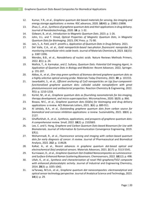
properties emerge due to quantum confinement phenomena, where the tiny size of the
quantum dots leads to discrete energy levels for electrons and holes [36] as shown in figure
1.
A) B)
Figure 1: (A Schematic diagram showing energy band structures in bulk semiconductor, quantum nanocrystals, and atom
and (B) The number of bonded atoms determines the electronic energy levels. The discrete energy levels of the atomic
orbitals mix into energy bands (seen here for a semiconducting material) when more atoms are blended together. As a
result, semiconducting quantum dots may be regarded as a hybrid between microscopic molecules and bulk material.](https://image.slidesharecdn.com/graphenequantumdotsbasedcompositesforbiomedicalapplications-240309175401-350d757d/85/Graphene-Quantum-Dots-Based-Composites-for-Biomedical-Applications-2-320.jpg)
![Page | 3
3 Graphene Quantum Dots-Based Composites for Biomedical Applications
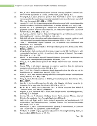
The schematically portrays of GQDs characterization, functionalization and the
methodology which incorporates synthesis via top-down and bottom-up methods, followed
by nanocomposite development and application assessment is shown in figure 2.
Figure 2: Schematic Workflow chart of the GQDs.
QDs are classified into 12 groups based on the position of their constituent elements in the
periodic table as shown in table 1. [37, 38].
Table 1: Chemical Composition Classification of QDs. Abbreviations: TMDCs are transition metal dichalcogenides;
while, P dots are semiconducting polymer dots.
Type Examples Reference(s)
VI A-I B
VII A-I B
VI A -II B
III A-V A
VI A-IV A
IV A
V A
III A-VI A-I B
P dot
TMDCs
Perovskite
MXene
Cu2S
Agbr
ZnSe, ZnO, CdS, CdTe, HgS
AlSb, AlP, GaAs, GaSb, InP, InAs
PbS, PbTe, PbSe
Graphene, C, Si (Graphene QDs in this article)
Black Phosphorus
CuInS2, CuInSe2, AgInS
NIR800
TiSe2, TaS2, MoSe2
CsPbI3
Nb2C, Ti3C2
[39]
[40]
[41]
[39]
[42]
[43]
[44]
[39]
[45]
[46]
[47]
[48]
Graphene Quantum Dots Distinctive Characteristics
Some of the significant features of quantum dots include photoluminescence, bandgap
tunability, High quantum yield, high photostability, biocompatibility, and much more. To
understand the properties of quantum dots we shall first describe the quantum confinement
effect [49].
Quantum Confinement Effect
According to De Broglie, A matter wave may be associated with any particle, and its
wavelength is inversely related to the linear momentum of the particle (λ = h / p). When a
physical system becomes comparable in size to the wavelength of the particles it interacts
with, quantum mechanics best describes the physics of the particles [50]. When the](https://image.slidesharecdn.com/graphenequantumdotsbasedcompositesforbiomedicalapplications-240309175401-350d757d/85/Graphene-Quantum-Dots-Based-Composites-for-Biomedical-Applications-3-320.jpg)
![Page | 4
4 Graphene Quantum Dots-Based Composites for Biomedical Applications
nanocrystal radius is equal to or smaller than the size of the exciton Bohr radius which
means r < rB, then both the holes and electrons are restricted to move within the dimensions
of the nanocrystal [51] best describe the "Quantum confinement" effect, refers to the
energy of confined electrons (electrons or holes) as illustrated in figure 3. In contrast to
bulk materials electron energy levels will not be continuous in nanocrystals [52].
Furthermore, by finding the constrained electron wave functions, they establish a
discrete collection of energy levels.
Figure 3: (A) schematic diagram of Bohr exciton radius (rBohr) and exciton radius (r) (B) quantum confinement in
GQDs.
Unique Optical Properties
GQDs exhibit various unique optical features such as strong photoluminescence,
adjustable band gap, quantum yield (QY), pure and saturated colors, limited bandwidth,
wide and strong absorption, narrow and symmetric emission [53].
Photoluminescence and Bandgap Engineering
Luminescence property is one of the major aspects of GQDs. In general, the electron-hole
pairs occur in semiconductors as a result of the absorption of photonic energy. The
diameter of a semiconductor plays a critical role in confining electrons and holes that lead
to the quantum confinement phenomenon [54]. The PL characteristics of graphene
quantum dots arise when excited electrons relax to the ground state and recombine with
the hole [55]. The emission wavelength is determined by the quantum dot size, for example
the bigger quantum dot has a greater emissive wavelength and the smaller quantum exhibits
a shorter emissive wavelength [56].
Another technique is to change the surface chemistry of the dots by inserting functional
groups or ligands to the edge, the edge structure might be zigzag or armchair configuration.
This can also modify the bandgap of the material, as well as its optical characteristics [57].
Due to the high confinement, the energy levels, in quantum dots resemble, those found in
atoms or molecules which is why they are commonly described as artificial atoms. During
the previous decade, numerous GQDs have been synthesized by diverse techniques and
reported with varying emission colors, ranging from ultraviolet (UV) to red region [58,
59]. In reality, solely graphene's zero energy bandgap does not exhibit PL. GQDs can only
be represented with a non-zero bandgap and hence exhibit PL by modifying factors such](https://image.slidesharecdn.com/graphenequantumdotsbasedcompositesforbiomedicalapplications-240309175401-350d757d/85/Graphene-Quantum-Dots-Based-Composites-for-Biomedical-Applications-4-320.jpg)
![Page | 5
5 Graphene Quantum Dots-Based Composites for Biomedical Applications
as adding surface groups, raising dopant concentrations, or developing the physical
dimensions [60].
GQDs, as a graphene derivative, have edge flaws, surface-active sites, and a larger surface
area. Due to edge effect and the quantum-confinement, GQDs commonly exhibit
electrochemical characteristics such as large current density, quick electron transferability,
and strong conductivity in addition to size tunable optical capabilities [28] as depicted in
figure 4.
Figure 4: Size-dependent fluorescence spectra of quantum dots.
Quantum Yield (Φ)
QY or quantum yield can be described as the ratio of photons released by a PL molecule
to photons absorbed by the material. The quantum yield measures the efficiency with which
photons are emitted by fluorescent or photoluminescent materials [61].
Quantum Yield (φ) =
nubmer of photons released
number of photons absorbed
x 100
Many limitations have limited the potential of GQDs for bioimaging both in the laboratory
and living cells. Low quantum yield and short wavelength emission are examples, as an
efficient synthesis technique that results in higher product yields and more homogeneous
GQDs. To be a feasible choice for bioimaging, GQDs would need to be made with uniform
size, high yield, and fluorescence. A large quantum yield would be required to provide a
greater signal-to-noise ratio, allowing for lower concentrations and higher-quality images.
In addition, lower energy emissions might offer increased imaging depth [62].
Biocompatibility and Low Toxicity
Another property of GQDs is their compatibility with living cells as well as reduced
toxicity, which are vital for the biological and biomedical applications of CQDs and GQDs,](https://image.slidesharecdn.com/graphenequantumdotsbasedcompositesforbiomedicalapplications-240309175401-350d757d/85/Graphene-Quantum-Dots-Based-Composites-for-Biomedical-Applications-5-320.jpg)
![Page | 6
6 Graphene Quantum Dots-Based Composites for Biomedical Applications
particularly for bioimaging and cellular imaging [10]. GQDs have the ability to interact
with biomolecules by forming π–π stacking interactions or through electrostatic
interactions. Unlike graphene sheets GQDs are more biocompatible and less toxic. This
makes them a practical choice, for delivering active probe into living organisms [13]. A
number of research have discovered that GQDs have the potential to cause cell death by
generating reactive oxygen species (ROS) in living cells. As a result, the toxicity of GQDs
is determined by their surface chemistry, size, dosage concentration, manufacturing
process and doping [29]. For example, in vitro studies have shown that GQDs have
minimal cytotoxicity owing to their ultra-small size and high oxygen content [63].
Additional in vivo biodistribution of GQDs investigations revealed no accumulation in any
of the main organs in mice and rapid graphene quantum dots clearance through the kidney
[64].
Since CQDs and GQDs display good biocompatibility, low toxicity, and strong
photoluminescence properties, they are extremely suitable for cancer therapy. Moreover,
the strong photoluminescence of both GQDs and CQDs assists in monitoring the
nanoparticles throughout the body to keep an eye on the release of the medicine at the
targeted organ [2]. Table 2 shows Toxicity of GQDs on different target organs or cells in
vivo and in vitro.
Table 2: GQDs and their cytotoxic properties
Type Target Toxicity Results References
Graphene
quantum dots
Toxicity in mice
in vivo
GQDs have a
low level of
dark toxicity.
At GQD
dosages of 10
and 15 mg/kg,
some modest
shifts were seen
in the liver and
pulmonary
system.
[65]
Carboxylated
GQD
In vivo and in
vitro studies on
spleen, tumor,
kidney, and liver
There was no
severe toxicity
between 5 and
10 mg/kg.
GQD deposition
in the spleen,
kidney liver,
and tumor of
mice 24 hours
after an
intravenous
infusion.
[66]
GQDs Injections into the
peritoneal cavity
in mice
There was no
serious toxicity
in rats
when given 300
µg of GQDs (15
mg/kg, per
head).
GC/Ds may be
eliminated from
the body's
system.
[67]](https://image.slidesharecdn.com/graphenequantumdotsbasedcompositesforbiomedicalapplications-240309175401-350d757d/85/Graphene-Quantum-Dots-Based-Composites-for-Biomedical-Applications-6-320.jpg)
![Page | 7
7 Graphene Quantum Dots-Based Composites for Biomedical Applications
GQDs 𝐶𝐷34+
cells
isolated from
blood using
leukapheresis
Low
cytotoxicity
SEPW1 is
lowered by a
factor of -5.
[68]
GQDs MCF-7 cells
alongside B16F10
cells
Surprisingly
low risk of
cytotoxicity
[69]
GQDs HeLa For 24 hours, 0-
400μg 𝑚𝑙−1
GQDs exhibited
low
cytotoxicity.
Cell survival
rate of at least an
80 percent
[70]
GQDs mice pulmonary
tissues
There was no
obvious acute
toxicity.
Following
swelling,
alveolar septa
enlarge in the
high-
dose sample.
[71]
Graphene Quantum Dots Synthesis
The current approaches for GQD synthesis may be classified broadly into top-down and
bottom-up. GQD properties have been discovered to change depending on the synthesis
procedure and the raw material that is used. Many GQD extraction techniques use carbon-
rich materials as Raw materials, namely fullerene, graphite carbon fibers, glucose,
graphene and carbon nanotubes [12]. It has been proven that the GQDs "green" production
can compensate for cytotoxicity owing to their biological compatibility and superior sized-
tuned emission characteristics [22]. Green luminous GQDs may be generated using a
straightforward synthesis technique for visualizing human hepatic cancer cells made from
graphite powder [72]. Later, uniform-sized, water-soluble GQDs with red fluorescence
(RF-GQDs) was shown as a vigorous biological marker for stem cells due to strong
biological imaging applicability, and noticeable red coloration [73]. Top-down and bottom-
up approaches have been shown in figure 5.](https://image.slidesharecdn.com/graphenequantumdotsbasedcompositesforbiomedicalapplications-240309175401-350d757d/85/Graphene-Quantum-Dots-Based-Composites-for-Biomedical-Applications-7-320.jpg)
![Page | 8
8 Graphene Quantum Dots-Based Composites for Biomedical Applications
Figure 5: Schematic illustration showing the synthesis of GQDs utilizing Bottom-up and Top-down techniques.
Biomedical Applications of Graphene Based Quantum Dots
Bioimaging
Bioimaging is a vital technique used in both research as well as clinical applications, which
facilitates the analysis of biological reactions like cellular uptake, targeted delivery, and
therapeutic biological distribution in an isolated, precise manner using multiple
wavelengths of the electromagnetic (EM) spectrum [66, 74, 75]. The early diagnosis of
illnesses improves patient survival, prompting researchers to develop highly sensitive, with
excellent specificity, and low-toxicity GQDs [23]. The luminous features of GQDs
distinguish them from graphene. In addition, GQDs' great luminous characteristics make
them an excellent alternative to organic dyes, and they perform very well in bioimaging
and disease identification, such as cancer [76]. Imaging plays an important role in cancer
diagnostics because sensitive imaging allows for earlier identification of tumor and also
early diagnosis of metastasis and future disease recurrence.
Fluorescence imaging is a vital technique in biological field that examines the distribution
of materials of interest in organs, tissues, cells, and complete organisms using visible light
and near-infrared spectra [77]. Organic or fluorescent dyes have been employed as
fluorescent probes in ex vivo, in vitro, and in vivo. Many fluorophores have low water
solubility [78]. If materials that cannot dissolve in water enter the body, they tend to clump
and get recognized by the body's defense system for elimination. In cases where someone
consumes a large amount of these materials, it could potentially lead to harm, like blocking
blood flow, when they enter the bloodstream in circulatory system. The inherent low
toxicity and water solubility of GQDs have been extensively characterized [79].
Magnetic resonance imaging (MRI) utilizes radio frequency (RF) pulses to modify the
spin of protons in the body. This modification helps in generating images and studying
physiological processes. Because of its noninvasive nature in clinical imaging technique,
due to its ability to penetrate deep into tissues, and high spatial resolution. Contrast agents
(CAs) can be employed during MRI examinations to enhance features making them appear
brighter (T1 CAs) or dimmer (T2 CAs). While T2 CAs have benefited from the
development of iron oxide nanoparticles T1 CAs mainly rely on transition metal ion](https://image.slidesharecdn.com/graphenequantumdotsbasedcompositesforbiomedicalapplications-240309175401-350d757d/85/Graphene-Quantum-Dots-Based-Composites-for-Biomedical-Applications-8-320.jpg)
![Page | 9
9 Graphene Quantum Dots-Based Composites for Biomedical Applications
chelates, particularly those containing gadolinium (Gd). However, it is important to note
that transition metals are generally recognized as being harmful, to the body and recent
studies have indicated that after prolonged usage of Gd based CAs may lead to fibrosis and
tissue accumulation [75, 80, 81].
Cellular Imaging happens when GQDs are absorbed by cells through endocytosis or
passive diffusion. Once within the cells, GQDs can relocate to certain cellular
compartments, such as the cytoplasm, nucleus, or mitochondria, depending on their
functionalization and characteristics [82]. GQDs have shown better PL characteristics for
bioimaging of standard organic and inorganic fluorophores, prompting scientists to choose
them for bioimaging of cancer cell. Bioimaging of HeLa cells has been extensively
investigated [83-85].
Some of the Used cell targeting agents include Hyaluronic acid (HA), arginine glycine
aspartic acid (RGD), folic acid (FA), and various proteins mentioned in the research [86].
Through endocytosis, FA has an attraction, to the receptor of folate (FR) found on the outer
layer of numerous cancer cells, in humans.
Diagnostic imaging: As well as the early identification and diagnosis of severe disorders
such as cancer, are critical in the medical business for lowering the mortality rate.
Previously, cancer treatment included chemotherapy, radiation, and surgery, but novel
kinds of treatment, namely photodynamic therapy (PDT) and photothermal therapy (PTT),
have recently been suggested [87]. In PDT through FR-mediated endocytosis, FA-
integrated GQDs (FA-GQDs) exhibit a high attraction to specific cancer cells, allowing
cancer cells to be distinguished from normal cells [88, 89]. The potency of the attraction
also aids in the cellular absorption of FA-GQDs to particular cells. Through the use of π−π
stacking and hydrophobic interactions, FA-GQDs can be utilized to package and transport
anticancer medication like doxorubicin (DOX) to tumor cells. Because of the complex's
exceptional PL behavior in relation to GQDs and DOX, this helps in real-time monitoring
of drug absorption and release to a specific spot may be seen [90, 91].
Biosensing
GQDs' optical properties can be utilized for biosensing as well as bioimaging. While the
PL of GQDs is utilized in both biosensing and bioimaging applications, require the
identification of emitted photons, the application of GQDs in bioimaging has enabled the
isolated monitoring of specific tissues as well as cells of interest [92]. The alteration, in the
GQDs electronic structure allows for the identification of an analyte when there is a change,
in the intensity of PL [80]. It has been proven that GQD-based biosensor devices can detect
ions, DNA, and a range of other substances [93, 94]. Biosensors can be divided into two
types as given in figure 6.](https://image.slidesharecdn.com/graphenequantumdotsbasedcompositesforbiomedicalapplications-240309175401-350d757d/85/Graphene-Quantum-Dots-Based-Composites-for-Biomedical-Applications-9-320.jpg)
![Page | 10
10 Graphene Quantum Dots-Based Composites for Biomedical Applications
Figure 6: Optical and Electrochemical types of Biosensors.
How GQDs Act as a Biosensor?
Fluorescence Properties: GQDs are strongly fluorescent and their fluorescence can be
quenched or enhanced upon interaction with target analytes. This allows for fluorescence-
based sensing.
Electrochemical Activity: GQDs have good electrochemical properties and electron
transfer ability. Their electrical signal (current/potential) changes upon binding target
molecules, enabling electrochemical biosensing.
GQDs have previously been used as sensors to identify tiny chemicals, metal ions, and
biomacromolecules (proteins, RNA, DNA, etc.) exhibiting improved sensitivity and
selectivity [95]. Biosensors exploiting the attraction of specific ions to certain functional
groups have previously been developed utilizing the PL properties of GQDs [96]. A GQDs
based 𝑁𝑖2+
sensor modified with ethylenediamine (E-GQDs) with a QY of 83% has been
discovered. The E-GQDs had a vivid yellow PL that was dramatically reduced when 𝑁𝑖2+
was added [97]. This was further confirmed by conducting in vitro experiments, in which
E-GQDs loaded onto mice adipocyte derived stem cells and observing the quenching in PL
signal when the cells were exposed to 𝑁𝑖2+
To detect an H2S attack, a turn-on sensor was
created using graphene quantum dots that were modified with (2,4 dinitrophenol) tyrosine
(DNPTYR). Abnormal levels of H2S in cells have been linked to diseases such as
Alzheimer's and malignancy [98, 99].
Due to efficient edge effects, and quantum confinement, graphene quantum dots are also
used as PL biosensors [100]. GQDs have excellent luminous performance, including
continuous light emission, strong photostability, simple modulation, significant quantum
yield, and good tissue compatibility [101, 102].
Fluorescence is a type of luminescence that occurs when a substance absorbs energy and
then emits it in the form of wavelengths, specifically shorter and longer ones. This
phenomenon has a short lifespan and is triggered by electromagnetic stimulation [103].
The period from absorption to emission event in fluorescence varies from 10−9
to 10−8
s,](https://image.slidesharecdn.com/graphenequantumdotsbasedcompositesforbiomedicalapplications-240309175401-350d757d/85/Graphene-Quantum-Dots-Based-Composites-for-Biomedical-Applications-10-320.jpg)
![Page | 11
11 Graphene Quantum Dots-Based Composites for Biomedical Applications
which is just a fraction of a second. When using GQDs for biomedical applications, in
addition to the excitation wavelength the ambient pH must be addressed, as it impacts the
fluorescence excitation’s quantum efficiency. Recently there have been discoveries of
GQD sensors that showcase the capabilities of their fluorescent properties [100]. Li et al
synthesized graphene quantum dots that were modified with pentaethylenehexamine and
histidine referred to as PEHA-GQD-His [104]. As a fluorescence probe the pre-made
PEHA-GQD-His served, as a fluorescence biosensing for microRNA (miRNA) in a
nanoplatform. Technique called beacon double cycle amplification is utilized in this
nanoplatform. Essentially the target microRNA binds to the beacon triggering both the
molecular beacon cycle and target cycle. This interaction on the film made of PEHA-GQD-
His leads to the formation of a DNA nano assembly. The G quadruplexes that are generated
can then bind to hemin effectively forming hemin/G quadruplex complexes. As a result,
the intensity of PEHA-GQD-His fluorescence emission is reduced through photoinduced
electron transfer by both an electron acceptor. Hemin present on its surface. This reduction
occurs due to an in-situ development caused by H2O2 decomposition thanks to the
performance of G quadruplex/hemin DNAzymes. The study demonstrates that employing
a cycle involving target and beacon sequences on the surface of PEHA-GQD-His promotes
DNA nano assembly. Furthermore, this assembly benefits from a double quenching
mechanism facilitated by DNAzymes for PEHA-GQD-His resulting in fluorescence
quenching ability. Additionally, histidine improves the activity of G quadruplex/hemin
DNAzymes, towards H2O2 while incorporating PEHA enhances the fluorescence emission
intensity of PEHA-GQD-His.
This biosensor has the capability to detect miRNA in serum with a sensitive fluorescence
response. It operates within a calibrating range of 1×10−18
×10−12
M. Limit of detection
(LOD) can detect concentrations as low, as 4.3×10⁻¹⁹
M [100].
Impedimetry, voltammetry, Amperometry, and electrochemical impedance spectroscopy
are employed to measure the output of a sensor. By incorporating GQDs into biosensing,
speed of electron transfer and redox reactions is enhanced, leading to improved sensitivity
in detecting target analysts [105].
Drug Delivery
Due to higher surface area, to volume ratio of GQDs, allows them to carry large quantity
of drugs compared to standard drug delivery devices. The chemical groups or components
associated with GQDs offer sites where other biomolecules like medicines and targeting
agents can be loaded. These biomolecules can be attached to the surface through bonding,
π-π interactions and hydrogen bonding. This makes GQDs a promising drug carriers with
active groups on the surface [7].
Doxorubicin (DOX) is a frequently used substance as an anticancer treatment. Such that to
the functional groups of GQDs a drug targeting ligand is connected to achieve delivery of
DOX, into cancer cells. Through p-p interaction the drug is then employed to transfer onto
the surface. This method of DOX and GQDs conjugation has shown results in delivering
drugs to the MCF-7 for treatment of breast cancer cell lines [12]. This research utilized
synthetic folic acid (FA) conjugated GQDs to load DOX. These nano assemblies have the
ability to differentiate between healthy cells and cancerous cells and efficiently transport
drugs to specific areas. The HeLa cells readily uptake the nano assemblies via receptor
mediated endocytosis although the release and accumulation of DOX require time [106].](https://image.slidesharecdn.com/graphenequantumdotsbasedcompositesforbiomedicalapplications-240309175401-350d757d/85/Graphene-Quantum-Dots-Based-Composites-for-Biomedical-Applications-11-320.jpg)
![Page | 12
12 Graphene Quantum Dots-Based Composites for Biomedical Applications
Another GQD employed with various surface molecules is the usage of a graphene
quantum dot (GQD) based nanomaterial termed N-GQD-DOX-APTES l (3-Aminopropyl)
triethoxysilane (APTES) for targeted drug delivery and photodynamic therapy [107].
The two chemotherapeutic drugs that are commonly used in clinical practice are DOX and
CDDP [108, 109]. Researcher used the nanoprobe of GQDs@GE11 and also incorporating
DOX and CDDP to it they created the anticancer nanoprobe GQDs@GE11/DOX/CDDP.
By taking advantage of the fluorescence imaging abilities of GQDs and DOX they
established a FRET system using these two components to investigate how medicines are
transported and released within cells.
Another way to approach cancer therapy is, by using Paclitaxel (PTX) which is widely
recognized as an option for treating different types of cancers. In this study conducted by
Olerile et al [110] explored the combination of ZnS/ CdS/CdTe QDs with PTX, which
were then placed into nanostructured lipid carriers. The aim was to create a theranostic
approach for cancer therapy, results showed that the encapsulation was 80% effective and
the amount of drug loaded was 4.68%. Additionally the tumor growth was suppressed by
77.85%.[111].
Graphene quantum dots (GQDs) have shown promise in combating Alzheimer’s disease.
In particular a type of GQD i.e., GQDGs which is graphene quantum dots doped glycine-
proline-glutamate were created and tested both on mice, and in lab settings. The results
revealed that GQDGs were able to inhibit the aggregation of amyloid β fibrils leading to
an increase, in the production of precursor cells and neurons [111].
Although GQDs offer an excellent technique to distribute chemotherapeutic with a high
drug loading further research, on how these drugs are released from the basal plane of
GQDs could enhance the realism of drug delivery systems based on GQDs in the future.
Graphene Quantum Dot-Based Composites
By incorporating GQDs with other materials, such as polymers, metals, or semiconductors,
researchers are able to create composites with specific characteristics for varied purposes
[112]. Nanohybrid materials containing graphene quantum dots (GQDs) have garnered
interest, in scientific fields particularly in biomedical studies. This is primarily due, to their
properties and remarkable biocompatibility when compared to other nanomaterials [113].
In the recent years scientists have been studying carbon-based nanomaterials, like graphene
and carbon nanotubes (CNT), along with their composites [114]. These materials have
shown to be potential as metal electrocatalysts due to their unique properties and stable
activity. For example, when these carbon nanomaterials are combined with nitrogen,
phosphorus, sulfur or boron atoms they form multifunctional electrocatalysts. This is
because these doped nanomaterials create active sites, within the carbon structure and
lower the reaction barrier [115]. Nanoparticles can serve various functions since they can
be utilized for diagnostics and treatment concurrently [113]. When exposed to light, metal
or metal oxide nanoparticles have the ability to produce ROS which can induce cell death
[116]. Photodynamic therapy (PDT) is a process where a photosensitizer generates (ROS)
upon exposure, to light leading to cell death [117]. Some of the GQDs-based composites
are mentioned in table 3.
Table 3: Applications of graphene quantum dots- based composites with advantages and disadvantages](https://image.slidesharecdn.com/graphenequantumdotsbasedcompositesforbiomedicalapplications-240309175401-350d757d/85/Graphene-Quantum-Dots-Based-Composites-for-Biomedical-Applications-12-320.jpg)
![Page | 13
13 Graphene Quantum Dots-Based Composites for Biomedical Applications
BINARY
COMPOSITE
TERTIARY
COMPOSITE
APPLICATIONS ADVANTAGES DISADVANTAGES REFERENC
ES
1 N-
GQDs/TiO2
It can be used
in
photodynami
c therapy for
cancer
treatment,
tissue
engineering,
sensing
It cannot
damage non-
cancerous cells,
Improve
photocatalytic
activity
and
biocompatibility
.
Past recombination of
electron-hole pairs.
[118]
2 N-
GQDs/TiO2/P
VA
It can be used
as an anti-UV
agent
Enhance power
conversion
efficiency in
solar cells,
Photoluminesce
nt and UV
properties with
UPF+50
provided on
cotton fabrics
toxic to certain organisms,
such as bacteria and yeast
[119]
3 GQDs/POLYM
ERS
It is used in
drug delivery,
biosensors,
imaging,
chemotherap
y, and
phototherapy
High drug
loading
capacity,
It improves the
mechanical,
electrical and
thermal
properties of
GQDs
Dispersibility challenges
limits the effectiveness of
GQDs.
Its synthesis required
complex and specialized
techniques
[120]
4 NH3/GQDs/H
A
utilized in
identifying
cancer cells
that have
been captured
on a
nanofibrous
membrane.
This composite
has high
selectivity and
specificity for
detecting cancer
cells and help in
precisely cancer
diagnosis.
NH3/GQDs composites
have been shown to induce
inflammatory cytokines in
cells, potentially causing
inflammation in vivo.
[121]
5 GQDs/Bacteria
l cellulose
It is used in
development
of potential
wound
dressings for
wound
healing.
Wound
disinfection
promotion
angiogenesis
and good wound
fluid absorption.
during synthesizing the
residual solvents and
reagents have the potential
to interact with cells and
tissues leading to
cytotoxicity and oxidative
stress.
[122,
123]
6 N/GQDs It can be used
in cancer
therapy as a
targeted drug
delivery,
Used in
biosensing
Efficient drug
loading and
delivery, good
biocompatible,
can be
synthesized in
low cost.
The scalability of N/GQDs
production may be
challenging in large scale,
Its synthesis needs
complex specialized tools.
[124],[1
25]
7 PEG/GQDs field of
regeneration
for
biomedical
therapies to
High
photoluminesce
nce ability,
tracking of cell
and imaging,
The long-term effects of
PEG/GQDs composites in
organisms are not yet fully
understood. Further
research is needed to
[126,
127]](https://image.slidesharecdn.com/graphenequantumdotsbasedcompositesforbiomedicalapplications-240309175401-350d757d/85/Graphene-Quantum-Dots-Based-Composites-for-Biomedical-Applications-13-320.jpg)
![Page | 14
14 Graphene Quantum Dots-Based Composites for Biomedical Applications
labeled and
tracked the
stem cells and
bioimaging.
low
cytotoxicity.
investigate the potential
accumulation and
persistence of PEG/GQDs
in different organs and
tissues over time.
8 Mycolic acid
/GQDs
It is used as a
biosensor for
tuberculosis
biomarker.
biological
compatibility,
high
fluorescence,
water solubility,
and minimal
cytotoxicity.
Mycolic acids are soluble
only in chloroform and
hexane, while GQDs are
water-soluble. The process
of linking mycolic acids to
GQDs may affect their
solubility properties,
potentially reducing their
effectiveness in biological
systems
[128,
129]
9 GQDs/PVA Used for
plasmonic
sensing
specifically
for the
detection of
carbaryl and
also used for
sensing
High surface
area improves
the sensitivity of
the sensor,
excellent
conductivity and
stability.
The biocompatibility of
GQDs/PVA composites is
an important consideration
for their use in organisms.
While some studies have
shown low toxicity and
minimal gene expression
changes.
[130,
131]
[132]
10 Polyindole/N-
GQDs
Used for the
detection of
dopamine
level in the
human body.
Improved
catalytic activity
for electrolytic
dopamine, high
selectivity and
sensitivity
Control over the size and
shape of the resultant
nanocomposites is
restricted.
[133]
RESULTS AND DISSCUSIONS
N-GQDs/TiO2 composite:
The cytotoxicity of the GQDs nanocomposite N-GQDs/TiO2 was examined, toxicity of
Nitrogen doped graphene quantum dots (N-GQDs) was observed to increase at greater
amounts (0.5 and 1.0 mg 𝑚𝑙−1
) in contrast to smaller amounts (0.01-0.1 mg 𝑚𝑙−1
) in both
malignant and non-cancerous cell lines. However, when N-GQDs were mixed with
titanium dioxide (TiO2) to produce nanocomposites, there was no significant increase in
toxicity after 24 hours of treatment. This shows that the combination of TiO2 with N-GQDs
may minimize the cytotoxic effects of N-GQDs. These findings have significance for the
use of nanomaterials in photodynamic treatment since precise dose management is
necessary to reduce any harmful effects and formation of ROS. The study indicated that
the concentration of nanocomposite and the period of light irradiation have a key effect in
triggering cell death. The nanocomposite led to an increase in intracellular ROS levels in
the cancer cells (MDA-MB-231), while little effects were found in the normal cells (HS27).
This shows that Cancer cells are more vulnerable to oxidative stress than normal cells. The
disturbance of redox equilibrium in cancer cells, induced by either an increase in ROS
formation or a reduction in ROS-scavenging ability, leads to oxidative damage to multiple
cellular components. On the other hand, normal cells have a lower basal ROS level and are
equipped with antioxidant defense mechanisms to maintain redox equilibrium and defend
against oxidative damage. Furthermore, the study studied the photokilling characteristics
of the nanocomposite on MDA-MB-231 cells under near-infrared (NIR) light irradiation.](https://image.slidesharecdn.com/graphenequantumdotsbasedcompositesforbiomedicalapplications-240309175401-350d757d/85/Graphene-Quantum-Dots-Based-Composites-for-Biomedical-Applications-14-320.jpg)
![Page | 15
15 Graphene Quantum Dots-Based Composites for Biomedical Applications
The findings showed that the nanocomposite reduced cell viability at lower concentrations
and shorter irradiation periods. Yet, higher concentrations of the nanocomposite and longer
irradiation periods resulted in a significant decrease in cell viability.When combined with
NIR light irradiation, the nanocomposite may cause cell death in MDA-MB-231 cells
through ROS production. This shows that the nanocomposite has promise as a
photosensitizer for photodynamic therapy (PDT) in cancer treatment. The effectiveness of
the therapy relies on the concentration of the nanocomposite and the time of light exposure.
NIR light is deemed safe and has the capacity to penetrate deep into tissues, making it a
viable alternative for PDT. The study conducted a comparative analysis on the hs27 cell
line to examine the selectivity of the therapy. The goal was to test the efficiency of the
light-based photodynamic therapy (PDT) treatment on human skin cells before reaching
the deeper layers of breast cancer cells. The results of the cytotoxicity experiment on the
photokilling effects showed that using the nanocomposites concentration of (0.05 mg/ml)
did not have an impact, on hs27 cells. However, as doses of 0.1 and 0.5 mg/ml of
nanocomposites were used there was a decrease of 45% and 60% in hs27 cells. Nonetheless
these reductions were smaller compared to the 72% and 65% decrease in cell sustainability
observed in MDA MB 231 cancer cells at the concentrations. This suggests that the N
GQDs/TIO2 NCs produced damage through exposure specifically in malignant MDA MB
231 cells compared to HS27 cells. The difference in Cytotoxicity between these cells might
be linked to changes in shape as structural and functional alterations, within their
mitochondria [118, 134].
N-GQDs/TIO2 /PVA composite:
PVA is a type of polymer that can form a film by covering the surface with PVA molecules.
However, it is difficult to detect N GQD and TiO2 NPs at magnification. To demonstrate
the presence of N GQD/TiO2, on the cotton fabric images of the fabrics surface were used
[119]. A. Zille et.al study the GQD-based nanocomposite N-GQDs/TIO2 /PVA to examine
the photoluminescent efficiency. Smaller N-GQD particles often display a blue-shifted
emission due to quantum confinement, whereas bigger particles exhibit a red-shifted
emission. Furthermore, the presence of defects or surface functional groups can potentially
alter the photoluminescence capabilities of N-GQDs. The photoluminescence spectra of N-
GQDs indicate a large emission peak centered roundabout 410 nm. This shows that the N-
GQDs sample produces blue light. The blue emission is caused by electron-hole pair
recombination inside the bandgap of N-GQDs. The emission intensity of N-GQDs is
governed by parameters such as surface passivation, defects, or surface charges, which may
impact the carrier recombination and radiative decay processes. Overall, the
photoluminescence and UV-visible absorption spectra give useful insights into the optical
characteristics of N-GQDs, allowing for a better understanding of their electrical structure
and possible applications in optoelectronic devices and sensors. Additionally, the presence
of defects and dopants in the nanomaterials can also impact the photoluminescence
intensity. The carbonization duration and temperature during the citric acid carbonization
process can alter the formation and position of sp clusters, consequently changing the
emission mechanism and intensity of the nanomaterials [119, 135].
Polymer/GQDs composite:
The GQDs-based composite polymer/GQDs are used in different medical applications
including bioimaging, medication delivery, gene delivery, light treatment, photodynamic](https://image.slidesharecdn.com/graphenequantumdotsbasedcompositesforbiomedicalapplications-240309175401-350d757d/85/Graphene-Quantum-Dots-Based-Composites-for-Biomedical-Applications-15-320.jpg)
![Page | 16
16 Graphene Quantum Dots-Based Composites for Biomedical Applications
therapy, and tissue engineering. The loading ability of curcumin in GQD-curcumin
composites was shown to be pH-dependent, with the highest concentration of curcumin
(40,800 mg/g) obtained in the composites. These composites displayed strong anti-cancer
efficacy. GQDs have been researched for targeted drug delivery and therapeutic uses,
leveraging their vast surface area and optical characteristics. They may be coupled with
ligands to target specific cells or tissues that have demonstrated amazing capacity in
delivering drugs to cancer cells and are biocompatible. They can be coupled with specific
ligand pharmaceuticals in nanomaterials to decrease toxicity and adverse effects. GQDs
have been examined as a platform for cancer treatment and drug delivery, with excellent
drug-loading capacity and the potential to conjugate with cationic polymers for gene
therapy applications. GQDs have been examined as photosensitizers for photodynamic
treatment (PDT), having the capacity to create reactive oxygen species (ROS) under
irradiation. Their intrinsic photoluminescence ability to emit light allows for the real time
monitoring of therapeutic payloads, within a living organism. The future of GQD-based
research in biotechnology and nanomedicine is optimistic, with continuous attempts to
enhance their physicochemical features and create safe and accessible production
techniques. However, difficulties such as precise characterization methods and
harmonizing theoretical models with actual results need to be solved [120, 136].
NH3/GQDs/HA composites:
In GQDs based composite NH3/GQDs/HA (hyaluronic acid) employed as a biosensor. The
GQD-HA biosensor revealed effective detection of cancer cells with CD44 receptor
overexpression. The biosensor was able to detect a range of cells from 500, to 50,000 with
limits of detection, for different types of cell lines. The fluorescent emission varied
depending on the quantity of CD44+ cells, with larger cell densities resulting in lower
emission owing to aggregation and reduced availability of CD44 receptors. The biosensor
was also successful in bioimaging cancer cells using confocal laser scanning microscopy,
proving its potential for cancer cell identification. The nanocomposite displayed good
sensitivity and specificity, with the lowest cytotoxicity seen in the investigated cell lines.
Overall, the GQD-HA nanocomposite shows potential for tailored gene/drug delivery in
cancer treatment [121, 137].
GQDs/BC (bacterial cellulose) composites:
Zoran M. Markovic et al examine the GQDs based composite GQDs/BC (bacterial
cellulose) utilized for wound healing. The chapter describes a study that offers novel
hydrogels constructed of GQDs/BC composite for use in wound dressings. These
hydrogels offer intriguing qualities, including shielding wounds, immobilizing wounded
skin, stimulating wound healing and angiogenesis, and being non-harmful to cells. The
loading ability of GQDs in the BC polymer matrix was determined to be 11.7 wt% after 48
hours, while 13.1% of GQDs were ejected from the hydrogels after 24 hours. The
composite hydrogels also demonstrate a high bactericidal impact against diverse bacteria
types, including MRSA. In vitro healing experiments demonstrated excellent cell migration
capacity, whereas antibacterial testing did not indicate a concentration-dependent impact.
Gene expression study demonstrated an increase and enhancement of angiogenesis
following the application of the hydrogels. Additional experiments indicated improved
moisture-holding abilities and great cell viability. This work is the first to effectively mix
BC hydrogel with GQDs, which are cost-effective and appropriate for large-scale](https://image.slidesharecdn.com/graphenequantumdotsbasedcompositesforbiomedicalapplications-240309175401-350d757d/85/Graphene-Quantum-Dots-Based-Composites-for-Biomedical-Applications-16-320.jpg)
![Page | 17
17 Graphene Quantum Dots-Based Composites for Biomedical Applications
manufacture. Overall, the findings imply that these composite hydrogels have potential
uses in wound healing dressings, notably for immobilization and disinfection toward drug-
resistant MRSA bacteria [122, 138].
PEG-GQDs composites:
The GQDs base composite (Polyethylene Glycol) PEG-GQDs demonstrated
biocompatibility and minimal cytotoxicity. The produced graphene quantum dots (GQDs)
and polyethylene glycol (PEG)-GQDs showed water-soluble characteristics and very
steady dispersion. The PEG coating on the GQDs helped to their stability by minimizing
aggregation and biofouling effects. The zeta potential of the PEG-GQDs was substantially
negative, indicating electrostatic stability. X-ray photoelectron spectroscopy (XPS)
examination of the carbon 1s spectra showed the existence of distinct surface functional
groups on the GQDs. The prominent signal at 284 eV showed the existence of 𝑠𝑝2
carbon
species, presumably from graphite or graphene samples. After the PEG coating, the fraction
of 𝑠𝑝3
carbon increased. There were other peaks matching to the PEG and amide groups
found. The chemical bonding on the surface of the GQDs was discovered using Fourier-
transform infrared spectroscopy (FT-IR). Changes in spectra revealed the effective
encapsulation of PEG on the GQDs. The presence of C-O stretching epoxide group in the
PEG-GQDs was indicated by the formation of a significant signal at 1024𝑐𝑚−1
. The ratio
between the stretching bands of C-H and C-O was adjusted, and the carboxyl groups
entirely vanished, further proving the effective alteration of the PEG layer on the GQDs.
These results give proof of the effective encapsulation of PEG on the GQDs and the
production of stable and water-soluble PEG-GQDs with desired characteristics for diverse
applications [126, 139].
MA-GQDs composite:
Lynne A. Pilcher et al studies on GQDs based (mycolic acid) MA-GQDs offer potential
for TB biomarker identification. The study examines how mycolic acid graphene quantum
dots (MA-GQDs) a composite of GQDs and MA behave in their ability to detect
biomarkers for tuberculosis (TB), especially anti-MA antibodies. The researchers observed
that both MA-GQDs and GQDs could pass through nitrocellulose membrane strips when
water was used as the eluent. This was verified by analyzing 437nm fluorescence emission
spectra of GQDs. 430 nm, for MA GQDs with an excitation wavelength of 360 nm. Prior
to the start of flow emission spectra were collected for MA-GQDs (450 nm) and GQDs
(435 nm) at the spot where the sample was applied on the strip. It was observed that the
intensity of the emission spectra for both MA-GQDs and GQDs slightly decreased after
flow, which was expected since the particles spread out over an area during elution. These
findings indicated that MA-GQDs and GQDs were dispersible, in water and capable of
flowing across a flow test membrane. As a result, mycolic acid graphene quantum dots by
utilizing nitrocellulose membrane flow method have the capability to identify anti-MA
antibodies. Evaluating the dispersibility of MA-GQDs in water with the semiconductor
quantum dot-based material utilized in a prior study, it was inferred that MA-GQDs
demonstrated greater dispersibility. The high solubility in water and minimal toxicology
of MA-GQDs make them the preferable material for prospective treatment of TB and
detection of anti-MA antibodies utilizing the nitrocellulose membrane flow method [128,
140].](https://image.slidesharecdn.com/graphenequantumdotsbasedcompositesforbiomedicalapplications-240309175401-350d757d/85/Graphene-Quantum-Dots-Based-Composites-for-Biomedical-Applications-17-320.jpg)
![Page | 18
18 Graphene Quantum Dots-Based Composites for Biomedical Applications
N-doped composites:
Polyindole (PIN) was used to make nitrogen-doped composite of graphene quantum dots,
known as N GQDs. N-GQDs-based electrochemical sensor identified dopamine with good
sensitivity and selectivity. Dopamine, also known as the " joyful hormone " can lead to
health issues such, as Epilepsy, senile dementia, schizophrenia, and Parkinson's disease
when present in excessive amounts within the body. A N-GQDs based electrochemical
sensor and glassy carbon electrode (GCE) was developed to detect dopamine (DA). This
sensor exhibited sensitivity by detecting concentrations as small as 0.15 nM and had a
linear range of 0.001–1000 µM. The stability, reproducibility and repeatability of the n
GQDs/GCE based sensor were thoroughly investigated to demonstrate its application, in
identifying dopamine molecules. Reproducibility was tested by creating five separate and
newly made n-GQDs/GCEs in 1.0 mM dopamine solution with 0.1 M PBS (pH 7.4), and
the cyclic voltammetry (CV) results were recorded. The anodic peak current relative
standard deviation (RSD) was 2.2%, showing outstanding uniformity in electrode
preparation and repeatability of the sensor. A series of repetitive experiments were
performed by conducting rounds of measurements using the GQDs/GCE while detecting
DA. The results showed variation, with a repeatability deviation of 2.7% (n = 10) showing
high consistency of the sensor. To assess stability CV responses were recorded in a 1 mM
DA solution in PBS (0.1 M) over cycles. The peak current only decreased by 1.08% from
the cycle to the cycle indicating that the n GQDs/GCE has excellent stability in detecting
DA molecules. Additionally good storage stability was observed when the GCE/ N-GQDs
was kept in 0.1 M PBS at ambient temperature and its CV response was measured in a 1
mM DA solution for 24 hours. The electrode retained 97.12% of its response demonstrating
its electrochemical sensing capabilities, for DA detection [133, 141]
GQDs-PVA composite:
Yap Wing Fen et al. studied a GQDs base GQDs-PVA composite thin film for plasmonic
sensing of carbaryl. The PVA/GQDs composite thin film was effectively produced and
analyzed utilizing several methods such as FTIR, AFM, and UV-Visible spectroscopy. The
composite thin film was analyzed using FT IR revealing the presence of groups, like
O=C=O stretching, C-O stretching, O-H stretching, and C-H stretching vibrations.
Additionally, after being exposed to carbaryl the surface roughness of the PVA/GQDs film
was observed to increase. To determine its absorbance tests were conducted on the
PVA/GQDs film resulting in a band gap energy value of 4.090 eV. Furthermore, to
investigate the detection potential of the PVA/GQDs thin film sensor for carbaryl, SPR
spectroscopy was employed. The sensor demonstrated a detection limit of 0.001 ppb for
carbaryl – lower than previously reported values. Other performance metrics such as width
at maximum detection accuracy and signal, to noise ratio were also assessed to evaluate
the effectiveness of this sensor [130, 142]
Conclusion and Future Perspectives
Graphene quantum dots have demonstrated promise in the field of biomedicine because of
their optical and electronic characteristics that result from quantum confinement. Their
bright, size-tunable photoluminescence and high photostability make GQDs excellent
fluorescence probes for bioimaging and biosensing. The low toxicity and good
biocompatibility of GQDs enable their use as non-invasive contrast agents and drug](https://image.slidesharecdn.com/graphenequantumdotsbasedcompositesforbiomedicalapplications-240309175401-350d757d/85/Graphene-Quantum-Dots-Based-Composites-for-Biomedical-Applications-18-320.jpg)