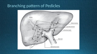
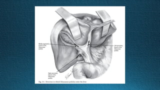
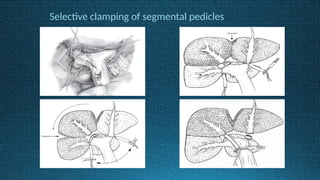
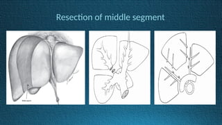
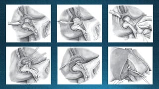
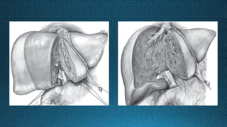
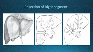
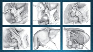
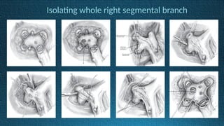
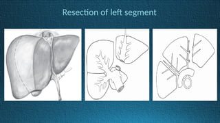
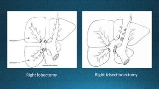
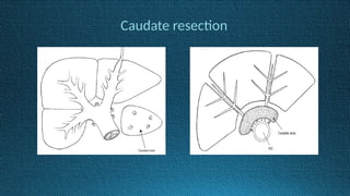
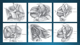
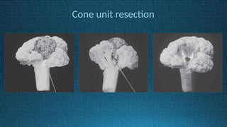
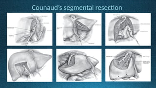
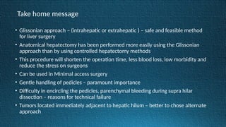
Glissonian pedicle approach
of
Liver Resection
History
• Partial liver resection and non-anatomical liver resection were common before 1950
• Lortat-Jacob and Robert and Honjo and Araki performed anatomical right hepatectomy with dissection of
the hepatic artery, portal vein and bile duct in the hepatoduodenal ligament around 1950 { Infrafascial
approach } – Controlled hepatectomy
• Extrafascial transfissural approach – Ton That Tung in 1960
• Glissonian pedicle(Extrafascial)approach at the hepatic hilus was reported by Takasaki et al. in 1986
• Launois and Jamieson - described the intrahepatic Glissonian pedicle approach to liver resection in 1992
• Makuuchi et al. - to puncture and inject dye to the portal pedicle for confirming the resection area under
intraoperative ultrasonography.
• Glisson’s capsule – Connective tissue wrap
around the portal vein, the hepatic artery
and the bile duct (Francis Glisson in 1654)
• Glisson’s capsule forms a thick plate at the
inferior part of the liver – ‘hilar plate’
• Hilar plate connects to Cystic plate, Umbilical
- plate, Arantius plate
• Laennec’s capsule – Proper membrane of liver
by Counaud
Branching pattern of Pedicles
Takasaki segmentation
• RHV, MHV – Intersegmental
• LHV drains exclusively Left segment
Relationship of segments with hepatic veins
Caudate area
• A – Superior part of IVC
• B – Bifurcation of Rt primary pedicle
• C – Origin of Lt segmental branch
• D – Edge of caudate process
• The border between the left segment and the
caudate area is determined by the anterior
paracaval triangle (Traingle ABC)
• The border between the right segment and
the caudate area is determined by the lateral
paracaval triangle (Triangle ABD)
Comparision with other classification systems
• Each Couinaud’s segment does not necessarily contain only one
single tertiary branch.
• One Couinaud’s segment consists of several cone units.
• Usually from two to eight tertiary branches ramify from a secondary
branch.
Position of Glissonian pedicles inside the segments
Relation between pedicles and hepatic vein
Upper part of liver Lower part of liver
Cone unit
• The area fed by each of the tertiary
branches is called a “cone unit”
• One cone unit is the smallest unit
for which a tertiary branch can be
transected selectively
Instruments
Boomerang
retractor
Side
retractor
Kent retractor
Glisson’s foreceps Sonosurg (CUSA)
Selective clamping of segmental pedicles
Resection of middle segment
Resection of Right segment
Isolating whole right segmental branch
Resection of left segment
Right lobectomy Right trisectionectomy
Caudate resection
Cone unit resection
Counaud’s segmental resection
Take home message
• Glissonian approach – (intrahepatic or extrahepatic ) – safe and feasible method
for liver surgery
• Anatomical hepatectomy has been performed more easily using the Glissonian
approach than by using controlled hepatectomy methods
• This procedure will shorten the operation time, less blood loss, low morbidity and
reduce the stress on surgeons
• Can be used in Minimal access surgery
• Gentle handling of pedicles – paramount importance
• Difficulty in encircling the pedicles, parenchymal bleeding during supra hilar
dissection – reasons for technical failure
• Tumors located immediately adjacent to hepatic hilum – better to chose alternate
approach
Thank you

Glissonian pedicle approach presentation.pptx

  • 1.
  • 2.
    History • Partial liverresection and non-anatomical liver resection were common before 1950 • Lortat-Jacob and Robert and Honjo and Araki performed anatomical right hepatectomy with dissection of the hepatic artery, portal vein and bile duct in the hepatoduodenal ligament around 1950 { Infrafascial approach } – Controlled hepatectomy • Extrafascial transfissural approach – Ton That Tung in 1960 • Glissonian pedicle(Extrafascial)approach at the hepatic hilus was reported by Takasaki et al. in 1986 • Launois and Jamieson - described the intrahepatic Glissonian pedicle approach to liver resection in 1992 • Makuuchi et al. - to puncture and inject dye to the portal pedicle for confirming the resection area under intraoperative ultrasonography.
  • 4.
    • Glisson’s capsule– Connective tissue wrap around the portal vein, the hepatic artery and the bile duct (Francis Glisson in 1654) • Glisson’s capsule forms a thick plate at the inferior part of the liver – ‘hilar plate’ • Hilar plate connects to Cystic plate, Umbilical - plate, Arantius plate • Laennec’s capsule – Proper membrane of liver by Counaud
  • 5.
  • 6.
  • 7.
    • RHV, MHV– Intersegmental • LHV drains exclusively Left segment Relationship of segments with hepatic veins
  • 8.
    Caudate area • A– Superior part of IVC • B – Bifurcation of Rt primary pedicle • C – Origin of Lt segmental branch • D – Edge of caudate process • The border between the left segment and the caudate area is determined by the anterior paracaval triangle (Traingle ABC) • The border between the right segment and the caudate area is determined by the lateral paracaval triangle (Triangle ABD)
  • 9.
    Comparision with otherclassification systems
  • 10.
    • Each Couinaud’ssegment does not necessarily contain only one single tertiary branch. • One Couinaud’s segment consists of several cone units. • Usually from two to eight tertiary branches ramify from a secondary branch.
  • 11.
    Position of Glissonianpedicles inside the segments
  • 12.
    Relation between pediclesand hepatic vein Upper part of liver Lower part of liver
  • 13.
    Cone unit • Thearea fed by each of the tertiary branches is called a “cone unit” • One cone unit is the smallest unit for which a tertiary branch can be transected selectively
  • 14.
  • 15.
  • 18.
    Selective clamping ofsegmental pedicles
  • 19.
  • 22.
  • 24.
    Isolating whole rightsegmental branch
  • 25.
  • 27.
    Right lobectomy Righttrisectionectomy
  • 28.
  • 30.
  • 31.
  • 32.
    Take home message •Glissonian approach – (intrahepatic or extrahepatic ) – safe and feasible method for liver surgery • Anatomical hepatectomy has been performed more easily using the Glissonian approach than by using controlled hepatectomy methods • This procedure will shorten the operation time, less blood loss, low morbidity and reduce the stress on surgeons • Can be used in Minimal access surgery • Gentle handling of pedicles – paramount importance • Difficulty in encircling the pedicles, parenchymal bleeding during supra hilar dissection – reasons for technical failure • Tumors located immediately adjacent to hepatic hilum – better to chose alternate approach
  • 33.