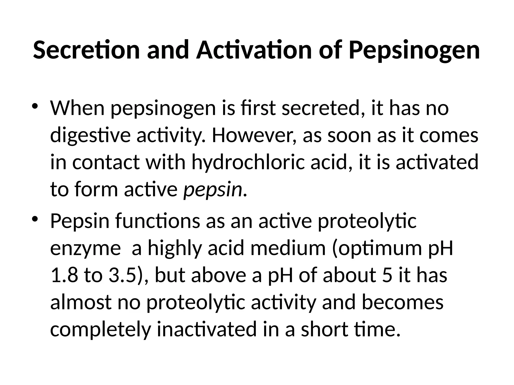
Gastrointestinal secretions
Saliva
Glands of Salivation
• Parotid glands: secrete entirely serous type
• Submandibular glands: both serous and
mucus type
• Sublingual glands: both serous and mucus
type
• Buccal glands secrete only mucus
Types of secretion
• Serous type of secretions contains ptyalin, an
enzyme for the digesting starches
• Mucus secretions contain mucin for
lubrication and for surface protective
purposes
Composition and function of saliva
• Sodium ions are actively reabsorbed from all the salivary ducts
and potassium ions are actively secreted in exchange for sodium.
• Bicarbonate ions are secreted by ductal epithelium into lumen of
the duct
• Ptyalin (α amylase) is responsible for the digestion of
carbohydrates
• Mucus, glycoproteins
• Thiocyanate ions
• Lysozymes
• Immunoglobulins (IgA)
Functions of Saliva
• Preparation of food for swallowing
• Appreciation of taste
• Digestive function
• Cleansing and protective functions
• Role in speech
• Excretory function
• Regulation of body temperature
• Regulation of water balance
Regulation of salivation
• Salivation is mainly controlled by autonomic
nervous system. Both branches of autonomic
nervous system stimulate salivation, but
parasympathetic nervous system stimulate
much more strongly than the sympathetic
nervous system
Reflex regulation of salivary secretion
• Unconditioned reflex
• Conditioned reflex
Nervous Regulation of Salivary Secretion
• Salivary glands are controlled mainly by parasympathetic
nervous signals all the way from the superior and inferior
salivatory nuclei in the brain stem.
• The salivatory nuclei are located approximately at the
juncture of the medulla and pons and are excited by both
taste and tactile stimuli from the tongue and other areas
of the mouth and pharynx. Many taste stimuli, especially
the sour taste (caused by acids), elicit copious secretion
of saliva—often 8 to 20 times the basal rate of secretion.
• Esophageal Secretion
• The esophageal secretions are entirely mucous
in character and principally provide lubrication
for swallowing. The main body of the
esophagus is lined with many simple mucous
glands. At the gastric end and to a lesser
extent in the initial portion of the esophagus,
there are also many compound mucous glands.
Gastric Secretion
• Oxyntic glands: these secrete hydrochloric
acid, pepsinogen, intrinsic factor and mucus.
These are located on the inside surfaces of
body and fundus of the stomach
• Pyloric glands: mainly secrete mucus and
hormone gastrin. These are located in the
antral portion of the stomach
Secretion from oxyntic glands
A typical oxyntic gland is composed of three types
of cells
• Mucus cells which secrete mainly mucus but also
some pepsinogen
• Peptic or chief cells which secrete mainly large
quantities of pepsinogen
• Parietal or oxyntic cells which secrete
hydrochloric acid and intrinsic factor
• Mucous Cells
• The thick mucous present in the gastric juice is
responsible for the protection of the gastric
wall. The mucous protects the stomach wall
from irritation or mechanical injury. It prevents
the back diffusion of hydrogen ions into the
gastric mucosa. Beneath the mucous layer, a
layer rich in bicarbonate ion neutralizes
hydrogen ions.
• Chief Cells
• Protein digestion begins in the stomach
because of the activity of chief cells. These
cells secrete the inactive precursor protein,
pepsinogen, which is activated to the
proteolytic enzyme, pepsin, in the presence of
acid and small amounts of active pepsin.
Pepsin functions optimally at a pH of
approximately 2.
Parietal Cells
Parietal cells
• Secrete hydrogen ions
• Activate protein digestive enzymes such as pepsinogen.
• Create a harsh environment for bacterial growth.
• Secrete intrinsic factor which binds vitamin B12 in
protein rich foods, such as meat, to prevent its
degradation in small intestine and to allow absorption
in the terminal ileum
Basic mechanism of hydrochloric acid secretion
• Parietal cells secrete HCl into the lumen of the stomach
and, concurrently, absorb bicarbonate ion into the blood
stream as follows
• In the parietal cells, CO2 and water are converted to
hydrogen ions and bicarbonate ions, catalyzed by carbonic
anhydrase
• Hydrogen ion is secreted into the lumen by H+
-K+
pump (H+
-
K+
ATPase). Chloride is secreted along with the hydrogen
ions, thus the secretion product of parietal cells is HCl
• The bicarbonate ions produced in the cells is
absorbed into the bloodstream in exchange
for chloride ions (Cl-
- HCO3
-
exchange). As
bicarbonate ion is added to the venous blood,
the pH of the blood increases (“alkaline
tide”). (Eventually this bicarbonate ion will be
secreted in pancreatic secretions to neutralize
hydrogen ions in the small intestine.)
Stimulation of gastric hydrogen ion secretion
• Vagal stimulation
– Increases hydrogen ion secretion by a direct
pathway and an indirect pathway.
– In the direct path, the vagus nerve innervates G
cells and stimulates gastrin secretion directly. The
neurotransmitter at these synapses is
Acetylcholine.
• In the indirect path, the vagus nerve innervates G
cells and stimulates gastrin secretion, which then
stimulates hydrogen ion secretion by an endocrine
action. The neurotransmitter at these synapses is
Gastrin releasing peptide.
Gastrin
– Is released in response to eating a meal (small
peptides, distention of the stomach, vagal
stimulation).
– Stimulates hydrogen ion secretion by interacting
with cholecystokininB (CCKB) receptor on the
parietal cells.
Histamine
– Is released from enterochromaffin-like (ECL) cells
in the gastric mucosa and diffuses to the nearby
parietal cells.
– Stimulates hydrogen ion secretion by activating H2
receptors on the parietal cell membrane.
Inhibition of gastric hydrogen ion secretion
• Negative feedback mechanisms inhibit the secretion of
hydrogen ions by parietal cells.
• The presence of food in the small intestine initiates a
reverse enterogastric reflex, transmitted through the
myenteric nervous system as well as through extrinsic
sympathetic and vagus nerves, that inhibits stomach
secretion. This reflex can be initiated by distending the
small bowel, by the presence of acid in the upper
intestine, by the presence of protein breakdown products,
or by irritation of the mucosa.
Somatostatin
– Inhibits gastric hydrogen ion secretion by a direct
pathway and an indirect pathway.
– In the direct pathway, somatostatin antagonizes
the stimulatory action of histamine on hydrogen
ion secretion.
– In the indirect pathway, somatostatin inhibits
release of histamine and gastrin, thus decreasing
hydrogen ion secretion indirectly.
Secretion and Activation of Pepsinogen
• When pepsinogen is first secreted, it has no
digestive activity. However, as soon as it comes
in contact with hydrochloric acid, it is activated
to form active pepsin.
• Pepsin functions as an active proteolytic
enzyme a highly acid medium (optimum pH
1.8 to 3.5), but above a pH of about 5 it has
almost no proteolytic activity and becomes
completely inactivated in a short time.
• Secretion of Intrinsic Factor. The substance intrinsic
factor, essential for absorption of vitamin B12 in the
ileum, is secreted by the parietal cells along with the
secretion of hydrochloric acid.
• When the acid-producing parietal cells of the
stomach are destroyed the person develops not only
achlorhydria (lack of stomach acid secretion) but
often also pernicious anemia because of failure of
maturation of the red blood cells in the absence of
vitamin B12 stimulation of the bone marrow.
Pancreas
• Pancreas is a dual organ having two functions
• Endocrine function: involves production of
hormones like insulin
• Exocrine function: involves secretion of
digestive juice- pancreatic juice
Pancreatic secretion
Anatomy of the exocrine part of the pancreas
• It is made up of acinar cells
• Acinar cells contain zymogen granules, which possess
digestive juices
• The ducts arising from acini join together to form
intralobular duct
• Intralobular ducts unite to form main duct of pancreas
called Wirsung’s duct
• Wirsung’s duct joins common bile duct to form ampulla
of vater which opens into the duodenum
Pancreatic secretion
• Contains high concentration of HCO3 whose
purpose is to neutralize the digestive enzymes
reaching the duodenum
• Contains enzymes for digesting all 3 major
types of food: proteins, carbohydrates and fats
Enzymatic components
• The more important of enzymes are
• Trypsin which is activated from trypsinogen in
the presence of enzyme called enterokinase.
Enterokinase is secreted by intestinal mucosa
when chyme comes on contact with mucosa
• Chymotrypsinogen is activated by trypsin to
form chymotrypsin
• Procarboxyploypeptidase is also activated in
the presence of trypsin to form
carboxypolypeptidase. It splits some peptides
into aminoacids.
• Pancreatic lipase hydrolyzes neutral fats into
fatty acids and monoglyceride
• Cholesterol esterase causes hydrolysis of
cholesterol ester and phospholipase splits fatty
acids from phospholipids.
• Pancreatic amylase hydrolyzes starches,
glycogen and most other carbohydrates
except cellulose to form disaccharides and a
few trisaccharides.
• Trypsin inhibitor is secreted into the acini of
pancreas and it prevents the activation of
trypsin both inside the secretory cells and in
the acini and ducts of the pancreas
Secretion of Bicarbonate ions
• Carbon dioxide diffuses to the interior of the
cell from the blood and combines with the
water in the presence of carbonic anhydrase to
form carbonic acid. This carbonic acid in turn
dissociates into bicarbonate ions and hydrogen
ions. The bicarbonate ions are actively
transported in exchange for the chloride ions
and enters into the lumen of the duct
Regulation of pancreatic secretion
• Stimuli of pancreatic secretion are
• Acetylcholine
• Choleccystokinin secreted when food enters small
intestine
• Secretin is secreted in response to acidic food
• Acetylcholine and cholecystokinin cause production of
large quantities of digestive enzymes whereas secretin
stimulates secretion of large quantities of water and
bicarbonate
Phases of pancreatic secretion
• Cephalic phase
• Gastric phase
• Intestinal phase
Secretions of bile by liver
• Normally 600-1200 ml /day bile is secreted by
the liver. Bile contains bile salts, phospholipids,
cholesterol, and bile pigments.
• Secretion of bile Bile is secreted in two stages
• Initial portion is secreted by liver hepatocytes.
This secretion contains large amounts of bile
acids, cholesterol and other organic
constituents. It is secreted into minute bile
canaliculi.
Storage of bile
• Most of the bile from liver enters the gallbladder
where it is stored. It is released from gallbladder
into the duodenum whenever required. The
maximum volume of the gallbladder is 30 -60 ml.
A large amount of water and electrolytes (except
calcium and potassium) are absorbed resulting in
high concentration of bile salts, bile pigments,
cholesterol, fatty acids and lecithin
Composition and function of bile
• The most abundant substances secreted in the
bile are bile salts, which account for about one
half of the total solutes also in the bile. Also
secreted or excreted in large concentrations
are bilirubin, cholesterol, lecithin, and the
usual electrolytes of plasma.
• Bile salts
• The liver cells synthesize about 6 grams of bile salts
daily. The precursor of the bile salts is cholesterol,
which is either present in the diet or synthesized in
the liver cells during the course of fat metabolism.
• Bile salts are amphipathic molecules and are
emulsifier. They are potassium or sodium salts of
bile acids, which are conjugated with glycine and to
lesser amount with taurine
• The precursor of the bile salts is cholesterol,
which is either present in the diet or
synthesized in the liver cells during the course
of fat metabolism.
• The cholesterol is first converted to cholic acid
or chenodeoxycholic acid in about equal
quantities.
These acids in turn combine principally with
glycine and to a lesser extent with taurine to
form glyco- and tauro conjugated bile acids.
The salts of these acids, mainly sodium salts,
are then secreted in the bile.
Due to bacterial action in the intestine the
primary bile acids are converted into secondary
bile acids which are transported back to the
liver through enterohepatic circulation.
• Role of Secretin in Helping to Control Bile
Secretion.
• In addition to the strong stimulating effect of
bile acids to cause bile secretion, the hormone
secretin that also stimulates pancreatic
secretion increases bile secretion, sometimes
more than doubling its secretion for several
hours after a meal.
• Bile pigments
• Bile pigments are the excretory products in the bile.
Bilirubin and biliverdin are the two bile pigments and
bilirubin is the major bile pigment in human being.
• Bilirubin: A major bile pigment, bilirubin is a lipid
soluble metabolite of haemoglobin. Transported to
the liver attached to the protein, it is then
conjugated and excreted as water soluble
glucuronides. These give a golden color to bile.
• Biliverdin: Heme splits into iron and pigment
biliverdin which then reduces to bilirubin.
• Stercobilin: It is produced from metabolism of
bilirubin by intestinal bacteria. It gives brown
color to the stool.
• Phospholipids (mainly lecithin)
• It is insoluble in water but are solubilized by
bile salt micelles
• Cholesterol
• It is present in small amount. It is insoluble in
water and must be solubilized by bile salt
micelles before it can be secreted in the bile.
Control of bile secretion and gall bladder
contraction
• Secretin causes secretion of bicarbonate ions and
fluid into bile canalicular ducts
• Secretion of bile salts by hepatocytes is directly
proportional to hepatic portal vein concentration
of bile salts
• Cholecystokinin causes gallbladder contraction
and sphincter of Oddi relaxation
Functions of bile salts
• Emulsification of fats
• Absorption of fats
• Choleretic action
• Cholagogue action
• Laxative action
• Prevention of gallstone formation
Small intestine secretions
Secretion of mucus by Brunner’s glands
• A large number of compound mucous glands,
called Brunner’s glands, is located in the wall
of the first few centimeters of the duodenum,
mainly between the pylorus of the stomach
and the papilla of Vater where pancreatic
secretion and bile empty into the duodenum.
• These glands secrete large amounts of alkaline
mucus in response to
• (1) tactile or irritating stimuli on the duodenal
mucosa;
• (2) vagal stimulation, which causes increased
Brunner’s glands secretion concurrently with
increase in stomach secretion; and
• (3) gastrointestinal hormones, especially secretin.
• The function of the mucus secreted by
Brunner’s glands is to protect the duodenal
wall from digestion by the highly acid gastric
juice emptying from the stomach.
• Mucus also contains large amount of
bicarbonate ions which neutralize the acid
entering from the stomach.
Secretion of Intestinal Digestive
Juices by the Crypts of Lieberkühn
• Over the entire surface of the small intestine are
located small pits called crypts of Lieberkühn
• These crypts lie between the intestinal villi. The
surfaces of both the crypts and the villi are
covered by an epithelium composed of two types
of cells:
• (1) a moderate number of goblet cells, which
secrete mucus that lubricates and protects the
• intestinal surfaces
• (2) a large number of enterocytes, which, in
the crypts, secrete large quantities of water
and electrolytes and, over the surfaces of
adjacent villi, reabsorb the water and
electrolytes along with end products of
digestion.
• Digestive Enzymes in the Small Intestinal
Secretion
• The enterocytes of the mucosa, especially
those that cover the villi, do contain digestive
enzymes that digest specific food substances
while they are being absorbed through the
epithelium.
• These enzymes are the following:
• (1) several peptidases for splitting small peptides
into amino acids
• (2) four enzymes—sucrase, maltase isomaltase,
and lactase—for splitting disaccharides into
monosaccharides
• (3) small amounts of intestinal lipase for splitting
neutral fats into glycerol and fatty acids.
• Regulating small intestine secretion are
controlled by local enteric nervous reflexes,
especially reflexes initiated by tactile or
irritative stimuli from the chyme in the
intestines.
Secretions of the Large Intestine
• Mucus Secretion. The mucosa of the large
intestine, like that of the small intestine, has
many crypts of Lieberkühn; however, unlike
the small intestine, there are no villi. The
epithelial cells contain almost no enzymes.
Instead, they consist mainly of mucous cells
that secrete only mucus.
• Stimulation of the pelvic nerves from the
spinal cord, which carry parasympathetic
innervation to the distal one half to two thirds
of the large intestine, also can cause marked
increase in mucus secretion. This occurs along
with increase in peristaltic motility of the
colon
Functions of mucus
• Mucus in the large intestine protects the
intestinal wall against excoriation,
• It provides an adherent medium for holding
fecal matter together.
• It protects the intestinal wall from the great
amount of bacterial activity that takes place
inside the feces
• The mucus plus the alkalinity of the secretion
(pH of 8.0 caused by large amounts of sodium
bicarbonate) provides a barrier to keep acids
formed in the feces from attacking the
intestinal wall.

gitsecretions physiology bds powerpoint.pptx

  • 1.
  • 3.
    Saliva Glands of Salivation •Parotid glands: secrete entirely serous type • Submandibular glands: both serous and mucus type • Sublingual glands: both serous and mucus type • Buccal glands secrete only mucus
  • 4.
    Types of secretion •Serous type of secretions contains ptyalin, an enzyme for the digesting starches • Mucus secretions contain mucin for lubrication and for surface protective purposes
  • 5.
    Composition and functionof saliva • Sodium ions are actively reabsorbed from all the salivary ducts and potassium ions are actively secreted in exchange for sodium. • Bicarbonate ions are secreted by ductal epithelium into lumen of the duct • Ptyalin (α amylase) is responsible for the digestion of carbohydrates • Mucus, glycoproteins • Thiocyanate ions • Lysozymes • Immunoglobulins (IgA)
  • 6.
    Functions of Saliva •Preparation of food for swallowing • Appreciation of taste • Digestive function • Cleansing and protective functions • Role in speech • Excretory function • Regulation of body temperature • Regulation of water balance
  • 7.
    Regulation of salivation •Salivation is mainly controlled by autonomic nervous system. Both branches of autonomic nervous system stimulate salivation, but parasympathetic nervous system stimulate much more strongly than the sympathetic nervous system
  • 8.
    Reflex regulation ofsalivary secretion • Unconditioned reflex • Conditioned reflex
  • 9.
    Nervous Regulation ofSalivary Secretion • Salivary glands are controlled mainly by parasympathetic nervous signals all the way from the superior and inferior salivatory nuclei in the brain stem. • The salivatory nuclei are located approximately at the juncture of the medulla and pons and are excited by both taste and tactile stimuli from the tongue and other areas of the mouth and pharynx. Many taste stimuli, especially the sour taste (caused by acids), elicit copious secretion of saliva—often 8 to 20 times the basal rate of secretion.
  • 10.
    • Esophageal Secretion •The esophageal secretions are entirely mucous in character and principally provide lubrication for swallowing. The main body of the esophagus is lined with many simple mucous glands. At the gastric end and to a lesser extent in the initial portion of the esophagus, there are also many compound mucous glands.
  • 11.
    Gastric Secretion • Oxynticglands: these secrete hydrochloric acid, pepsinogen, intrinsic factor and mucus. These are located on the inside surfaces of body and fundus of the stomach • Pyloric glands: mainly secrete mucus and hormone gastrin. These are located in the antral portion of the stomach
  • 12.
    Secretion from oxynticglands A typical oxyntic gland is composed of three types of cells • Mucus cells which secrete mainly mucus but also some pepsinogen • Peptic or chief cells which secrete mainly large quantities of pepsinogen • Parietal or oxyntic cells which secrete hydrochloric acid and intrinsic factor
  • 13.
    • Mucous Cells •The thick mucous present in the gastric juice is responsible for the protection of the gastric wall. The mucous protects the stomach wall from irritation or mechanical injury. It prevents the back diffusion of hydrogen ions into the gastric mucosa. Beneath the mucous layer, a layer rich in bicarbonate ion neutralizes hydrogen ions.
  • 14.
    • Chief Cells •Protein digestion begins in the stomach because of the activity of chief cells. These cells secrete the inactive precursor protein, pepsinogen, which is activated to the proteolytic enzyme, pepsin, in the presence of acid and small amounts of active pepsin. Pepsin functions optimally at a pH of approximately 2.
  • 15.
    Parietal Cells Parietal cells •Secrete hydrogen ions • Activate protein digestive enzymes such as pepsinogen. • Create a harsh environment for bacterial growth. • Secrete intrinsic factor which binds vitamin B12 in protein rich foods, such as meat, to prevent its degradation in small intestine and to allow absorption in the terminal ileum
  • 17.
    Basic mechanism ofhydrochloric acid secretion • Parietal cells secrete HCl into the lumen of the stomach and, concurrently, absorb bicarbonate ion into the blood stream as follows • In the parietal cells, CO2 and water are converted to hydrogen ions and bicarbonate ions, catalyzed by carbonic anhydrase • Hydrogen ion is secreted into the lumen by H+ -K+ pump (H+ - K+ ATPase). Chloride is secreted along with the hydrogen ions, thus the secretion product of parietal cells is HCl
  • 18.
    • The bicarbonateions produced in the cells is absorbed into the bloodstream in exchange for chloride ions (Cl- - HCO3 - exchange). As bicarbonate ion is added to the venous blood, the pH of the blood increases (“alkaline tide”). (Eventually this bicarbonate ion will be secreted in pancreatic secretions to neutralize hydrogen ions in the small intestine.)
  • 20.
    Stimulation of gastrichydrogen ion secretion • Vagal stimulation – Increases hydrogen ion secretion by a direct pathway and an indirect pathway. – In the direct path, the vagus nerve innervates G cells and stimulates gastrin secretion directly. The neurotransmitter at these synapses is Acetylcholine.
  • 21.
    • In theindirect path, the vagus nerve innervates G cells and stimulates gastrin secretion, which then stimulates hydrogen ion secretion by an endocrine action. The neurotransmitter at these synapses is Gastrin releasing peptide.
  • 22.
    Gastrin – Is releasedin response to eating a meal (small peptides, distention of the stomach, vagal stimulation). – Stimulates hydrogen ion secretion by interacting with cholecystokininB (CCKB) receptor on the parietal cells.
  • 23.
    Histamine – Is releasedfrom enterochromaffin-like (ECL) cells in the gastric mucosa and diffuses to the nearby parietal cells. – Stimulates hydrogen ion secretion by activating H2 receptors on the parietal cell membrane.
  • 24.
    Inhibition of gastrichydrogen ion secretion • Negative feedback mechanisms inhibit the secretion of hydrogen ions by parietal cells. • The presence of food in the small intestine initiates a reverse enterogastric reflex, transmitted through the myenteric nervous system as well as through extrinsic sympathetic and vagus nerves, that inhibits stomach secretion. This reflex can be initiated by distending the small bowel, by the presence of acid in the upper intestine, by the presence of protein breakdown products, or by irritation of the mucosa.
  • 25.
    Somatostatin – Inhibits gastrichydrogen ion secretion by a direct pathway and an indirect pathway. – In the direct pathway, somatostatin antagonizes the stimulatory action of histamine on hydrogen ion secretion. – In the indirect pathway, somatostatin inhibits release of histamine and gastrin, thus decreasing hydrogen ion secretion indirectly.
  • 26.
    Secretion and Activationof Pepsinogen • When pepsinogen is first secreted, it has no digestive activity. However, as soon as it comes in contact with hydrochloric acid, it is activated to form active pepsin. • Pepsin functions as an active proteolytic enzyme a highly acid medium (optimum pH 1.8 to 3.5), but above a pH of about 5 it has almost no proteolytic activity and becomes completely inactivated in a short time.
  • 27.
    • Secretion ofIntrinsic Factor. The substance intrinsic factor, essential for absorption of vitamin B12 in the ileum, is secreted by the parietal cells along with the secretion of hydrochloric acid. • When the acid-producing parietal cells of the stomach are destroyed the person develops not only achlorhydria (lack of stomach acid secretion) but often also pernicious anemia because of failure of maturation of the red blood cells in the absence of vitamin B12 stimulation of the bone marrow.
  • 28.
    Pancreas • Pancreas isa dual organ having two functions • Endocrine function: involves production of hormones like insulin • Exocrine function: involves secretion of digestive juice- pancreatic juice
  • 29.
    Pancreatic secretion Anatomy ofthe exocrine part of the pancreas • It is made up of acinar cells • Acinar cells contain zymogen granules, which possess digestive juices • The ducts arising from acini join together to form intralobular duct • Intralobular ducts unite to form main duct of pancreas called Wirsung’s duct • Wirsung’s duct joins common bile duct to form ampulla of vater which opens into the duodenum
  • 30.
    Pancreatic secretion • Containshigh concentration of HCO3 whose purpose is to neutralize the digestive enzymes reaching the duodenum • Contains enzymes for digesting all 3 major types of food: proteins, carbohydrates and fats
  • 31.
    Enzymatic components • Themore important of enzymes are • Trypsin which is activated from trypsinogen in the presence of enzyme called enterokinase. Enterokinase is secreted by intestinal mucosa when chyme comes on contact with mucosa • Chymotrypsinogen is activated by trypsin to form chymotrypsin
  • 32.
    • Procarboxyploypeptidase isalso activated in the presence of trypsin to form carboxypolypeptidase. It splits some peptides into aminoacids. • Pancreatic lipase hydrolyzes neutral fats into fatty acids and monoglyceride • Cholesterol esterase causes hydrolysis of cholesterol ester and phospholipase splits fatty acids from phospholipids.
  • 33.
    • Pancreatic amylasehydrolyzes starches, glycogen and most other carbohydrates except cellulose to form disaccharides and a few trisaccharides. • Trypsin inhibitor is secreted into the acini of pancreas and it prevents the activation of trypsin both inside the secretory cells and in the acini and ducts of the pancreas
  • 34.
    Secretion of Bicarbonateions • Carbon dioxide diffuses to the interior of the cell from the blood and combines with the water in the presence of carbonic anhydrase to form carbonic acid. This carbonic acid in turn dissociates into bicarbonate ions and hydrogen ions. The bicarbonate ions are actively transported in exchange for the chloride ions and enters into the lumen of the duct
  • 36.
    Regulation of pancreaticsecretion • Stimuli of pancreatic secretion are • Acetylcholine • Choleccystokinin secreted when food enters small intestine • Secretin is secreted in response to acidic food • Acetylcholine and cholecystokinin cause production of large quantities of digestive enzymes whereas secretin stimulates secretion of large quantities of water and bicarbonate
  • 37.
    Phases of pancreaticsecretion • Cephalic phase • Gastric phase • Intestinal phase
  • 38.
    Secretions of bileby liver • Normally 600-1200 ml /day bile is secreted by the liver. Bile contains bile salts, phospholipids, cholesterol, and bile pigments. • Secretion of bile Bile is secreted in two stages • Initial portion is secreted by liver hepatocytes. This secretion contains large amounts of bile acids, cholesterol and other organic constituents. It is secreted into minute bile canaliculi.
  • 40.
    Storage of bile •Most of the bile from liver enters the gallbladder where it is stored. It is released from gallbladder into the duodenum whenever required. The maximum volume of the gallbladder is 30 -60 ml. A large amount of water and electrolytes (except calcium and potassium) are absorbed resulting in high concentration of bile salts, bile pigments, cholesterol, fatty acids and lecithin
  • 41.
    Composition and functionof bile • The most abundant substances secreted in the bile are bile salts, which account for about one half of the total solutes also in the bile. Also secreted or excreted in large concentrations are bilirubin, cholesterol, lecithin, and the usual electrolytes of plasma.
  • 43.
    • Bile salts •The liver cells synthesize about 6 grams of bile salts daily. The precursor of the bile salts is cholesterol, which is either present in the diet or synthesized in the liver cells during the course of fat metabolism. • Bile salts are amphipathic molecules and are emulsifier. They are potassium or sodium salts of bile acids, which are conjugated with glycine and to lesser amount with taurine
  • 44.
    • The precursorof the bile salts is cholesterol, which is either present in the diet or synthesized in the liver cells during the course of fat metabolism. • The cholesterol is first converted to cholic acid or chenodeoxycholic acid in about equal quantities.
  • 45.
    These acids inturn combine principally with glycine and to a lesser extent with taurine to form glyco- and tauro conjugated bile acids. The salts of these acids, mainly sodium salts, are then secreted in the bile. Due to bacterial action in the intestine the primary bile acids are converted into secondary bile acids which are transported back to the liver through enterohepatic circulation.
  • 46.
    • Role ofSecretin in Helping to Control Bile Secretion. • In addition to the strong stimulating effect of bile acids to cause bile secretion, the hormone secretin that also stimulates pancreatic secretion increases bile secretion, sometimes more than doubling its secretion for several hours after a meal.
  • 47.
    • Bile pigments •Bile pigments are the excretory products in the bile. Bilirubin and biliverdin are the two bile pigments and bilirubin is the major bile pigment in human being. • Bilirubin: A major bile pigment, bilirubin is a lipid soluble metabolite of haemoglobin. Transported to the liver attached to the protein, it is then conjugated and excreted as water soluble glucuronides. These give a golden color to bile.
  • 48.
    • Biliverdin: Hemesplits into iron and pigment biliverdin which then reduces to bilirubin. • Stercobilin: It is produced from metabolism of bilirubin by intestinal bacteria. It gives brown color to the stool.
  • 49.
    • Phospholipids (mainlylecithin) • It is insoluble in water but are solubilized by bile salt micelles • Cholesterol • It is present in small amount. It is insoluble in water and must be solubilized by bile salt micelles before it can be secreted in the bile.
  • 50.
    Control of bilesecretion and gall bladder contraction • Secretin causes secretion of bicarbonate ions and fluid into bile canalicular ducts • Secretion of bile salts by hepatocytes is directly proportional to hepatic portal vein concentration of bile salts • Cholecystokinin causes gallbladder contraction and sphincter of Oddi relaxation
  • 51.
    Functions of bilesalts • Emulsification of fats • Absorption of fats • Choleretic action • Cholagogue action • Laxative action • Prevention of gallstone formation
  • 52.
    Small intestine secretions Secretionof mucus by Brunner’s glands • A large number of compound mucous glands, called Brunner’s glands, is located in the wall of the first few centimeters of the duodenum, mainly between the pylorus of the stomach and the papilla of Vater where pancreatic secretion and bile empty into the duodenum.
  • 53.
    • These glandssecrete large amounts of alkaline mucus in response to • (1) tactile or irritating stimuli on the duodenal mucosa; • (2) vagal stimulation, which causes increased Brunner’s glands secretion concurrently with increase in stomach secretion; and • (3) gastrointestinal hormones, especially secretin.
  • 54.
    • The functionof the mucus secreted by Brunner’s glands is to protect the duodenal wall from digestion by the highly acid gastric juice emptying from the stomach. • Mucus also contains large amount of bicarbonate ions which neutralize the acid entering from the stomach.
  • 55.
    Secretion of IntestinalDigestive Juices by the Crypts of Lieberkühn
  • 56.
    • Over theentire surface of the small intestine are located small pits called crypts of Lieberkühn • These crypts lie between the intestinal villi. The surfaces of both the crypts and the villi are covered by an epithelium composed of two types of cells: • (1) a moderate number of goblet cells, which secrete mucus that lubricates and protects the • intestinal surfaces
  • 57.
    • (2) alarge number of enterocytes, which, in the crypts, secrete large quantities of water and electrolytes and, over the surfaces of adjacent villi, reabsorb the water and electrolytes along with end products of digestion.
  • 58.
    • Digestive Enzymesin the Small Intestinal Secretion • The enterocytes of the mucosa, especially those that cover the villi, do contain digestive enzymes that digest specific food substances while they are being absorbed through the epithelium.
  • 59.
    • These enzymesare the following: • (1) several peptidases for splitting small peptides into amino acids • (2) four enzymes—sucrase, maltase isomaltase, and lactase—for splitting disaccharides into monosaccharides • (3) small amounts of intestinal lipase for splitting neutral fats into glycerol and fatty acids.
  • 60.
    • Regulating smallintestine secretion are controlled by local enteric nervous reflexes, especially reflexes initiated by tactile or irritative stimuli from the chyme in the intestines.
  • 61.
    Secretions of theLarge Intestine • Mucus Secretion. The mucosa of the large intestine, like that of the small intestine, has many crypts of Lieberkühn; however, unlike the small intestine, there are no villi. The epithelial cells contain almost no enzymes. Instead, they consist mainly of mucous cells that secrete only mucus.
  • 62.
    • Stimulation ofthe pelvic nerves from the spinal cord, which carry parasympathetic innervation to the distal one half to two thirds of the large intestine, also can cause marked increase in mucus secretion. This occurs along with increase in peristaltic motility of the colon
  • 63.
    Functions of mucus •Mucus in the large intestine protects the intestinal wall against excoriation, • It provides an adherent medium for holding fecal matter together. • It protects the intestinal wall from the great amount of bacterial activity that takes place inside the feces
  • 64.
    • The mucusplus the alkalinity of the secretion (pH of 8.0 caused by large amounts of sodium bicarbonate) provides a barrier to keep acids formed in the feces from attacking the intestinal wall.