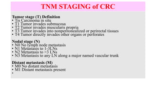
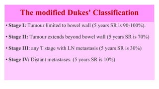
EXAMINATION
 DRE: lower rectal mass or ulcer
Liver : hepatomegaly in metastasis
Percussion: Ascites
Rarely: Sister Joseph or Verchow’s
LN
IMPORTANCE OF DRE
TNM STAGING of CRC
Tumor stage (T) Definition
• Tis Carcinoma in situ
• T1 Tumor invades submucosa
• T2 Tumor invades muscularis propria
• T3 Tumor invades into nonperitonealized or perirectal tissues
• T4 Tumor directly invades other organs or perforates
Nodal stage (N)
• N0 No lymph node metastasis
• N1 Metastasis to 1-3LNs
• N2 Metastasis to >3 LNs
• N3 Metastasis to any LN along a major named vascular trunk
Distant metastasis (M)
• M0 No distant metastasis
• M1 Distant metastasis present
•
The modified Dukes' Classification
• Stage I: Tumour limited to bowel wall (5 years SR is 90-100%).
• Stage II: Tumour extends beyond bowel wall (5 years SR is 70%)
• Stage III: any T stage with LN metastasis (5 years SR is 30%)
• Stage IV: Distant metastases. (5 years SR is 10%)
INVESTIGATIONS
1. Full colonoscopy to detect synchronous tumours (5%) and
metachronous tumours (10-40%)
2. EUS
3. A chest/abdominal/pelvic CT scan
4. MRI
5. Water-soluble contrast study (obstructing tumours).
6. PET scan
7. Preoperative CEA
CT SCAN
PET SCAN
CRC TREATMENT
SURGICAL TREATMENT is the curative treatment option
TYPES:
A.Open
B. Laparoscopic
 Hand-assisted
 Laparoscopic assisted
 Totally laparoscopic
LAPAROSCOPIC COLECTOMY
SURGICAL TREATMENT OF CRC
1. Caecal and ascending colon carcinoma → right hemicolectomy (RHC)
2. Hepatic flexure and transverse colon cancer → extended RHC
3. Splenic flexure and descending colon cancer → LHC
4. Sigmoid colon cancer → sigmoid colectomy (SC)
RHC & Extended RHC
LHC & SIGMOID COLECTOMY
SUBTOTAL & TOTAL COLECTOMY
TREATMENT OF CRC
Rectal cancer are treated:
A.Upper third cancer: high anterior resection (AR) with (TME)
B.Middle third cancer: low anterior resection (AR) with (TME)
C.Lower third cancer that is more than 2 cm above anal spincter: extended low
anterior resection (AR) with (TME).
D.Lower third cancer that is less than 2 cm above anal spincter:
abdominoperineal resection (APR) with (TME)
E.Locally advanced cancer or recurrent cancer, pelvic exenteration.
ANTERIOR RESECTION & AP RESECTION
1.Those with isolated systemic metastasis as pulmonary or
hepatic can be treated with excision of primary cancer
along with segmental or lobar hepatic or pulmonary
resection.
2.If the tumour is inoperable: diversion colostomy or
ileostomy or a bypass procedure is indicated.
3.Early cancer (CIS and malignant polyp with no deeper
invasion) can be treated with local excision or
polypectomy
Adjuvant Therapy
1. Systemic chemotherapy
2. Chemoradiation
3. Intraoperative radiation therapy (usually brachytherapy)
4. Target therapy
• Bevacizumab (anti-VEGF)
• Erlotinib (anti-EGF molecule)
• Cetuximab (anti-EGF monoclonal Ab)
BEVACIZUMAB, ERLOTINIB, & CETUXIMAB
Prevention and Screening
1. Fecal occult blood testing (FOBT) annually.
2. Flexible sigmoidoscopy (every 5 years)
3. Combination of FOBT annually with flexible sigmoidoscopy every 5
years
4. Colonoscopy (every 10 years) best method and allows biopsy as well as
it may be therapeutic to remove a polyp, stop a bleeder, or stent an
obstruction
5. Double-contrast barium enema
6. Computed tomographic colonography (virtual colonoscopy)
Premalignant anal conditions
Anal intraepithelial neoplasia (AIN or Bowen’s disease)
squamous cell carcinoma in situ of the anus
HPV types 16 and 18,
HIV infection.
Treatment include topical immunomodulators as imiquimod and topical
cytotoxic agents as 5-FU and surgical ablation.
Malignant anal conditions
1. Adenocarcinoma
2. Epidermoid Carcinoma
3. Buschke-Lowenstein tumour (variant of condylomata accuminata)
4. Basal cell carcinoma
5. Melanoma
Treatment
1. Chemoradiation
2. Sphincter-preserving wide local excision (WLE)
3. Radical excision (APR)

GIT onc 4.pptx

  • 2.
    EXAMINATION  DRE: lowerrectal mass or ulcer Liver : hepatomegaly in metastasis Percussion: Ascites Rarely: Sister Joseph or Verchow’s LN
  • 3.
  • 4.
    TNM STAGING ofCRC Tumor stage (T) Definition • Tis Carcinoma in situ • T1 Tumor invades submucosa • T2 Tumor invades muscularis propria • T3 Tumor invades into nonperitonealized or perirectal tissues • T4 Tumor directly invades other organs or perforates Nodal stage (N) • N0 No lymph node metastasis • N1 Metastasis to 1-3LNs • N2 Metastasis to >3 LNs • N3 Metastasis to any LN along a major named vascular trunk Distant metastasis (M) • M0 No distant metastasis • M1 Distant metastasis present •
  • 5.
    The modified Dukes'Classification • Stage I: Tumour limited to bowel wall (5 years SR is 90-100%). • Stage II: Tumour extends beyond bowel wall (5 years SR is 70%) • Stage III: any T stage with LN metastasis (5 years SR is 30%) • Stage IV: Distant metastases. (5 years SR is 10%)
  • 6.
    INVESTIGATIONS 1. Full colonoscopyto detect synchronous tumours (5%) and metachronous tumours (10-40%) 2. EUS 3. A chest/abdominal/pelvic CT scan 4. MRI 5. Water-soluble contrast study (obstructing tumours). 6. PET scan 7. Preoperative CEA
  • 7.
  • 8.
  • 9.
    CRC TREATMENT SURGICAL TREATMENTis the curative treatment option TYPES: A.Open B. Laparoscopic  Hand-assisted  Laparoscopic assisted  Totally laparoscopic
  • 10.
  • 11.
    SURGICAL TREATMENT OFCRC 1. Caecal and ascending colon carcinoma → right hemicolectomy (RHC) 2. Hepatic flexure and transverse colon cancer → extended RHC 3. Splenic flexure and descending colon cancer → LHC 4. Sigmoid colon cancer → sigmoid colectomy (SC)
  • 12.
  • 13.
    LHC & SIGMOIDCOLECTOMY
  • 14.
  • 15.
    TREATMENT OF CRC Rectalcancer are treated: A.Upper third cancer: high anterior resection (AR) with (TME) B.Middle third cancer: low anterior resection (AR) with (TME) C.Lower third cancer that is more than 2 cm above anal spincter: extended low anterior resection (AR) with (TME). D.Lower third cancer that is less than 2 cm above anal spincter: abdominoperineal resection (APR) with (TME) E.Locally advanced cancer or recurrent cancer, pelvic exenteration.
  • 16.
  • 17.
    1.Those with isolatedsystemic metastasis as pulmonary or hepatic can be treated with excision of primary cancer along with segmental or lobar hepatic or pulmonary resection. 2.If the tumour is inoperable: diversion colostomy or ileostomy or a bypass procedure is indicated. 3.Early cancer (CIS and malignant polyp with no deeper invasion) can be treated with local excision or polypectomy
  • 18.
    Adjuvant Therapy 1. Systemicchemotherapy 2. Chemoradiation 3. Intraoperative radiation therapy (usually brachytherapy) 4. Target therapy • Bevacizumab (anti-VEGF) • Erlotinib (anti-EGF molecule) • Cetuximab (anti-EGF monoclonal Ab)
  • 19.
  • 20.
    Prevention and Screening 1.Fecal occult blood testing (FOBT) annually. 2. Flexible sigmoidoscopy (every 5 years) 3. Combination of FOBT annually with flexible sigmoidoscopy every 5 years 4. Colonoscopy (every 10 years) best method and allows biopsy as well as it may be therapeutic to remove a polyp, stop a bleeder, or stent an obstruction 5. Double-contrast barium enema 6. Computed tomographic colonography (virtual colonoscopy)
  • 21.
    Premalignant anal conditions Analintraepithelial neoplasia (AIN or Bowen’s disease) squamous cell carcinoma in situ of the anus HPV types 16 and 18, HIV infection. Treatment include topical immunomodulators as imiquimod and topical cytotoxic agents as 5-FU and surgical ablation.
  • 22.
    Malignant anal conditions 1.Adenocarcinoma 2. Epidermoid Carcinoma 3. Buschke-Lowenstein tumour (variant of condylomata accuminata) 4. Basal cell carcinoma 5. Melanoma Treatment 1. Chemoradiation 2. Sphincter-preserving wide local excision (WLE) 3. Radical excision (APR)