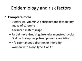
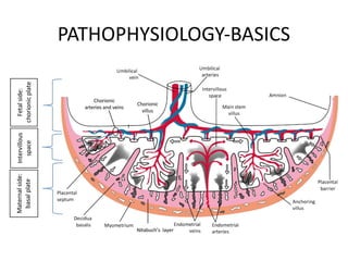
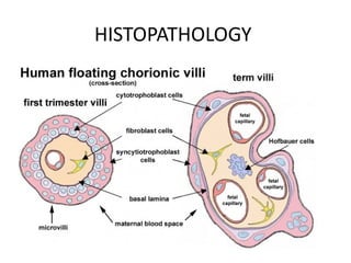
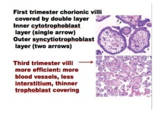
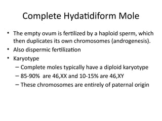
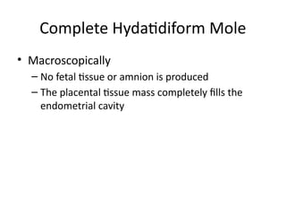
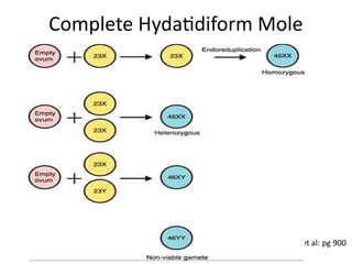
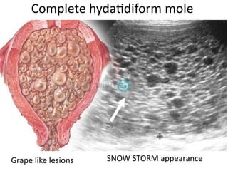
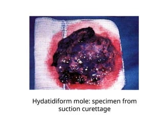
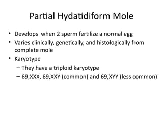
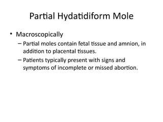
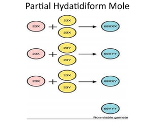
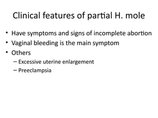
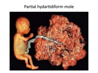
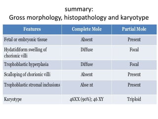
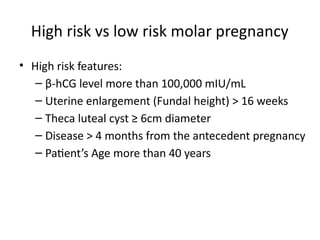
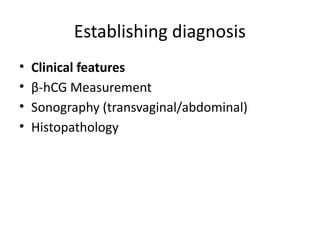
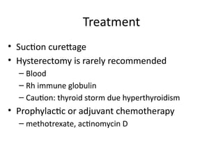
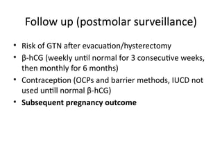
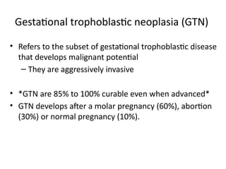
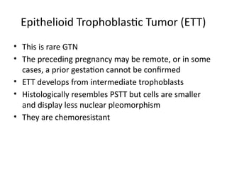
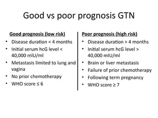
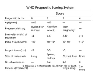
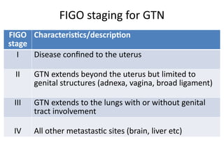
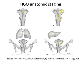
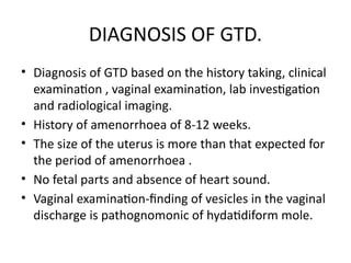
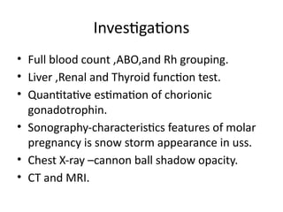
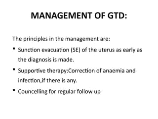
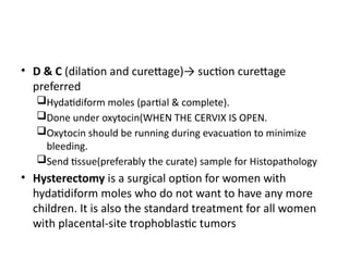
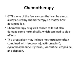
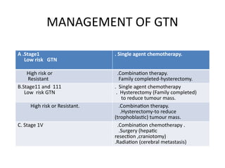
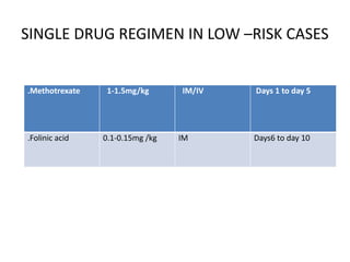
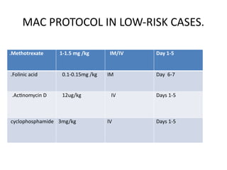
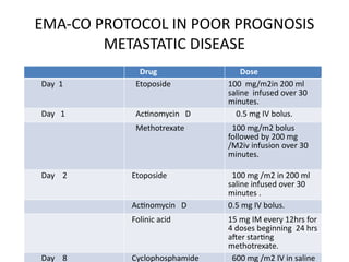
Gestational trophoblastic diseases
(GTDs)
1:Joshua mlungwana
2:Seth miyego
3:Gloria mrosso
Facilitator: Dr. Shayo
Dr. Matilda
Outline
• Objectives
• Introduction
• Epidemiology and risk factors
• Basic pathophysiology
• classification of GTDs
• Hydatidiform moles
• Gestational trophoblastic neoplasia
• Summary
• References
Learning objectives
• By the end of the session students should be
able to:-
– Define GTD and GTN
– Describe the epidemiology and risk factors of GTDs
– Classify different GTDs
– Explain about the hydatidiform moles
– Explain about the GTNs
– List treatment options different types of GTDs
Introduction
• Gestational trophoblastic disease:
– refers to a spectrum of interrelated but histologically
distinct tumors originating from the placenta
– Spectrum of abnormal growth and proliferation of the
trophoblasts that continue even beyond the end of
pregnancy
• Reliable tumor marker, β-hCG
• Gestational trophoblastic neoplasia
– Refers to the subset of gestational trophoblastic
disease that develops malignant sequelae
– They have potential for local invasion and
metastases
Introduction…
Introduction….
Source: www.google.com/url?sa=i&rct=j&q=&esrc=s&source=images&cd
• Syncytiotrophoblasts
• Cytotrophoblasts
• Intermediate
trophoblasts
Epidemiology and risk factors
Incidence
• The incidence of GTD is approximately 1 to 2 per 1000
deliveries in North America and Europe (Drake, 2006; et al)
• Some ethnic groups appear to be at higher risk of GTD
• Hispanics and Native Americans living in the United States
reportedly have an increased incidence (Drake, 2006; et al)
• They are also very common in south east Asia.
Risk factors
• Age
– Maternal age ≤ 15 years or ≥ 45 years
– Older paternal age
• Obstetric History
– Previous spontaneous abortion doubles the risk of
molar pregnancy
– Personal history of GTD – 1 molar, risk is 2%,if has
history of 2 molar pregancies the risk is 20%
Epidemiology and risk factors
• Complete mole
– Dietary, eg, vitamin A deficiency and low dietary
intake of carotene
– Advanced maternal age
– Partial mole -Smoking, irregular menstrual cycles
Oral contraceptive pills-no proven association
– H/o spontaneous abortion or infertility
– Women with blood type A or AB
Epidemiology and risk factors
PATHOPHYSIOLOGY-BASICS
HISTOPATHOLOGY
PATHOPHYSIOLOGY BASICS
•Imprinting contributes to GTD
•Paternal gene controls placental growth
•Maternal gene controls fetal Growth
•Exaggerated paternal genetic material -Excessive
placental tissue
The pathologies in Gestational Trophoblastic
diseases(GTDs) includes
•Cystic swelling of chorionic villi( Hydropic
degeneration of villi)
•Trophoblastic proliferation.
Modified WHO Classification of GTD, 2011
• Hydatidiform moles
– Hydatidiform mole (partial and complete)
– Invasive mole
• Trophoblastic tumors
– Choriocarcinoma
– Placental site trophoblastic tumor
– Epithelioid trophoblastic tumor
Hydatidiform mole (molar pregnancy)
• Abnormal pregnancies characterized histologically by
aberrant changes within the placenta.
• Results from abnormalities in fertilization
• Classical Placenta appearance
– Abnormal trophoblastic proliferation
– Edema of the chorionic villi
• Cytogenetics have shown chromosomal abnormalities
• Three types:
– Complete hydatidiform mole
– Partial hydatidiform mole
– Invasive mole (chorioadenoma destruens)
• They are essentially benign, but carry an
increased risk of persistent or malignant GTN
– Complete mole 15-20%, partial mole 1-5%
Hydatidiform moles
Complete Hydatidiform Mole
• The empty ovum is fertilized by a haploid sperm, which
then duplicates its own chromosomes (androgenesis).
• Also dispermic fertilization
• Karyotype
– Complete moles typically have a diploid karyotype
– 85-90% are 46,XX and 10-15% are 46,XY
– These chromosomes are entirely of paternal origin
• Macroscopically
– No fetal tissue or amnion is produced
– The placental tissue mass completely fills the
endometrial cavity
Complete Hydatidiform Mole
Source: Williams GYNECOLOGY, 2nd EDITION, by Barbara L. Hoffman, MD, et al: pg 900
Complete Hydatidiform Mole
Complete hydatidiform mole
Grape like lesions SNOW STORM appearance
Hydatidiform mole: specimen from
suction curettage
Clinical features of complete H. mole
• Vaginal bleeding the most common symptom
• Uterine size greater than dates
• β-hCG levels are commonly greater than expected
• Sometimes passing of grape like lesions(more specific)
• Others
– hyperemesis gravidarum
– Preeclampsia
– Theca-lutein cysts
– Hyperthyroidism (Palpitations, Tachycardia,
sweating,Tremors etc)
– Anemia
Partial Hydatidiform Mole
• Develops when 2 sperm fertilize a normal egg
• Varies clinically, genetically, and histologically from
complete mole
• Karyotype
– They have a triploid karyotype
– 69,XXX, 69,XXY (common) and 69,XYY (less common)
• Macroscopically
– Partial moles contain fetal tissue and amnion, in
addition to placental tissues.
– Patients typically present with signs and
symptoms of incomplete or missed abortion.
Partial Hydatidiform Mole
Clinical features of partial H. mole
• Have symptoms and signs of incomplete abortion
• Vaginal bleeding is the main symptom
• Others
– Excessive uterine enlargement
– Preeclampsia
Partial hydartidiform mole
summary:
Gross morphology, histopathology and karyotype
High risk vs low risk molar pregnancy
• High risk features:
– β-hCG level more than 100,000 mIU/mL
– Uterine enlargement (Fundal height) > 16 weeks
– Theca luteal cyst ≥ 6cm diameter
– Disease > 4 months from the antecedent pregnancy
– Patient’s Age more than 40 years
Establishing diagnosis
• Clinical features
• β-hCG Measurement
• Sonography (transvaginal/abdominal)
• Histopathology
Treatment
• Suction curettage
• Hysterectomy is rarely recommended
– Blood
– Rh immune globulin
– Caution: thyroid storm due hyperthyroidism
• Prophylactic or adjuvant chemotherapy
– methotrexate, actinomycin D
Follow up (postmolar surveillance)
• Risk of GTN after evacuation/hysterectomy
• β-hCG (weekly until normal for 3 consecutive weeks,
then monthly for 6 months)
• Contraception (OCPs and barrier methods, IUCD not
used untill normal β-hCG)
• Subsequent pregnancy outcome
Gestational trophoblastic neoplasia (GTN)
• Refers to the subset of gestational trophoblastic disease
that develops malignant potential
– They are aggressively invasive
• *GTN are 85% to 100% curable even when advanced*
• GTN develops after a molar pregnancy (60%), abortion
(30%) or normal pregnancy (10%).
Types of GTN
• Invasive mole
• Choriocarcinoma
• Placental site trophoblastic tumor
• Epithelioid trophoblastic tumor
Invasive Mole
• An invasive mole (formerly known as chorioadenoma
destruens) is a hydatidiform mole that penetrates the
muscular wall of the uterus (myometrium).
• It is benign but locally destructive
• Invades myometrium by hydropic chorionic villi
associated with proliferation of both cytotrophoblast
and syncytiotrophoblasts
• If the tumor or mole grows through the full thickness
of the myometrium, it may result in a hole in the
uterus that can bleed heavily.
Invasive mole: the tissue invades into the myometrial
layer. No obvious borderline, with obvious bleeding.
Gestational Choriocarcinoma
• It is extremely malignant tumor, comprised of
– sheets of anaplastic cytotrophoblasts and
syncytiotrophoblasts
– hemorrhage, necrosis, and vascular invasion
– chorionic villi are characteristically absent
• Initially invade the endometrium and myometrium
• High potential of early blood-borne metastases
• Most cases (50%) follow molar pregnancy, the rest follow
a nonmolar pregnancy.
• Nonmolar choriocarcinoma, 2/3 follow term pregnancies,
and 1/3 follow abortions & ectopic
• Following term pregnancies carry higher mortality
• Sites of metastases are lungs (80%), anterior vaginal wall
(30%), brain (10%), liver (10%)
Gestational Choriocarcinoma
Gross specimen of
choriocarcinoma
FIGO diagnostic criteria for post-molar GTN
• Serum levels of βhcG
– ≥ four values of plateaued hcG (+/-10%) over at 3
weeks time (day 1, 7, 14 and 21)
– A rise of hcG of > 10% for > 3 values over 2 weeks
time (day 1, 7 and 14)
– Persistence of hcG > 6 months of mole evacuation
• Histologic diagnosis of choriocarcinoma
Placental-site Trophoblastic
Tumor
• Placental-site trophoblastic tumor (PSTT) is a rare form
of GTD that develops where the placenta attaches to
the uterus.
• This tumor most often develops after a normal
pregnancy or abortion
• Most placental-site tumors do not spread to other sites
in the body
• Although most forms of GTD are very sensitive to
chemotherapy drugs, placental-site tumors are not and,
therefore, must be completely removed by surgery.
Epithelioid Trophoblastic Tumor (ETT)
• This is rare GTN
• The preceding pregnancy may be remote, or in some
cases, a prior gestation cannot be confirmed
• ETT develops from intermediate trophoblasts
• Histologically resembles PSTT but cells are smaller
and display less nuclear pleomorphism
• They are chemoresistant
Good vs poor prognosis GTN
Good prognosis (low risk)
• Disease duration < 4 months
• Initial serum hcG level <
40,000 mlU/ml
• Metastasis limited to lung and
vagina
• No prior chemotherapy
• WHO score ≤ 6
Poor prognosis (high risk)
• Disease duration > 4 months
• Initial serum hcG level >
40,000 mIU/ml
• Brain or liver metastasis
• Failure of prior chemotherapy
• Following term pregnancy
• WHO score ≥ 7
WHO Prognostic Scoring System
Score
Prognostic factor 0 1 2 4
Age(years) ≤40 >40 — —
Pregnancy history
Hydatidifor
m mole
Abortion,
ectopic
Term
pregnancy
—
Interval (months) of
treatment
<4 4-6 7-12 >12
Initial hCG(mIU/ml) <103
103
-104
104
-105
>105
Largest tumor(cm) <3 3-5 >5 —
Sites of metastasis Lung
Spleen,
kidney
GI tract, liver Brain
No. of metastasis — 1-4 4-8 8
Previous (treatment) — — Single drug
2 or
more
0-4 low risk, 5-7 intermediate risk, >8 high risk for death
FIGO
stage
Characteristics/description
I Disease confined to the uterus
II GTN extends beyond the uterus but limited to
genital structures (adnexa, vagina, broad ligament)
III GTN extends to the lungs with or without genital
tract involvement
IV All other metastastic sites (brain, liver etc)
FIGO staging for GTN
FIGO anatomic staging
Source: Williams GYNECOLOGY, 2nd EDITION, by Barbara L. Hoffman, MD, et al: pg 914
DIAGNOSIS OF GTD.
• Diagnosis of GTD based on the history taking, clinical
examination , vaginal examination, lab investigation
and radiological imaging.
• History of amenorrhoea of 8-12 weeks.
• The size of the uterus is more than that expected for
the period of amenorrhoea .
• No fetal parts and absence of heart sound.
• Vaginal examination-finding of vesicles in the vaginal
discharge is pathognomonic of hydatidiform mole.
Investigations
• Full blood count ,ABO,and Rh grouping.
• Liver ,Renal and Thyroid function test.
• Quantitative estimation of chorionic
gonadotrophin.
• Sonography-characteristics features of molar
pregnancy is snow storm appearance in uss.
• Chest X-ray –cannon ball shadow opacity.
• CT and MRI.
MANAGEMENT OF GTD:
The principles in the management are:
 Sunction evacuation (SE) of the uterus as early as
the diagnosis is made.
 Supportive therapy:Correction of anaemia and
infection,if there is any.
 Councelling for regular follow up
• D & C (dilation and curettage)→ suction curettage
preferred
Hydatidiform moles (partial & complete).
Done under oxytocin(WHEN THE CERVIX IS OPEN.
Oxytocin should be running during evacuation to minimize
bleeding.
Send tissue(preferably the curate) sample for Histopathology
• Hysterectomy is a surgical option for women with
hydatidiform moles who do not want to have any more
children. It is also the standard treatment for all women
with placental-site trophoblastic tumors
Chemotherapy
• GTN is one of the few cancers that can be almost
always cured by chemotherapy no matter how
advanced it is.
• Chemotherapy drugs kill cancer cells but also
damage some normal cells, which can lead to side
effects
• The drugs given may include methotrexate (often
combined with leucovorin), actinomycin-D,
cyclophosphamide (Cytoxan), vincristine, etoposide,
and cisplatin.
MANAGEMENT OF GTN
A .Stage1
Low risk GTN
. Single agent chemotherapy.
High risk or
Resistant
.Combination therapy.
Family completed-hysterectomy.
B.Stage11 and 111
Low risk GTN
. Single agent chemotherapy
. Hysterectomy (Family completed)
to reduce tumour mass.
High risk or Resistant. .Combination therapy.
.Hysterectomy-to reduce
(trophoblastic) tumour mass.
C. Stage 1V .Combination chemotherapy .
.Surgery (hepatic
resection ,craniotomy)
.Radiation (cerebral metastasis)
SINGLE DRUG REGIMEN IN LOW –RISK CASES
.Methotrexate 1-1.5mg/kg IM/IV Days 1 to day 5
.Folinic acid 0.1-0.15mg /kg IM Days6 to day 10
MAC PROTOCOL IN LOW-RISK CASES.
.Methotrexate 1-1.5 mg /kg IM/IV Day 1-5
.Folinic acid 0.1-0.15mg /kg IM Day 6-7
.Actinomycin D 12ug/kg IV Days 1-5
cyclophosphamide 3mg/kg IV Days 1-5
EMA-CO PROTOCOL IN POOR PROGNOSIS
METASTATIC DISEASE
Drug Dose
Day 1 Etoposide 100 mg/m2in 200 ml
saline infused over 30
minutes.
Day 1 Actinomycin D 0.5 mg IV bolus.
Methotrexate 100 mg/m2 bolus
followed by 200 mg
/M2iv infusion over 30
minutes.
Day 2 Etoposide 100 mg /m2 in 200 ml
saline infused over 30
minutes .
Actinomycin D 0.5 mg IV bolus.
Folinic acid 15 mg IM every 12hrs for
4 doses beginning 24 hrs
after starting
methotrexate.
Day 8 Cyclophosphamide 600 mg /m2 IV in saline
Follow up
• Weekly β-hCG measurements
• Contraception
• Subsequent Pregnancy Outcome
Follow up of the patient
– Check hCG levels before evacuation, and counsel the patient to
return immediately if any bleeding occurs
– Check hCG levels weekly till you get 3negative readings then see
monthly for 6-12 months
– Keep patient on oral contraceptive during this period
– Patient should not conceive for 12 months
– Do CXR prior to evacuation; then 4wks interval till remission,
then 3 months interval for the follow-up period
– Close observation of future pregnancies –do pelvic
– USS in the first trimester After 6 weeks, repeat U/S pelvis,
CT/MRI if they were abnormal.
Complications of GTDs
• Hemorrhage → Anemia
• Persistence and metastases
• Trophoblastic embolism
• Infertility
• Infections
• Preeclampsia/ eclampsia
• Hyperthyroidism
Summary
• GTD is characterized by abnormal trophoblastic proliferation
• Benign GTD includes complete or partial H. mole
• GTN includes invasive mole, choriocarcinoma, PSTT or ETT
• β-hCG is the reliable maker for GTDs surveillance
• Pesistence occurs 20% of patients after molar evacuation
• Most GTN follow molar and some follow nonmolar preg
• GTN after nonmolar pregnancy is commonly a
choriocarcinoma
REFERENCES
• 1. Almeida, C. E. D. d., et al. (2011). "Thyrotoxic Crisis Associated with Gestational Trophoblastic Disease." Brazilian Journal of
Anesthesiology 61(5): 604-609.
• 2. Erturk, E., et al. (2007). "Total intravenous anesthesia for evacuation of a hydatidiform mole and termination of pregnancy
in a patient with thyrotoxicosis." International Journal of Obstetric Anesthesia 16(4): 363-366.
• 3. van Cromvoirt, S. M. E., et al. "Identification of patients with persistent trophoblastic disease after complete hydatidiform
mole by using a normal 24-hour urine hCG regression curve." Gynecologic Oncology(0).
• 4. Lurain, J. R. (2011). "Gestational trophoblastic disease II: classification and management of gestational trophoblastic
neoplasia." American Journal of Obstetrics and Gynecology 204(1): 11-18.
• 5. Kale, A., et al. (2001). "Expressions of proliferation markers (Ki-67, proliferating cell nuclear antigen, and silver-staining
nucleolar organizer regions) and of p53 tumor protein in gestational trophoblastic disease." American Journal of Obstetrics
and Gynecology 184(4): 567-574.
• 6. Khanlian, S. A., et al. (2003). "Persistent low levels of human chorionic gonadotropin: A premalignant gestational
trophoblastic disease." American Journal of Obstetrics and Gynecology 188(5): 1254-1259.
• 7. Zalel, Y. and R. Dgani (1997). "Gestational trophoblastic disease following the evacuation of partial hydatidiform mole: a
review of 66 cases." European Journal of Obstetrics & Gynecology and Reproductive Biology 71(1): 67-71.
• 8. Kurdi MS. Hydratidiform mole: A sour encounter with a grapy case. Indian Journal of anaesthesia 2011;55: 171-173
• 9. Daniel Celeski. Anaesthetic implications of a partial molar pregnancy and associated complications AANA Feb 2001; 69:
49-53
• 10. Robert, C. R. (2009). Problems of early pregnancy. Obstetric anesthesia: principles and practice. D. H. Chestnut, L. C.
Tsen, L. S. Polley and C. A. Wong, Mosby Elsevier: 319-
• Barbara L. Hoffman M, John O. Schorge M, Joseph I. Schaffer M, Lisa M. Halvorson M, Karen D. Bradshaw M, F. Gary
Cunningham M. Chapter 37: Gestational Trophoblastic Disease. In: William Gynecology (2nd Edition). 2012. p. 898–914
• https://www.uptodate.com/contents/gestational-trophoblastic-neoplasia
• .

Gestational trophoblastic diseases doc.pptx

  • 1.
    Gestational trophoblastic diseases (GTDs) 1:Joshuamlungwana 2:Seth miyego 3:Gloria mrosso Facilitator: Dr. Shayo Dr. Matilda
  • 2.
    Outline • Objectives • Introduction •Epidemiology and risk factors • Basic pathophysiology • classification of GTDs • Hydatidiform moles • Gestational trophoblastic neoplasia • Summary • References
  • 3.
    Learning objectives • Bythe end of the session students should be able to:- – Define GTD and GTN – Describe the epidemiology and risk factors of GTDs – Classify different GTDs – Explain about the hydatidiform moles – Explain about the GTNs – List treatment options different types of GTDs
  • 4.
    Introduction • Gestational trophoblasticdisease: – refers to a spectrum of interrelated but histologically distinct tumors originating from the placenta – Spectrum of abnormal growth and proliferation of the trophoblasts that continue even beyond the end of pregnancy • Reliable tumor marker, β-hCG
  • 5.
    • Gestational trophoblasticneoplasia – Refers to the subset of gestational trophoblastic disease that develops malignant sequelae – They have potential for local invasion and metastases Introduction…
  • 6.
  • 7.
    Epidemiology and riskfactors Incidence • The incidence of GTD is approximately 1 to 2 per 1000 deliveries in North America and Europe (Drake, 2006; et al) • Some ethnic groups appear to be at higher risk of GTD • Hispanics and Native Americans living in the United States reportedly have an increased incidence (Drake, 2006; et al) • They are also very common in south east Asia.
  • 8.
    Risk factors • Age –Maternal age ≤ 15 years or ≥ 45 years – Older paternal age • Obstetric History – Previous spontaneous abortion doubles the risk of molar pregnancy – Personal history of GTD – 1 molar, risk is 2%,if has history of 2 molar pregancies the risk is 20% Epidemiology and risk factors
  • 9.
    • Complete mole –Dietary, eg, vitamin A deficiency and low dietary intake of carotene – Advanced maternal age – Partial mole -Smoking, irregular menstrual cycles Oral contraceptive pills-no proven association – H/o spontaneous abortion or infertility – Women with blood type A or AB Epidemiology and risk factors
  • 10.
  • 11.
  • 13.
    PATHOPHYSIOLOGY BASICS •Imprinting contributesto GTD •Paternal gene controls placental growth •Maternal gene controls fetal Growth •Exaggerated paternal genetic material -Excessive placental tissue The pathologies in Gestational Trophoblastic diseases(GTDs) includes •Cystic swelling of chorionic villi( Hydropic degeneration of villi) •Trophoblastic proliferation.
  • 14.
    Modified WHO Classificationof GTD, 2011 • Hydatidiform moles – Hydatidiform mole (partial and complete) – Invasive mole • Trophoblastic tumors – Choriocarcinoma – Placental site trophoblastic tumor – Epithelioid trophoblastic tumor
  • 15.
    Hydatidiform mole (molarpregnancy) • Abnormal pregnancies characterized histologically by aberrant changes within the placenta. • Results from abnormalities in fertilization • Classical Placenta appearance – Abnormal trophoblastic proliferation – Edema of the chorionic villi • Cytogenetics have shown chromosomal abnormalities
  • 16.
    • Three types: –Complete hydatidiform mole – Partial hydatidiform mole – Invasive mole (chorioadenoma destruens) • They are essentially benign, but carry an increased risk of persistent or malignant GTN – Complete mole 15-20%, partial mole 1-5% Hydatidiform moles
  • 17.
    Complete Hydatidiform Mole •The empty ovum is fertilized by a haploid sperm, which then duplicates its own chromosomes (androgenesis). • Also dispermic fertilization • Karyotype – Complete moles typically have a diploid karyotype – 85-90% are 46,XX and 10-15% are 46,XY – These chromosomes are entirely of paternal origin
  • 18.
    • Macroscopically – Nofetal tissue or amnion is produced – The placental tissue mass completely fills the endometrial cavity Complete Hydatidiform Mole
  • 19.
    Source: Williams GYNECOLOGY,2nd EDITION, by Barbara L. Hoffman, MD, et al: pg 900 Complete Hydatidiform Mole
  • 20.
    Complete hydatidiform mole Grapelike lesions SNOW STORM appearance
  • 21.
    Hydatidiform mole: specimenfrom suction curettage
  • 22.
    Clinical features ofcomplete H. mole • Vaginal bleeding the most common symptom • Uterine size greater than dates • β-hCG levels are commonly greater than expected • Sometimes passing of grape like lesions(more specific) • Others – hyperemesis gravidarum – Preeclampsia – Theca-lutein cysts – Hyperthyroidism (Palpitations, Tachycardia, sweating,Tremors etc) – Anemia
  • 23.
    Partial Hydatidiform Mole •Develops when 2 sperm fertilize a normal egg • Varies clinically, genetically, and histologically from complete mole • Karyotype – They have a triploid karyotype – 69,XXX, 69,XXY (common) and 69,XYY (less common)
  • 24.
    • Macroscopically – Partialmoles contain fetal tissue and amnion, in addition to placental tissues. – Patients typically present with signs and symptoms of incomplete or missed abortion. Partial Hydatidiform Mole
  • 25.
  • 26.
    Clinical features ofpartial H. mole • Have symptoms and signs of incomplete abortion • Vaginal bleeding is the main symptom • Others – Excessive uterine enlargement – Preeclampsia
  • 27.
  • 28.
  • 29.
    High risk vslow risk molar pregnancy • High risk features: – β-hCG level more than 100,000 mIU/mL – Uterine enlargement (Fundal height) > 16 weeks – Theca luteal cyst ≥ 6cm diameter – Disease > 4 months from the antecedent pregnancy – Patient’s Age more than 40 years
  • 30.
    Establishing diagnosis • Clinicalfeatures • β-hCG Measurement • Sonography (transvaginal/abdominal) • Histopathology
  • 31.
    Treatment • Suction curettage •Hysterectomy is rarely recommended – Blood – Rh immune globulin – Caution: thyroid storm due hyperthyroidism • Prophylactic or adjuvant chemotherapy – methotrexate, actinomycin D
  • 32.
    Follow up (postmolarsurveillance) • Risk of GTN after evacuation/hysterectomy • β-hCG (weekly until normal for 3 consecutive weeks, then monthly for 6 months) • Contraception (OCPs and barrier methods, IUCD not used untill normal β-hCG) • Subsequent pregnancy outcome
  • 33.
    Gestational trophoblastic neoplasia(GTN) • Refers to the subset of gestational trophoblastic disease that develops malignant potential – They are aggressively invasive • *GTN are 85% to 100% curable even when advanced* • GTN develops after a molar pregnancy (60%), abortion (30%) or normal pregnancy (10%).
  • 34.
    Types of GTN •Invasive mole • Choriocarcinoma • Placental site trophoblastic tumor • Epithelioid trophoblastic tumor
  • 35.
    Invasive Mole • Aninvasive mole (formerly known as chorioadenoma destruens) is a hydatidiform mole that penetrates the muscular wall of the uterus (myometrium). • It is benign but locally destructive • Invades myometrium by hydropic chorionic villi associated with proliferation of both cytotrophoblast and syncytiotrophoblasts • If the tumor or mole grows through the full thickness of the myometrium, it may result in a hole in the uterus that can bleed heavily.
  • 36.
    Invasive mole: thetissue invades into the myometrial layer. No obvious borderline, with obvious bleeding.
  • 37.
    Gestational Choriocarcinoma • Itis extremely malignant tumor, comprised of – sheets of anaplastic cytotrophoblasts and syncytiotrophoblasts – hemorrhage, necrosis, and vascular invasion – chorionic villi are characteristically absent • Initially invade the endometrium and myometrium • High potential of early blood-borne metastases
  • 38.
    • Most cases(50%) follow molar pregnancy, the rest follow a nonmolar pregnancy. • Nonmolar choriocarcinoma, 2/3 follow term pregnancies, and 1/3 follow abortions & ectopic • Following term pregnancies carry higher mortality • Sites of metastases are lungs (80%), anterior vaginal wall (30%), brain (10%), liver (10%) Gestational Choriocarcinoma
  • 39.
  • 40.
    FIGO diagnostic criteriafor post-molar GTN • Serum levels of βhcG – ≥ four values of plateaued hcG (+/-10%) over at 3 weeks time (day 1, 7, 14 and 21) – A rise of hcG of > 10% for > 3 values over 2 weeks time (day 1, 7 and 14) – Persistence of hcG > 6 months of mole evacuation • Histologic diagnosis of choriocarcinoma
  • 41.
    Placental-site Trophoblastic Tumor • Placental-sitetrophoblastic tumor (PSTT) is a rare form of GTD that develops where the placenta attaches to the uterus. • This tumor most often develops after a normal pregnancy or abortion • Most placental-site tumors do not spread to other sites in the body • Although most forms of GTD are very sensitive to chemotherapy drugs, placental-site tumors are not and, therefore, must be completely removed by surgery.
  • 42.
    Epithelioid Trophoblastic Tumor(ETT) • This is rare GTN • The preceding pregnancy may be remote, or in some cases, a prior gestation cannot be confirmed • ETT develops from intermediate trophoblasts • Histologically resembles PSTT but cells are smaller and display less nuclear pleomorphism • They are chemoresistant
  • 43.
    Good vs poorprognosis GTN Good prognosis (low risk) • Disease duration < 4 months • Initial serum hcG level < 40,000 mlU/ml • Metastasis limited to lung and vagina • No prior chemotherapy • WHO score ≤ 6 Poor prognosis (high risk) • Disease duration > 4 months • Initial serum hcG level > 40,000 mIU/ml • Brain or liver metastasis • Failure of prior chemotherapy • Following term pregnancy • WHO score ≥ 7
  • 44.
    WHO Prognostic ScoringSystem Score Prognostic factor 0 1 2 4 Age(years) ≤40 >40 — — Pregnancy history Hydatidifor m mole Abortion, ectopic Term pregnancy — Interval (months) of treatment <4 4-6 7-12 >12 Initial hCG(mIU/ml) <103 103 -104 104 -105 >105 Largest tumor(cm) <3 3-5 >5 — Sites of metastasis Lung Spleen, kidney GI tract, liver Brain No. of metastasis — 1-4 4-8 8 Previous (treatment) — — Single drug 2 or more 0-4 low risk, 5-7 intermediate risk, >8 high risk for death
  • 45.
    FIGO stage Characteristics/description I Disease confinedto the uterus II GTN extends beyond the uterus but limited to genital structures (adnexa, vagina, broad ligament) III GTN extends to the lungs with or without genital tract involvement IV All other metastastic sites (brain, liver etc) FIGO staging for GTN
  • 46.
    FIGO anatomic staging Source:Williams GYNECOLOGY, 2nd EDITION, by Barbara L. Hoffman, MD, et al: pg 914
  • 47.
    DIAGNOSIS OF GTD. •Diagnosis of GTD based on the history taking, clinical examination , vaginal examination, lab investigation and radiological imaging. • History of amenorrhoea of 8-12 weeks. • The size of the uterus is more than that expected for the period of amenorrhoea . • No fetal parts and absence of heart sound. • Vaginal examination-finding of vesicles in the vaginal discharge is pathognomonic of hydatidiform mole.
  • 48.
    Investigations • Full bloodcount ,ABO,and Rh grouping. • Liver ,Renal and Thyroid function test. • Quantitative estimation of chorionic gonadotrophin. • Sonography-characteristics features of molar pregnancy is snow storm appearance in uss. • Chest X-ray –cannon ball shadow opacity. • CT and MRI.
  • 49.
    MANAGEMENT OF GTD: Theprinciples in the management are:  Sunction evacuation (SE) of the uterus as early as the diagnosis is made.  Supportive therapy:Correction of anaemia and infection,if there is any.  Councelling for regular follow up
  • 50.
    • D &C (dilation and curettage)→ suction curettage preferred Hydatidiform moles (partial & complete). Done under oxytocin(WHEN THE CERVIX IS OPEN. Oxytocin should be running during evacuation to minimize bleeding. Send tissue(preferably the curate) sample for Histopathology • Hysterectomy is a surgical option for women with hydatidiform moles who do not want to have any more children. It is also the standard treatment for all women with placental-site trophoblastic tumors
  • 51.
    Chemotherapy • GTN isone of the few cancers that can be almost always cured by chemotherapy no matter how advanced it is. • Chemotherapy drugs kill cancer cells but also damage some normal cells, which can lead to side effects • The drugs given may include methotrexate (often combined with leucovorin), actinomycin-D, cyclophosphamide (Cytoxan), vincristine, etoposide, and cisplatin.
  • 52.
    MANAGEMENT OF GTN A.Stage1 Low risk GTN . Single agent chemotherapy. High risk or Resistant .Combination therapy. Family completed-hysterectomy. B.Stage11 and 111 Low risk GTN . Single agent chemotherapy . Hysterectomy (Family completed) to reduce tumour mass. High risk or Resistant. .Combination therapy. .Hysterectomy-to reduce (trophoblastic) tumour mass. C. Stage 1V .Combination chemotherapy . .Surgery (hepatic resection ,craniotomy) .Radiation (cerebral metastasis)
  • 53.
    SINGLE DRUG REGIMENIN LOW –RISK CASES .Methotrexate 1-1.5mg/kg IM/IV Days 1 to day 5 .Folinic acid 0.1-0.15mg /kg IM Days6 to day 10
  • 54.
    MAC PROTOCOL INLOW-RISK CASES. .Methotrexate 1-1.5 mg /kg IM/IV Day 1-5 .Folinic acid 0.1-0.15mg /kg IM Day 6-7 .Actinomycin D 12ug/kg IV Days 1-5 cyclophosphamide 3mg/kg IV Days 1-5
  • 55.
    EMA-CO PROTOCOL INPOOR PROGNOSIS METASTATIC DISEASE Drug Dose Day 1 Etoposide 100 mg/m2in 200 ml saline infused over 30 minutes. Day 1 Actinomycin D 0.5 mg IV bolus. Methotrexate 100 mg/m2 bolus followed by 200 mg /M2iv infusion over 30 minutes. Day 2 Etoposide 100 mg /m2 in 200 ml saline infused over 30 minutes . Actinomycin D 0.5 mg IV bolus. Folinic acid 15 mg IM every 12hrs for 4 doses beginning 24 hrs after starting methotrexate. Day 8 Cyclophosphamide 600 mg /m2 IV in saline
  • 56.
    Follow up • Weeklyβ-hCG measurements • Contraception • Subsequent Pregnancy Outcome
  • 57.
    Follow up ofthe patient – Check hCG levels before evacuation, and counsel the patient to return immediately if any bleeding occurs – Check hCG levels weekly till you get 3negative readings then see monthly for 6-12 months – Keep patient on oral contraceptive during this period – Patient should not conceive for 12 months – Do CXR prior to evacuation; then 4wks interval till remission, then 3 months interval for the follow-up period – Close observation of future pregnancies –do pelvic – USS in the first trimester After 6 weeks, repeat U/S pelvis, CT/MRI if they were abnormal.
  • 58.
    Complications of GTDs •Hemorrhage → Anemia • Persistence and metastases • Trophoblastic embolism • Infertility • Infections • Preeclampsia/ eclampsia • Hyperthyroidism
  • 59.
    Summary • GTD ischaracterized by abnormal trophoblastic proliferation • Benign GTD includes complete or partial H. mole • GTN includes invasive mole, choriocarcinoma, PSTT or ETT • β-hCG is the reliable maker for GTDs surveillance • Pesistence occurs 20% of patients after molar evacuation • Most GTN follow molar and some follow nonmolar preg • GTN after nonmolar pregnancy is commonly a choriocarcinoma
  • 60.
    REFERENCES • 1. Almeida,C. E. D. d., et al. (2011). "Thyrotoxic Crisis Associated with Gestational Trophoblastic Disease." Brazilian Journal of Anesthesiology 61(5): 604-609. • 2. Erturk, E., et al. (2007). "Total intravenous anesthesia for evacuation of a hydatidiform mole and termination of pregnancy in a patient with thyrotoxicosis." International Journal of Obstetric Anesthesia 16(4): 363-366. • 3. van Cromvoirt, S. M. E., et al. "Identification of patients with persistent trophoblastic disease after complete hydatidiform mole by using a normal 24-hour urine hCG regression curve." Gynecologic Oncology(0). • 4. Lurain, J. R. (2011). "Gestational trophoblastic disease II: classification and management of gestational trophoblastic neoplasia." American Journal of Obstetrics and Gynecology 204(1): 11-18. • 5. Kale, A., et al. (2001). "Expressions of proliferation markers (Ki-67, proliferating cell nuclear antigen, and silver-staining nucleolar organizer regions) and of p53 tumor protein in gestational trophoblastic disease." American Journal of Obstetrics and Gynecology 184(4): 567-574. • 6. Khanlian, S. A., et al. (2003). "Persistent low levels of human chorionic gonadotropin: A premalignant gestational trophoblastic disease." American Journal of Obstetrics and Gynecology 188(5): 1254-1259. • 7. Zalel, Y. and R. Dgani (1997). "Gestational trophoblastic disease following the evacuation of partial hydatidiform mole: a review of 66 cases." European Journal of Obstetrics & Gynecology and Reproductive Biology 71(1): 67-71. • 8. Kurdi MS. Hydratidiform mole: A sour encounter with a grapy case. Indian Journal of anaesthesia 2011;55: 171-173 • 9. Daniel Celeski. Anaesthetic implications of a partial molar pregnancy and associated complications AANA Feb 2001; 69: 49-53 • 10. Robert, C. R. (2009). Problems of early pregnancy. Obstetric anesthesia: principles and practice. D. H. Chestnut, L. C. Tsen, L. S. Polley and C. A. Wong, Mosby Elsevier: 319- • Barbara L. Hoffman M, John O. Schorge M, Joseph I. Schaffer M, Lisa M. Halvorson M, Karen D. Bradshaw M, F. Gary Cunningham M. Chapter 37: Gestational Trophoblastic Disease. In: William Gynecology (2nd Edition). 2012. p. 898–914 • https://www.uptodate.com/contents/gestational-trophoblastic-neoplasia • .