- The study examined 90 pregnancies in 53 women with congenital heart disease between 1998-2004.
- Cardiac complications occurred in 19.4% of ongoing pregnancies, most commonly pulmonary edema (16.7%).
- Impaired subpulmonary ventricular systolic function and/or severe pulmonary regurgitation were independent risk factors for adverse maternal cardiac outcomes.
- Adverse neonatal outcomes such as preterm delivery and respiratory distress occurred in 27.8% of ongoing pregnancies. A subaortic ventricular outflow tract gradient >30 mm Hg independently predicted adverse neonatal outcomes.
3. heart failure, endocarditis, arrhythmia), New York Heart Association
(NYHA) functional class, blood pressure, heart rate, oxygen satura-
tion, maximum oxygen uptake (mL/kg per minute), laboratory
values (eg, blood urea nitrogen, creatinine, hematocrit), echocardio-
graphic assessment including Doppler quantification of obstructive
and regurgitant lesions and systolic pulmonary artery pressure
estimates, and cardiac MRI findings. Follow-up data were obtained
from clinical visits throughout pregnancy and the puerperium and
included changes in medication, NYHA functional class, provision
of high-risk care, onset of cardiac care, and formal genetic
counseling.
Cardiac, obstetric, and neonatal events were classified according
to previously proposed definitions determined by Siu and col-
leagues.4 Cardiac complications were subdivided into primary and
secondary events. A primary cardiac event consisted of cardiac
death, cardiac arrest, stroke, symptomatic sustained bradyarrhythmia
or tachyarrhythmia requiring therapy, or pulmonary edema docu-
mented by physical examination or radiography. Decline in Ն2
NYHA functional classes, need for urgent invasive cardiac interven-
tions during pregnancy or within 6 weeks postpartum, and symp-
tomatic nonsustained bradyarrhythmia or tachyarrhythmia requiring
therapy were considered secondary cardiac events. Obstetric events
consisted of preeclampsia, defined as sustained systolic or diastolic
blood pressure Ն140 or Ն90 mm Hg, respectively, with concurrent
proteinuria; postpartum hemorrhage, defined as estimated blood loss
Ͼ500 mL for vaginal delivery or Ͼ1 L for cesarean delivery
accompanied by a Ն10 g/L drop in hemoglobin or requiring
transfusion; and noncardiac death. Neonatal events were defined as
preterm delivery (Ͻ37 weeks of gestation), birth weight small for
gestational age (Ͻ10th percentile), respiratory distress syndrome,
cerebral intraventricular hemorrhage, fetal death (Ն20 weeks of
gestation), or neonatal death (within the first month after birth).
Spontaneous abortion was defined as fetal loss before 20 weeks of
gestation. The protocol was approved by the institutional review
boards of both hospitals. The authors had full access to the data and
take full responsibility for its integrity. All authors have read and
agree to the manuscript as written.
The validity of a previously proposed maternal cardiac risk index
from the Cardiac Disease in Pregnancy (CARPREG) investigators,4
generated in a cohort of women with a variety of congenital and
acquired forms of heart disease, was tested in the study population.
One point was assigned for each of the following variables: arrhyth-
mia or prior cardiac event (heart failure, transient ischemic attack, or
stroke before pregnancy); baseline NYHA functional class III or IV
or cyanosis (oxygen saturation Ͻ90%); systemic heart obstruction
(atrioventricular valve area of the subaortic ventricle Ͻ2 cm2
, aortic
valve area Ͻ1.5 cm2
, or peak outflow tract gradient of the subaortic
ventricle Ͼ30 mm Hg); and subaortic ventricular ejection fraction
Ͻ40%.
Statistical Analyses
Dichotomous variables are presented as percentage and continuous
variables as meanϮSD or median and range depending on their
distribution. Given that each woman could have Ͼ1 pregnancy,
analyses took into consideration the nonindependent nature of the
data structure. Generalized estimating equations were used to pro-
duce regression marginal models for cluster sampling data by
specifying link and distribution functions. Univariate and multivar-
iate predictors of cardiovascular, neonatal, and obstetric events were
assessed. Highly correlated (rՆ0.6) statistically significant univari-
ate predictors were combined for consideration in multivariate
models. In validating the previously proposed CARPREG index,
actual versus expected primary cardiac event rates were compared by
2
goodness-of-fit tests. A generalized estimating equation model
that controlled for the CARPREG index assessed the additional
predictive ability of candidate risk factors for adverse maternal
cardiac risk. Two-tailed probability values Ͻ0.05 were considered
statistically significant. Statistical testing was performed with the use
of SAS software Version 8 (SAS Institute).
Results
Baseline Characteristics
During the study period, 614 women with congenital heart
disease between 12 and 50 years of age were seen in the
outpatient division of the BACH service. Of these women,
53 had a total of 90 pregnancies at age 27.7Ϯ6.1 years.
Underlying congenital heart lesions and maternal baseline
characteristics are summarized in Tables 1 and 2, respec-
tively. Gestational age at the first prenatal visit was
12.8Ϯ7.9 weeks. Overall, 41% of pregnancies were con-
sidered planned. Genetic counseling was received in 26%,
cardiology counseling before pregnancy in 71%, and
high-risk obstetric care in 92%.
Complete information on cardiac and obstetric events
was available for all pregnancies, and 98% of pregnancies
had complete information on neonatal events. Echocardio-
graphic data were available in 92% of pregnancies, and
cardiac MRI findings were available in 11%. When both
were performed, the degree of atrioventricular and aortic
regurgitation, subaortic ventricular function, and subaortic
TABLE 1. Distribution by Primary Type of Congenital
Heart Disease
Congenital Lesion No. (%)
Septal defect 6 (11.3)
Atrial septal defect 2 (3.8)
Unoperated 1 (1.9)
Surgically repaired 1 (1.9)
Ventricular septal defect 2 (3.8)
Unoperated 1 (1.9)
Surgically repaired 1 (1.9)
Surgically repaired atrioventricular canal defect 2 (3.8)
Right-sided obstructive lesion 3 (5.7)
Pulmonary stenosis, unoperated* 1 (1.9)
Pulmonary stenosis with surgical valvotomy 1 (1.9)
Pulmonary atresia, ventricular septal defect, and RV-PA conduit 1 (1.9)
Left-sided obstructive lesion 11 (17.0)
Aortic stenosis 5 (9.4)
Unoperated (excluding patent ductus arteriosus ligation)† 2 (3.8)
Transcatheter valvuloplasty 2 (3.8)
Aortic valve replacement with coarctation repair 1 (1.9)
Surgically repaired aortic coarctation 6 (11.3)
Tetralogy of Fallot after repair 15 (28.3)
TGA 10 (18.9)
D-TGA with Mustard or Senning 8 (15.1)
D-TGA, ventricular septal defect, pulmonary stenosis with Rastelli 1 (1.9)
Unoperated congenitally corrected TGA 1 (1.9)
Single ventricle with Fontan physiology 4 (7.5)
Ebstein’s anomaly 2 (3.8)
Repaired anomalous left coronary artery from pulmonary artery 2 (3.8)
RV-PA indicates right ventricle to pulmonary artery; TGA, transposition of the
great arteries.
*Pulmonary stenosis gradient of 30 mm Hg.
†Aortic stenosis gradients of 60 and 40 mm Hg.
518 Circulation January 31, 2006
by on July 13, 2008circ.ahajournals.orgDownloaded from
4. ventricular outflow tract obstruction were concordant in all
cases. Pulmonary regurgitation was graded severe by
echocardiography and mild by MRI in one patient and
moderate by echocardiography and severe by MRI in
another. In a third patient, subpulmonary ventricular sys-
tolic function was deemed normal by echocardiography
and mildly dysfunctional by cardiac MRI. In all cases of
discrepancy, MRI data were retained.
Cardiac Events
Qualifying cardiac events occurred in 25.0% of ongoing
pregnancies and were limited to heart failure with pulmonary
edema, symptomatic arrhythmias, and need for urgent inva-
sive intervention (Table 3). Predictors of primary cardiac
events are summarized in Table 4. There were no maternal
deaths. Four patients experienced deterioration by 1 NYHA
functional class. One patient with Turner syndrome, surgi-
cally repaired aortic coarctation, and normal biventricular
systolic function had a troponin leak in the setting of
preeclampsia with hemolysis, elevated liver enzymes, and
low platelets (HELLP) syndrome.12 She had invasive moni-
toring to assist with volume management during
hospitalization.
The most common adverse cardiac event was pulmonary
edema, documented in 12 pregnancies. Underlying con-
TABLE 2. Maternal Baseline Characteristics for the
90 Pregnancies
Demographics
Maternal age, y 27.7Ϯ6.1
Ethnicity
Asian 1 (1.1%)
Black 6 (6.7%)
White 75 (83.3%)
Hispanic 8 (8.9%)
Clinical examination
Oxygen saturation, % 98.6Ϯ0.9
NYHA functional class
Class I 79 (87.8)
Class II 11 (12.2)
Past medical history
Sustained arrhythmia 12 (13.3%)
Atrial 8 (8.9%)
Ventricular 8 (8.9%)
Permanent pacemaker 3 (3.3%)
Internal cardioverter-defibrillator 2 (2.2%)
Subacute bacterial endocarditis 2 (2.2%)
Congestive heart failure 7 (7.8%)
Cerebrovascular accident 2 (2.2%)
Hypertension 3 (3.3%)
Smoking history 9 (10.0%)
Medications at first prenatal visit
Diuretic 7 (7.8%)
Antiplatelet 10 (11.1%)
Warfarin 2 (2.2%)
Digoxin 14 (15.6%)
Angiotensin-converting enzyme inhibitor 8 (8.9%)
Antiarrhythmic 14 (15.6%)
Hemodynamics
Moderate or severe mitral regurgitation 2 (2.7%)
Moderate or severe tricuspid regurgitation 9 (12.0%)
Moderate or severe aortic regurgitation 5 (6.7%)
Moderate or severe pulmonary regurgitation 18 (25.4%)
Pulmonary outflow tract gradient, mm Hg 0 (0, 150)*
Ͼ30 mm Hg 11 (14.9%)
Aortic outflow tract gradient, mm Hg 0 (0, 70)*
Ͼ30 mm Hg 13 (17.1%)
Subaortic ventricular systolic dysfunction
Mild 9 (12.0%)
Moderate 0 (0%)
Severe 0 (0%)
Subpulmonary ventricular systolic dysfunction
Mild 6 (8.2%)
Moderate 5 (6.8%)
Severe 0 (0%)
*Median (range).
TABLE 3. Cardiac, Obstetric, and Neonatal Outcomes
No. (%)
Aborted pregnancy 18 (20.0)
Spontaneous 11 (12.2)
Induced 7 (7.8)
Maternal cardiac event (at least 1 of the following)* 18 (25.0)
Primary 14 (19.4)
Pulmonary edema 12 (16.7)
Sustained symptomatic arrhythmia requiring therapy 2 (2.8)
Cerebrovascular accident 0 (0)
Cardiac arrest 0 (0)
Cardiac death 0 (0)
Secondary 9 (12.5)
Change in NYHA class by Ն2 0 (0.0)
Need for urgent invasive intervention 4 (5.6)
Nonsustained symptomatic arrhythmia requiring therapy 6 (8.3)
Obstetric event (at least 1 of the following)* 8 (11.1)
Preeclampsia 2 (2.8)
Postpartum hemorrhage 7 (7.8)
Noncardiac maternal death 0 (0)
Neonatal event (at least 1 of the following)* 20 (27.8)
Preterm delivery 15 (20.8)
Small for gestational age 6 (8.3)
Respiratory distress syndrome 6 (8.3)
Intraventricular hemorrhage 1 (1.4)
Intrauterine fetal demise 2 (2.8)
Neonatal death 1 (1.4)
*The denominator for cardiac, obstetric, and neonatal events excludes
aborted pregnancies.
Khairy et al Pregnancy Outcomes in Congenital Heart Disease 519
by on July 13, 2008circ.ahajournals.orgDownloaded from
5. genital cardiac diagnoses were as follows: tetralogy of
Fallot (nϭ5) with severe pulmonary regurgitation in all
and subpulmonary ventricular systolic dysfunction in 3
patients, 1 of whom had biventricular systolic dysfunction;
transposition of the great arteries (nϭ2) with a Mustard
(nϭ1) or Senning (nϭ1) baffle; bicuspid aortic valve with
mixed aortic valve disease (nϭ2), 1 of whom had aortic
valve replacement; bicuspid aortic valve with surgically
corrected aortic coarctation (nϭ2), 1 of whom had con-
comitant surgery for a supravalvar mitral ring; and surgi-
cally repaired primum atrial septal defect and cleft mitral
valve (nϭ1). All episodes of heart failure responded to
medical therapy that included diuretics, except for the
patient with mixed aortic valve disease and increasing
aortic stenosis (gradient Ͼ100 mm Hg) who underwent
urgent aortic valve replacement at 20 weeks of gestation.
Intrauterine fetal demise occurred on the third postopera-
tive day.
Symptomatic arrhythmias were documented in 8 preg-
nancies, 2 of which were supraventricular and sustained. A
patient with a Rastelli repair was cardioverted for atrial
flutter, and a patient with a stenotic bicuspid aortic valve
was successfully treated medically for supraventricular
tachycardia. Six patients had palpitations with documented
nonsustained monomorphic ventricular tachycardia. A pa-
tient with repaired tetralogy of Fallot had a 17-beat run of
ventricular tachycardia on Holter monitoring requested for
palpitations and dizziness. Sustained ventricular
tachycardia was not inducible on an electrophysiological
study at 22 weeks of gestation, and -blocker therapy was
initiated. One patient with pulmonary atresia and right
ventricle to pulmonary artery conduit had an 18-beat run of
ventricular tachycardia on Holter monitoring but refused
medical therapy. Four other patients had 4- to 10-beat runs
of ventricular tachycardia treated with -blockers. Under-
lying congenital lesions were tetralogy of Fallot (nϭ1),
pulmonary stenosis and atrial septal defect (nϭ1), trans-
position of the great arteries with a Mustard baffle (nϭ1),
and hypoplastic right ventricle with a modified Fontan
(nϭ1).
In further analyses of independent risk factors for
primary or secondary cardiac events, smoking was associ-
ated with pulmonary edema (odds ratio [OR], 9.5; 95% CI,
1.8 to 50.5; Pϭ0.0082) and symptomatic arrhythmias (OR,
9.0; 95% CI, 1.6 to 52.0; Pϭ0.0140). Similarly, subpul-
monary ventricular dysfunction and/or severe pulmonary
regurgitation was predictive of both symptomatic arrhyth-
mias (OR, 6.9; 95% CI, 1.1 to 42.1; Pϭ0.0358) and
pulmonary edema (OR, 4.6; 95% CI, 1.2 to 17.9;
Pϭ0.0283). This latter association persisted after adjust-
ment for subaortic ventricular systolic dysfunction (OR,
4.1; 95% CI, 1.0 to 16.1; Pϭ0.0486).
Obstetric and Neonatal Events
Eighteen pregnancies (20.0%) were aborted: 11 (12.2%)
spontaneously and 7 (7.8%) electively. Nine spontaneous
abortions occurred in the first trimester and 2 in the second
trimester, at a mean of 10.8Ϯ3.7 weeks. Univariate predictors
of spontaneous abortion were maternal hypertension (OR,
17.8; 95% CI, 1.4 to 218.3; Pϭ0.0247), antiplatelet agent
(OR, 7.3; 95% CI, 1.6 to 33.4; Pϭ0.0100), and antiarrhyth-
mic medication (OR, 5.3; 95% CI, 1.2 to 23.0; Pϭ0.0249).
All ongoing 72 pregnancies were singletons except for 1 twin
pregnancy.
There were 2 intrauterine fetal demises occurring at 20
and 26 weeks of gestation. The demise at 20 weeks of
gestation is described above. The other took place in a
patient with a stenotic bicuspid aortic valve whose preg-
nancy was complicated by ovarian vein thrombosis and
pulmonary embolism. The only neonatal death also oc-
curred in this patient’s prior pregnancy. That pregnancy
was complicated by premature rupture of membranes at
24.5 weeks of gestation requiring urgent cesarean delivery
for chorioamnionitis. The neonate expired 9 days after
delivery from complications of prematurity.
Seventeen pregnancies (23.6%) were delivered by cesar-
ean, and 55 (76.4%) had successful vaginal deliveries. All
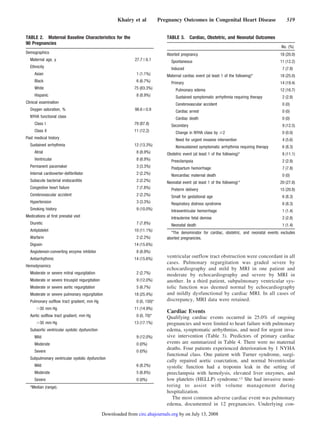
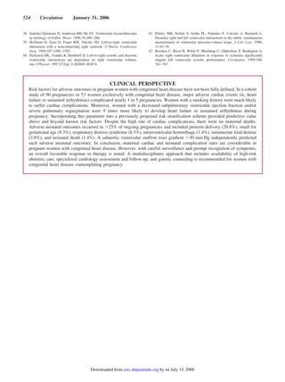
TABLE 4. Maternal Predictors of Primary Cardiac Events During Pregnancy
OR 95% CI P
Univariate predictor
Baseline NYHA class Ն2 5.4 1.2, 25.2 0.0320
Weight, kg 1.1 1.02, 1.14 0.0098
Prior history of heart failure 15.5 1.5, 163.6 0.0223
Smoking history 15.6 2.6, 92.7 0.0026
Pulmonary regurgitation 1.8 1.1, 3.1 0.0440
Severe pulmonary regurgitation 4.6 1.1, 19.5 0.0372
Depressed subpulmonary ventricular EF 7.7 1.5, 40.2 0.0159
Depressed morphological right ventricular EF 4.6 1.4, 15.2 0.0130
Multivariate predictor
Severe pulmonary regurgitation or depressed
subpulmonary ventricular EF
9.0 1.5, 53.1 0.0158
Smoking history 27.2 1.9, 384.6 0.0145
EF indicates ejection fraction.
520 Circulation January 31, 2006
by on July 13, 2008circ.ahajournals.orgDownloaded from
6. patients without an obstetric contraindication attempted a
trial of labor. All but 1 patient received epidural anesthe-
sia, and all labored primarily in the left lateral decubitus
position. Twenty vaginal deliveries were spontaneous,
whereas 35 were assisted by forceps (nϭ22) or vacuum
(nϭ13). All cesarean deliveries were performed for routine
obstetric indications: breech (nϭ5), elective repeated ce-
sarean (nϭ3), cephalopelvic disproportion (nϭ3), and
other (nϭ6). Obstetric and neonatal events are summarized
in Table 3, and predictors of neonatal events are summa-
rized in Table 5.
Of the 70 live births, the following cardiac anomalies
were diagnosed in 5 progeny (7.1%): tetralogy of Fallot,
ventricular septal defect, dilated cardiomyopathy, cor tria-
triatum, and interrupted aortic arch with ventricular septal
defect. All cases were identified by fetal cardiac ultra-
sonography before birth. Additional congenital malforma-
tions included single umbilical artery, anomalous left
subclavian artery, and gonadal dysgenesis.
Validation of Cardiac Risk Index
Actual versus predicted primary cardiac event rates in pa-
tients with 0, 1, and Ն2 predictors from the previously
proposed CARPREG risk index4 were 12%, 30%, and 100%
versus 5%, 27%, and 75%, respectively. Differences between
actual and predicted rates, depicted in the Figure, panel A,
were not statistically significant (Pϭ0.5904). Further strati-
fication according to whether or not patients had severe
pulmonary regurgitation and/or reduced subpulmonary ven-
tricular systolic function further improved overall risk assess-
ment (Pϭ0.0111). As illustrated in the Figure, panel B, this
was most marked in patients with 1 predictor (83% versus
8%; Pϭ0.0010). In a multiple regression model that con-
trolled for the CARPREG index score, smoking history (OR,
19.3; 95% CI, 2.6 to 146.3; Pϭ0.0041) and subpulmonary
ventricular dysfunction and/or severe pulmonary regurgita-
tion (OR, 10.3; 95% CI, 1.9 to 55.9; Pϭ0.0069) had inde-
pendent predictive ability. A 1-point increase in the
CARPREG index was associated with a 5-fold increased risk
for maternal cardiac complications (OR, 5.1; 95% CI, 1.4 to
18.1; Pϭ0.0113).
Discussion
As an increasing number of women with congenital heart
disease contemplate pregnancy, caregivers are faced with the
difficult task of estimating maternal and fetal risks to counsel
patients regarding safety issues and plan antenatal care. In the
absence of clear data, recommendations are often based on
comprehensive clinical assessments with speculations as to
how particular underlying cardiac substrates are likely to
respond to physiological changes imposed by pregnancy.
This study examines a large contemporary cohort of pregnan-
cies in women exclusively with congenital heart disease.
The spontaneous abortion rate of 12.2% of clinically
recognized pregnancies is likely underestimated because
those occurring before medical attention and referral are
not tabulated. Nevertheless, this rate is consistent with
those described in women without heart malformations, in
whom the incidence of miscarriage is 12% to 15% for
clinical pregnancies and 17% to 22% if early pregnancy
TABLE 5. Maternal Predictors of Neonatal Events
OR 95% CI P
Univariate predictor
Oxygen saturation (per 1% decrease) 2.1 1.1, 4.1 0.0322
Smoking history 8.0 1.4, 45.6 0.0191
Subaortic ventricular outflow tract gradient, mm Hg 1.03 1.01, 1.06 0.0139
Gradient Ͼ30 mm Hg 5.3 1.3, 21.7 0.0220
Symptomatic arrhythmia during pregnancy 5.2 1.1, 24.5 0.0360
Multivariate predictor
Subaortic ventricular outflow tract gradient Ͼ30 mm Hg 7.5 1.5, 37.6 0.0147
A, Actual vs predicted primary cardiac event rates with varying
numbers of predictors. B, Further stratification according to
presence of severe pulmonary regurgitation (PR) and/or reduced
subpulmonary ventricular ejection fraction (SPVEF).
Khairy et al Pregnancy Outcomes in Congenital Heart Disease 521
by on July 13, 2008circ.ahajournals.orgDownloaded from
7. losses are included.13 Associations between miscarriages,
hypertension,14 and aspirin15 have been previously re-
ported. Class III antiarrhythmic agents such as sotalol have
been shown to increase the incidence of embryonic deaths
in animal studies.16 Of live offspring, the 7% rate of
cardiac anomalies is identical to a previously reported
recurrence rate in mothers with congenital heart disease4
and 10-fold higher than the population at large.17
The primary cardiac event rate of 19.4% observed in this
study is comparable to the 23.5% incidence described in a
large cohort of pregnant women with heart disease, of
whom 19.1% had underlying congenital heart malforma-
tions.5 The remaining patients had rheumatic heart disease
(55.7%), Chagas’ disease (8.5%), cardiac arrhythmias
(5.1%), cardiomyopathies (4.3%), and others (7.3%).5 In a
prospective multicenter study of pregnancy outcomes in
women with heart disease, Siu et al4 reported a 13%
incidence of primary cardiac events. This lower event rate
may be explained in part by differences in study popula-
tions because 22% of women had acquired forms of heart
disease and 4% had structurally normal hearts with ar-
rhythmias. In addition, lower-risk patients are less likely to
be referred to a tertiary care center.
As in the present study, the most common cardiac
complications have consistently been congestive heart
failure followed by arrhythmias.4,5 Responses to medical
therapy have generally been favorable. Among risk factors
for adverse cardiac outcomes, advanced NYHA functional
class and prior history of heart failure have been previ-
ously described.4 Detrimental effects of smoking on car-
diovascular hemodynamics are well known and include
increases in heart rate, systemic vascular resistance, sys-
temic arteriolar resistance, blood pressure, pulmonary
artery pressure, pulmonary vascular resistance, right and
left atrial pressures, and arrhythmias.18 –20 In addition to
previously described risk factors, the present study found
an increased incidence of adverse cardiac events in preg-
nant women with depressed subpulmonary ventricular
ejection fraction and/or severe pulmonary regurgitation.
Perhaps these risk factors were not previously identified
because of the strong influence of acquired forms of heart
disease on risk factor identification. In CARPREG, a
notable 69% of heart failure events occurred in patients
with acquired valve disease or noncongenital
cardiomyopathy.4
Neonatal complications occurred in 27.8% of pregnan-
cies, comparable to the 20% reported in CARPREG.4 In
both studies, premature births were the most common
events, followed by birth weight small for gestational age
and respiratory distress syndrome. Identified risk factors
including smoking during pregnancy, cyanosis, and mater-
nal systemic heart obstruction were similar in both studies.
When differences in study populations are considered,
on the whole, our findings are consistent with prior reports
of risk factors in women with congenital and other forms
of heart disease. In fact, the CARPREG index4 had
independent predictive value, providing further validation
of this risk score. However, in women exclusively with
congenital heart disease, risk stratification could be refined
by considering the presence of severe pulmonary regurgi-
tation and/or impaired subpulmonary ventricular function.
This finding is further supported by a recent study in
pregnant women with tetralogy of Fallot in whom 5 of 6
patients with adverse cardiovascular events had hemody-
namically significant lesions including severe pulmonary
regurgitation with subpulmonary ventricular dysfunction,
severe pulmonary hypertension with marked subpulmo-
nary ventricular dilation, subpulmonary ventricular hyper-
tension due to hypoplastic pulmonary arteries, and peri-
partum subaortic ventricular dysfunction.7
During the course of pregnancy, hormonally mediated
changes result in an increase in blood volume, red blood
cell mass, and heart rate.21,22 Systolic subaortic ventricular
function decreases near term and early postpartum because
of reduced contractility and decreased preload.23 Interest-
ingly, different degrees of chamber enlargement in sys-
temic and pulmonary circulations have been observed.
Whereas subaortic ventricular size increases by 6%, the
right atrium and subpulmonary ventricle increase by
Ϸ20%.24,25 Morphological characteristics of an already
compromised subpulmonary ventricle may enhance sus-
ceptibility to further pregnancy-induced volume load-
ing.26 –34 Moreover, the additional volume load on a sub-
pulmonary ventricle exposed to hemodynamic or hypoxic
stress and surgical scars may predispose to such cardiac
complications as atrial or ventricular arrhythmias.26,35–37
Indeed, the association between ventricular arrhythmias
and severe pulmonary regurgitation with subpulmonary
ventricular dilation is well established in patients with
repaired tetralogy of Fallot.35,36
In our study, pulmonary edema complicating pregnancy
was associated with subpulmonary ventricular dysfunction
and/or severe pulmonary regurgitation independent of
subaortic ventricular systolic function. Previously demon-
strated right to left ventricular interactions may be ampli-
fied in congenitally malformed hearts. Whereas deeper
myocardial fiber layers are separated, superficial layers
encircling normal right and left ventricles are shared.28 In
some malformations, even deeper layers of right and left
ventricles may be contiguous within the interventricular
septum.38 In experimental studies, gradual enlargement of
a noncontractile right ventricular free wall resulted in
progressive reduction in left ventricular mechanical
work.39 Moreover, right ventricular volumes may modulate
left ventricular indices of diastolic compliance.40,41 Acute
right ventricular dilation, even in the absence of an intact
pericardium, is associated with substantial reductions in
load-independent measures of left ventricular myocardial
contractility.42 These changes could not be explained by
alterations in left ventricular geometry and likely reflect
abnormalities of myocardial “cross talk.”28
Limitations
Data were retrospectively collected. Follow-up, outcome
assessment, and treatment strategies were not standardized.
However, information bias was likely minimized by the
completeness of data, absence of losses to follow-up, and
uniformity of obstetric and cardiology caregivers from a
522 Circulation January 31, 2006
by on July 13, 2008circ.ahajournals.orgDownloaded from
8. single center. In addition, patients at low or negligible risk
may not have been referred to a regional center, and those
deemed at highest risk may have been counseled against
pregnancy. For example, only 2 patients in this cohort had a
primary diagnosis of secundum atrial septal defect, and no
patient had cyanosis, a NYHA functional class of III or IV, or
more than mild subaortic ventricular dysfunction.
Conclusion
In a large single-center cohort of pregnant women with
congenital heart disease, maternal cardiac and neonatal com-
plications were considerable. Patients with impaired subpul-
monary ventricular systolic function and/or severe pulmonary
regurgitation are at increased risk for adverse cardiac out-
comes. Despite this high maternal cardiac complication rate,
with careful surveillance and prompt recognition of symp-
toms, an overall favorable response to therapy was noted,
with no maternal deaths. A multidisciplinary approach that
includes availability of high-risk obstetric care, specialized
cardiology assessment and follow-up, and genetic counseling
is recommended for women with congenital heart disease
contemplating pregnancy.
Acknowledgments
This study was supported in part by the Canada Research Chair in
Adult Congenital Heart Disease and Electrophysiology (Dr Khairy).
The authors thank Dr Samuel Siu for his expert assistance in
reviewing an earlier draft of the manuscript for content and statistical
issues.
Disclosures
None.
References
1. Shime J, Mocarski EJ, Hastings D, Webb GD, McLaughlin PR. Con-
genital heart disease in pregnancy: short- and long-term implications.
Am J Obstet Gynecol. 1987;156:313–322.
2. Daliento L, Somerville J, Presbitero P, Menti L, Brach-Prever S, Rizzoli
G, Stone S. Eisenmenger syndrome: factors relating to deterioration and
death. Eur Heart J. 1998;19:1845–1855.
3. Presbitero P, Somerville J, Stone S, Aruta E, Spiegelhalter D, Rabajoli F.
Pregnancy in cyanotic congenital heart disease: outcome of mother and
fetus. Circulation. 1994;89:2673–2676.
4. Siu SC, Sermer M, Colman JM, Alvarez AN, Mercier LA, Morton BC,
Kells CM, Bergin ML, Kiess MC, Marcotte F, Taylor DA, Gordon EP,
Spears JC, Tam JW, Amankwah KS, Smallhorn JF, Farine D, Sorensen S.
Prospective multicenter study of pregnancy outcomes in women with
heart disease. Circulation. 2001;104:515–521.
5. Avila WS, Rossi EG, Ramires JA, Grinberg M, Bortolotto MR, Zugaib
M, da Luz PL. Pregnancy in patients with heart disease: experience with
1,000 cases. Clin Cardiol. 2003;26:135–142.
6. Connolly HM, Warnes CA. Ebstein’s anomaly: outcome of pregnancy.
J Am Coll Cardiol. 1994;23:1194–1198.
7. Veldtman GR, Connolly HM, Grogan M, Ammash NM, Warnes CA.
Outcomes of pregnancy in women with tetralogy of Fallot. J Am Coll
Cardiol. 2004;44:174–180.
8. Connolly HM, Grogan M, Warnes CA. Pregnancy among women with
congenitally corrected transposition of great arteries. J Am Coll Cardiol.
1999;33:1692–1695.
9. Genoni M, Jenni R, Hoerstrup SP, Vogt P, Turina M. Pregnancy after
atrial repair for transposition of the great arteries. Heart. 1999;81:
276–277.
10. Zuber M, Gautschi N, Oechslin E, Widmer V, Kiowski W, Jenni R.
Outcome of pregnancy in women with congenital shunt lesions. Heart.
1999;81:271–275.
11. Guedes A, Mercier LA, Leduc L, Berube L, Marcotte F, Dore A. Impact
of pregnancy on the systemic right ventricle after a Mustard operation for
transposition of the great arteries. J Am Coll Cardiol. 2004;44:433–437.
12. Morton A. Raised cardiac troponins: troponin is raised in pre-eclampsia.
BMJ. 2004;329:111.
13. Garcia-Enguidanos A, Calle ME, Valero J, Luna S, Dominguez-Rojas V.
Risk factors in miscarriage: a review. Eur J Obstet Gynecol Reprod Biol.
2002;102:111–119.
14. Chianchiano N, Rossi C, Bertolino G, Simonaro C, Perino A, Cittadini E.
Uteroplacental blood flow in pregnant women with hypertension and
recurrent spontaneous abortion. Acta Eur Fertil. 1991;22:151–152.
15. Li DK, Liu L, Odouli R. Exposure to non-steroidal anti-inflammatory
drugs during pregnancy and risk of miscarriage: population based cohort
study. BMJ. 2003;327:368–373.
16. Skold AC, Danielsson BR. Developmental toxicity in the pregnant rabbit
by the class III antiarrhythmic drug sotalol. Pharmacol Toxicol. 2001;88:
34–39.
17. Hoffman JI, Kaplan S. The incidence of congenital heart disease. J Am
Coll Cardiol. 2002;39:1890–1900.
18. Walsh RA. Effects of maternal smoking on adverse pregnancy outcomes:
examination of the criteria of causation. Hum Biol. 1994;66:1059–1092.
19. Peters RW, Brooks MM, Todd L, Liebson PR, Wilhelmsen L. Smoking
cessation and arrhythmic death: the CAST experience. J Am Coll Cardiol.
1995;26:1287–1292.
20. Nicolozakes AW, Binkley PF, Leier CV. Hemodynamic effects of
smoking in congestive heart failure. Am J Med Sci. 1988;296:377–380.
21. Siu SC, Colman JM. Heart disease and pregnancy. Heart. 2001;85:
710–715.
22. Longo LD. Maternal blood volume and cardiac output during pregnancy:
a hypothesis of endocrinologic control. Am J Physiol. 1983;245(pt
1):R720–R729.
23. Mone SM, Sanders SP, Colan SD. Control mechanisms for physiological
hypertrophy of pregnancy. Circulation. 1996;94:667–672.
24. Robson SC, Hunter S, Boys RJ, Dunlop W. Serial study of factors
influencing changes in cardiac output during human pregnancy.
Am J Physiol. 1989;256(pt 2):H1060–H1065.
25. Campos O. Doppler echocardiography during pregnancy: physiological
and abnormal findings. Echocardiography. 1996;13:135–146.
26. Hijazi ZM, Hellenbrand WE. The right ventricle in congenital heart
disease. Cardiol Clin. 1992;10:91–110.
27. Fogel MA, Rychik J. Right ventricular function in congenital heart
disease: pressure and volume overload lesions. Prog Cardiovasc Dis.
1998;40:343–356.
28. Redington AN. Right ventricular function. Cardiol Clin. 2002;20:
341–349.
29. Chadwick RS, Azancot-Benisty A, Ohayon J, Ito Y. Right and left
ventricle interaction and remodeling in congenital heart disease. Adv Exp
Med Biol. 1993;346:373–381.
30. Ishii M, Eto G, Tei C, Tsutsumi T, Hashino K, Sugahara Y, Himeno W,
Muta H, Furui J, Akagi T, Fukiyama R, Toyoda O, Kato H. Quantitation
of the global right ventricular function in children with normal heart and
congenital heart disease: a right ventricular myocardial performance
index. Pediatr Cardiol. 2000;21:416–421.
31. Tulevski II, Dodge-Khatami A, Groenink M, van der Wall EE, Romkes
H, Mulder BJ. Right ventricular function in congenital cardiac disease:
noninvasive quantitative parameters for clinical follow-up. Cardiol
Young. 2003;13:397–403.
32. Farb A, Burke AP, Virmani R. Anatomy and pathology of the right
ventricle (including acquired tricuspid and pulmonic valve disease).
Cardiol Clin. 1992;10:1–21.
33. Mills NL, Ochsner JL. Right ventricular failure: observations of the
interrelationships affecting RV failure after aortocoronary bypass, valve
surgery, and congenital heart defect repair. Cardiovasc Clin. 1987;17:
45–49.
34. Mittal SR, Agrawal D, Mathur D. Right ventricular regional wall motion
abnormality in congenital heart disease. Int J Cardiol. 1996;54:76–80.
35. Gatzoulis MA, Balaji S, Webber SA, Siu SC, Hokanson JS, Poile C,
Rosenthal M, Nakazawa M, Moller JH, Gillette PC, Webb GD, Redington
AN. Risk factors for arrhythmia and sudden cardiac death late after repair
of tetralogy of Fallot: a multicentre study. Lancet. 2000;356:975–981.
36. Khairy P, Landzberg MJ, Gatzoulis MA, Lucron H, Lambert J, Marcon F,
Alexander ME, Walsh EP. Value of programmed ventricular stimulation
after tetralogy of Fallot repair: a multicenter study. Circulation. 2004;
109:1994–2000.
37. Kanter RJ, Garson A Jr. Atrial arrhythmias during chronic follow-up of
surgery for complex congenital heart disease. Pacing Clin Electrophysiol.
1997;20(pt 2):502–511.
Khairy et al Pregnancy Outcomes in Congenital Heart Disease 523
by on July 13, 2008circ.ahajournals.orgDownloaded from
9. 38. Sanchez-Quintana D, Anderson RH, Ho SY. Ventricular myoarchitecture
in tetralogy of Fallot. Heart. 1996;76:280–286.
39. Hoffman D, Sisto D, Frater RW, Nikolic SD. Left-to-right ventricular
interaction with a noncontracting right ventricle. J Thorac Cardiovasc
Surg. 1994;107:1496–1502.
40. Dickstein ML, Todaka K, Burkhoff D. Left-to-right systolic and diastolic
ventricular interactions are dependent on right ventricular volume.
Am J Physiol. 1997;272(pt 2):H2869–H2874.
41. Pinsky MR, Perlini S, Solda PL, Pantaleo P, Calciati A, Bernardi L.
Dynamic right and left ventricular interactions in the rabbit: simultaneous
measurement of ventricular pressure-volume loops. J Crit Care. 1996;
11:65–76.
42. Brookes C, Ravn H, White P, Moeldrup U, Oldershaw P, Redington A.
Acute right ventricular dilatation in response to ischemia significantly
impairs left ventricular systolic performance. Circulation. 1999;100:
761–767.
CLINICAL PERSPECTIVE
Risk factors for adverse outcomes in pregnant women with congenital heart disease have not been fully defined. In a cohort
study of 90 pregnancies in 53 women exclusively with congenital heart disease, major adverse cardiac events (ie, heart
failure or sustained arrhythmias) complicated nearly 1 in 5 pregnancies. Women with a smoking history were much likely
to suffer cardiac complications. Moreover, women with a decreased subpulmonary ventricular ejection fraction and/or
severe pulmonary regurgitation were 9 times more likely to develop heart failure or sustained arrhythmias during
pregnancy. Incorporating this parameter into a previously proposed risk stratification scheme provided predictive value
above and beyond known risk factors. Despite the high rate of cardiac complications, there were no maternal deaths.
Adverse neonatal outcomes occurred in Ͼ25% of ongoing pregnancies and included preterm delivery (20.8%), small for
gestational age (8.3%), respiratory distress syndrome (8.3%), intraventricular hemorrhage (1.4%), intrauterine fetal demise
(2.8%), and neonatal death (1.4%). A subaortic ventricular outflow tract gradient Ͼ30 mm Hg independently predicted
such adverse neonatal outcomes. In conclusion, maternal cardiac and neonatal complication rates are considerable in
pregnant women with congenital heart disease. However, with careful surveillance and prompt recognition of symptoms,
an overall favorable response to therapy is noted. A multidisciplinary approach that includes availability of high-risk
obstetric care, specialized cardiology assessment and follow-up, and genetic counseling is recommended for women with
congenital heart disease contemplating pregnancy.
524 Circulation January 31, 2006
by on July 13, 2008circ.ahajournals.orgDownloaded from