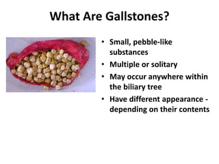
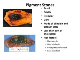
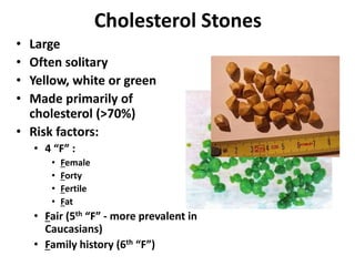
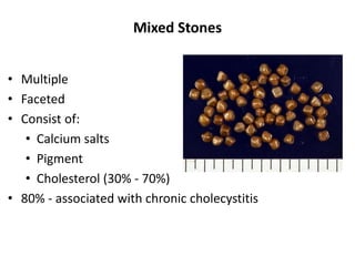
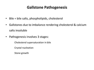
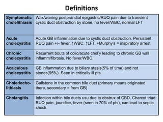
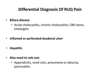
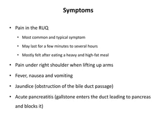
Gall Stone disease:
Etiology, Clinical features, diagnosis,
Complications
Cholecystectomy:
Indications,
Procedure, Complications
Dr Amit Gupta
Associate Professor
Dept of Surgery
What Are Gallstones?
• Small, pebble-like
substances
• Multiple or solitary
• May occur anywhere within
the biliary tree
• Have different appearance -
depending on their contents
Pigment Stones
• Small
• Friable
• Irregular
• Dark
• Made of bilirubin and
calcium salts
• Less than 20% of
cholesterol
• Risk factors:
• Haemolysis
• Liver cirrhosis
• Biliary tract infections
• Ileal resection
Cholesterol Stones
• Large
• Often solitary
• Yellow, white or green
• Made primarily of
cholesterol (>70%)
• Risk factors:
• 4 “F” :
• Female
• Forty
• Fertile
• Fat
• Fair (5th “F” - more prevalent in
Caucasians)
• Family history (6th “F”)
Mixed Stones
• Multiple
• Faceted
• Consist of:
• Calcium salts
• Pigment
• Cholesterol (30% - 70%)
• 80% - associated with chronic cholecystitis
Gallstone Prevalence
• 10% of people over 40 yrs.
• 90% “silent stones”
• Risk factors for becoming symptomatic:
• Smoking
• Parity
Risk Factors
• Women
• Age > 60 years
• American Indians & Mexican Americans
• Overweight or obese men and women
• People who tend to fast or lose weight quickly
• Family history of gallstones
• Diabetes
• Diet high in cholesterol
• Use of OCPs
• Pregnancy
Gallstone Pathogenesis
• Bile = bile salts, phospholipids, cholesterol
• Gallstones due to imbalance rendering cholesterol & calcium
salts insoluble
• Pathogenesis involves 3 stages:
Cholesterol supersaturation in bile
Crystal nucleation
Stone growth
Definitions
Symptomatic
cholelithiasis
Wax/waning postprandial epigastric/RUQ pain due to transient
cystic duct obstruction by stone, no fever/WBC, normal LFT
Acute
cholecystitis
Acute GB inflammation due to cystic duct obstruction. Persistent
RUQ pain +/- fever, ↑WBC, ↑LFT, +Murphy’s = inspiratory arrest
Chronic
cholecystitis
Recurrent bouts of colic/acute chol’y leading to chronic GB wall
inflamm/fibrosis. No fever/WBC.
Acalculous
cholecystitis
GB inflammation due to biliary stasis(5% of time) and not
stones(95%). Seen in critically ill pts
Choledocho-
lithiasis
Gallstone in the common bile duct (primary means originated
there, secondary = from GB)
Cholangitis Infection within bile ducts usu due to obstrux of CBD. Charcot triad:
RUQ pain, jaundice, fever (seen in 70% of pts), can lead to septic
shock
Differential Diagnosis Of RUQ Pain
• Biliary disease
• Acute cholecystitis, chronic cholecystitis, CBD stone,
cholangitis
• Inflamed or perforated duodenal ulcer
• Hepatitis
• Also need to rule out:
• Appendicitis, renal colic, pneumonia or pleurisy,
pancreatitis
Symptoms
• Pain in the RUQ
• Most common and typical symptom
• May last for a few minutes to several hours
• Mostly felt after eating a heavy and high-fat meal
• Pain under right shoulder when lifting up arms
• Fever, nausea and vomiting
• Jaundice (obstruction of the bile duct passage)
• Acute pancreatitis (gallstone enters the duct leading to pancreas
and blocks it)
Murphy’s Sign: Inspiratory arrest with manual pressure
below the gallbladder
Complications Of Gallstones
• In the GB:
• Biliary colic
• Acute and chronic cholecystitis
• Empyema
• Mucocoele
• Carcinoma
• In the bile ducts:
• Obstructive jaundice
• Pancreatitis
• Cholangitis
• In the gut:
• Gallstone ileus
0.1–0.7% of patients who have gallstones
Csendes classification :
• Type 1: external compression of the common bile duct – 11%
• Type 2: cholecystobiliary fistula is present involving <1/3 rd the circumference
of the bile duct – 41%
• Type 3: a fistula is present involving upto 2/3 the circumference of the bile
duct – 44%
• Type 4: a fistula is present with complete destruction of the wall of the bile
duct – 4%
Mirizzi syndrome
Diagnosis
• Ultrasound
• Computerized tomography (CT) scan
• May show gallstones or complications, such as infection and rupture
of GB or bile ducts
• Cholescintigraphy (HIDA scan)
• Used to diagnose abnormal contraction of gallbladder or obstruction
of bile ducts
• Endoscopic retrograde cholangiopancreatography (ERCP)
• Used to locate and remove stones in bile ducts
• Blood tests
• Performed to look for signs of infection, obstruction, pancreatitis, or
jaundice
USG CT Scan
• Asymptomatic gallstones do not require operation
• Whilst awaiting for surgery
• Low fat diet
• Dissolution therapy (ursodeoxycholic acid) generally useless
Management
• Cholecystostomy
• Subtotal cholecystectomy
• Open cholecystectomy
• Laparoscopic cholecystectomy
Surgical options
Cholecystostomy
• Patients at high risk related to multisystem organ failure
• Severe pulmonary, renal, or cardiac disease
• Recent myocardial infarction
• Cirrhosis with portal hypertension
• Acalculus cholecystitis after severe trauma, burns, or surgery
• Empyema or gangrene of the gallbladder
Subtotal Cholecystectomy
• Severe inflammation renders identification of the
anatomy impossible, eg. Gangrenous cholecystitis
• Scarred partially intrahepatic gallbladder
• Severe cirrhosis and portal hypertension
Cholecystectomy
Laparoscopic Surgery
• Advantages:
• Less post-op pain
• Shorter hospital stay
• Quicker return to normal activities
• Disadvantages:
• Learning curve
• Inexperience at performing open cholecystectomies
• After acute cholecystitis, cholecystectomy traditionally performed after 6
weeks
• Arguments for 6 weeks later
• Laparoscopic dissection more difficult when acutely inflammed
• Surgery not optimal when patient septic/dehydrated
• Logistical difficulties (theatre space, lack of surgeons)
• Arguments for same admission
• Research suggests same admission lap chole as safe as elective chole (conversion to
open maybe higher)
• Waiting increases risk of further attacks/complications which can be life threatening
• Risk of failure of conservative management and development of dangerous
complication such as empyema, gangrene and perforation can be avoided
• National guidelines state any patient with attack of gallstone pancreatitis
should have lap chole within 3 weeks of the attack
Cholecystectomy when to perform?
Complications of Lap Cholecystectomy
• Trocar/Veress needle injury
• Hemorrhage
• Wound infection and/or abscess
• Ileus
• Bile leak
• Gallstone spillage
• Deep vein thrombosis
• Retained common bile duct (CBD) stone
• CBD injury & stricture
• Pancreatitis
• Conversion to open procedure
• Nonsurgical treatment:
• Only in special situations
• When a patient has a serious medical condition preventing surgery
• Only for cholesterol stones
• Oral dissolution therapy
• Ursodeoxycholic acid - to dissolve cholesterol gallstones
• Months or years of treatment may be necessary before all stones
dissolve
• Contact dissolution therapy
• Experimental procedure
• Involves injecting a drug directly into the gallbladder to dissolve
cholesterol stones
Prevention
A sensible diet is the best way to prevent gall stones
Avoid crash diet or very low intake of calories
Eat good sources of fiber

gall_stones.ppt

  • 1.
    Gall Stone disease: Etiology,Clinical features, diagnosis, Complications Cholecystectomy: Indications, Procedure, Complications Dr Amit Gupta Associate Professor Dept of Surgery
  • 2.
    What Are Gallstones? •Small, pebble-like substances • Multiple or solitary • May occur anywhere within the biliary tree • Have different appearance - depending on their contents
  • 3.
    Pigment Stones • Small •Friable • Irregular • Dark • Made of bilirubin and calcium salts • Less than 20% of cholesterol • Risk factors: • Haemolysis • Liver cirrhosis • Biliary tract infections • Ileal resection
  • 4.
    Cholesterol Stones • Large •Often solitary • Yellow, white or green • Made primarily of cholesterol (>70%) • Risk factors: • 4 “F” : • Female • Forty • Fertile • Fat • Fair (5th “F” - more prevalent in Caucasians) • Family history (6th “F”)
  • 5.
    Mixed Stones • Multiple •Faceted • Consist of: • Calcium salts • Pigment • Cholesterol (30% - 70%) • 80% - associated with chronic cholecystitis
  • 6.
    Gallstone Prevalence • 10%of people over 40 yrs. • 90% “silent stones” • Risk factors for becoming symptomatic: • Smoking • Parity
  • 7.
    Risk Factors • Women •Age > 60 years • American Indians & Mexican Americans • Overweight or obese men and women • People who tend to fast or lose weight quickly • Family history of gallstones • Diabetes • Diet high in cholesterol • Use of OCPs • Pregnancy
  • 8.
    Gallstone Pathogenesis • Bile= bile salts, phospholipids, cholesterol • Gallstones due to imbalance rendering cholesterol & calcium salts insoluble • Pathogenesis involves 3 stages: Cholesterol supersaturation in bile Crystal nucleation Stone growth
  • 9.
    Definitions Symptomatic cholelithiasis Wax/waning postprandial epigastric/RUQpain due to transient cystic duct obstruction by stone, no fever/WBC, normal LFT Acute cholecystitis Acute GB inflammation due to cystic duct obstruction. Persistent RUQ pain +/- fever, ↑WBC, ↑LFT, +Murphy’s = inspiratory arrest Chronic cholecystitis Recurrent bouts of colic/acute chol’y leading to chronic GB wall inflamm/fibrosis. No fever/WBC. Acalculous cholecystitis GB inflammation due to biliary stasis(5% of time) and not stones(95%). Seen in critically ill pts Choledocho- lithiasis Gallstone in the common bile duct (primary means originated there, secondary = from GB) Cholangitis Infection within bile ducts usu due to obstrux of CBD. Charcot triad: RUQ pain, jaundice, fever (seen in 70% of pts), can lead to septic shock
  • 10.
    Differential Diagnosis OfRUQ Pain • Biliary disease • Acute cholecystitis, chronic cholecystitis, CBD stone, cholangitis • Inflamed or perforated duodenal ulcer • Hepatitis • Also need to rule out: • Appendicitis, renal colic, pneumonia or pleurisy, pancreatitis
  • 11.
    Symptoms • Pain inthe RUQ • Most common and typical symptom • May last for a few minutes to several hours • Mostly felt after eating a heavy and high-fat meal • Pain under right shoulder when lifting up arms • Fever, nausea and vomiting • Jaundice (obstruction of the bile duct passage) • Acute pancreatitis (gallstone enters the duct leading to pancreas and blocks it)
  • 12.
    Murphy’s Sign: Inspiratoryarrest with manual pressure below the gallbladder
  • 13.
    Complications Of Gallstones •In the GB: • Biliary colic • Acute and chronic cholecystitis • Empyema • Mucocoele • Carcinoma • In the bile ducts: • Obstructive jaundice • Pancreatitis • Cholangitis • In the gut: • Gallstone ileus
  • 14.
    0.1–0.7% of patientswho have gallstones Csendes classification : • Type 1: external compression of the common bile duct – 11% • Type 2: cholecystobiliary fistula is present involving <1/3 rd the circumference of the bile duct – 41% • Type 3: a fistula is present involving upto 2/3 the circumference of the bile duct – 44% • Type 4: a fistula is present with complete destruction of the wall of the bile duct – 4% Mirizzi syndrome
  • 15.
    Diagnosis • Ultrasound • Computerizedtomography (CT) scan • May show gallstones or complications, such as infection and rupture of GB or bile ducts • Cholescintigraphy (HIDA scan) • Used to diagnose abnormal contraction of gallbladder or obstruction of bile ducts • Endoscopic retrograde cholangiopancreatography (ERCP) • Used to locate and remove stones in bile ducts • Blood tests • Performed to look for signs of infection, obstruction, pancreatitis, or jaundice
  • 16.
  • 17.
    • Asymptomatic gallstonesdo not require operation • Whilst awaiting for surgery • Low fat diet • Dissolution therapy (ursodeoxycholic acid) generally useless Management
  • 18.
    • Cholecystostomy • Subtotalcholecystectomy • Open cholecystectomy • Laparoscopic cholecystectomy Surgical options
  • 19.
    Cholecystostomy • Patients athigh risk related to multisystem organ failure • Severe pulmonary, renal, or cardiac disease • Recent myocardial infarction • Cirrhosis with portal hypertension • Acalculus cholecystitis after severe trauma, burns, or surgery • Empyema or gangrene of the gallbladder
  • 20.
    Subtotal Cholecystectomy • Severeinflammation renders identification of the anatomy impossible, eg. Gangrenous cholecystitis • Scarred partially intrahepatic gallbladder • Severe cirrhosis and portal hypertension
  • 21.
    Cholecystectomy Laparoscopic Surgery • Advantages: •Less post-op pain • Shorter hospital stay • Quicker return to normal activities • Disadvantages: • Learning curve • Inexperience at performing open cholecystectomies
  • 23.
    • After acutecholecystitis, cholecystectomy traditionally performed after 6 weeks • Arguments for 6 weeks later • Laparoscopic dissection more difficult when acutely inflammed • Surgery not optimal when patient septic/dehydrated • Logistical difficulties (theatre space, lack of surgeons) • Arguments for same admission • Research suggests same admission lap chole as safe as elective chole (conversion to open maybe higher) • Waiting increases risk of further attacks/complications which can be life threatening • Risk of failure of conservative management and development of dangerous complication such as empyema, gangrene and perforation can be avoided • National guidelines state any patient with attack of gallstone pancreatitis should have lap chole within 3 weeks of the attack Cholecystectomy when to perform?
  • 24.
    Complications of LapCholecystectomy • Trocar/Veress needle injury • Hemorrhage • Wound infection and/or abscess • Ileus • Bile leak • Gallstone spillage • Deep vein thrombosis • Retained common bile duct (CBD) stone • CBD injury & stricture • Pancreatitis • Conversion to open procedure
  • 25.
    • Nonsurgical treatment: •Only in special situations • When a patient has a serious medical condition preventing surgery • Only for cholesterol stones • Oral dissolution therapy • Ursodeoxycholic acid - to dissolve cholesterol gallstones • Months or years of treatment may be necessary before all stones dissolve • Contact dissolution therapy • Experimental procedure • Involves injecting a drug directly into the gallbladder to dissolve cholesterol stones
  • 26.
    Prevention A sensible dietis the best way to prevent gall stones Avoid crash diet or very low intake of calories Eat good sources of fiber