Growth and Development of Craniofacial Complex
•
0 likes•150 views

The Indian Dental Academy is the Leader in continuing dental education , training dentists in all aspects of dentistry and offering a wide range of dental certified courses in different formats.for more details please visit www.indiandentalacademy.com

Recommended
Recommended
More Related Content
What's hot
What's hot (6)
Similar to Growth and Development of Craniofacial Complex
Similar to Growth and Development of Craniofacial Complex (20)
More from Indian dental academy
More from Indian dental academy (20)
Recently uploaded
Recently uploaded (20)
Growth and Development of Craniofacial Complex
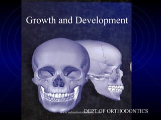
- 1. Growth and Development DEPT OF ORTHODONTICSwww.indiandentalacademy.com
- 2. INTRODUCTION DEFINITIONS METHODS OF STUDYING GROWTH CONCEPTS OF GROWTH PROCESS {THEORIES} GROWTH OF CRANIOFACIAL COMPLEX CLINICAL IMPLICATIONS OF GROWTH www.indiandentalacademy.com
- 3. Introduction • Importance of studying growth and development www.indiandentalacademy.com
- 4. Definition of growth :- GROWTH IS DEFINED AS A SERIES OF ANATOMIC AND PHYSIOLOGIC CHANGES TAKING PLACE FROM THE BEGINNING OF PRENATAL LIFE TO CLOSE OF SENILITY • Growth refers to increase in size – Todd • Growth may be defined as the normal changes in the amount of living substance. - Moyer's www.indiandentalacademy.com
- 5. SALZMAN; GROWTH IS A PHYSIO CHEMICAL PROCESS WHICH RELATES STRUCTURE COMPOSITION SIZE SHAPE OXFORD ; GROWTH IS INCREASE IN SIZE QUANTITY AND NUMBER HUXLEY; GROWTH IS CELL MULTIPLICATION •PROFFIT;Growth usually refers to an increase in size and the number - www.indiandentalacademy.com
- 6. Definition of Development • Development is progress towards maturity” - Todd • Development connotes a maturation process involving progressive differentiation at the cellular and tissue levels www.indiandentalacademy.com
- 7. Correlation between growth and development • Growth is basically anatomic phenomenon and quantitative in nature. • Development is basically physiologic phenomenon and qualitative in nature. www.indiandentalacademy.com
- 8. Normal features of growth and development • Differential growth • Pattern • Variability • Timing, rate and direction. www.indiandentalacademy.com
- 9. Differential growth Not all tissue system s of the body grow at the same rate. Different tissues and in term different organs grow at different rates. This process is called differential growth. www.indiandentalacademy.com
- 10. Scammons Curve As the graph indicates, growth of the neural tissues is nearly complete by 6 or 7 years of age. General body tissue, including muscle, bone and viscera, show and S-shaped curve, with a definite slowing of the rate of growth during childhood and an acceleration at puberty. Lymphoid tissues proliferate far beyond the adult amount in late childhood, and then undergo involution at the same time that growth of the genital tissues accelerates rapidly.www.indiandentalacademy.com
- 12. Cephalocaudal Gradient of growth • Represents the changes in over all body proportions during normal growth and development. • In fetal life, at about the third month of intrauterine development, the head takes up almost 50% of the total body length. At this stage, the cranium is large relative to the face and represents more than half the total head.www.indiandentalacademy.com
- 15. In contrast, the limbs are still rudimentary and the trunk is underdeveloped. By the time of birth, the trunk and limbs have grown faster than the head and face, so that the proportion of the entire body devoted to the head has decreased to about 30%. www.indiandentalacademy.com
- 16. The overall pattern of growth thereafter follows this course, with a progressive reduction of the relative size of the head to about 12% the adult. Thus “Cephalocaudal gradient of growth”. This simply means that there is an axis of increased growth extending from the head towards the feet. www.indiandentalacademy.com
- 17. Growth Spurts Refers to Sudden increase in growth of general Body. Woodside in his study of Burlington Group showed. Girls Boys Just after birth 3 yrs 3 yrs Juvenile growth Spurt 6-7yrs 7-9yrs Pubertal growth spurt 10-12yrs 12-15yrs www.indiandentalacademy.com
- 18. Importance of Growth Spurts: Pubertal increments offers best time for large number of cases for the orthodontic and orthopedic treatment. It also helps in determining the predictability, growth direction, patient management and total treatment time. www.indiandentalacademy.com
- 19. Orthopedic correction of maxilla and Mandible. Understanding the growth, predictability of future growth of maxilla, mandible and alveolar process helps in diagnosing and achieving excellent results of the mal- occlusion. www.indiandentalacademy.com
- 20. Growth spurts serve as excellent indicators for timing of orthodontic treatment Correlation of a. Skeletal age, b. Dental age c. Chronological age. With on set of puberty. www.indiandentalacademy.com
- 21. Biological changes seen during puberty. Biological changes differ with boys and girls In Boys : Stage I: - Fat spurt - Initially maturing boy gains weight and becomes chubby –production of estrogen before production of testosterone. www.indiandentalacademy.com
- 22. Stage II - Spurt in height, development of secondary sexual characteristics. - Occurs 1 year after the Stage I Stage III - Occurs 8-10months after stage II and coincides with the peak velocity with gain in height - At this stage auxillary hair appears and facial hair appears on upper lip. Spurt in muscle growth occurs. www.indiandentalacademy.com
- 23. Stage IV: - Occurs from 15-24 months after stage III - Spurt of growth in height ends. Facial hair on chin and upper lip. This indicates growth is almost complete. In Girls: (9-12 yrs) Stage I: - Beginning of growth spurt appearance secondary sexual characteristics .www.indiandentalacademy.com
- 24. Stage II: - Occurs 1 year after stage I coincides with peak velocity physical growth. Stage III: - Occurs 1-1½ years later stage II. marked by on set of menstruation. - By this time growth spurt all but complete. www.indiandentalacademy.com
- 25. Methods of Studying Growth A • Experimental approach • Measurement approach B • Longitudinal study • Cross sectional study. • Semi longitudinal study. www.indiandentalacademy.com
- 26. Longitudinal study: • Advantages: Temporary problems are smoothen with time, variability in development within a group is put in proper perspective, serial comparison makes study of specific developmental pattern of individual possible. www.indiandentalacademy.com
- 27. • Disadvantages: Time consuming, expensive, sample loss or attrition, averaging. • These are measurements made of the same person or group at regular intervals through time. www.indiandentalacademy.com
- 28. Cross sectional studies : • These are measurements made of different samples or different individuals and studied at different periods. • Advantages: Quicker, less expensive, statistical treatment of data is easier. Studies can be readily repeated. Method can be used in archeological data. www.indiandentalacademy.com
- 29. • Disadvantages: It must be assumed that groups being measured and compared are similar. Cross sectional group averages tend to obscure individual variations. www.indiandentalacademy.com
- 30. ( • Longitudinal and cross sectional studies can be combined to seek the advantages of both. In this way one might compress 15 years of study into 3 years of gathering growth data. www.indiandentalacademy.com
- 31. Measuremental approach Non destructive Technique for measuring growth in living animals including man The Species will be available for additional measurements at another time. www.indiandentalacademy.com
- 32. Measurement Approaches: Craniometry : Based on measurement of skulls found among human skeletal remains. From such skeletal material, it has been possible to piece together a great deal of knowledge Advantage: Craniometry has the rather precise measurements can be made on dry skulls. Disadvantage: It is a Cross Sectional study. www.indiandentalacademy.com
- 33. • Cross-Sectional means that although different ages are represented in the population, the same individual can be measured at only one point in time. Antropometric Study: This measurement can be made on either a dried skull or a living individual, but results would be different because of the soft tissue thickness overlying both landmarks. Advantages: This produces longitudinal data: repeated measures of the same individual. www.indiandentalacademy.com
- 34. Cephalometric radiology: is of considerable importance not only in the study of growth but also in clinical evaluation of orthodontic patients. This approach can combine the advantages of craniometry and anthropometry. It allows a direct measurement of bony skeletal dimensions, but it also allows the same individual to be followed over time. www.indiandentalacademy.com
- 35. Experimental approach a. Detailed study may be destructive b. Largely restricted to non human beings www.indiandentalacademy.com
- 36. Experimental approaches: 1. Vital Staining: John Hunter originated this method. In eighteenth century. Hunter observed that bones of Pigs that are occasionally fed with textile waste were often stained in an interesting way. He observed that active agent was a dye called alizarin which still used for vital staining studies. www.indiandentalacademy.com
- 37. Vital staining is method of studying skeletal growth in which dyes that stain mineralizing tissues are injected in to animal. These dyes remain in teeth and bones and can be detected later after the sacrifice of animal. www.indiandentalacademy.com
- 38. These dyes react strongly at sites where bone calcification is occurring. Since these are the sites of active skeletal growth, the dye marks the locations at which active growth is occurring. www.indiandentalacademy.com
- 39. Tetracycline was discovered too late in 1960’s as an excellent vital stain that binds to calcium often resulting in discoloration of tooth Vital staining method can not be used in humans. www.indiandentalacademy.com
- 40. Alizarin Red 5 Tetracycline Used for vital Lead Acetate staining. Trypon Blue. www.indiandentalacademy.com
- 41. Radio-Isotopes With development of radio active tracers it is possible to use radio actively labeled metabolite that becomes incorporate in tissues as sort of vital stain. The location of radio active element is detected from tissues by the weak radio-activity given off at the site where material was incorporated. www.indiandentalacademy.com
- 42. • Isotope technetum (Tc99m ) (Gamma Emitting) • 33 MTc • Calcium 45 • Potassium 32 • 14 C Proline • 3 H Thymidine This radioactivity in tissues of experimental animals is detected in tissue culture by method called Autoradiographywww.indiandentalacademy.com
- 43. In Autoradiography a film emulsion is placed over a this section of tissue containing the isotope and then it is exposed in dark by the radiation that indicates where growth is occurring. www.indiandentalacademy.com
- 44. Implant Radiography: - This method of study developed by professor Arne Bjork and Coworkers at Royal Dental College in Copenhagen Denmark in 1969. This has provided important new information about growth pattern of jaws. www.indiandentalacademy.com
- 45. In this technique, Innert metal pins are placed in bones of face and Jaws. These metal pins are innert and are well tolerated by skeleton. Cephalometric reading taken periodically and superimposition of radiographs provide precise changes in position of bone and Jaw growths. www.indiandentalacademy.com
- 46. Natural markers. • The persistence of certain developmental features has led to their use as natural markers by means of serial radiography. • Eg: trabaculae,nutrient canals and lines of arrested growth can be used for reference to study deposition, resorption and remodeling. • Certain natural markers are used as cephalometric landmarks. www.indiandentalacademy.com
- 47. Functional Matrix theory Melvin Moss (1965) www.indiandentalacademy.com
- 48. Implant markers. • Bjork devised a method of implanting tiny bits of tantalum or biologically inert alloys into growing bone which served as radiographic reference markers for serial cephalometric study. • The method allows precise orientation of serial cephalograms and information on the amount and sites of bone growth. www.indiandentalacademy.com
- 49. Theories of Growth and Development www.indiandentalacademy.com
- 50. According to BAUME: Growth Center: Is a site of endochondral ossification with tissue separating force,contributing to the increase of skeletal mass. Growth Site: Regions of Periosteal or suture bone formation and modeling resorption adaptive to environmental influences. www.indiandentalacademy.com
- 51. GROWTH THEORIES 1) Genetic Theory 2) Sutural Theory 3) Cartilagnous theory 4) Functional Matrix thory 5) Cybernetic Theory 6) Composite Theory www.indiandentalacademy.com
- 52. Genetic Theory 1950’s to 1970’s: -Mainly based on observations -No evident scientific data -Lacked scientific understanding and soon replaced by other theories. www.indiandentalacademy.com
- 53. Sutural Theory: Proposed by Sicher in 1955: According to Sicher -“The primary event in sutural growth is the proliferation of the connective tissue between the two bones. If sutural tissue proliferates, it creates the space for appositional growth at the border of the bones”. www.indiandentalacademy.com
- 54. We now know that functions of future are : 1. Unite the bone 2. Absorb the forces, 3. Act as a joint 4. Act as a growth site and not growth centre www.indiandentalacademy.com
- 55. Evidences Against Sicher’s Theory: 1. Auto transplants of sutures fail to grow in cultural medium though provided with same environment and conditions. 2. Extripation of sutures has no appreciable effect on growth of skeletal. 3. The shape and growth within sutures is dependent on external stimuli. 4. It is possible to bring the sutural grwoth to halt by mechanical stresse applied across the sutures. www.indiandentalacademy.com
- 56. Cartilagenous Theory (James Scott-1956) The fact that, for many bones of the hand and legs, cartilagedoes the growing while bone merely replaces it makes this theory attractive for the bones of the jaws. According the Scott:- -Spheno-occipital synchondrosis cartilage -responsible for the growth of cranial base. -Nasal septal cartilage – Responsible for the growth of maxilla -Condylar cartilage – Responsible for the growth of mandible www.indiandentalacademy.com
- 58. Synchondroses of the cranial base 1. Spheno ethmoidal 2. Inter-sphenoidal 3. Spheno-occipital www.indiandentalacademy.com
- 59. Spheno-occipital Synchodrosis: -Important growth center of craniofacial skeleton, especially cranial base. Cartilage of Nasal Septum: -Growth of maxilla is difficult to explain on the cartilage theory. Proponents of the cartilage theory hypothesize that the cartilaginous nasal septum serves as a pacemaker for other aspects of maxillary growth.www.indiandentalacademy.com
- 60. -Two kinds of experiments have been carried out to test the idea that cartilage can serve as a true growth center. 1. Transplanting nasal cartilage to cultural medium or any other place did not give equivocal results, that is sometime it grew, sometimes it did not. Indicating doubtful growth potential of the nasal septal cartilage whereas, if a piece of the epiphyseal plate of a long bone is transplanted, it will continue to grow in a new location or in culture, indicating that these cartilages do have innate potential. www.indiandentalacademy.com
- 61. Effect of removal of the cartilaginous nasal septum on forward growth of the snout in the rabbit. A B www.indiandentalacademy.com
- 62. Profile view of man whose cartilaginous nasal septum was removed at age 8, after an injury. www.indiandentalacademy.com
- 63. Condylar Cartilage and Mandibular Growth •Since longtime, its being hypothesized that condylar cartilage is the growth center for the growth of mandible. •Experiments of transplanting condylar cartilage showed little or No growth potential. •It is no clear that condylar cartilage is secondary cartilage, which grows by appositions and not by intestitial deposition. Whereas, epiphyseal cartilage is primary cartilage. • Mandibular condylar thus do not have innate growth potential and not a growth center. www.indiandentalacademy.com
- 65. • The fact that after the condylar fracture in children do not all together inhibit growth of mandible indicates that condyle is not a growth center. • Studies carried out in Scndinavia in 1960’s demonstrated that after the fracture of mandibular condyle in child, there was an excellent chance that the condylar process would regenerate to approximately its original size and small chance that it would overgrow after the injury. www.indiandentalacademy.com
- 67. • THE ORIGIN,GROWTH AND MAINTENANCE OF ALL SKELETAL TISSUES AND ORGANS ARE ALWAYS SECONDARY,COMPENSATORY AND OBLIGATORY TO TEMPORALLY AND OPERATIONAL PRIOR EVENTS OR PROCESSES THAT OCCUR IN SPECIFICALLY RELATED NON-SKELETAL TISSUES,ORGANS OR FUNCTIONAL SPACES www.indiandentalacademy.com
- 68. • Each of these function is completely carried out by FUNCTIONAL CRANIAL COMPONENT www.indiandentalacademy.com
- 69. Functional cranial component Skeletal unit Functional matrices Macroskeletal Eg-coronoid, angular Microskeletal Eg-endocranial surface Of calvaria Periosteal Eg-teeth and muscles Capsular Eg-orofacial, neurocranial www.indiandentalacademy.com
- 70. Skeletal unit • Composed of –bone, cartilage and tendinous tissue MACROSKELETAL UNIT- • Adjoining portions of number of neighbouring bones carrying out a single function eg- endocrainal surface of calvaria www.indiandentalacademy.com
- 72. MICROSKELETAL UNIT bones consisting of number of small skeletal units MAXILLA-orbital -pneumatic -palatal -basal MANDIBLE-coronoid -angular -alveolar -basal www.indiandentalacademy.com
- 74. FUNCTIONAL MATRICES • This consist of soft tissue- muscle,gland,nerve,vessels,fat and teeth as well as non skeletal cartilages DIVIDE INTO TWO TYPES- • Periosteal matrices • Capsular matrices www.indiandentalacademy.com
- 75. PERIOSTEAL MATRICES • All non skeletal functional units adjacent to skeletal unit form the periostel matrices • They act by bringing transformation of the related skeletal units • Best explanation – coronoid process and temporalis muscle • Removal,denervation, postinfectively- decrease in the size or total disappearance www.indiandentalacademy.com
- 76. • Hence in simple terms it can be stated- Coronoid process does not grow itself first and thus provide a platform upon which the temporalis muscle can alter its function but it is the opposite which is true www.indiandentalacademy.com
- 77. CAPSULAR MATRICES FOUR CAPSULES ARE PRESENT- • NEURO CRANIAL • ORO FACIAL • OTIC • ORBITAL www.indiandentalacademy.com
- 78. • Each of these capsules is an envelop containing functional cranial component • Sandwitched between two covering layers • Capsules expands due to volumetric increase of capsular matrix • This results in the translative movement of the embedded bones www.indiandentalacademy.com
- 79. NEUROCRAINAL CAPSULE • Sandwiched between-skin and dura mater • Consists of-5 layers of scalp -bone -two layer dura mater www.indiandentalacademy.com
- 81. ORO FACIAL MATRIX • Surround and protect oronasopharyngeal space. • Surrounded by skin and mucous membrane on either side. • Originates by process of enclosure. • Volumetric growth of these spaces is the primary morphogenetic event in facial skull growth www.indiandentalacademy.com
- 83. • Primary function is maintaining airway this is accomplished by “AIRWAY MAINTENANCE SYSTEM”-BOSMA • Growth of functional spaces-increase in the size of capsule • Followed by passive movement of functional cranial component www.indiandentalacademy.com
- 84. Servosystem theory or Cybernetic theory: Proposed by petrovic accordingly, the growth of maxilla and mandible and cranial base depends upon cybernetic control. This cybernetic control is mainly by Secretion of hormones. These hormones mainly include growth hormone - somatomedin, testosterone and estrogen. www.indiandentalacademy.com
- 85. Author describes the secretion of hormones is by the signal established independed of the feedback system. •This signal secretion is described as COMMAND . •This signal is transmitted to the Reference input elements. In maxilla they include septal cartilage, septopremaxillary frenum, the labionarinary muscles and the maxillary bones. In mandible reference input elements include muscle attachments to the mandible that is lateral pterygoid, medial pterygoid and tempralis muscles. www.indiandentalacademy.com
- 86. The commanding signal is first established in the maxilla through the above quoted reference input elements and thus maxilla grows in sagittal and vertical direction. The corresponding actuating signal followed to the above process is felt in the mandible through the reference input elements and mandible growth occurs. www.indiandentalacademy.com
- 87. Functional matrix theory revisited Melwin Moss in 1997 proposed continuation of his classical functional matrix theory with the new concept. He published series of articles in American Journal of Orthodontics in 1997. According to this concept the mechanical stimulus is pursued by the specialized cells by process called as mechanoperception. Then these signals are transmitted through the tissues by way mechanoconduction or mechanotrasmision. Finally, these signals are transmitted to the genome of the bone were protein synthesis is taking place.www.indiandentalacademy.com
- 88. These signals alter the protein metabolism depending upon the severity and longativity of the mechanical stimulus. In short the earlier concept of FMH theory remained same as form is determine by the function. Moss also recognizes the important role of genetics and human genome in determining the ultimate size and shape of the craniofacial skeleton. He quotes reference of human genome project which is being carried in a mega scale allover the world. According to the human genome project human chromosomes contain the genetic informations necessary for buildingup of entire human body. www.indiandentalacademy.com
- 89. Genes now beyond doubt have been proved to effect the physical growth of the person, behavior of person and psychology of person. Thus Moss FMH revisited theory states the ultimate growth controlling factor of the craniofacial skeleton depends on two factors. 1. Genetic factors 2. Environment factors. www.indiandentalacademy.com
- 90. Mechanisms of growth • 3 mechanisms at the cellular level • Hyperplasia • Hypertrophy • Secretion of extracellular matter www.indiandentalacademy.com
- 91. Mechanism of growth in soft tissues • In soft tissues growth occurs by a combination of two mechanisms namely hyperplasia and hypertrophy – Interstitial growth www.indiandentalacademy.com
- 92. Mechanism of growth in hard tissue It is of two types Intramembranous Endochondral Bone formation. Bone formation www.indiandentalacademy.com
- 93. Mechanism of growth in hard tissues • Intramembranous bone formation: • Occurs in areas exposed to tension • It differs from endochondral bone formation by formation of bone directly from mesenchymal tissue • Seen in areas like • Cranial vault • Maxilla • Mandible except condylar cartilage www.indiandentalacademy.com
- 94. Undifferentiated mesenchymal cells differentiate Osteoblasts Osteoid Matrix Calcification and formation of bone results www.indiandentalacademy.com
- 96. Mechanism of growth in hard tissues • Endochondral bone formation: • Occurs in regions exposed to high levels of compression • In craniofacial region it is seen in areas like • Synchondrosis at the cranial base • Condylar cartilage • Nasal septal cartilage www.indiandentalacademy.com
- 97. Cartilage cells undergo hypertrophy Marix become calcified. Cells Degenerate Osteogenic tissues invade disintegrating cartilage and replace it by formation of bone. www.indiandentalacademy.com
- 99. Bone metabolism • Bone is the primary calcium reservoir of the body (99% stored in skeleton) •Bone structure is sacrificed to maintain the critical serum calcium levels at 10mg % www.indiandentalacademy.com
- 100. Bone metabolism Calcium homeostasis is supported by 3 mechanisms : 1. Rapid instantaneous flux of calcium from bonefluid (seconds) by selective transfer of calcium ions into and out of bone fluid. 2. Shorterm control of serum calcium levels affects rates of bone formation $ resorption 3. Longterm regulation of metabolism- have effects on skeleton. www.indiandentalacademy.com
- 101. Types of Bones • Woven bone – The first bone formed in response to orthodontic loading usually is the woven type. It is weak, disorganized, and poorly mineralized • Lamellar bone – a strong, highly organized, well-mineralized tissue www.indiandentalacademy.com
- 102. Types of bones • Composite bone – is an osseous tissue formed by the deposition of lamellar bone within a woven bone lattice, a process called Cancellous compaction. This is the quickest means of producing relatively strong bone • Bundle bone - is a functional adaptation of lamellar structure to allow attachment of tendons and ligaments www.indiandentalacademy.com
- 103. Mechanisms of bone growth • Modeling • Remodeling • Displacement • Combination of remodeling & displacement • Rotation www.indiandentalacademy.com
- 104. MODELING • Bone modeling involves independent sites of resorption and formation that change the size and shape of a bone. www.indiandentalacademy.com
- 106. Remodelling • A process involving deposition and resorption occuring on opposite ends • Four types • Biochemical remodelling • Haversian remodelling • Pathologic remodelling • Growth remodelling www.indiandentalacademy.com
- 107. Functions of Remodelling 1. Progressively change the size of whole bone 2.Sequentially relocate each component of the whole bone 3.Progressively change the shape of the bone to accommodate its various functions www.indiandentalacademy.com
- 108. 4. Progressive fine tune fitting of all the separate bones to each other and to their contiguous ,growing, functioning soft tissues 5. Carry out continuous structural adjustments to adapt to the intrinsic and extrinsic changes in conditions . www.indiandentalacademy.com
- 109. Displacement • Refers to a shift in the position of the bone • Two types • Primary displacement • Secondary displacement www.indiandentalacademy.com
- 110. Rotation • According to Enlow, growth rotation is due to diagonally placed areas of deposition and resorption • Two types • Remodelling rotations • Displacement rotations www.indiandentalacademy.com
- 111. ‘V’ - Principle • Deposition occurs on the inner side and resorption on the outerside of the bones causing enlargement and displacement. • The displacement is towards wide end of ‘V’ • Examples • Neck of the condyle • Palatal process of maxilla www.indiandentalacademy.com
- 113. Growth equivalent principle This principle proposed by Hunter & Enlow relates the effects of cranial base growth on the facial bone Growth. www.indiandentalacademy.com