SlideShare a Scribd company logo
1 of 8
Download to read offline
doi: 10.1111/j.1365-4632.2010.04663.x
Report
Full-spectrum light phototherapy for atopic dermatitis
Hee Jin Byun1, MD, Hye In Lee2, MD, Beomjoon Kim2, MD, PhD, Myeung Nam Kim2,
                      3               3              3                1
MD, PhD, Hyuckki Hong , Yeonshik Choi , Youngchang Jo , Kwang Hyun Cho , MD, PhD
                   4
and Seog-Kyun Mun , MD, PhD


1
  Department of Dermatology, College of      Summary
Medicine, Seoul National University,         Background Atopic dermatitis (AD) is a chronic skin disease resulting in a profound deteri-
Seoul, 2Department of Dermatology,
                                             oration in quality of life. The FSLÒ is a newly developed phototherapy device generating
College of Medicine, Chung-Ang
                                             full-spectrum light (FSL) with a continuous wavelength (320–5000 nm). This study aimed to
University, Seoul, 3Medical IT Research
Center, Korea Electronics Technology         evaluate the efficacy and safety of FSLÒ phototherapy in AD.
Institute, Gyeonggi-do, South Korea,         Methods We enrolled 38 patients with moderate to severe AD in this open, randomized,
and 4Department of                           controlled, prospective study. In the FSL-irradiated group (20 patients), irradiation was
Otorhinolaryngology - Head and Neck
                                             conducted twice per week for 4 consecutive weeks. In the control group (18 patients), only
Surgery, College of Medicine, Chung-
                                             emollient application was allowed. SCORing Atopic Dermatis (SCORAD) values were
Ang University, Seoul,Korea
                                             obtained at baseline, week 4 and 8. Patients were asked to give subjective assessments of
Correspondence                               improvement and laboratory tests including serum eosinophil counts, ECP levels, IgE levels
Seog-Kyun Mun, MD, PHD.                      and 22 cytokine levels were performed.
Department of Otorhinolaryngology-
                                             Results In the FSL-irradiated group, the mean SCORAD value decreased significantly after
Head and Neck Surgery
                                             4 weeks of phototherapy and remained reduced for a further 4 weeks after the cessation of
Chung-Ang University
Yongsan Hospital                             treatment. In the control group, the mean SCORAD value did not change significantly over
65-207, Hangangro 3-ka                       the study period. Patients’ subjective assessments indicated good to excellent responses
Yongsan-gu                                   in 75% of the FSL-irradiated group, by contrast with 50% of the control group. The mean
Seoul 140-757
                                             values for serum eosinophil counts, IL-4 and IL-5 levels decreased significantly after FLS
South Korea
                                             phototherapy. No serious adverse effects were reported.
E-mail: entdoctor@cau.ac.kr
                                             Conclusions In this study, we showed that FSLÒ phototherapy can be an effective and
Funding: This study was supported by a       safe treatment option in AD.
research grant from Chung-Ang
University awarded in 2010.
Conflicts of interest: None.


                                                                        At present, mainly 311-nm narrowband UVB (NBUVB),
Introduction
                                                                        UVA1 and psoralen with UVA (PUVA) are used.3 In the
Atopic dermatitis (AD) is a chronically relapsing skin dis-             management of AD, PUVA is thought to be the most
ease, characterized by age-related distribution and severe              effective and to induce the longest remission period
pruritus, and resulting in a profound deterioration in                  (£12 weeks4). After PUVA, high- and medium-dose (sin-
quality of life. Multiple treatment modalities, including               gle-exposure doses of 80–130 J/cm2 and 50–70 J/cm2,
topical and systemic immunosuppressants, and photother-                 respectively) UVA1 and NBUVB are thought to be
apy at various wavelengths, have been used in the treat-                equally effective.4–9 Combined UVA/UVB has been shown
ment of AD. However, current therapies are often                        to be less effective than medium-dose UVA1, and low-
unsatisfactory and cannot be used continuously because                  dose (single-exposure doses of 10 J/cm2) UVA1 has rarely
they carry a risk for adverse effects.1 Therefore, efforts to           been effective in AD.4,10 However, conventional photo-
develop novel treatment modalities with high levels of                  therapies have limitations and cannot be used in all
efficacy and safety are ongoing.                                         patients. Despite its excellent efficacy, the use of PUVA
  In 1948, Nexman2 reported that ultraviolet (UV) light                 has decreased continuously as a result of longterm haz-
emitted by a carbon arc lamp significantly improved AD.                  ards, such as the development of skin cancers, and the
Since then, numerous reports have shown that photother-                 difficulties involved in taking oral medication on a timely
apies are potent modalities for moderate to severe AD.                  basis and shielding the skin from sunlight for several                 1


ª 2010 The International Society of Dermatology                                                    International Journal of Dermatology 2010
2   Report   Full-spectrum light phototherapy for atopic dermatitis                                                               Byun et al.


    days.3,5,10 UVA1 has a rapid onset of action, but fewer             Table 1 Demographic patient data
    than 50% of affected individuals respond to UVA1 treat-
    ment and the response is usually short-lived.3,10                                                FSL-irradiated group     Control group
       Natural sunlight itself has also been used for photother-
                                                                        Patients, n                  20                       18
    apy under the designations of ‘‘heliotherapy’’ (after               Age, years (mean ± SD)       25.68 ± 7.69             25.63 ± 8.41
    Helios, the personification of the sun in Greek mythology)           Sex, n (male/female)         8/12                     9/9
    or climatotherapy. Heliotherapy has been demonstrated to            SCORAD (mean ± SD)           47.87 ± 15.45            39.79 ± 9.76
    be highly effective in AD. Two-week heliotherapy in the             Mean duration of AD, years   11.09                    10.95
    Canary Islands reduced mean SCORing Atopic Dermatis
    (SCORAD) values by 70–74% in previous reports, indi-                FSL, full-spectrum light; SD, standard deviation; AD, atopic
    cating a level of efficacy higher than that of PUVA, which           dermatitis.
    has been reported to reduce baseline SCORAD by 54%
    after 15 exposures.4,11,12 However, heliotherapy can be             ranged from 25 to 48 years. The mean SCORAD index at the
    performed only in specific regions with strong sunlight,             beginning of the study was 47.87 points in the FSL-irradiated
    such as the Canary Islands and the area around the Dead             group and 39.79 points in the control group. There was no
    Sea, and is usually conducted in the form of a ‘‘heliothera-        statistically significant difference in baseline SCORAD values
    py trip’’ organized as a 2- or 3-week tour. Therefore,              between the two groups (P = 0.167, Mann–Whitney U-test). All
    despite its high efficacy, the application of heliotherapy is        patients were Korean and had skin of phototypes III or IV.
    limited as a result of the interruption it causes to daily life     Demographic data for the patients are shown in Table 1.
    and the cost of travel expenses.11,12                               Informed consent was obtained from each patient prior to
       To overcome the limitations of heliotherapy, we used             enrollment.
    the FSLÒ (BMC Co. Ltd, Anyang-si, South Korea) for                    The FSLÒ phototherapy device generates light on a
    treatment of AD. The FSLÒ is a newly developed photo-               continuous wavelength of 320–5000 nm, from an electrical arc
    therapy device which generates full-spectrum light (FSL)            between two carbon rods (Ark RodsÒ; BMC Co. Ltd) at a
    on a continuous wavelength spectrum of 320–5000 nm.                 temperature of >3000 °C. An Ark RodÒ is made of carbon
    The light profile emitted by the FSLÒ is nearly identical            compounds including 16 confidential chemical elements.
    to that of sunlight except that it omits UVB; it includes           A calibrated quartz tungsten halogen lamp (model 63355;
    UVA, visible light and infrared light. Given the high effi-          Newport Corp., Stratford, CT, USA), a monochromator
    cacy of heliotherapy in AD treatment, we expected that              (Cornerstone 260Ô; Newport Corp.) and a digital lock-in
    FSL phototherapy would also be effective. Phototherapy              radiometry system (MerlineÔ; Newport Corp.) were used to
    devices emitting FSL, like the FSLÒ, have been used                 make light with a continuous wavelength. A two-color
    mainly in neuropsychiatric diseases, such as depression             photodetector (IGA030-TE2-H; Electro Optical Systems, Inc.,
    and sleep disturbance.13–15 So far, there have been no              Phoenixville, PA, USA), a TE-cooler (PS/TC-1; Newport Corp.)
    reports on the application of FSL devices in AD.                    and a TE-cooled HgCdZnTe detector (MIR8025Ô; Newport
       In this study, we aimed to evaluate the efficacy and              Corp.) were used as a detection system.
    safety of FSLÒ phototherapy in patients with moderate to              In the FSL-irradiated group, irradiation was performed with
    severe AD.                                                          the FSLÒ phototherapy device twice per week for 4 consecutive
                                                                        weeks, resulting in a total of eight applications of irradiation. We
                                                                        performed a pilot study to determine the appropriate exposure
    Materials and methods
                                                                        time. From the pilot study results, we concluded that exposures
    This was an open, randomized, controlled, prospective study.        of 20 min are most effective and safe (data not shown).
    We recruited 38 patients diagnosed with extrinsic AD according      Therefore, in each phototherapy session, the anterior side of
    to previously published definitions,8,9 from August 2007 to July     the body was irradiated for 20 min and the posterior side was
    2008. Only patients with moderate to severe AD with SCORAD          subsequently irradiated for 20 min. The fluence of each
    index values of >25 were included. The following subjects were      irradiation was 530 J/cm2, including 121 J/cm2 of UVA and
    excluded: patients aged <18 years; patients who had been            409 J/cm2 of visible and infrared light. During the entire study
    treated with systemic corticosteroid, any type of phototherapy or   period, the use of any topical or systemic agents other than the
    photosensitizing drugs in the 3 months prior to enrollment;         designated emollient cream (PhysiogelÒ; Stiefel-Korea Ltd,
    pregnant or lactating women, and patients with a history of         Seoul, South Korea) was disallowed. In the control group, the
    neoplasm or photosensitive dermatosis. Patients were screened       emollient, PhysiogelÒ, was applied twice per day without any
    to ensure they fulfilled inclusion and exclusion criteria and        other treatment.
    randomly assigned to either the FSL-irradiated group (20              The SCORAD index was used to assess disease severity.10
    patients) or to the control group (18 patients). Patient ages       SCORAD values in each patient were assessed three times


    International Journal of Dermatology 2010                                                 ª 2010 The International Society of Dermatology
Byun et al.                                                                 Full-spectrum light phototherapy for atopic dermatitis   Report


during the study period: (i) prior to the beginning of treatment                (a)
(at baseline); (ii) after 4 weeks of FSL phototherapy (at week
4), and (iii) 4 weeks after the cessation of phototherapy (at
week 8). All assessments were performed by the same
dermatologist. Patients’ subjective assessments of clinical
improvement were obtained at week 8, using a 4-point scale
with the following descriptors: ‘‘poor response’’ (0–25%
improvement); ‘‘fair response’’ (26–50% improvement); ‘‘good
response’’ (51–75% improvement), and ‘‘excellent response’’
(76–100% improvement).
  Laboratory tests were carried out in those patients who
agreed to them. The tests were performed twice, at baseline
and at week 8, and included serum eosinophil counts,
eosinophil cationic protein (ECP) levels, immunoglobulin E (IgE)
levels and levels of 22 human cytokines. Serum cytokine levels
were determined using the Luminex 200 SystemÒ (Luminex
Corp., Austin, TX, USA), which uses xMAP technology to
combine sandwich immunoassay and fluorescent bead-based
technology, and the Beadlyte Human 22-plex Multi-cytokine
                                                                                (b)
Detection SystemÒ (Millipore Corp., Billerica, MA, USA). The 22
human cytokines were: interleukin-1a (IL-1a); IL-1b; IL-2; IL-3;
IL-4; IL-5; IL-6; IL-7; IL-8; IL-10; p40 subunit of IL-12; p70
subunit of IL-12; IL-13; IL-15; GM-CSF (granulocyte-
macrophage colony-stimulating factor); interferon-c (IFN-c);
tumor necrosis factor-a (TNF-a); eotaxin; monocyte chemotactic
protein-1 (MCP-1); RANTES (regulated upon activation, normal
T cell expressed and secreted); macrophage inflammatory
protein-1a (MIP-1a), and interferon-inducible protein-10 (IP-10).
  The Wilcoxon signed rank test was used to compare clinical
and laboratory values at baseline and at weeks 4 or 8. P-values
of < 0.05 were considered statistically significant.


Results
Clinical evaluation
In the FSL-irradiated group, the mean SCORAD value
was 47.87 at baseline and decreased significantly to 36.81
at week 4 (reduced by 23.1%; P < 0.01). It reduced fur-             Figure 1 (a) Before treatment, several eczematous patches
ther to 30.76 at week 8 (reduced by 35.7% from base-                with some erosive lesions could be seen on the face. (b) After
line), 4 weeks after the cessation of treatment (Figs 1 and 2).     4 weeks of full-spectrum light phototherapy, facial lesions
In the control group, mean SCORAD values did not                    were significantly improved, leaving only slight erythema and
change significantly during the entire study period (39.79           hyperpigmentation
at baseline, 35.39 at week 4, 33.85 at week 8; P = 0.236)
(Fig. 2).                                                           the control group. Serum ECP and IgE levels were
   Patients’ subjective assessments are summarized in               checked in 18 patients in the FSL-irradiated group and 16
Table 2. Good to excellent responses (51–100% improve-              patients in the control group. Serum cytokine levels were
ment) were found in 75% of the FSL-irradiated group, by             checked in 13 patients in the FSL-irradiated group and 11
contrast with 50% of the control group.                             patients in the control group.
                                                                      In the FSL-irradiated group, the mean values of serum
Laboratory tests                                                    eosinophil counts showed a significant decrease at week 8
Laboratory tests were performed in patients who con-                compared with baseline values, although both were
sented to them. Serum eosinophil counts were checked in             within normal limits. Mean IL-4 and IL-5 levels also
19 patients in the FSL-irradiated group and 17 patients in          showed a significant decrease at week 8 compared with

ª 2010 The International Society of Dermatology                                                  International Journal of Dermatology 2010
4   Report   Full-spectrum light phototherapy for atopic dermatitis                                                                  Byun et al.


                                                                                                 Figure 2 SCORAD index values
                                                                                                 (mean ± SD) at baseline, at week 4 of
                                                                                                 phototherapy, and at week 8 (4 weeks
                                                                                                 after the cessation of phototherapy).
                                                                                                 Scores in the full-spectrum light (FSL)-
                                                                                                 irradiated group at weeks 4 and 8, but
                                                                                                 not in the control group, show a signifi-
                                                                                                 cant decrease from baseline. *P < 0.01;
                                                                                                  
                                                                                                  P < 0.001


                                                                              ous reports which used the SCORAD index for evaluating
    Table 2 Patients’ subjective assessments of clinical improve-
    ment                                                                      the efficacy of phototherapy are as follows. Tzaneva
                                                                              et al.4 reported mean reductions in SCORAD values of
                                           FSL-irradiated      Control
                                                                              54% (from 62.5 to 28.8) after 15 administrations of
                                           group, n (%)        group, n (%)   PUVA treatment and of 37% (from 63.7 to 40.1) after
                                                                              medium-dose UVA1 treatment. The same group of
    Excellent (76–100% improvement)         6   (30.0)          2   (11.1)    authors also reported that baseline SCORAD was reduced
    Good (51–75% improvement)               9   (45.0)          7   (38.9)
                                                                              by 35% by high-dose UVA1 and by 28% by medium-
    Fair (26–50% improvement)               4   (20.0)          8   (44.4)
    Poor (0–25% improvement)                1   (5.0)           1   (5.6)     dose UVA1 after five treatments per week for 3 weeks, in
    Total                                  20   (100)          18   (100)     10 patients with severe AD with a median baseline SCO-
                                                                              RAD value of 67.9 In other reports, medium-dose UVA1
    FSL, full-spectrum light.                                                 and low-dose UVA1 reduced SCORAD values from 64 at
                                                                              baseline to 47 (reduced by 27%) and 60 (reduced by
    baseline values (P < 0.001 for all) (Fig. 3). Serum ECP,                  6%), respectively.16,17 Narrowband UVB therapy was
    IgE and other cytokine levels did not show significant                     reported to reduce SCORAD values from 71.2 to 48.8
    changes over the entire study period.                                     (reduced by 33%) after 4.3 accumulated joules of therapy
      None of the laboratory values in the control group                      in 10 patients with severe AD.18 Der-Petrossian et al.19
    showed significant changes over the entire study period                    reported more promising results with NBUVB, which
    (Fig. 3).                                                                 showed SCORAD reductions of 24% after 2 weeks and
                                                                              45% after 4 weeks of therapy, in 10 patients with severe
    Safety and adverse effects                                                AD with a mean baseline SCORAD value of 67.9.
    The FSL irradiation was generally well tolerated by all                   Vähävihu et al.11 reported that a 2-week course of helio-
    patients. Among the 20 patients in the FSL-irradiated                     therapy in the Canary Islands reduced SCORAD values
    group, erythema was reported in six patients (30%), dry-                  from 34 to 9 (reduced by 74%) in January and from 30
    ness in six patients (30%), pruritus in four patients                     to 9 (reduced by 70%) in March in 23 AD patients. Au-
    (20%), and burning sensation in two patients (10%).                       tio et al.12 reported a mean reduction of 70% in SCO-
    Transient exacerbation of AD was observed in six                          RAD values after 2 weeks of heliotherapy and noted that
    patients (30%) during the first 2 weeks of FSL photother-                  mean SCORAD values remained at 45% of baseline val-
    apy. Dryness and pruritus were controllable with the                      ues at 3 months after therapy. In summary, the reduction
    emollient, and the erythema and burning sensation sub-                    from baseline SCORAD values after 2–4 weeks of treat-
    sided within 1 week of the cessation of phototherapy. No                  ment was about 70–74% in heliotherapy, 54% in PUVA,
    serious adverse effects were reported during the study.                   35% in high-dose UVA1, 27–37% in medium-dose
                                                                              UVA1, 24–45% in NBUVB, and 6% in low-dose UVA1.
                                                                                 In this study, mean SCORAD values dropped from
    Discussion                                                                47.87 at baseline to 36.81 (reduced by 23.1%) after
    The SCORAD index is a viable parameter which can be                       4 weeks of FSL phototherapy with a total of eight irradia-
    used for comparing the results of different studies. Previ-               tion treatments, and fell further to 30.76 (reduced by

    International Journal of Dermatology 2010                                                    ª 2010 The International Society of Dermatology
Byun et al.                                                             Full-spectrum light phototherapy for atopic dermatitis   Report


(a)                                                             35.7% from baseline) at 4 weeks after the cessation of
                                                                treatment. From these results, FSL phototherapy would
                                                                appear to be less effective than heliotherapy, contrary to
                                                                our expectations. This discrepancy in efficacy between
                                                                FSL phototherapy and heliotherapy may result from dif-
                                                                ferences in light doses or light profiles. However, the total
                                                                energy of the irradiated light does not seem to represent
                                                                the cause of the difference in efficacy because total light
                                                                energy irradiated over 2 weeks of heliotherapy was
                                                                reported to about 60–109 standard erythema dose corre-
                                                                sponding to 0.60–1.09 J/cm2, whereas the energy irradi-
                                                                ated during even a single exposure to FSL phototherapy
                                                                was 530 J/cm2 and included 121 J/cm2 of UVA and
                                                                409 J/cm2 of visible and infrared light.11 Instead, the lack
(b)                                                             of UVB in FSL may represent a cause of decreased effi-
                                                                cacy. Furthermore, the therapeutic effects of heliotherapy
                                                                do not derive solely from light. Air, temperature, humid-
                                                                ity and barometric pressure also contribute to the thera-
                                                                peutic effects. Although there are no standardized
                                                                programs of heliotherapy, most programs commonly
                                                                include relaxation activities such as yoga, lectures on skin
                                                                care, time for the exchange of personal experiences, and,
                                                                in some cases, psychological consultation, as well as sun-
                                                                bathing time.11,12,20 The various relaxation activities
                                                                included may also contribute to the therapeutic effects of
                                                                the treatment, and the fact that patients are removed
                                                                from the stress of daily life may also play a positive role.
                                                                Compared with the known efficacies of other photothera-
                                                                py modalities, the efficacy of FSL phototherapy, with
                                                                SCORAD reductions of 23.1% at week 4 and 35.7% at
                                                                week 8, was lower than that of PUVA and comparable
(c)
                                                                with those of NBUVB, high-dose and medium-dose
                                                                UVA1. However, in terms of the number of treatment
                                                                sessions required, FSL proved superior to the other
                                                                phototherapy modalities. In previous reports, PUVA and
                                                                UVA1 were usually irradiated five times per week for
                                                                4 weeks resulting in a total of 20 irradiation
                                                                treatments, and NBUVB was delivered three times per
                                                                week for 2–6 weeks for a total of six to 18 irradiation
                                                                treatments.3–10,16–19 Full-spectrum light was administered
                                                                twice per week for 4 weeks and therefore only eight
                                                                irradiation treatments were delivered to achieve the
                                                                results reported herein. However, it may not be appropri-
                                                                ate to directly compare the results of previous reports
                                                                with those of our study because the mean SCORAD
                                                                index at baseline in patients in most of the previous
Figure 3 At week 8, 4 weeks after the cessation of photo-       studies was > 60–70, much higher than that of 47.87 in
therapy, significant decreases from baseline levels are seen     our patients.3–10,16–19 The discrepancy in the severity of
in mean values of (a) eosinophil count, (b) IL-4 and (c) IL-5   disease in enrolled patients may result in apparent differ-
in the full-spectrum light (FSL)-irradiated group, but not      ences in efficacy. Further, in many of the previous
in the control group. Data are shown as mean ± SD.              reports,3–10,16–19 use of topical steroids was allowed,
*P < 0.01                                                       whereas only emollient application was permitted in our
                                                                study. Further well-designed comparative studies are

ª 2010 The International Society of Dermatology                                              International Journal of Dermatology 2010
6   Report   Full-spectrum light phototherapy for atopic dermatitis                                                          Byun et al.


    needed to precisely compare the efficacy of FSL photo-             number and function of Langerhans cells, and the induc-
    therapy with those of conventional phototherapies.                tion of immunomodulatory cytokines such as IL-10.24–27
       Remission periods have been reported to extend to 4            UVA has also been known to inhibit histamine release
    and 12 weeks after medium-dose UVA1 and PUVA,                     from human basophils and mast cells.28 In addition, both
    respectively, although not many reports have included             UVA and UVB light have been shown to have antimicro-
    this.4 In this study, patients were followed until 4 weeks        bial effects.29–32 Because acute exacerbation of AD is
    after FSL phototherapy and therefore the exact length of          often associated with bacterial super-infection, UV photo-
    remission could not be evaluated. However, the reduced            therapy, especially high-dose UVA1, is effective in this
    SCORAD was maintained to 4 weeks after the cessation              context in AD.32 Visible and infrared light have mostly
    of treatment and therefore we can assume that FSL                 been used for the treatment of chronic and acute wounds
    phototherapy is at least comparable with medium-dose              and for the acceleration of skin regeneration after chemi-
    UVA1 in terms of remission length. Further longterm               cal or laser peeling.33–36 This spectrum of light is
    studies are needed to evaluate the length of the remission        absorbed by cellular photosensitizers, such as cyto-
    period after FSL phototherapy more precisely.                     chromes, flavins/riboflavins and NADP (nicotinamide
       There are few reports on changes in cytokine levels            adenine dinucleotide phosphate), and then induces the
    after phototherapy in AD patients. Skin biopsy specimens          production of low levels of reactive oxygen species. Low
    used to evaluate mRNA expression showed significant                levels of reactive oxygen species play important roles in
    reductions in expression of IL-5, IL-13 and IL-31 after           redox signaling, leading to the acceleration of cell growth
    UVA1 phototherapy and a reduction in IFN-c in patients            and regeneration, which can be helpful for the regenera-
    who underwent various treatments, including UVA1.21–23            tion of damaged skin in AD.33,34,36,37 Infrared light also
    By contrast, we used patients’ serum to evaluate cytokine         induces heating of the irradiated skin, leading to vessel
    levels, a process that is thought to reflect systemic cyto-        dilatation and an increase in blood circulation, which can
    kine levels more than skin specimens. Changes in serum            result in the wash-up of inflammatory molecules and the
    cytokine levels after phototherapy in AD patients have            acceleration of tissue regeneration.38 Thus, visible and
    not previously been reported. In our results, the mean val-       infrared light can play positive roles in the treatment of
    ues of eosinophil counts, IL-4 and IL-5 levels showed sig-        AD, although they may not be very effective in isolation.
    nificant decreases after FSL phototherapy. Eosinophil is           Combined irradiation with several types of light may lead
    known to play an important role in the chronic mainte-            to complex interactions in the irradiated skin that extend
    nance of AD, contributing to allergic inflammation by the          beyond simple synergism. However, this complex interac-
    secretion of cytokines and mediators and inducing tissue          tion has not been studied very much. Everett et al.35
    injury through the production of reactive oxygen interme-         reported that prior heating of the skin increased the UV
    diates and the release of toxic granule proteins. IL-4 and        threshold erythema dose. Similarly, heating of the skin by
    IL-5 are the most important cytokines produced by Th 2            the infrared light included in FSL may modulate the
    cells, which play dominant roles in the pathogenesis of           effects of UV light irradiated at the same time. Further
    AD. IL-4 is especially important in acute AD, mediating           studies are needed to elucidate the precise interactions of
    immunoglobulin isotype switching to IgE synthesis and             various types of light in irradiated skin.
    upregulating expression of adhesion molecules on endo-               The common adverse effects of conventional photother-
    thelial cells. By contrast, IL-5 predominates in chronic          apy are dryness and first-degree burn presenting as ery-
    AD and is involved in eosinophil development.1 There-             thema.10 The adverse effects observed in this study were
    fore, we can conclude that FSL phototherapy improves              similar to those of conventional phototherapy and
    not only the clinical symptoms, but also the laboratory           included erythema, dryness, pruritus and a burning
    profiles that are important in both the acute and chronic          sensation. Dryness and pruritus were controllable with
    phases of AD.                                                     the emollient, and the erythema and burning sensation
       The therapeutic effects of the FSL used in this study          subsided within 1 week of the cessation of phototherapy.
    (320–5000 nm) are thought to derive from the combined             No serious adverse effects were reported during the study.
    effects of UVA, visible light and infrared light. This is the     We suggest that more intensive use of emollients and deli-
    advantage of FSL therapy, which can offer benefits from            cate dose adjustment (a fixed dose was adopted in this
    the various types of light simultaneously. UV light has           study) may minimize these reactions. Phototherapy can
    been most widely used as phototherapy for various                 also result in serious complications such as skin cancers.
    inflammatory skin diseases, including AD. The major                Skin carcinogenesis is induced by UV light in photothera-
    therapeutic mechanisms of UV phototherapy in AD                   py.3 Therefore, FSL with a lower proportion of UV can
    involve immunosuppression and include the induction of            be safer than conventional UV phototherapies such as
    apoptosis in skin-infiltrating T cells, a reduction in the         PUVA, UVA1 and NBUVB. However, FSL phototherapy

    International Journal of Dermatology 2010                                            ª 2010 The International Society of Dermatology
Byun et al.                                                             Full-spectrum light phototherapy for atopic dermatitis   Report


should also be used cautiously, especially if it is used over   13 Praschak-Rieder N, Willeit M, Neumeister A, et al.
a long period, and should be avoided in patients at high           Therapeutic sleep deprivation and phototherapy. Wien
risk for developing skin cancers.                                  Med Wochenschr 1999; 149: 520–524.
   In this study, we showed that FSL phototherapy can be        14 Neumeister A, Kapitany T, Rieder N, et al. Fall/winter
                                                                   depression and its therapy. Wien Klin Wochenschr 1994;
an effective and safe treatment option for moderate to
                                                                   106: 665–670.
severe AD. Further large-scale studies are needed to
                                                                15 Lingjaerde O, Reichborn-Kjennerud T, Haggag A, et al.
confirm the efficacy and safety of FSL phototherapy and
                                                                   Treatment of winter depression in Norway. I. Short- and
to conclude the optimal therapeutic schedule.                      long-term effects of 1500-lux white light for 6 days. Acta
                                                                   Psychiatr Scand 1993; 88: 292–299.
                                                                16 Krutmann J, Czech W, Diepgen T, et al. High-dose
References
                                                                   UVA1 therapy in the treatment of patients with atopic
 1 Leung DYM, Eichenfield LF, Boguniewicz M. Atopic                 dermatitis. J Am Acad Dermatol 1992; 26: 225–230.
   dermatitis. In: Wolff K, Goldsmith LA, Kats SI, et al.,      17 Krutmann J, Diepgen TL, Luger TA, et al. High-dose
   eds. Fitzpatrick’s Dermatitis in General Medicine, 7th          UVA1 therapy for atopic dermatitis: results of a
   edn. New York, NY: McGraw-Hill, 2008: 146–158.                  multicenter trial. J Am Acad Dermatol 1998; 38: 589–
 2 Nexman PH. Clinical Studies of Besnier’s Prurigo,               593.
   Dissertation. Copenhagen: Rosenkilde & Bagger                18 Silva SH, Guedes AC, Gontijo B, et al. Influence of
   Publishers, 1948.                                               narrowband UVB phototherapy on cutaneous microbiota
 3 Gambichler T. Management of atopic dermatitis using             of children with atopic dermatitis. J Eur Acad Dermatol
   photo(chemo)therapy. Arch Dermatol Res 2009; 301:               Venereol 2006; 20: 1114–1120.
   197–203.                                                     19 Der-Petrossian M, Seeber A, Hönigsmann H, et al. Half-
 4 Tzaneva S, Kittler H, Holzer G, et al. 5-Methoxypsoralen        side comparison study on the efficacy of 8-
   plus ultraviolet (UV) A is superior to medium-dose UVA1         methoxypsoralen bath-PUVA versus narrowband
   in the treatment of severe atopic dermatitis: a randomized      ultraviolet B phototherapy in patients with severe chronic
   crossover trial. Br J Dermatol 2010; 162: 655–660.              atopic dermatitis. Br J Dermatol 2000; 142: 39–43.
 5 von Kobyletzki G, Pieck C, Hoffmann K, et al. Medium-        20 Harari M, Shani J, Seidl V, et al. Climatotherapy of
   dose UVA1 cold-light phototherapy in the treatment of           atopic dermatitis at the Dead Sea: demographic
   severe atopic dermatitis. J Am Acad Dermatol 1999; 41:          evaluation and cost-effectiveness. Int J Dermatol 2000;
   931–937.                                                        39: 59–69.
 6 Legat FJ, Hofer A, Brabek E, et al. Narrowband UV-B          21 Gambichler T, Kreuter A, Tomi NS, et al. Gene
   vs. medium-dose UV-A1 phototherapy in chronic atopic            expression of cytokines in atopic eczema before and after
   dermatitis. Arch Dermatol 2003; 139: 223–224.                   ultraviolet A1 phototherapy. Br J Dermatol 2008; 158:
 7 Gambichler T, Othlinghaus N, Tomi NS, et al. Medium-            1117–1120.
   dose ultraviolet (UV) A1 vs. narrowband UVB                  22 Grewe M, Gyufko K, Schöpf E, et al. Lesional expression
   phototherapy in atopic eczema: a randomized crossover           of interferon-r in atopic eczema. Lancet 1994; 343: 25–
   study. Br J Dermatol 2009; 160: 652–658.                        26.
 8 Majoie I, Oldhoff J, van Weelden H, et al. Narrowband        23 Krutmann J. Ultraviolet A-1 irradiation as a tool to study
   ultraviolet B and medium-dose ultraviolet A1 are equally        the pathogenesis of atopic dermatitis. Methods Enzymol
   effective in the treatment of moderate to severe atopic         2000; 319: 296–302.
   dermatitis. J Am Acad Dermatol 2009; 60: 77–84.              24 Vermeer BJ, Hurks M. The clinical relevance of
 9 Tzaneva S, Seeber A, Schwaiger M, et al. High-dose              immunosuppression by UV irradiation. J Photochem
   versus medium-dose UVA1 phototherapy for patients               Photobiol 1994; 24: 149–154.
   with severe generalized atopic dermatitis. J Am Acad         25 Kalimo K, Koulu L, Jansen CT. Effect of a single UVB or
   Dermatol 2001; 45: 503–507.                                     PUVA exposure on immediate and delayed skin
10 Meduri NB, Vandergriff T, Rasmussen H, et al.                   hypersensitivity reactions in humans: correlation to
   Phototherapy in the management of atopic dermatitis: a          erythemal response and Langerhans cell depletion. Arch
   systematic review. Photodermatol Photoimmunol                   Dermatol Res 1983; 275: 374–378.
   Photomed 2007; 23: 106–112.                                  26 Garssen J, Vandebriel RJ, De Gruijl FR, et al. UVB
11 Vähävihu K, Ylianttila L, Salmelin R, et al. Heliotherapy       exposure-induced systemic modulation of Th1- and Th2-
   improves vitamin D balance and atopic dermatitis. Br J          mediated immune responses. Immunology 1999; 97:
   Dermatol 2008; 158: 1323–1328.                                  506–514.
12 Autio P, Komulainen P, Larni HM. Heliotherapy in             27 Nghiem DX, Kazimi N, Mitchell DL, et al. Mechanisms
   atopic dermatitis: a prospective study on climatotherapy        underlying the suppression of established immune
   using the SCORAD index. Acta Derm Venereol 2002;                responses by ultraviolet radiation. J Invest Dermatol
   82: 436–440.                                                    2002; 119: 600–608.


ª 2010 The International Society of Dermatology                                              International Journal of Dermatology 2010
8   Report   Full-spectrum light phototherapy for atopic dermatitis                                                            Byun et al.


    28 Kronauer C, Eberlein-Konig B, Ring J, et al. Influence of            acute exacerbation of atopic eczema. Acta Derm
       UVB, UVA and UVA1 irradiation on histamine release                  Venereol (Suppl) 1992; 176: 120–122.
       from human basophils and mast cells in vitro in the            33   Conlan MJ, Rapley JW, Cobb CM. Biostimulation of
       presence and absence of antioxidants. Photochem                     wound healing by low-energy laser irradiation. J Clin
       Photobiol 2003; 77: 531–534.                                        Periodontol 1996; 23: 492–496.
    29 Yoshimura-Mishima M, Akamatsu H, Namura S, et al.              34   Grossman N, Schneid N, Reuveni H, et al. 780-nm low
       Suppressive effect of ultraviolet (UVB and PUVA)                    power diode laser irradiation stimulates proliferation of
       radiation on superantigen production by                             keratinocyte cultures: involvement of reactive oxygen
       Staphylococcus aureus. J Dermatol Sci 1999; 19: 31–                 species. Lasers Surg Med 1998; 22: 212–218.
       36.                                                            35   Everett MA, Doran CK, Everett HD, et al. Modification
    30 Atherton DJ, Carabott F, Glover MT, et al. The role of              of sunburn by infrared ray. JAMA 1963; 23: 778–779.
       psoralen photochemotherapy (PUVA) in the treatment of          36   Lipovsky A, Nitzan Y, Lubart R. A possible mechanism
       severe atopic eczema in adolescents. Br J Dermatol 1988;            for visible light-induced wound healing. Lasers Surg Med
       118: 791–795.                                                       2008; 40: 509–514.
    31 Jekler J, Larkö O. UVB phototherapy of atopic                  37   Rhee SG. Redox signaling: hydrogen peroxide as
       dermatitis. Br J Dermatol 1988; 119: 697–705.                       intracellular messenger. Exp Mol Med 1999; 31: 53–59.
    32 Krutmann J, Schöpf E. High-dose-UVA 1 phototherapy: a          38   Schriber WJ. A Manual of Electrotherapy, 4th edn.
       novel and highly effective approach for the treatment of            Philadelphia, PA: Lea & Febiger: 1975, 128–129.


    International Journal of Dermatology 2010                                              ª 2010 The International Society of Dermatology

More Related Content

What's hot

Radiation therapy
Radiation therapyRadiation therapy
Radiation therapyRad Tech
 
Precautions and challenges in delivering intense radiotherapy protocols with/...
Precautions and challenges in delivering intense radiotherapy protocols with/...Precautions and challenges in delivering intense radiotherapy protocols with/...
Precautions and challenges in delivering intense radiotherapy protocols with/...Pramod Tike
 
Green journal update AUGUST 2020
Green journal  update AUGUST 2020Green journal  update AUGUST 2020
Green journal update AUGUST 2020Kanhu Charan
 
Chemical modifiers of radiotherapy
Chemical modifiers of radiotherapyChemical modifiers of radiotherapy
Chemical modifiers of radiotherapyDr.Saadvik Raghuram
 
Alma 2016 Clinical Publication - Harmony XL Pro Treats Photodamaged Skin
Alma 2016 Clinical Publication - Harmony XL Pro Treats Photodamaged SkinAlma 2016 Clinical Publication - Harmony XL Pro Treats Photodamaged Skin
Alma 2016 Clinical Publication - Harmony XL Pro Treats Photodamaged SkinHamutal Gozlan
 
Meta analysis of clinical efficacy of pulsed radio frequency
Meta analysis of clinical efficacy of pulsed radio frequencyMeta analysis of clinical efficacy of pulsed radio frequency
Meta analysis of clinical efficacy of pulsed radio frequencyPainezee Specialist
 
Radiobiology behind dose fractionation
Radiobiology behind dose fractionationRadiobiology behind dose fractionation
Radiobiology behind dose fractionationfondas vakalis
 
Potential role of ultra violet radiation therapy in ameliorating the pathogen...
Potential role of ultra violet radiation therapy in ameliorating the pathogen...Potential role of ultra violet radiation therapy in ameliorating the pathogen...
Potential role of ultra violet radiation therapy in ameliorating the pathogen...wael ezzat
 
Treatment of Brain Metastases Using Gamma Knife Radiosurgery – The Gold Standard
Treatment of Brain Metastases Using Gamma Knife Radiosurgery – The Gold StandardTreatment of Brain Metastases Using Gamma Knife Radiosurgery – The Gold Standard
Treatment of Brain Metastases Using Gamma Knife Radiosurgery – The Gold StandardElekta Oncology and Neuroscience
 
Photo dynamic therapy
Photo dynamic therapyPhoto dynamic therapy
Photo dynamic therapyLalitha Kavya
 
ZMPCHW070000.11 Varying frequency and intensity in TENS
ZMPCHW070000.11 Varying frequency and intensity in TENSZMPCHW070000.11 Varying frequency and intensity in TENS
ZMPCHW070000.11 Varying frequency and intensity in TENSPainezee Specialist
 
History of radiation therapy and application
History of radiation therapy and applicationHistory of radiation therapy and application
History of radiation therapy and applicationKanhu Charan
 
Radiation Oncology Speciality by Dr.Majed Alghamdi
Radiation Oncology Speciality by Dr.Majed AlghamdiRadiation Oncology Speciality by Dr.Majed Alghamdi
Radiation Oncology Speciality by Dr.Majed AlghamdiMajed Alghamdi
 
Radiation protection in nuclear medicine.ppt 2
Radiation protection in nuclear medicine.ppt 2Radiation protection in nuclear medicine.ppt 2
Radiation protection in nuclear medicine.ppt 2Rad Tech
 

What's hot (20)

Radiation therapy
Radiation therapyRadiation therapy
Radiation therapy
 
Precautions and challenges in delivering intense radiotherapy protocols with/...
Precautions and challenges in delivering intense radiotherapy protocols with/...Precautions and challenges in delivering intense radiotherapy protocols with/...
Precautions and challenges in delivering intense radiotherapy protocols with/...
 
Green journal update AUGUST 2020
Green journal  update AUGUST 2020Green journal  update AUGUST 2020
Green journal update AUGUST 2020
 
Chemical modifiers of radiotherapy
Chemical modifiers of radiotherapyChemical modifiers of radiotherapy
Chemical modifiers of radiotherapy
 
Alma 2016 Clinical Publication - Harmony XL Pro Treats Photodamaged Skin
Alma 2016 Clinical Publication - Harmony XL Pro Treats Photodamaged SkinAlma 2016 Clinical Publication - Harmony XL Pro Treats Photodamaged Skin
Alma 2016 Clinical Publication - Harmony XL Pro Treats Photodamaged Skin
 
Proton beam therapy
Proton beam therapyProton beam therapy
Proton beam therapy
 
Post operative pain study
Post operative pain studyPost operative pain study
Post operative pain study
 
Meta analysis of clinical efficacy of pulsed radio frequency
Meta analysis of clinical efficacy of pulsed radio frequencyMeta analysis of clinical efficacy of pulsed radio frequency
Meta analysis of clinical efficacy of pulsed radio frequency
 
radiotherapy
radiotherapyradiotherapy
radiotherapy
 
Radiobiology behind dose fractionation
Radiobiology behind dose fractionationRadiobiology behind dose fractionation
Radiobiology behind dose fractionation
 
Potential role of ultra violet radiation therapy in ameliorating the pathogen...
Potential role of ultra violet radiation therapy in ameliorating the pathogen...Potential role of ultra violet radiation therapy in ameliorating the pathogen...
Potential role of ultra violet radiation therapy in ameliorating the pathogen...
 
Treatment of Brain Metastases Using Gamma Knife Radiosurgery – The Gold Standard
Treatment of Brain Metastases Using Gamma Knife Radiosurgery – The Gold StandardTreatment of Brain Metastases Using Gamma Knife Radiosurgery – The Gold Standard
Treatment of Brain Metastases Using Gamma Knife Radiosurgery – The Gold Standard
 
Photo dynamic therapy
Photo dynamic therapyPhoto dynamic therapy
Photo dynamic therapy
 
BASICS RADIOBIOLOGY FOR RADIOTHERAPY
BASICS RADIOBIOLOGY FOR RADIOTHERAPYBASICS RADIOBIOLOGY FOR RADIOTHERAPY
BASICS RADIOBIOLOGY FOR RADIOTHERAPY
 
ZMPCHW070000.11 Varying frequency and intensity in TENS
ZMPCHW070000.11 Varying frequency and intensity in TENSZMPCHW070000.11 Varying frequency and intensity in TENS
ZMPCHW070000.11 Varying frequency and intensity in TENS
 
History of radiation therapy and application
History of radiation therapy and applicationHistory of radiation therapy and application
History of radiation therapy and application
 
RADIOTHERAPY MANAGEMENT OF ORAL CANCER
RADIOTHERAPY MANAGEMENT OF ORAL CANCERRADIOTHERAPY MANAGEMENT OF ORAL CANCER
RADIOTHERAPY MANAGEMENT OF ORAL CANCER
 
Radiation Oncology Speciality by Dr.Majed Alghamdi
Radiation Oncology Speciality by Dr.Majed AlghamdiRadiation Oncology Speciality by Dr.Majed Alghamdi
Radiation Oncology Speciality by Dr.Majed Alghamdi
 
Radiation protection in nuclear medicine.ppt 2
Radiation protection in nuclear medicine.ppt 2Radiation protection in nuclear medicine.ppt 2
Radiation protection in nuclear medicine.ppt 2
 
4 rs of radiobiology
4 rs of radiobiology4 rs of radiobiology
4 rs of radiobiology
 

Similar to Full-spectrum light therapy effective for eczema

Photodynamic therapy in treatment of oral lichen planus: Dr Aparna
Photodynamic therapy in treatment of oral lichen planus: Dr AparnaPhotodynamic therapy in treatment of oral lichen planus: Dr Aparna
Photodynamic therapy in treatment of oral lichen planus: Dr AparnaAparna Srivastava
 
Clinical use of the sono photo-dynamic therapy
Clinical use of the sono photo-dynamic therapyClinical use of the sono photo-dynamic therapy
Clinical use of the sono photo-dynamic therapyRenkang Hospital
 
Topic radiation safety
Topic radiation safetyTopic radiation safety
Topic radiation safetyMai Parachy
 
Radiotherapy in gynaecology
Radiotherapy in gynaecologyRadiotherapy in gynaecology
Radiotherapy in gynaecologydrmcbansal
 
Laser & Light Therapy
Laser & Light TherapyLaser & Light Therapy
Laser & Light TherapyJLS10
 
Modalities Poster Presentation
Modalities Poster PresentationModalities Poster Presentation
Modalities Poster PresentationLauren Slayton
 
Sonodynamic and photodynamic therapy in advanced breast carcinoma: a report o...
Sonodynamic and photodynamic therapy in advanced breast carcinoma: a report o...Sonodynamic and photodynamic therapy in advanced breast carcinoma: a report o...
Sonodynamic and photodynamic therapy in advanced breast carcinoma: a report o...Renkang Hospital
 
Brief introduction to pulsed electro magnetic fields, preview of our workshop...
Brief introduction to pulsed electro magnetic fields, preview of our workshop...Brief introduction to pulsed electro magnetic fields, preview of our workshop...
Brief introduction to pulsed electro magnetic fields, preview of our workshop...ServaneCollette
 
Resistant urticaria tutorial ppt.
Resistant urticaria tutorial ppt.Resistant urticaria tutorial ppt.
Resistant urticaria tutorial ppt.Dr Daulatram Dhaked
 
Combined Photodynamic and Radiotherapy Synergistic Effect in Cancer Treatment...
Combined Photodynamic and Radiotherapy Synergistic Effect in Cancer Treatment...Combined Photodynamic and Radiotherapy Synergistic Effect in Cancer Treatment...
Combined Photodynamic and Radiotherapy Synergistic Effect in Cancer Treatment...CrimsonpublishersCancer
 
The effects of ultraviolet light emitting diodes with different wavelengths o...
The effects of ultraviolet light emitting diodes with different wavelengths o...The effects of ultraviolet light emitting diodes with different wavelengths o...
The effects of ultraviolet light emitting diodes with different wavelengths o...Nay Aung
 
Optimizing Radiation Therapy for Paediatric Cancers: A Case Study of Medullob...
Optimizing Radiation Therapy for Paediatric Cancers: A Case Study of Medullob...Optimizing Radiation Therapy for Paediatric Cancers: A Case Study of Medullob...
Optimizing Radiation Therapy for Paediatric Cancers: A Case Study of Medullob...Victor Ekpo
 

Similar to Full-spectrum light therapy effective for eczema (20)

my paper published
my paper publishedmy paper published
my paper published
 
Photodynamic therapy in treatment of oral lichen planus: Dr Aparna
Photodynamic therapy in treatment of oral lichen planus: Dr AparnaPhotodynamic therapy in treatment of oral lichen planus: Dr Aparna
Photodynamic therapy in treatment of oral lichen planus: Dr Aparna
 
Clinical use of the sono photo-dynamic therapy
Clinical use of the sono photo-dynamic therapyClinical use of the sono photo-dynamic therapy
Clinical use of the sono photo-dynamic therapy
 
Topic radiation safety
Topic radiation safetyTopic radiation safety
Topic radiation safety
 
Radiotherapy in gynaecology
Radiotherapy in gynaecologyRadiotherapy in gynaecology
Radiotherapy in gynaecology
 
American Journal of Nanotechnology & Nanomedicine
American Journal of Nanotechnology & NanomedicineAmerican Journal of Nanotechnology & Nanomedicine
American Journal of Nanotechnology & Nanomedicine
 
Hypofractionation in hnc
Hypofractionation in hncHypofractionation in hnc
Hypofractionation in hnc
 
Laser & Light Therapy
Laser & Light TherapyLaser & Light Therapy
Laser & Light Therapy
 
Modalities Poster Presentation
Modalities Poster PresentationModalities Poster Presentation
Modalities Poster Presentation
 
Tomasz zuzak1
Tomasz zuzak1Tomasz zuzak1
Tomasz zuzak1
 
Sonodynamic and photodynamic therapy in advanced breast carcinoma: a report o...
Sonodynamic and photodynamic therapy in advanced breast carcinoma: a report o...Sonodynamic and photodynamic therapy in advanced breast carcinoma: a report o...
Sonodynamic and photodynamic therapy in advanced breast carcinoma: a report o...
 
Organic Semiconductor Optoelectronics.
Organic Semiconductor Optoelectronics.Organic Semiconductor Optoelectronics.
Organic Semiconductor Optoelectronics.
 
Brief introduction to pulsed electro magnetic fields, preview of our workshop...
Brief introduction to pulsed electro magnetic fields, preview of our workshop...Brief introduction to pulsed electro magnetic fields, preview of our workshop...
Brief introduction to pulsed electro magnetic fields, preview of our workshop...
 
Resistant urticaria tutorial ppt.
Resistant urticaria tutorial ppt.Resistant urticaria tutorial ppt.
Resistant urticaria tutorial ppt.
 
PENTOCLO
PENTOCLOPENTOCLO
PENTOCLO
 
Combined Photodynamic and Radiotherapy Synergistic Effect in Cancer Treatment...
Combined Photodynamic and Radiotherapy Synergistic Effect in Cancer Treatment...Combined Photodynamic and Radiotherapy Synergistic Effect in Cancer Treatment...
Combined Photodynamic and Radiotherapy Synergistic Effect in Cancer Treatment...
 
The effects of ultraviolet light emitting diodes with different wavelengths o...
The effects of ultraviolet light emitting diodes with different wavelengths o...The effects of ultraviolet light emitting diodes with different wavelengths o...
The effects of ultraviolet light emitting diodes with different wavelengths o...
 
Radiation dose reduction
Radiation dose reduction Radiation dose reduction
Radiation dose reduction
 
Optimizing Radiation Therapy for Paediatric Cancers: A Case Study of Medullob...
Optimizing Radiation Therapy for Paediatric Cancers: A Case Study of Medullob...Optimizing Radiation Therapy for Paediatric Cancers: A Case Study of Medullob...
Optimizing Radiation Therapy for Paediatric Cancers: A Case Study of Medullob...
 
Age reversing modalities
Age reversing modalitiesAge reversing modalities
Age reversing modalities
 

Recently uploaded

Z Score,T Score, Percential Rank and Box Plot Graph
Z Score,T Score, Percential Rank and Box Plot GraphZ Score,T Score, Percential Rank and Box Plot Graph
Z Score,T Score, Percential Rank and Box Plot GraphThiyagu K
 
Introduction to ArtificiaI Intelligence in Higher Education
Introduction to ArtificiaI Intelligence in Higher EducationIntroduction to ArtificiaI Intelligence in Higher Education
Introduction to ArtificiaI Intelligence in Higher Educationpboyjonauth
 
Beyond the EU: DORA and NIS 2 Directive's Global Impact
Beyond the EU: DORA and NIS 2 Directive's Global ImpactBeyond the EU: DORA and NIS 2 Directive's Global Impact
Beyond the EU: DORA and NIS 2 Directive's Global ImpactPECB
 
Organic Name Reactions for the students and aspirants of Chemistry12th.pptx
Organic Name Reactions  for the students and aspirants of Chemistry12th.pptxOrganic Name Reactions  for the students and aspirants of Chemistry12th.pptx
Organic Name Reactions for the students and aspirants of Chemistry12th.pptxVS Mahajan Coaching Centre
 
Introduction to AI in Higher Education_draft.pptx
Introduction to AI in Higher Education_draft.pptxIntroduction to AI in Higher Education_draft.pptx
Introduction to AI in Higher Education_draft.pptxpboyjonauth
 
POINT- BIOCHEMISTRY SEM 2 ENZYMES UNIT 5.pptx
POINT- BIOCHEMISTRY SEM 2 ENZYMES UNIT 5.pptxPOINT- BIOCHEMISTRY SEM 2 ENZYMES UNIT 5.pptx
POINT- BIOCHEMISTRY SEM 2 ENZYMES UNIT 5.pptxSayali Powar
 
SOCIAL AND HISTORICAL CONTEXT - LFTVD.pptx
SOCIAL AND HISTORICAL CONTEXT - LFTVD.pptxSOCIAL AND HISTORICAL CONTEXT - LFTVD.pptx
SOCIAL AND HISTORICAL CONTEXT - LFTVD.pptxiammrhaywood
 
Software Engineering Methodologies (overview)
Software Engineering Methodologies (overview)Software Engineering Methodologies (overview)
Software Engineering Methodologies (overview)eniolaolutunde
 
How to Make a Pirate ship Primary Education.pptx
How to Make a Pirate ship Primary Education.pptxHow to Make a Pirate ship Primary Education.pptx
How to Make a Pirate ship Primary Education.pptxmanuelaromero2013
 
Paris 2024 Olympic Geographies - an activity
Paris 2024 Olympic Geographies - an activityParis 2024 Olympic Geographies - an activity
Paris 2024 Olympic Geographies - an activityGeoBlogs
 
Measures of Central Tendency: Mean, Median and Mode
Measures of Central Tendency: Mean, Median and ModeMeasures of Central Tendency: Mean, Median and Mode
Measures of Central Tendency: Mean, Median and ModeThiyagu K
 
Kisan Call Centre - To harness potential of ICT in Agriculture by answer farm...
Kisan Call Centre - To harness potential of ICT in Agriculture by answer farm...Kisan Call Centre - To harness potential of ICT in Agriculture by answer farm...
Kisan Call Centre - To harness potential of ICT in Agriculture by answer farm...Krashi Coaching
 
Activity 01 - Artificial Culture (1).pdf
Activity 01 - Artificial Culture (1).pdfActivity 01 - Artificial Culture (1).pdf
Activity 01 - Artificial Culture (1).pdfciinovamais
 
Interactive Powerpoint_How to Master effective communication
Interactive Powerpoint_How to Master effective communicationInteractive Powerpoint_How to Master effective communication
Interactive Powerpoint_How to Master effective communicationnomboosow
 
Nutritional Needs Presentation - HLTH 104
Nutritional Needs Presentation - HLTH 104Nutritional Needs Presentation - HLTH 104
Nutritional Needs Presentation - HLTH 104misteraugie
 
Contemporary philippine arts from the regions_PPT_Module_12 [Autosaved] (1).pptx
Contemporary philippine arts from the regions_PPT_Module_12 [Autosaved] (1).pptxContemporary philippine arts from the regions_PPT_Module_12 [Autosaved] (1).pptx
Contemporary philippine arts from the regions_PPT_Module_12 [Autosaved] (1).pptxRoyAbrique
 
CARE OF CHILD IN INCUBATOR..........pptx
CARE OF CHILD IN INCUBATOR..........pptxCARE OF CHILD IN INCUBATOR..........pptx
CARE OF CHILD IN INCUBATOR..........pptxGaneshChakor2
 
Employee wellbeing at the workplace.pptx
Employee wellbeing at the workplace.pptxEmployee wellbeing at the workplace.pptx
Employee wellbeing at the workplace.pptxNirmalaLoungPoorunde1
 
Call Girls in Dwarka Mor Delhi Contact Us 9654467111
Call Girls in Dwarka Mor Delhi Contact Us 9654467111Call Girls in Dwarka Mor Delhi Contact Us 9654467111
Call Girls in Dwarka Mor Delhi Contact Us 9654467111Sapana Sha
 

Recently uploaded (20)

Z Score,T Score, Percential Rank and Box Plot Graph
Z Score,T Score, Percential Rank and Box Plot GraphZ Score,T Score, Percential Rank and Box Plot Graph
Z Score,T Score, Percential Rank and Box Plot Graph
 
Introduction to ArtificiaI Intelligence in Higher Education
Introduction to ArtificiaI Intelligence in Higher EducationIntroduction to ArtificiaI Intelligence in Higher Education
Introduction to ArtificiaI Intelligence in Higher Education
 
Beyond the EU: DORA and NIS 2 Directive's Global Impact
Beyond the EU: DORA and NIS 2 Directive's Global ImpactBeyond the EU: DORA and NIS 2 Directive's Global Impact
Beyond the EU: DORA and NIS 2 Directive's Global Impact
 
Organic Name Reactions for the students and aspirants of Chemistry12th.pptx
Organic Name Reactions  for the students and aspirants of Chemistry12th.pptxOrganic Name Reactions  for the students and aspirants of Chemistry12th.pptx
Organic Name Reactions for the students and aspirants of Chemistry12th.pptx
 
Introduction to AI in Higher Education_draft.pptx
Introduction to AI in Higher Education_draft.pptxIntroduction to AI in Higher Education_draft.pptx
Introduction to AI in Higher Education_draft.pptx
 
POINT- BIOCHEMISTRY SEM 2 ENZYMES UNIT 5.pptx
POINT- BIOCHEMISTRY SEM 2 ENZYMES UNIT 5.pptxPOINT- BIOCHEMISTRY SEM 2 ENZYMES UNIT 5.pptx
POINT- BIOCHEMISTRY SEM 2 ENZYMES UNIT 5.pptx
 
TataKelola dan KamSiber Kecerdasan Buatan v022.pdf
TataKelola dan KamSiber Kecerdasan Buatan v022.pdfTataKelola dan KamSiber Kecerdasan Buatan v022.pdf
TataKelola dan KamSiber Kecerdasan Buatan v022.pdf
 
SOCIAL AND HISTORICAL CONTEXT - LFTVD.pptx
SOCIAL AND HISTORICAL CONTEXT - LFTVD.pptxSOCIAL AND HISTORICAL CONTEXT - LFTVD.pptx
SOCIAL AND HISTORICAL CONTEXT - LFTVD.pptx
 
Software Engineering Methodologies (overview)
Software Engineering Methodologies (overview)Software Engineering Methodologies (overview)
Software Engineering Methodologies (overview)
 
How to Make a Pirate ship Primary Education.pptx
How to Make a Pirate ship Primary Education.pptxHow to Make a Pirate ship Primary Education.pptx
How to Make a Pirate ship Primary Education.pptx
 
Paris 2024 Olympic Geographies - an activity
Paris 2024 Olympic Geographies - an activityParis 2024 Olympic Geographies - an activity
Paris 2024 Olympic Geographies - an activity
 
Measures of Central Tendency: Mean, Median and Mode
Measures of Central Tendency: Mean, Median and ModeMeasures of Central Tendency: Mean, Median and Mode
Measures of Central Tendency: Mean, Median and Mode
 
Kisan Call Centre - To harness potential of ICT in Agriculture by answer farm...
Kisan Call Centre - To harness potential of ICT in Agriculture by answer farm...Kisan Call Centre - To harness potential of ICT in Agriculture by answer farm...
Kisan Call Centre - To harness potential of ICT in Agriculture by answer farm...
 
Activity 01 - Artificial Culture (1).pdf
Activity 01 - Artificial Culture (1).pdfActivity 01 - Artificial Culture (1).pdf
Activity 01 - Artificial Culture (1).pdf
 
Interactive Powerpoint_How to Master effective communication
Interactive Powerpoint_How to Master effective communicationInteractive Powerpoint_How to Master effective communication
Interactive Powerpoint_How to Master effective communication
 
Nutritional Needs Presentation - HLTH 104
Nutritional Needs Presentation - HLTH 104Nutritional Needs Presentation - HLTH 104
Nutritional Needs Presentation - HLTH 104
 
Contemporary philippine arts from the regions_PPT_Module_12 [Autosaved] (1).pptx
Contemporary philippine arts from the regions_PPT_Module_12 [Autosaved] (1).pptxContemporary philippine arts from the regions_PPT_Module_12 [Autosaved] (1).pptx
Contemporary philippine arts from the regions_PPT_Module_12 [Autosaved] (1).pptx
 
CARE OF CHILD IN INCUBATOR..........pptx
CARE OF CHILD IN INCUBATOR..........pptxCARE OF CHILD IN INCUBATOR..........pptx
CARE OF CHILD IN INCUBATOR..........pptx
 
Employee wellbeing at the workplace.pptx
Employee wellbeing at the workplace.pptxEmployee wellbeing at the workplace.pptx
Employee wellbeing at the workplace.pptx
 
Call Girls in Dwarka Mor Delhi Contact Us 9654467111
Call Girls in Dwarka Mor Delhi Contact Us 9654467111Call Girls in Dwarka Mor Delhi Contact Us 9654467111
Call Girls in Dwarka Mor Delhi Contact Us 9654467111
 

Full-spectrum light therapy effective for eczema

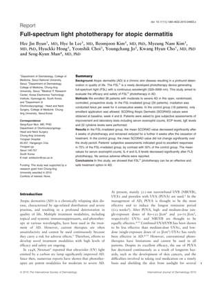
  • 1. doi: 10.1111/j.1365-4632.2010.04663.x Report Full-spectrum light phototherapy for atopic dermatitis Hee Jin Byun1, MD, Hye In Lee2, MD, Beomjoon Kim2, MD, PhD, Myeung Nam Kim2, 3 3 3 1 MD, PhD, Hyuckki Hong , Yeonshik Choi , Youngchang Jo , Kwang Hyun Cho , MD, PhD 4 and Seog-Kyun Mun , MD, PhD 1 Department of Dermatology, College of Summary Medicine, Seoul National University, Background Atopic dermatitis (AD) is a chronic skin disease resulting in a profound deteri- Seoul, 2Department of Dermatology, oration in quality of life. The FSLÒ is a newly developed phototherapy device generating College of Medicine, Chung-Ang full-spectrum light (FSL) with a continuous wavelength (320–5000 nm). This study aimed to University, Seoul, 3Medical IT Research Center, Korea Electronics Technology evaluate the efficacy and safety of FSLÒ phototherapy in AD. Institute, Gyeonggi-do, South Korea, Methods We enrolled 38 patients with moderate to severe AD in this open, randomized, and 4Department of controlled, prospective study. In the FSL-irradiated group (20 patients), irradiation was Otorhinolaryngology - Head and Neck conducted twice per week for 4 consecutive weeks. In the control group (18 patients), only Surgery, College of Medicine, Chung- emollient application was allowed. SCORing Atopic Dermatis (SCORAD) values were Ang University, Seoul,Korea obtained at baseline, week 4 and 8. Patients were asked to give subjective assessments of Correspondence improvement and laboratory tests including serum eosinophil counts, ECP levels, IgE levels Seog-Kyun Mun, MD, PHD. and 22 cytokine levels were performed. Department of Otorhinolaryngology- Results In the FSL-irradiated group, the mean SCORAD value decreased significantly after Head and Neck Surgery 4 weeks of phototherapy and remained reduced for a further 4 weeks after the cessation of Chung-Ang University Yongsan Hospital treatment. In the control group, the mean SCORAD value did not change significantly over 65-207, Hangangro 3-ka the study period. Patients’ subjective assessments indicated good to excellent responses Yongsan-gu in 75% of the FSL-irradiated group, by contrast with 50% of the control group. The mean Seoul 140-757 values for serum eosinophil counts, IL-4 and IL-5 levels decreased significantly after FLS South Korea phototherapy. No serious adverse effects were reported. E-mail: entdoctor@cau.ac.kr Conclusions In this study, we showed that FSLÒ phototherapy can be an effective and Funding: This study was supported by a safe treatment option in AD. research grant from Chung-Ang University awarded in 2010. Conflicts of interest: None. At present, mainly 311-nm narrowband UVB (NBUVB), Introduction UVA1 and psoralen with UVA (PUVA) are used.3 In the Atopic dermatitis (AD) is a chronically relapsing skin dis- management of AD, PUVA is thought to be the most ease, characterized by age-related distribution and severe effective and to induce the longest remission period pruritus, and resulting in a profound deterioration in (£12 weeks4). After PUVA, high- and medium-dose (sin- quality of life. Multiple treatment modalities, including gle-exposure doses of 80–130 J/cm2 and 50–70 J/cm2, topical and systemic immunosuppressants, and photother- respectively) UVA1 and NBUVB are thought to be apy at various wavelengths, have been used in the treat- equally effective.4–9 Combined UVA/UVB has been shown ment of AD. However, current therapies are often to be less effective than medium-dose UVA1, and low- unsatisfactory and cannot be used continuously because dose (single-exposure doses of 10 J/cm2) UVA1 has rarely they carry a risk for adverse effects.1 Therefore, efforts to been effective in AD.4,10 However, conventional photo- develop novel treatment modalities with high levels of therapies have limitations and cannot be used in all efficacy and safety are ongoing. patients. Despite its excellent efficacy, the use of PUVA In 1948, Nexman2 reported that ultraviolet (UV) light has decreased continuously as a result of longterm haz- emitted by a carbon arc lamp significantly improved AD. ards, such as the development of skin cancers, and the Since then, numerous reports have shown that photother- difficulties involved in taking oral medication on a timely apies are potent modalities for moderate to severe AD. basis and shielding the skin from sunlight for several 1 ª 2010 The International Society of Dermatology International Journal of Dermatology 2010
  • 2. 2 Report Full-spectrum light phototherapy for atopic dermatitis Byun et al. days.3,5,10 UVA1 has a rapid onset of action, but fewer Table 1 Demographic patient data than 50% of affected individuals respond to UVA1 treat- ment and the response is usually short-lived.3,10 FSL-irradiated group Control group Natural sunlight itself has also been used for photother- Patients, n 20 18 apy under the designations of ‘‘heliotherapy’’ (after Age, years (mean ± SD) 25.68 ± 7.69 25.63 ± 8.41 Helios, the personification of the sun in Greek mythology) Sex, n (male/female) 8/12 9/9 or climatotherapy. Heliotherapy has been demonstrated to SCORAD (mean ± SD) 47.87 ± 15.45 39.79 ± 9.76 be highly effective in AD. Two-week heliotherapy in the Mean duration of AD, years 11.09 10.95 Canary Islands reduced mean SCORing Atopic Dermatis (SCORAD) values by 70–74% in previous reports, indi- FSL, full-spectrum light; SD, standard deviation; AD, atopic cating a level of efficacy higher than that of PUVA, which dermatitis. has been reported to reduce baseline SCORAD by 54% after 15 exposures.4,11,12 However, heliotherapy can be ranged from 25 to 48 years. The mean SCORAD index at the performed only in specific regions with strong sunlight, beginning of the study was 47.87 points in the FSL-irradiated such as the Canary Islands and the area around the Dead group and 39.79 points in the control group. There was no Sea, and is usually conducted in the form of a ‘‘heliothera- statistically significant difference in baseline SCORAD values py trip’’ organized as a 2- or 3-week tour. Therefore, between the two groups (P = 0.167, Mann–Whitney U-test). All despite its high efficacy, the application of heliotherapy is patients were Korean and had skin of phototypes III or IV. limited as a result of the interruption it causes to daily life Demographic data for the patients are shown in Table 1. and the cost of travel expenses.11,12 Informed consent was obtained from each patient prior to To overcome the limitations of heliotherapy, we used enrollment. the FSLÒ (BMC Co. Ltd, Anyang-si, South Korea) for The FSLÒ phototherapy device generates light on a treatment of AD. The FSLÒ is a newly developed photo- continuous wavelength of 320–5000 nm, from an electrical arc therapy device which generates full-spectrum light (FSL) between two carbon rods (Ark RodsÒ; BMC Co. Ltd) at a on a continuous wavelength spectrum of 320–5000 nm. temperature of >3000 °C. An Ark RodÒ is made of carbon The light profile emitted by the FSLÒ is nearly identical compounds including 16 confidential chemical elements. to that of sunlight except that it omits UVB; it includes A calibrated quartz tungsten halogen lamp (model 63355; UVA, visible light and infrared light. Given the high effi- Newport Corp., Stratford, CT, USA), a monochromator cacy of heliotherapy in AD treatment, we expected that (Cornerstone 260Ô; Newport Corp.) and a digital lock-in FSL phototherapy would also be effective. Phototherapy radiometry system (MerlineÔ; Newport Corp.) were used to devices emitting FSL, like the FSLÒ, have been used make light with a continuous wavelength. A two-color mainly in neuropsychiatric diseases, such as depression photodetector (IGA030-TE2-H; Electro Optical Systems, Inc., and sleep disturbance.13–15 So far, there have been no Phoenixville, PA, USA), a TE-cooler (PS/TC-1; Newport Corp.) reports on the application of FSL devices in AD. and a TE-cooled HgCdZnTe detector (MIR8025Ô; Newport In this study, we aimed to evaluate the efficacy and Corp.) were used as a detection system. safety of FSLÒ phototherapy in patients with moderate to In the FSL-irradiated group, irradiation was performed with severe AD. the FSLÒ phototherapy device twice per week for 4 consecutive weeks, resulting in a total of eight applications of irradiation. We performed a pilot study to determine the appropriate exposure Materials and methods time. From the pilot study results, we concluded that exposures This was an open, randomized, controlled, prospective study. of 20 min are most effective and safe (data not shown). We recruited 38 patients diagnosed with extrinsic AD according Therefore, in each phototherapy session, the anterior side of to previously published definitions,8,9 from August 2007 to July the body was irradiated for 20 min and the posterior side was 2008. Only patients with moderate to severe AD with SCORAD subsequently irradiated for 20 min. The fluence of each index values of >25 were included. The following subjects were irradiation was 530 J/cm2, including 121 J/cm2 of UVA and excluded: patients aged <18 years; patients who had been 409 J/cm2 of visible and infrared light. During the entire study treated with systemic corticosteroid, any type of phototherapy or period, the use of any topical or systemic agents other than the photosensitizing drugs in the 3 months prior to enrollment; designated emollient cream (PhysiogelÒ; Stiefel-Korea Ltd, pregnant or lactating women, and patients with a history of Seoul, South Korea) was disallowed. In the control group, the neoplasm or photosensitive dermatosis. Patients were screened emollient, PhysiogelÒ, was applied twice per day without any to ensure they fulfilled inclusion and exclusion criteria and other treatment. randomly assigned to either the FSL-irradiated group (20 The SCORAD index was used to assess disease severity.10 patients) or to the control group (18 patients). Patient ages SCORAD values in each patient were assessed three times International Journal of Dermatology 2010 ª 2010 The International Society of Dermatology
  • 3. Byun et al. Full-spectrum light phototherapy for atopic dermatitis Report during the study period: (i) prior to the beginning of treatment (a) (at baseline); (ii) after 4 weeks of FSL phototherapy (at week 4), and (iii) 4 weeks after the cessation of phototherapy (at week 8). All assessments were performed by the same dermatologist. Patients’ subjective assessments of clinical improvement were obtained at week 8, using a 4-point scale with the following descriptors: ‘‘poor response’’ (0–25% improvement); ‘‘fair response’’ (26–50% improvement); ‘‘good response’’ (51–75% improvement), and ‘‘excellent response’’ (76–100% improvement). Laboratory tests were carried out in those patients who agreed to them. The tests were performed twice, at baseline and at week 8, and included serum eosinophil counts, eosinophil cationic protein (ECP) levels, immunoglobulin E (IgE) levels and levels of 22 human cytokines. Serum cytokine levels were determined using the Luminex 200 SystemÒ (Luminex Corp., Austin, TX, USA), which uses xMAP technology to combine sandwich immunoassay and fluorescent bead-based technology, and the Beadlyte Human 22-plex Multi-cytokine (b) Detection SystemÒ (Millipore Corp., Billerica, MA, USA). The 22 human cytokines were: interleukin-1a (IL-1a); IL-1b; IL-2; IL-3; IL-4; IL-5; IL-6; IL-7; IL-8; IL-10; p40 subunit of IL-12; p70 subunit of IL-12; IL-13; IL-15; GM-CSF (granulocyte- macrophage colony-stimulating factor); interferon-c (IFN-c); tumor necrosis factor-a (TNF-a); eotaxin; monocyte chemotactic protein-1 (MCP-1); RANTES (regulated upon activation, normal T cell expressed and secreted); macrophage inflammatory protein-1a (MIP-1a), and interferon-inducible protein-10 (IP-10). The Wilcoxon signed rank test was used to compare clinical and laboratory values at baseline and at weeks 4 or 8. P-values of < 0.05 were considered statistically significant. Results Clinical evaluation In the FSL-irradiated group, the mean SCORAD value was 47.87 at baseline and decreased significantly to 36.81 at week 4 (reduced by 23.1%; P < 0.01). It reduced fur- Figure 1 (a) Before treatment, several eczematous patches ther to 30.76 at week 8 (reduced by 35.7% from base- with some erosive lesions could be seen on the face. (b) After line), 4 weeks after the cessation of treatment (Figs 1 and 2). 4 weeks of full-spectrum light phototherapy, facial lesions In the control group, mean SCORAD values did not were significantly improved, leaving only slight erythema and change significantly during the entire study period (39.79 hyperpigmentation at baseline, 35.39 at week 4, 33.85 at week 8; P = 0.236) (Fig. 2). the control group. Serum ECP and IgE levels were Patients’ subjective assessments are summarized in checked in 18 patients in the FSL-irradiated group and 16 Table 2. Good to excellent responses (51–100% improve- patients in the control group. Serum cytokine levels were ment) were found in 75% of the FSL-irradiated group, by checked in 13 patients in the FSL-irradiated group and 11 contrast with 50% of the control group. patients in the control group. In the FSL-irradiated group, the mean values of serum Laboratory tests eosinophil counts showed a significant decrease at week 8 Laboratory tests were performed in patients who con- compared with baseline values, although both were sented to them. Serum eosinophil counts were checked in within normal limits. Mean IL-4 and IL-5 levels also 19 patients in the FSL-irradiated group and 17 patients in showed a significant decrease at week 8 compared with ª 2010 The International Society of Dermatology International Journal of Dermatology 2010
  • 4. 4 Report Full-spectrum light phototherapy for atopic dermatitis Byun et al. Figure 2 SCORAD index values (mean ± SD) at baseline, at week 4 of phototherapy, and at week 8 (4 weeks after the cessation of phototherapy). Scores in the full-spectrum light (FSL)- irradiated group at weeks 4 and 8, but not in the control group, show a signifi- cant decrease from baseline. *P < 0.01;   P < 0.001 ous reports which used the SCORAD index for evaluating Table 2 Patients’ subjective assessments of clinical improve- ment the efficacy of phototherapy are as follows. Tzaneva et al.4 reported mean reductions in SCORAD values of FSL-irradiated Control 54% (from 62.5 to 28.8) after 15 administrations of group, n (%) group, n (%) PUVA treatment and of 37% (from 63.7 to 40.1) after medium-dose UVA1 treatment. The same group of Excellent (76–100% improvement) 6 (30.0) 2 (11.1) authors also reported that baseline SCORAD was reduced Good (51–75% improvement) 9 (45.0) 7 (38.9) by 35% by high-dose UVA1 and by 28% by medium- Fair (26–50% improvement) 4 (20.0) 8 (44.4) Poor (0–25% improvement) 1 (5.0) 1 (5.6) dose UVA1 after five treatments per week for 3 weeks, in Total 20 (100) 18 (100) 10 patients with severe AD with a median baseline SCO- RAD value of 67.9 In other reports, medium-dose UVA1 FSL, full-spectrum light. and low-dose UVA1 reduced SCORAD values from 64 at baseline to 47 (reduced by 27%) and 60 (reduced by baseline values (P < 0.001 for all) (Fig. 3). Serum ECP, 6%), respectively.16,17 Narrowband UVB therapy was IgE and other cytokine levels did not show significant reported to reduce SCORAD values from 71.2 to 48.8 changes over the entire study period. (reduced by 33%) after 4.3 accumulated joules of therapy None of the laboratory values in the control group in 10 patients with severe AD.18 Der-Petrossian et al.19 showed significant changes over the entire study period reported more promising results with NBUVB, which (Fig. 3). showed SCORAD reductions of 24% after 2 weeks and 45% after 4 weeks of therapy, in 10 patients with severe Safety and adverse effects AD with a mean baseline SCORAD value of 67.9. The FSL irradiation was generally well tolerated by all Vähävihu et al.11 reported that a 2-week course of helio- patients. Among the 20 patients in the FSL-irradiated therapy in the Canary Islands reduced SCORAD values group, erythema was reported in six patients (30%), dry- from 34 to 9 (reduced by 74%) in January and from 30 ness in six patients (30%), pruritus in four patients to 9 (reduced by 70%) in March in 23 AD patients. Au- (20%), and burning sensation in two patients (10%). tio et al.12 reported a mean reduction of 70% in SCO- Transient exacerbation of AD was observed in six RAD values after 2 weeks of heliotherapy and noted that patients (30%) during the first 2 weeks of FSL photother- mean SCORAD values remained at 45% of baseline val- apy. Dryness and pruritus were controllable with the ues at 3 months after therapy. In summary, the reduction emollient, and the erythema and burning sensation sub- from baseline SCORAD values after 2–4 weeks of treat- sided within 1 week of the cessation of phototherapy. No ment was about 70–74% in heliotherapy, 54% in PUVA, serious adverse effects were reported during the study. 35% in high-dose UVA1, 27–37% in medium-dose UVA1, 24–45% in NBUVB, and 6% in low-dose UVA1. In this study, mean SCORAD values dropped from Discussion 47.87 at baseline to 36.81 (reduced by 23.1%) after The SCORAD index is a viable parameter which can be 4 weeks of FSL phototherapy with a total of eight irradia- used for comparing the results of different studies. Previ- tion treatments, and fell further to 30.76 (reduced by International Journal of Dermatology 2010 ª 2010 The International Society of Dermatology
  • 5. Byun et al. Full-spectrum light phototherapy for atopic dermatitis Report (a) 35.7% from baseline) at 4 weeks after the cessation of treatment. From these results, FSL phototherapy would appear to be less effective than heliotherapy, contrary to our expectations. This discrepancy in efficacy between FSL phototherapy and heliotherapy may result from dif- ferences in light doses or light profiles. However, the total energy of the irradiated light does not seem to represent the cause of the difference in efficacy because total light energy irradiated over 2 weeks of heliotherapy was reported to about 60–109 standard erythema dose corre- sponding to 0.60–1.09 J/cm2, whereas the energy irradi- ated during even a single exposure to FSL phototherapy was 530 J/cm2 and included 121 J/cm2 of UVA and 409 J/cm2 of visible and infrared light.11 Instead, the lack (b) of UVB in FSL may represent a cause of decreased effi- cacy. Furthermore, the therapeutic effects of heliotherapy do not derive solely from light. Air, temperature, humid- ity and barometric pressure also contribute to the thera- peutic effects. Although there are no standardized programs of heliotherapy, most programs commonly include relaxation activities such as yoga, lectures on skin care, time for the exchange of personal experiences, and, in some cases, psychological consultation, as well as sun- bathing time.11,12,20 The various relaxation activities included may also contribute to the therapeutic effects of the treatment, and the fact that patients are removed from the stress of daily life may also play a positive role. Compared with the known efficacies of other photothera- py modalities, the efficacy of FSL phototherapy, with SCORAD reductions of 23.1% at week 4 and 35.7% at week 8, was lower than that of PUVA and comparable (c) with those of NBUVB, high-dose and medium-dose UVA1. However, in terms of the number of treatment sessions required, FSL proved superior to the other phototherapy modalities. In previous reports, PUVA and UVA1 were usually irradiated five times per week for 4 weeks resulting in a total of 20 irradiation treatments, and NBUVB was delivered three times per week for 2–6 weeks for a total of six to 18 irradiation treatments.3–10,16–19 Full-spectrum light was administered twice per week for 4 weeks and therefore only eight irradiation treatments were delivered to achieve the results reported herein. However, it may not be appropri- ate to directly compare the results of previous reports with those of our study because the mean SCORAD index at baseline in patients in most of the previous Figure 3 At week 8, 4 weeks after the cessation of photo- studies was > 60–70, much higher than that of 47.87 in therapy, significant decreases from baseline levels are seen our patients.3–10,16–19 The discrepancy in the severity of in mean values of (a) eosinophil count, (b) IL-4 and (c) IL-5 disease in enrolled patients may result in apparent differ- in the full-spectrum light (FSL)-irradiated group, but not ences in efficacy. Further, in many of the previous in the control group. Data are shown as mean ± SD. reports,3–10,16–19 use of topical steroids was allowed, *P < 0.01 whereas only emollient application was permitted in our study. Further well-designed comparative studies are ª 2010 The International Society of Dermatology International Journal of Dermatology 2010
  • 6. 6 Report Full-spectrum light phototherapy for atopic dermatitis Byun et al. needed to precisely compare the efficacy of FSL photo- number and function of Langerhans cells, and the induc- therapy with those of conventional phototherapies. tion of immunomodulatory cytokines such as IL-10.24–27 Remission periods have been reported to extend to 4 UVA has also been known to inhibit histamine release and 12 weeks after medium-dose UVA1 and PUVA, from human basophils and mast cells.28 In addition, both respectively, although not many reports have included UVA and UVB light have been shown to have antimicro- this.4 In this study, patients were followed until 4 weeks bial effects.29–32 Because acute exacerbation of AD is after FSL phototherapy and therefore the exact length of often associated with bacterial super-infection, UV photo- remission could not be evaluated. However, the reduced therapy, especially high-dose UVA1, is effective in this SCORAD was maintained to 4 weeks after the cessation context in AD.32 Visible and infrared light have mostly of treatment and therefore we can assume that FSL been used for the treatment of chronic and acute wounds phototherapy is at least comparable with medium-dose and for the acceleration of skin regeneration after chemi- UVA1 in terms of remission length. Further longterm cal or laser peeling.33–36 This spectrum of light is studies are needed to evaluate the length of the remission absorbed by cellular photosensitizers, such as cyto- period after FSL phototherapy more precisely. chromes, flavins/riboflavins and NADP (nicotinamide There are few reports on changes in cytokine levels adenine dinucleotide phosphate), and then induces the after phototherapy in AD patients. Skin biopsy specimens production of low levels of reactive oxygen species. Low used to evaluate mRNA expression showed significant levels of reactive oxygen species play important roles in reductions in expression of IL-5, IL-13 and IL-31 after redox signaling, leading to the acceleration of cell growth UVA1 phototherapy and a reduction in IFN-c in patients and regeneration, which can be helpful for the regenera- who underwent various treatments, including UVA1.21–23 tion of damaged skin in AD.33,34,36,37 Infrared light also By contrast, we used patients’ serum to evaluate cytokine induces heating of the irradiated skin, leading to vessel levels, a process that is thought to reflect systemic cyto- dilatation and an increase in blood circulation, which can kine levels more than skin specimens. Changes in serum result in the wash-up of inflammatory molecules and the cytokine levels after phototherapy in AD patients have acceleration of tissue regeneration.38 Thus, visible and not previously been reported. In our results, the mean val- infrared light can play positive roles in the treatment of ues of eosinophil counts, IL-4 and IL-5 levels showed sig- AD, although they may not be very effective in isolation. nificant decreases after FSL phototherapy. Eosinophil is Combined irradiation with several types of light may lead known to play an important role in the chronic mainte- to complex interactions in the irradiated skin that extend nance of AD, contributing to allergic inflammation by the beyond simple synergism. However, this complex interac- secretion of cytokines and mediators and inducing tissue tion has not been studied very much. Everett et al.35 injury through the production of reactive oxygen interme- reported that prior heating of the skin increased the UV diates and the release of toxic granule proteins. IL-4 and threshold erythema dose. Similarly, heating of the skin by IL-5 are the most important cytokines produced by Th 2 the infrared light included in FSL may modulate the cells, which play dominant roles in the pathogenesis of effects of UV light irradiated at the same time. Further AD. IL-4 is especially important in acute AD, mediating studies are needed to elucidate the precise interactions of immunoglobulin isotype switching to IgE synthesis and various types of light in irradiated skin. upregulating expression of adhesion molecules on endo- The common adverse effects of conventional photother- thelial cells. By contrast, IL-5 predominates in chronic apy are dryness and first-degree burn presenting as ery- AD and is involved in eosinophil development.1 There- thema.10 The adverse effects observed in this study were fore, we can conclude that FSL phototherapy improves similar to those of conventional phototherapy and not only the clinical symptoms, but also the laboratory included erythema, dryness, pruritus and a burning profiles that are important in both the acute and chronic sensation. Dryness and pruritus were controllable with phases of AD. the emollient, and the erythema and burning sensation The therapeutic effects of the FSL used in this study subsided within 1 week of the cessation of phototherapy. (320–5000 nm) are thought to derive from the combined No serious adverse effects were reported during the study. effects of UVA, visible light and infrared light. This is the We suggest that more intensive use of emollients and deli- advantage of FSL therapy, which can offer benefits from cate dose adjustment (a fixed dose was adopted in this the various types of light simultaneously. UV light has study) may minimize these reactions. Phototherapy can been most widely used as phototherapy for various also result in serious complications such as skin cancers. inflammatory skin diseases, including AD. The major Skin carcinogenesis is induced by UV light in photothera- therapeutic mechanisms of UV phototherapy in AD py.3 Therefore, FSL with a lower proportion of UV can involve immunosuppression and include the induction of be safer than conventional UV phototherapies such as apoptosis in skin-infiltrating T cells, a reduction in the PUVA, UVA1 and NBUVB. However, FSL phototherapy International Journal of Dermatology 2010 ª 2010 The International Society of Dermatology
  • 7. Byun et al. Full-spectrum light phototherapy for atopic dermatitis Report should also be used cautiously, especially if it is used over 13 Praschak-Rieder N, Willeit M, Neumeister A, et al. a long period, and should be avoided in patients at high Therapeutic sleep deprivation and phototherapy. Wien risk for developing skin cancers. Med Wochenschr 1999; 149: 520–524. In this study, we showed that FSL phototherapy can be 14 Neumeister A, Kapitany T, Rieder N, et al. Fall/winter depression and its therapy. Wien Klin Wochenschr 1994; an effective and safe treatment option for moderate to 106: 665–670. severe AD. Further large-scale studies are needed to 15 Lingjaerde O, Reichborn-Kjennerud T, Haggag A, et al. confirm the efficacy and safety of FSL phototherapy and Treatment of winter depression in Norway. I. Short- and to conclude the optimal therapeutic schedule. long-term effects of 1500-lux white light for 6 days. Acta Psychiatr Scand 1993; 88: 292–299. 16 Krutmann J, Czech W, Diepgen T, et al. High-dose References UVA1 therapy in the treatment of patients with atopic 1 Leung DYM, Eichenfield LF, Boguniewicz M. Atopic dermatitis. J Am Acad Dermatol 1992; 26: 225–230. dermatitis. In: Wolff K, Goldsmith LA, Kats SI, et al., 17 Krutmann J, Diepgen TL, Luger TA, et al. High-dose eds. Fitzpatrick’s Dermatitis in General Medicine, 7th UVA1 therapy for atopic dermatitis: results of a edn. New York, NY: McGraw-Hill, 2008: 146–158. multicenter trial. J Am Acad Dermatol 1998; 38: 589– 2 Nexman PH. Clinical Studies of Besnier’s Prurigo, 593. Dissertation. Copenhagen: Rosenkilde & Bagger 18 Silva SH, Guedes AC, Gontijo B, et al. Influence of Publishers, 1948. narrowband UVB phototherapy on cutaneous microbiota 3 Gambichler T. Management of atopic dermatitis using of children with atopic dermatitis. J Eur Acad Dermatol photo(chemo)therapy. Arch Dermatol Res 2009; 301: Venereol 2006; 20: 1114–1120. 197–203. 19 Der-Petrossian M, Seeber A, Hönigsmann H, et al. Half- 4 Tzaneva S, Kittler H, Holzer G, et al. 5-Methoxypsoralen side comparison study on the efficacy of 8- plus ultraviolet (UV) A is superior to medium-dose UVA1 methoxypsoralen bath-PUVA versus narrowband in the treatment of severe atopic dermatitis: a randomized ultraviolet B phototherapy in patients with severe chronic crossover trial. Br J Dermatol 2010; 162: 655–660. atopic dermatitis. Br J Dermatol 2000; 142: 39–43. 5 von Kobyletzki G, Pieck C, Hoffmann K, et al. Medium- 20 Harari M, Shani J, Seidl V, et al. Climatotherapy of dose UVA1 cold-light phototherapy in the treatment of atopic dermatitis at the Dead Sea: demographic severe atopic dermatitis. J Am Acad Dermatol 1999; 41: evaluation and cost-effectiveness. Int J Dermatol 2000; 931–937. 39: 59–69. 6 Legat FJ, Hofer A, Brabek E, et al. Narrowband UV-B 21 Gambichler T, Kreuter A, Tomi NS, et al. Gene vs. medium-dose UV-A1 phototherapy in chronic atopic expression of cytokines in atopic eczema before and after dermatitis. Arch Dermatol 2003; 139: 223–224. ultraviolet A1 phototherapy. Br J Dermatol 2008; 158: 7 Gambichler T, Othlinghaus N, Tomi NS, et al. Medium- 1117–1120. dose ultraviolet (UV) A1 vs. narrowband UVB 22 Grewe M, Gyufko K, Schöpf E, et al. Lesional expression phototherapy in atopic eczema: a randomized crossover of interferon-r in atopic eczema. Lancet 1994; 343: 25– study. Br J Dermatol 2009; 160: 652–658. 26. 8 Majoie I, Oldhoff J, van Weelden H, et al. Narrowband 23 Krutmann J. Ultraviolet A-1 irradiation as a tool to study ultraviolet B and medium-dose ultraviolet A1 are equally the pathogenesis of atopic dermatitis. Methods Enzymol effective in the treatment of moderate to severe atopic 2000; 319: 296–302. dermatitis. J Am Acad Dermatol 2009; 60: 77–84. 24 Vermeer BJ, Hurks M. The clinical relevance of 9 Tzaneva S, Seeber A, Schwaiger M, et al. High-dose immunosuppression by UV irradiation. J Photochem versus medium-dose UVA1 phototherapy for patients Photobiol 1994; 24: 149–154. with severe generalized atopic dermatitis. J Am Acad 25 Kalimo K, Koulu L, Jansen CT. Effect of a single UVB or Dermatol 2001; 45: 503–507. PUVA exposure on immediate and delayed skin 10 Meduri NB, Vandergriff T, Rasmussen H, et al. hypersensitivity reactions in humans: correlation to Phototherapy in the management of atopic dermatitis: a erythemal response and Langerhans cell depletion. Arch systematic review. Photodermatol Photoimmunol Dermatol Res 1983; 275: 374–378. Photomed 2007; 23: 106–112. 26 Garssen J, Vandebriel RJ, De Gruijl FR, et al. UVB 11 Vähävihu K, Ylianttila L, Salmelin R, et al. Heliotherapy exposure-induced systemic modulation of Th1- and Th2- improves vitamin D balance and atopic dermatitis. Br J mediated immune responses. Immunology 1999; 97: Dermatol 2008; 158: 1323–1328. 506–514. 12 Autio P, Komulainen P, Larni HM. Heliotherapy in 27 Nghiem DX, Kazimi N, Mitchell DL, et al. Mechanisms atopic dermatitis: a prospective study on climatotherapy underlying the suppression of established immune using the SCORAD index. Acta Derm Venereol 2002; responses by ultraviolet radiation. J Invest Dermatol 82: 436–440. 2002; 119: 600–608. ª 2010 The International Society of Dermatology International Journal of Dermatology 2010
  • 8. 8 Report Full-spectrum light phototherapy for atopic dermatitis Byun et al. 28 Kronauer C, Eberlein-Konig B, Ring J, et al. Influence of acute exacerbation of atopic eczema. Acta Derm UVB, UVA and UVA1 irradiation on histamine release Venereol (Suppl) 1992; 176: 120–122. from human basophils and mast cells in vitro in the 33 Conlan MJ, Rapley JW, Cobb CM. Biostimulation of presence and absence of antioxidants. Photochem wound healing by low-energy laser irradiation. J Clin Photobiol 2003; 77: 531–534. Periodontol 1996; 23: 492–496. 29 Yoshimura-Mishima M, Akamatsu H, Namura S, et al. 34 Grossman N, Schneid N, Reuveni H, et al. 780-nm low Suppressive effect of ultraviolet (UVB and PUVA) power diode laser irradiation stimulates proliferation of radiation on superantigen production by keratinocyte cultures: involvement of reactive oxygen Staphylococcus aureus. J Dermatol Sci 1999; 19: 31– species. Lasers Surg Med 1998; 22: 212–218. 36. 35 Everett MA, Doran CK, Everett HD, et al. Modification 30 Atherton DJ, Carabott F, Glover MT, et al. The role of of sunburn by infrared ray. JAMA 1963; 23: 778–779. psoralen photochemotherapy (PUVA) in the treatment of 36 Lipovsky A, Nitzan Y, Lubart R. A possible mechanism severe atopic eczema in adolescents. Br J Dermatol 1988; for visible light-induced wound healing. Lasers Surg Med 118: 791–795. 2008; 40: 509–514. 31 Jekler J, Larkö O. UVB phototherapy of atopic 37 Rhee SG. Redox signaling: hydrogen peroxide as dermatitis. Br J Dermatol 1988; 119: 697–705. intracellular messenger. Exp Mol Med 1999; 31: 53–59. 32 Krutmann J, Schöpf E. High-dose-UVA 1 phototherapy: a 38 Schriber WJ. A Manual of Electrotherapy, 4th edn. novel and highly effective approach for the treatment of Philadelphia, PA: Lea & Febiger: 1975, 128–129. International Journal of Dermatology 2010 ª 2010 The International Society of Dermatology