Fluid Mechanics inCirculating
Tumour cell
Course:Biofluid Mechanics and Heat Transfer
Presented by………
Jannatun Noor Promi Jannatul Raisa Istiak Ahmed Saju
ID: 23167008 ID: 23167011 ID: 23167013
2.
Content
• Introduction
• WorkflowProcess of Tumour cells
• Tumor Vasculature & Fluid Mechanics (Primary Tumor)
• Angiogenesis and fluid mechanics
• Tumour Vascularity
• Flows in the tiny blood vessels of tumours
• Microenvironmental characteristics of malignant tumors
• Flow mediated Tumor Metastases
3.
Content
• Lymphatic andintravascular spread of tumour cells
• Spreading of components that originate from the tumour
• CTCs' intravascular journey
• Therapy
• Observation
• Conclusion
4.
Introduction
Cancer metastasis isa key reason for reduced life
expectancy in patients, as tumor cells spread through the
blood and lymph systems. Fluid dynamics—such as flow,
shear stress, and pressure—affect how cancer cells behave
and spread. By understanding these forces, we can create
new strategies to target metastasis and improve treatment
outcomes. This presentation focuses on how fluid
dynamics influence cancer metastasis and how this
knowledge could lead to better therapies.
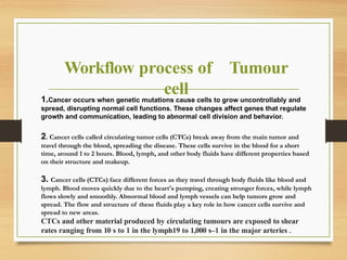
Workflow process ofTumour
cell
1.Cancer occurs when genetic mutations cause cells to grow uncontrollably and
spread, disrupting normal cell functions. These changes affect genes that regulate
growth and communication, leading to abnormal cell division and behavior.
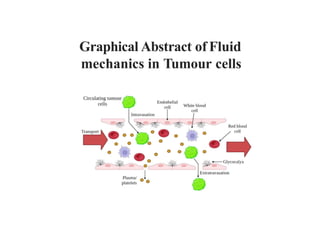
2. Cancer cells called circulating tumor cells (CTCs) break away from the main tumor and
travel through the blood, spreading the disease. These cells survive in the blood for a short
time, around 1 to 2 hours. Blood, lymph, and other body fluids have different properties based
on their structure and makeup.
3. Cancer cells (CTCs) face different forces as they travel through body fluids like blood and
lymph. Blood moves quickly due to the heart's pumping, creating stronger forces, while lymph
flows slowly and smoothly. Abnormal blood and lymph vessels can help tumors grow and
spread. The flow and structure of these fluids play a key role in how cancer cells survive and
spread to new areas.
CTCs and other material produced by circulating tumours are exposed to shear
rates ranging from 10 s to 1 in the lymph19 to 1,000 s–1 in the major arteries .
7.
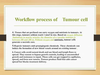
Workflow process ofTumour cell
4. Tissues that are perfused can carry oxygen and nutrients to tumours. At
this stage, tumours seldom reach 1 mm3 in size. Based on oxygen diffusion
limitations in nearby arteries, the diameter of a vascular tumours is
between 100 and 200 m. Without oxygen and nutrients, tumour cells
generate a necrotic core.
5.Hypoxic tumours emit proangiogenic chemicals. These chemicals can
induce the formation of new blood vessels around an existing tumour .
6. Cancer cells avoid natural death and use blood and lymph flows to
spread. They mutate to bypass growth controls, leading to uncontrolled
growth and resistance to repair. These cells can get trapped in capillaries,
spread, and form new tumors. Tumors produce fluid that aids cancer
spread but blocks treatment delivery.
8.
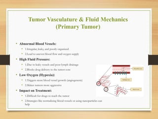
Tumor Vasculature &Fluid Mechanics
(Primary Tumor)
• Abnormal Blood Vessels:
• 1.Irregular, leaky, and poorly organized
• 2.Lead to uneven blood flow and oxygen supply
• High Fluid Pressure:
• 1.Due to leaky vessels and poor lymph drainage
• 2.Blocks drug delivery to the tumor core
• Low Oxygen (Hypoxia):
• 1.Triggers more blood vessel growth (angiogenesis)
• 2.Makes tumors more aggressive
• Impact on Treatment:
• 1.Difficult for drugs to reach the tumor
• 2.Strategies like normalizing blood vessels or using nanoparticles can
help
9.
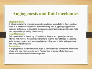
Angiogenesis and fluidmechanics
Angiogenesis:
Angiogenesis is the process by which new blood vessels form from existing
ones. It is essential for growth, wound healing, and supplying oxygen and
nutrients to tissues. In diseases like cancer, abnormal angiogenesis can help
tumors grow by providing blood supply.
Fluid Mechanics:
Fluid mechanics is the study of how fluids (liquids and gases) move and
interact with forces. It explains phenomena like the flow of blood in vessels,
water through pipes, and air around planes. Key concepts include pressure,
flow rate, and resistance.
Connection:
In angiogenesis, fluid mechanics plays a crucial role as blood flow influences
where and how new vessels form. Proper flow ensures efficient oxygen
delivery and healthy tissue development.
10.
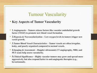
Tumour Vascularity
• KeyAspects of Tumor Vascularity
• 1.Angiogenesis – Tumors release factors like vascular endothelial growth
factor (VEGF) to promote new blood vessel formation.
• 2.Hypoxia & Neovascularization – Low oxygen levels in tumors trigger new
vessel growth.
• 3.Tumor Blood Vessel Characteristics – Tumor vessels are often irregular,
leaky, and poorly organized compared to normal vessels.
• 4.Imaging & Assessment – Doppler ultrasound, CT angiography, MRI, and
PET scans help assess vascularity.
• 5.Clinical Significance – Highly vascular tumors may grow and spread more
aggressively, but also respond better to anti-angiogenic therapies (e.g.,
bevacizumab).
11.
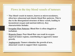
Flows in thetiny blood vessels of tumours
• Tiny blood vessels in tumors, known as microvasculature,
often have abnormal and chaotic blood flow patterns. This is
due to the disorganized structure of these vessels, leading to
inconsistent oxygen and nutrient delivery. Key
characteristics include:
• Irregular Flow Patterns: Blood flow is often sluggish or
turbulent.
• Hypoxia Zones: Poor blood flow can result in oxygen-
deprived tumor regions, contributing to aggressive tumor
growth.
• Angiogenesis: Tumors stimulate the growth of new,
abnormal vessels to support their expansion.
12.
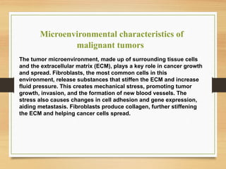
Microenvironmental characteristics of
malignanttumors
The tumor microenvironment, made up of surrounding tissue cells
and the extracellular matrix (ECM), plays a key role in cancer growth
and spread. Fibroblasts, the most common cells in this
environment, release substances that stiffen the ECM and increase
fluid pressure. This creates mechanical stress, promoting tumor
growth, invasion, and the formation of new blood vessels. The
stress also causes changes in cell adhesion and gene expression,
aiding metastasis. Fibroblasts produce collagen, further stiffening
the ECM and helping cancer cells spread.
13.
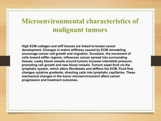
Microenvironmental characteristics of
malignanttumors
High ECM collagen and stiff tissues are linked to breast cancer
development. Changes in matrix stiffness caused by ECM remodeling
encourage cancer cell growth and migration. Durotaxis, the movement of
cells toward stiffer regions, influences cancer spread into surrounding
tissues. Leaky blood vessels around tumors increase interstitial pressure,
promoting cell growth and new blood vessels. Tumors expel fluid via the
lymphatic system, which alters fibroblasts and stiffens the ECM. Fluid flow
changes cytokine gradients, directing cells into lymphatic capillaries. These
mechanical changes in the tumor microenvironment affect cancer
progression and treatment outcomes.
14.
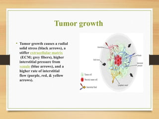
Tumor growth
• Tumorgrowth causes a radial
solid stress (black arrows), a
stiffer extracellular matrix
(ECM; grey fibers), higher
interstitial pressure from
venule (blue arrows), and a
higher rate of interstitial
flow (purple, red, & yellow
arrows).
15.
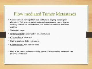
Flow mediated TumorMetastases
• Cancer spreads through the blood and lymph, helping tumors grow
elsewhere. This process, called metastasis, causes most cancer deaths.
Primary tumors are easier to treat, but metastatic cancer is harder to
control.
• Metastasis steps:
• Intravasation: Cancer enters blood or lymph.
• Circulation: Cells travel.
• Extravasation: Cells exit vessels.
• Colonization: New tumors form.
• Only a few cancer cells successfully spread. Understanding metastasis can
improve treatments.
16.
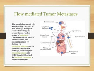
Flow mediated TumorMetastases
• The spread of metastatic cells
is regulated by a network of
fluid pathways. Biological
and mechanical signals
govern the non-random
process of metastasis.
Common metastatic patterns
for colon, breast, and
pancreatic cancers are
depicted via
anatomical structure and the
accompanying vascular
pathways, illustrating how
circulating tumour cells
(CTCs) exploit blood and
the lymphatic circulation to
reach distant organs.
17.
Lymphatic and intravascular
spreadof tumour cells
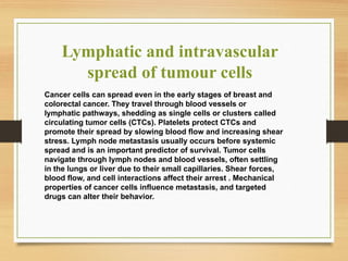
Cancer cells can spread even in the early stages of breast and
colorectal cancer. They travel through blood vessels or
lymphatic pathways, shedding as single cells or clusters called
circulating tumor cells (CTCs). Platelets protect CTCs and
promote their spread by slowing blood flow and increasing shear
stress. Lymph node metastasis usually occurs before systemic
spread and is an important predictor of survival. Tumor cells
navigate through lymph nodes and blood vessels, often settling
in the lungs or liver due to their small capillaries. Shear forces,
blood flow, and cell interactions affect their arrest . Mechanical
properties of cancer cells influence metastasis, and targeted
drugs can alter their behavior.
18.
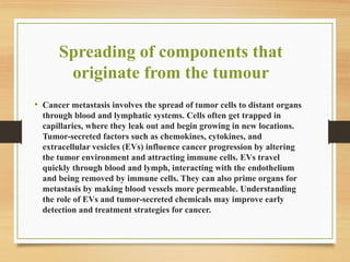
Spreading of componentsthat
originate from the tumour
• Cancer metastasis involves the spread of tumor cells to distant organs
through blood and lymphatic systems. Cells often get trapped in
capillaries, where they leak out and begin growing in new locations.
Tumor-secreted factors such as chemokines, cytokines, and
extracellular vesicles (EVs) influence cancer progression by altering
the tumor environment and attracting immune cells. EVs travel
quickly through blood and lymph, interacting with the endothelium
and being removed by immune cells. They can also prime organs for
metastasis by making blood vessels more permeable. Understanding
the role of EVs and tumor-secreted chemicals may improve early
detection and treatment strategies for cancer.
19.
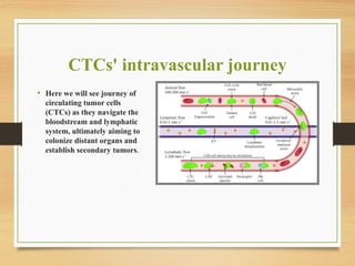
CTCs' intravascular journey
•Here we will see journey of
circulating tumor cells
(CTCs) as they navigate the
bloodstream and lymphatic
system, ultimately aiming to
colonize distant organs and
establish secondary tumors.
20.
Shear Stress: ADouble-Edged Sword:
Shear stress is a complex factor, with its impact depending not
only on the magnitude but also the duration and type of flow
encountered.
• The Power of Partnerships:
CTCs clusters and interact with blood components like platelets
and neutrophils. These interactions can protect CTCs from
immune attacks and shear stress, while platelets may enhance
their survival and adhesion.
21.
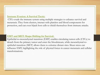
Immune Evasion: ASurvival Tactic:
CTCs evade the immune system using multiple strategies to enhance survival and
metastasis. They form clusters, interact with platelets and blood components for
protection, and can even hijack host cells to shield themselves from immune attacks.
EMT and MET: Shape-Shifting for Survival:
Epithelial-to-mesenchymal transition (EMT) enables circulating tumor cells (CTCs) to
detach from the primary tumor and enter the bloodstream, while mesenchymal-to-
epithelial transition (MET) allows them to colonize distant sites. Shear stress can
influence EMT, highlighting the role of physical forces in cancer metastasis and cellular
transformations.
22.
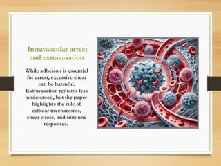
Intravascular arrest
and extravasation
Whileadhesion is essential
for arrest, excessive shear
can be harmful.
Extravasation remains less
understood, but the paper
highlights the role of
cellular mechanisms,
shear stress, and immune
responses.
23.
Intravascular Arrest:
CTCs canget stuck in small blood vessels either by blocking them or sticking to the walls.
Shear stress helps them stick but can also detach or damage them. The strength of adhesion
molecules like CD44 and integrin 1 is key to how tightly they stick.
Shear Stress and Extravasation:
Shear stress can activate platelets and endothelial cells, potentially influencing extravasation.
The endothelial cells can respond to and clear blockages, which might remove CTCs. Shear
stress in lung metastasis can lead to the shedding of CTC fragments, affecting the immune
response.
Cell Death and Emboli:
CTCs can undergo apoptosis or necroptosis during extravasation. They can also form
emboli within blood vessels.
24.
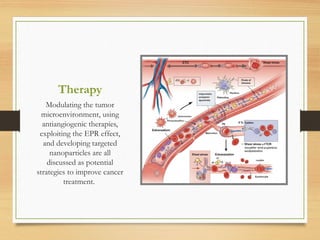
Therapy
Modulating the tumor
microenvironment,using
antiangiogenic therapies,
exploiting the EPR effect,
and developing targeted
nanoparticles are all
discussed as potential
strategies to improve cancer
treatment.
25.
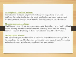
Challenges in TraditionalTherapy
Current cancer treatments target the whole body, but drug delivery to tumors is
inefficient due to barriers like irregular blood vessels, abnormal tumor structure, and
impaired lymphatic drainage. These obstacles limit drug transport and effectiveness.
Microenvironment as a Target
Modifying the tumor microenvironment can enhance drug delivery by normalizing blood
vessels, breaking down the extracellular matrix, increasing blood flow, and restoring
lymphatic function. The timing of these interventions is crucial for effectiveness.
Antiangiogenic Therapy
This approach targets endothelial cells in new blood vessels to inhibit tumor growth. It
has side effects like high blood pressure and potential tumor aggressiveness. Combining
antiangiogenic drugs with chemotherapy has shown some success.
26.
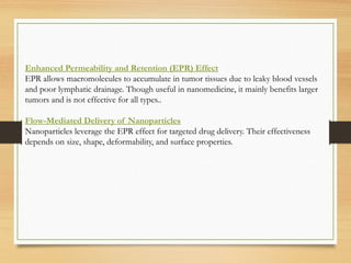
Enhanced Permeability andRetention (EPR) Effect
EPR allows macromolecules to accumulate in tumor tissues due to leaky blood vessels
and poor lymphatic drainage. Though useful in nanomedicine, it mainly benefits larger
tumors and is not effective for all types..
Flow-Mediated Delivery of Nanoparticles
Nanoparticles leverage the EPR effect for targeted drug delivery. Their effectiveness
depends on size, shape, deformability, and surface properties.
27.
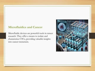
Microfluidics and Cancer
Microfluidicdevices are powerful tools in cancer
research. They offer a means to isolate and
characterize CTCs, providing valuable insights
into cancer metastasis.
28.
Importance of CTCs:
CTCsare important for understanding cancer spread and improving genetic analysis of
tumors (liquid biopsies). Isolating and characterizing CTCs can provide valuable
information about metastasis.
Microfluidic Devices for CTC Isolation:
Microfluidic devices offer a promising approach for CTC isolation due to their ability to
handle small sample volumes and control flow conditions.
Biomarker-Based Selection
Many microfluidic devices use biomarkers like EpCAM to identify and capture CTCs.
However, EpCAM expression can vary, particularly during epithelial-mesenchymal
transition (EMT), limiting this method’s effectiveness.
29.
Filterless Microfluidic Devices
Certaindevices use inertial focusing to separate CTCs based on size and other physical
characteristics, eliminating the need for filters and reducing clogging risks.
Microfluidics for Studying Cell Behavior
Microfluidic devices also help analyze cancer cell behavior, including movement and
responses to stimuli, by mimicking aspects of the tumor microenvironment in a controlled
setting.
Flow and Cell Adhesion
Microfluidics allows researchers to study how fluid flow affects CTC adhesion to vessel
walls. Flow influences cell rolling (mediated by glycoproteins and selectins) and strengthens
adhesion, with mechanical forces playing a key role in these processes.
30.
Observation
This paper explainsthat the chaotic fluid in tumors makes drug delivery
harder, but understanding fluid dynamics could improve treatments. It
also suggests that fluid forces could affect cancer spread and immune
responses, offering ways to enhance immunotherapies. By combining
fluid dynamics with liquid biopsy methods, we could improve cancer
diagnosis and treatment. The authors call for a more complete approach
to cancer research, combining fluid mechanics with genetics and
biochemistry to improve patient outcomes.
31.
Conclusion
This paper highlightsthe important but underexplored role of
fluid dynamics in cancer spread. While much focus has been on
genetic and biochemical factors, blood and lymph flow also play a
crucial role. The movement of fluids around tumors could help
spread cancer cells, and studying how these fluids affect circulating
tumor cells (CTCs) may lead to new ways to predict or treat
metastasis. Understanding how fluid forces influence CTCs could
open doors to better diagnostics and therapies.
32.
References
Fluid mechanics incirculating tumour cells:
https://doi.org/10.1016/j.medidd.2023.100158
Genetic alteration and gene expression modulation during cancer progression :
https://doi.org/10.1186/1476-4598-3-9
Transmural coupling of fluid flow in microcirculatory network and interstitium in
tumors : https://doi.org/10.1006/mvre.1996.2005