Plant Biotechnology: Introduction, Scope and Application
Final Report
1. 1
Chapter 1
INTRODUCTION
1.1 Sunflower
The cultivated sunflower (Helianthus annuus L.) is an annual plant with high nutrition
value. Sunflower (Helianthus annuus L.) as an economical important crop, is relatively young,
having been selected and cultivated on a large scale since the latter part of the nineteenth
century. Presently, it is the fourth most important oilseed crop in the world. It was grown
worldwide on over 21 million ha in 2000, in intermediate, temperate, subtropical and parts of
tropical climates. Sunflower species are allelopathic in nature and this crop appears to have a
bright future, especially if the scientists can translate the cutting-edge research into technologies
that will reduce the reliance on synthetic herbicides, pesticides, and crop protection chemicals.
Sunflower oil world production now ranks 4th after soybean. It is a major target for the
food and feed industry. It is not only known for its richness in polyunsaturated fatty acids but
also for its relative high content of miner constituents such as tocopherols (vitamin E)and
phytosterols, known to lower plasma cholesterol levels (Hewezi et al 2004).
Sunflower seeds are the achenes fruits of sunflower plants (Helianthus annus L.), They
are grayish –green with an outer shell (pericarp) that appears black, white or striated, depending
on the variety of plant.
Sunflower seeds contain an important source of key nutrients in a healthy diet: vitamin E,
vegetable protein, potassium, phosphorous, calcium, iron, magnesium, thiamin, riboflavin,
niacin, with very low intake of saturated fatty acids.
In recent years, there has been a very interesting assessment of the content of sunflower
seeds and their medicinal properties (Liu et al., 2011).Tissue culture technology of sunflower has
also been under investigation since the 1980s, but regeneration of this crop is still limited to date
(Liu et al., 2011).
2. 2
1.2 Agrobacterium tumefaciens
Agrobacterium tumefacians (updated scientific name: Rhizobium radiobacter) is the
causal agent of crown gall disease (the formation of tumours) in over 140 species of eudicots. It
is a rod-shaped, Gram - negative soil bacterium. Symptoms are caused by the insertion of a small
segment of DNA (known as the T-DNA, for ‘transfer the DNA’), from plasmid, into the plant
cell, which is incorporated at semi-random location into the plant genome. Some
Agrobacterium cells carry only one vector but some of them have two, each carrying two
different combination of gene.
There are several significant advantages to transferring DNA via Agrobacterium,
including a reduction in transgene copy number, the stable integration with fewer rearrangements
of long molecules of DNA with defined ends and the ability to generate lines free from selectable
marker genes (Jones et al 2005).
1.3 Plant transformation methods
Agrobacterium mediated transformation.
Biolistic (or) particle bombardment.
1.3.1 Agrobacterium mediated transformation
Agrobacterium mediated transformation method are thought to induce less rearrangement
of the transgene. This method produces lower transgene copy number than direct DNA delivery
method. It is capable of transferring large fragments of DNA very efficiently without substantial
rearrangement. The stabily of gene transferred is excellent.
1.3.2 Biolistic (or) particle bombardment.
This method can be use to transform all plant species. No binary vector is required.
Transformation protocol is relatively simple. High velocity micro projectile were utilize to
deliver nucleic acids into loving cells. This method uses the instrument called as biolistic gun or
gene gun.
3. 3
AIM, OBJECTIVES AND SCOPE
AIM
The Aim for this study is to standardize a simple protocol for inplanta transformation of
sunflower (Helianthus annuus L.)
OBJECTIVES
Transformation of pCAMBIA 1305.2 vector into Agrobacterium tumefaciens Strain
LBA4404.
Standardization of genetic transformation protocol for Helianthus annuus L.
Screening of transgenic Helianthus annuus L.
SCOPE
Once a simple protocol for inplanta transformation is standardized, this method can be
utilized to do genetic engineering in sunflower easily with any given gene of interest.
4. 4
CHAPTER 2
REVIEW OF LITERATURE
Agrobacterium mediated transformation is one of the most widely used mode of
transformation in plants. Several Researchers throughout the world are involved in this research
working with several plants. This is more preferred method of transformation because it is
economical, does not require any high end instrumentation, and also produces stable lines of
transformants. The choice of tissue or organ in the plant of study has been found varying.
Feldmann and Marks (1987) developed a non-tissue culture approach of Agrobacterium-
mediated transformation in germinating seeds of Arabidopsis thaliana. Stable transformed lines
were obtained from apical shoots of sunflower (Burrus et al 1996) and callus in Vicia faba
(Bottinger et al., 2001). Keshamma et al., (2008) successfully developed an inplata method of
Agrobacterium mediated transformation in Cotton (Gossypium hirsutum L.). This tissue culture
independent method was found to be an effective method of obtaining stable transformants in
recalcitrant plant species like cotton.
2.1 Genetic transformation in plants
Protocols were developed by Mukopadhyay et al (1992) for efficient shoot regeneration
from hypocotyl and cotyledon explants of oilseed Brassica campestris (brown sarson) cv. ‘Pusa
Kalyani’. These were used for genetic transformation by an Agrobacterium based binary vector
carrying neomycin phosphotransferase (npt) gene and β-glucuronidase (gus)-intron gene for
plant cell specific expression. Transformed plants were recovered from hypocotyl explants at a
frequency of 7–13%. Fursova et al (2012) transiently expressed three hydrolase genes in
Brachypodium distachyon plants using specially designed vectors that express the gene product
of interest and target it to the plant cell wall. Expression of functional hydrolases in genotyped
plants was confirmed using western blotting, activity assays, cell wall compositional analysis and
digestibility tests.
5. 5
2.2 Genetic transformations in Helianthus annuus L.
Lappara et al (1995) evaluated three methods of transformation in sunflower viz., direct
gene transfer into protoplasts, particle bombardment and Agrobacterium co-culture. All
techniques allowed efficient short-term or transient expression of the introduced gene(s) in the
respective tissues, stable transformation was only observed after transformation with
Agrobacterium. Burrus et al (1996) developed stable lines of Helianthus annuus L. through
agrobacterium mediated transformation in the apical shoots. In 1999, Rao and Rohini developed
a very simple protocol of transformation in sunflower using Agrobacterium. In this method, two
days old seedlings with one cotyledon detached were infected with Agrobacterium, this resulted
in stable transformation. Weber et al (2003) assessed the macerating enzymes and sonication
methods of treatment in improving Agrobacterium -mediated transformation of sunflower
(Helianthus annuus L.). Liu et al (2011) optimized Agrobacterium mediated transformation in
Helianthus annuus L. using immature embryos.
6. 6
CHAPTER 3
METHODOLOGY
3. a Requirements for the project
Collection of samples:
The Sunflower seeds of variety CO4 were obtained from Tamil Nadu Agricultural
University, Coimbatore. Few of these seeds were sown in our college garden for further
studies.
Bacterial strain: Agrobacterium tumefaciens strain LBA4404
Agrobacterium tumefaciens (Rhizobium radiobactor) is capable of T-DNA transfer to
plant cells. The T-DNA (transfer DNA) is located in the Ti plasmid and is capable of
integration into the host plant chromosomal DNA. Integrated genes derived from T-DNA
are expressed and the transformed plant cells typically become Crown gall tumor
cells.The strain LBA4404 has rifampicin resistance gene present in its chromosome and
streptomycin resistance gene on the Ti plasmid.
Plasmid Vector: pCAMBIA 1305.2
These vectors contain minimal heterologous sequences for plant transformation and
selection of transformants; they allow insertion of desired genes for transformation into
plants but require all promoter and terminator sequences for plant expression of newly
cloned genes.
Vector contains kanamycin resistance gene for bacterial selection and hygromycin
B resistance gene for plant selection.
It also incorporates the GusPlus reporter gene.
The reporter gene of pCAMBIA 1305.2 lacks the bacterial ribosome binding site and
shows no expression in Agrobacterium but good expression in plant cells.
7. 7
3.1 TRANSFORMATION OF pCAMBIA 1305.2 VECTOR INTO Agrobacterium
tumefaciens STRAIN LBA4404
3.1.1 Preparation of Competant cells of Agrobacterium tumefaciens strain LBA4404
Most species of bacteria take up only limited amounts of DNA under normal
circumstances. For efficient uptake, the bacteria have to undergo some form of physical and /or
chemical treatment that enhances their ability to take up DNA. Cells that have undergone this
treatment are said to be competent. The fact that Agrobacterium cells that are soaked in an ice-
cold salt solution are more efficient at DNA uptake than unsocked cells. Traditionally, a solution
of CaCl2 is used is used to make competent Agrobacterium cells.
Materials:
YEP broth
100mM CaCl2 solution
250 ml conical flask
1.5 ml centrifuge tube
Microtips and 1.5ml microfuge tubes
Protocol:
Agrobacterium strain LBA4404 was grown over night at 28˚c in YEP medium containing
50 μgmlˉ¹ rifampicin.
The overnight culture was then chilled on ice for 30 mins.
It was then transferred into prechilled 15ml falcon tubes and was then centrifuged at 4000
rpm at 4ºC for 10 mins.
The supernatant was then discarded and the pellet was dissolved in 10ml 100mM ice cold
CaCl2.
The tubes were then incubated on ice for 20 mins.
Later it was centrifuged at 4000 rpm at 4ºC for 10 mins.
The supernatant was then discarded and the pellet was gently resuspended in 2 ml
100mM ice cold CaCl2.
8. 8
Aliquots of 200µl were transferred to prechilled 1.5 ml tubes and was then used for
transformation.
3.1.2 Transformation of pCAMBIA 1305.2 vector into Agrobacterium tumefaciens strain
LBA4404 by free-thaw method
Transformation broadly means uptake of any DNA molecule especially plasmid by living
cell like bacteria. Agrobacterium cells that are soaked in CaCl2 solution affects only DNA
binding, and not the actual uptake into cell. The actual movement of DNA into competent cells is
stimulated by briefly raising the temperature to 37˚c by heat shock treatment.
Materials:
Competent cells (200µl)
Plant transformation vector – pCAMBIA 1305.2
YEP broth
YEP agar plates with Kanamycin and Rifampicin each 50μg/ml.
Sterile Microtips
IPTG
X-Gal
Protocol:
Plasmid DNA, 500ng (3μl) was added to the tube of competent cells of Agrobacterium.
This was then mixed well and incubated in -20 ºC for 30 mins.
Heat shock treatment was then given by immediately transferring into 37ºc water bath for
5 mins and was soon placed on ice for 5 mins.
YEP broth, 800 μl was added and then was incubated at 28ºC for 3-4 hrs.
After incubation the culture was spread plated YEP plate with Rif + Kan containing IPTG
and X-Gal
These plates were then incubated at 28ºC for 2 days.
3.1.3 Selection of transformed colonies
Selection of transformed colonies was done by picking the blue colored colonies. The
selected colonies were then again grown on YEP agar plates with Rif + Kan by streaking. These
9. 9
plates were then incubated at 28ºC for 2 days. Then the transformed colonies were confirmed by
plasmid isolation.
3.1.4 Isolation of plasmid from transformed colonies containing the pCAMBIA 1305.2
vector
Plasmid is a double stranded, circular extra chromosomal DNA of bacterium. It is used in
recombinant DNA experiments to clone genes from other organisms and make large quantities of
their DNA. Plasmid can be transferred between same species or between different species. Size
of plasmids range from 1-1000 kilo base pairs. Plasmids are part of mobilomes (total of all
mobile genetic elements in a genome) like transposons or prophages and are associated with
conjugation. Even the largest plasmids are considerably smaller than the chromosomal DNA of
the bacterium, which can contain several million base pairs.
The term 'plasmid' was introduced by an American molecular biologist Joshua Lederberg.
Plasmids are considered as transferrable genetic elements or 'replicons'. They are actually naked
DNA. Plasmids are important tools in genetic and biotechnology labs where they are commonly
used to multiply or express particular genes. Plasmids are also used to make large amounts of
proteins.
Plasmids encoding Zinc Finger Nucleases are used to deliver therapeutic genes to a
preselected chromosomal site with a frequency higher than that of random integration. Mainly
there are two types of plasmids: conjugative and non conjugative. Conjugative plasmids have tra-
genes (tra-transfer) and can perform conjugation. Non conjugative plasmids cannot perform
conjugation. There is an intermediate class of plasmid called mobilizable plasmid. Mobilizable
plasmid can carry only a subset of genes required for transfer. They can parasitize a conjugative
plasmid transferring at high frequency only in its presence.
3.1.4.1 Plasmid Isolation by Mini-Prep Method
Mini-Prep method is commonly used protocol for plasmid isolation.
Materials:
Bacterial culture
YEP broth with kanamycin
Solution I (Suspension Buffer)
10. 10
Solution II (Lysis Buffer)
Solution III (DNA neutralization buffer)
Sterile Microtips
Sterile microfuge tubes
Isopropanol
70% ethanol
Protocol:
1. Five ml of sterile medium was incubated with a single bacterial colony and kept
overnight at 280C.
2. Next day, 2 ml of bacterial culture was taken in microfuge tubes and centrifuged for 10
min at 10,000 rpm.
3. The supernatant was decanted till the last drop.
4. The cells were in 100μl of ice cold DNA suspension buffer (Solution I). The bacterial
cells were completely suspended by vortexing until no cells clumps remain.
5. DNA Lysis buffer (Solution II) 200μlwas added and mixed gently by inverting 3-4 times.
6. Immediately 150μl of prechilled DNA neutralization buffer (Solution III) was added. It
was then mixed immediately by gently inverting the vial 3-4 times and incubated in ice
for 10 min.
7. The tubes were then centrifuged at 10,000 rpm for 12 min at 40C. The clear supernatant
containing plasmid was collected into a fresh centrifuge tube.
8. If the supernatant was turbid, it was re-centrifuged at 10,000 rpm for 10 min at 40C. The
cleared supernatant containing plasmid was collected into a fresh centrifuge tube.
9. Equal volume of isopropanol was added to the supernatant and was mixed properly.
10. It was then centrifuged at 10,000 rpm for 15 min at room temperature. Supernatant was
discarded and the pellet was washed with 70% ethanol.
11. The pellet was dried at 370C for 10 min and was then suspended in 50-100μl of glass-
distilled water (or) nuclease free water.
11. 11
3.2 STANDARDIZATION OF GENETIC TRANSFORMATION PROTOCOL FOR
Helianthus annuus L.
3.2.1 Sterilization of seeds
Materials:
1% Bavistin
0.1% HgCl2
Steriile petri dishes
Germination sheets (Sterile)
Sterile forceps and conical flasks
Sterile distilled water
Protocol:
The seeds of sunflower were soaked overnight in distilled water and were surface
sterilized first with 1% Bavastin for 10 mins.
It was followed by distilled water wash thrice.
Later it was treated with 0.1% HgCl2 for 30 seconds and washed thoroughly with distilled
water.
The seeds were then transferred onto sterile petri plates with moist germination sheets
and was allowed to germinate on petriplates 30˚C in dark.
Two-day old seedlings were taken as explants for Agrobacterium infection.
3.2.2 Transformation of sunflower seeds
Materials:
Agrobacterium strain LBA4404 + pCAMBIA1305.2 culture
YEP broth with Kanamycin
½ MS media
Soilrite
Sterile Tissue culture bottles
Sterile forceps and conical flasks
Sterile distilled water
12. 12
Protocol:
A loopful of Agrobacterium strain LBA4404 containing pCAMBIA1305.2 culture was
inoculated in 20 ml YEP media supplemented with kanamycin for overnight
Overnight culture, 5ml was resuspended into 100 ml of YEP with kanamycin media and
incubated for 2 days.
The culture was centrifuged and the pellet was resuspended into 100 ml ½ strength MS
media and acetosyringone to final concentration of 50µM was added and incubation was
continued at 28˚c for 5 hours.
The seeds with emerging plumule were infected by separating the cotyledons without
damaging the meristem with a sterile sewing needle.
Subsequently the seeds were dunked into Agrobacterium culture in ½ MS media and was
incubated at 28˚c for 60 mins.
Then the seedlings were washed with Sterile water and few of them were transferred into
autoclaved soilrite and remaining few in ½ MS agar for germination under aseptic
condition (5 seedling per Jar).
The growth chamber was maintained at 26-28˚c under photoperiod of 14hours with
florescent light of intensity of approximately 2500lux.
3.3 SCREENING OF TRANSGENIC Helianthus annuus L.
3.3.1 GUS assay
Plants contain endogenous β-galactosidase activity, so lacZ is not generally a useful
reporter gene for plants. A widely used reporter gene in plants is the uidA, or gusA, gene that
encodes the enzyme β-glucuronidase (GUS). This enzyme can cleave the chromogenic
(colorgenerating) substrate X-gluc (5-bromo-4-chloro-3-indolyl β-D-glucuronic acid; Fig. 2),
resulting in the production of an insoluble blue color in those plant cells displaying GUS activity.
Plant cells themselves do not contain any GUS activity, so the production of a blue color when
stained with X-gluc in particular cells indicates the activity of the promoter that drives the
transcription of the gusA-chimeric gene in that particular cell. The GUS assay is easy to perform,
sensitive, relatively inexpensive, highly reliable, safe, requires no specialized equipment, and is
highly visual (Jefferson 1987; Jefferson et al. 1987; Jefferson and Wilson 1991)
13. 13
3.3.1.1 Expression of β-glucuronidase
Materials:
0.1% HgCl2
GUS staining solution.
Sterile distilled water
75% ethanol
Protocol:
Five days old transformants were screened for expression of β-glucuronidase.
For analysis of the transformants, tissue that were tested and found free of residual
Agrobacterium were used.
The persistence of Agrobacterium in the putative transformants was largely controlled by
a brief agitation of the co-cultivated seedlings with 0.1% HgCl2 for 30s followed by
through washes with distilled water.
The method of Jefferson (1987) was used to assess histochemical assay of uid A gene
expression in the tissues of primary transformants,
The complete seedlings as such were incubated overnight at 37˚C in GUS staining
solution.
The next day they were washed in water and later soaked with 75% ethanol to clear
chlorophyll.
14. 14
CHAPTER 4
RESULT AND DISCUSSION
4.1 TRANSFORMATION OF pCAMBIA 1305.2 VECTOR INTO Agrobacterium
tumefaciens STRAIN LBA4404
The current study was initiated by transforming pCAMBIA 1305.2 vector into
Agrobacterium tumefaciencs strain LBA4404. For this standard method of competent
preparation was followed and competent cells for Agrobacterium tumefaciencs strain LBA4404
was prepared. The prepared competent cells were used for transformation of the plasmid vector.
The transformation of plasmid into Agrobacteium was performed by freeze thaw method which
is also called as heat shock method. Successfully, 330 transformed colonies were obtained (Fig.
4.1). The transformation efficiency was calculated as 2.75x10³ transformants / μg DNA. Further
all the point colonies obtained on the selection plate were blue in colour since IPTG and X-Gal
were added onto the plate for screening of transformants. The presence of blue colored colonies
meant that the plasmid did not have any gene inserted within its Multiple Cloning Site (MCS)
region and thus the LacZ gene was functional and was able to utilize the X-Gal in presence of
IPTG, thus releasing the chromogenic blue substrate resulting in blue colour.
4.2 STANDARDIZATION OF INPLANTA GENETIC TRANSFORMATION
PROTOCOL FOR Helianthus annuus L.
The method of seed sterilization published by Keshamma et al., (2008) was modified and
used for this study. The sterilization procedure was repeated thrice with different number of
seeds and resulted in overall percentage of germination as 84%. Also it was found the overall
percentage of contamination was 1.33%. Thus this protocol for sterilization of seeds proved to be
efficient as it resulted in lesser contamination with greater percentage of germination. The
efficiency of this protocol, after considering the rate of germination as well as contamination
resulted as 82.16%. Data shown in Table 4.1.
The overall transformation efficiency for the inplanta transformation method that we
adopted was 11.32%. But the transformation efficiency for the plants cultured in ½ MS media
grew better and gave better results with transformation efficiency of 19.35%. The lower overall
percentage of transformation was due to the transformants grown in soilrite. The growth in all
the plants after transformation which were grown in soilrite was stagnant. This was because the
15. 15
roots of the plants put in the soilrite started decaying and hence the plants did not grew and
resulted in no transformation. So ½ MS proved to be a better medium of potting soon after
transformation in invitro studies. Data shown in Table 4.2 and Fig. 4.2.
4.3 Screening of transgenic Helianthus annuus L.
Though the transformation efficiency was less, still the GUS staining showed that there
was uniform expression of GUS in the transformed plants above the hypocotyls (Fig. 4.3).
In the present study, a tissue culture-independent in planta transformation protocol was
used to develop transformants (Rohini and Sankara Rao, 2000a; Rohini and Sankara Rao, 2000b;
Rohini and Sankara Rao, 2001). Such in planta transformation techniques have also been
standardized in other crops like, buckwheat (Kojima et al., 2000), mulberry (Ping et al., 2003),
kenaf (Kojima et al., 2004), soybean (Chee et al., 1989) and rice (Supartana et al., 2005) etc. In
this method, Agrobacterium is targeted to the wounded apical meristem of the differentiated seed
embryo. Therefore, Agrobacterium tumefaciens transfers the gene into the genome of diverse
cells which are already destined develop into specific organs and the meristematic cells still to be
differentiated. This results in the primary transformants being chimeric in nature.
The method therefore is advantageous because it avoids the need for tissue culture.
Nevertheless, transformability depends on the susceptibility of the variety to Agrobacterium. A
number of factors affect transformability by inplanta transformation. Finally, all these factors
including stability of the transgene can only be assessed after getting the T1 and T2 generations.
16. 16
No.
of
Seeds
(a)
No. of Seeds
Contaminated
(b)
No. of
Seeds
Germinated
(c)
Percentage of
Contamination
(b/a*100)
Percentage
of
Germination
(c/a*100)
Efficiency
[(c-
b)/a*100]
I 18 1 18 5.5% 100% 94.44%
II 30 0 22 0.0% 73.33% 73.33%
III 27 0 23 0.0% 85.16% 85.16%
T 75 1 63 1.33% 84% 82.76%
Table 4.1 Efficiency of the sterilization protocol after considering the rate of germination
and rate of contamination
Media No of seedling
used for
transformation
(a)
No. of
transformed
seedlings
(b)
No. of
seedlings not
transformed
(c)
Transformation
percentage
(b/a*100)
Soilrite 35 0 35 0.00%
½ MS 18 6 12 33.33%
Total 53 6 47 11.32%
Table 4.2 Total inplanta transformation percentage.
17. 17
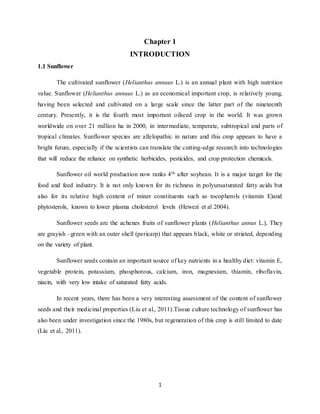
Fig. 4.1a Fig. 4.1b
Kanamycin resistant blue colonies of Negative control plate without any growth of
Agrobacteriumtumefaciens transformed competent cells due to the presence of
with pCAMBIA 1305.2 antibiotic Kanamycin
Figure 4.1: Transformation of pCAMBIA 1305.2 vector into Agrobacterium tumefaciens
Strain LBA4404
18. 18
Figure 4.2. Sunflower seedlings after inplanta transformation
Seedlings to the left are control plants and the one to the right are putative transformants
(a) (b)
Figure 4.3. Expression of GUS enzyme
(a) Control showing no blue colour stain after GUS staining
(b) Transformats stained blue above hypocotyls after GUS staining
19. 19
REFERENCES
Bottinger P., Steinmetz, Schieder O. and Pickardt T. 2001. Agrobacterium-mediated
transformation of Vicia faba. Molecular Breeding Volume 8, Issue 3, pp 243-254
Burrus M., Molinier J., Himber C ., Hunold R., Bronner R., Rousselin P. and
Hahne G. 1996. Agrobacterium-mediated transformation of sunflower (Helianthus
annuus L.) shoot apices: transformation patterns. Molecular Breeding, Volume 2, Issue 4,
pp 329-338
Chee, P.P., A.K. Fober, and L.J. Slightom, 1989. Transformation of soybean (Glycine
max (L.) Merrill) by infecting germinating seeds with Agrobacterium tumefaciens, Plant
Physiol. 91: 1212-1218.
Feldmann K. A and Marks M.D. 1987,Agrobacterium-mediated transformation of
germinating seeds of Arabidopsis thaliana: A non-tissue culture approach. Molecular and
General Genetics Volume 208, Issue 1-2, pp 1-9
Fursova.O, Pogorelko.G and Zabotina.O.A 2012. An efficient method for transient
gene expression in monocots applied to modify the Brachypodium distachyon cell wall,
Annals of Botany, 1-10.
Heweli.T, Alibert.G and Kallerhoff.J 2004. Genetic transformation of sunflower
(Helianthus annuus L.) transgenic Crops of the World p435-451.
Jefferson, R. A., T. A. Kavanagh, and M.W. Bevan. 1987. GUS fusions: β-
glucuronidase as a sensitive and versatile gene fusion marker in higher plants. EMBO
Journal 6: 3901- 3907.
Jefferson, R.A. 1987. Assaying chimeric genes in plants: The GUS gene fusion system.
Plant Molecular Biology Reporter 5: 387-405.
Jefferson, R.A., and K.J. Wilson. 1991. The GUS gene fusion system. In Gelvin, S.B.
and R.A. Schilperoort (eds.) Plant Molecular Biology Manual, Kluwer Academic
Publishers (Dordrecht). B14: 1-33.
20. 20
Jones.H.D, Doherty.A and Wu.H 2005. Review of methodologies and a protocol for the
Agrobacterium-mediated transformation of wheat, Plant method 2005, 1:5
doi:10.1186/1746-4811-1-5.
Keshamma.E, Rohini.S, Rao.K.S, Madhusudhan.B, and kumar.M 2008.Tissue
culture-independent inplanta transformation strategy: An agrobacterium tumefaciens-
mediated gene transfer method to overcome recalcitrance in cotton (Gossypium hirsutum
L.) The Journal of Cotton Science 12:264-272.
Khan M. M. robin A. B. M. A. H. K, nazim-ud-dowla.M.A.N, Talukder. S. K. and
Hassan.L 2009. Agrobacterium-mediated genetic transformation of two varieties of
brassica: Bangladesh J. Agril. Res. 34(2) : 287-301.
Kojima, M., H. Shioiri, M. Nogawa, M. Nozue, D. Matsumoto, A. Wada, Y. Saiki, K.
Kiguchi, 2004. In planta transformation of kenaf plants (Hibiscus cannabinus var.
aokawa no.3) by Agrobacterium tumefaciens, J. Biosci. Bioeng. 98: 136-139.
Kojima, M., Y. Arai, N. Iwase, K. Shiratori, H. Shioiri, and M. Nozue, 2000.
Development of a simple and efficient method for transformation of buckwheat plants
(Fugopyrum esculentum) using Agrobacterium tumefaciens, Biosci. Biotechnol.
Biochem. 64: 845-847.
Kumar.M, shukla.A.K, singh.H, verma.P.C and Singh.P.K 2013. A Genotype-
independent agrobacterium mediated transformation of germinated embryo of cotton
(Gossypium hirsutum L.), International Journal of Bio-technology and Research (IJBTR)
vol.3,issue1,81-90.
Lappara H., Burris M., Hunold R., Damm B., Bravo-Angel A.M., Bronner R., and
Hahne G. 1995, Expression of foreign genes in sunflower (Helianthus annuus L.) —
Evaluation of three gene transfer methods. Euphytica Volume 85, Issue 1-3, pp 63-74
Liu.H, Xie.X, Sun.S, Zhu.W, Ji.J, Wang.G 2011. Optimization of agrobacterium-
mediated transformation of sunflower (Heliathus annuus L.) AJCS(12):1616-1621.
Mukhopadhyay A., Arumugam N., Nandakumar P.B.A., Pradhan A.K., Gupta V
and Pental D. 1992. Agrobacterium-mediated genetic transformation of oilseed Brassica
campestris: Transformation frequency is strongly influenced by the mode of shoot
regeneration. Plant Cell Reports, Volume 11, Issue 10, pp 506-513
21. 21
Ping, L.X., M.Nogawa, M. Nozue, M. Makita, M.Takeda, L. Bao and M. Kojima,
2003. In planta transformation of mulberry trees (Morus alba L.) by Agrobactetium
tumefaciens, J. Insect Biotechnol. Sericol. 72: 177-184.
Piqueras.A, Alburquerque.N and Folta K.M 2010.Explants used for the Generation of
transgenic plants. DOI 10.1007/978-3-642-04809-8_2.
Radonic.L.M, Zimmermann.J.M, Lopez.D.Z.N, Bilbao.M.L 2008. Introduction of
antifungal genes in sunflower via Agrobacterium. Electronic Journal of Biotechnology
ISSN:0717-3458.
Rao.K and Rohini.V.K 1999. Agrobacterium-mediated transformation of Sunflower
(Helianthus annuus L.): Annals of Botany 83: 347-354.
Rohini, V.K. and K. Sankara Rao, 2000a. Transformation of peanut (Arachis hypogeae
L.): a non-tissue culture based approach for generating transgenic plants, Plant Sci.
150:41-49.
Rohini, V.K., and K. Sankara Rao, 2000b. Embryo transformation, a practical
approach for realizing transgenic plants of safflower (Carthamus tinctorius L.), Annals of
Botany 86: 1043-1049.
Rohini, V.K., and K. Sankara Rao, 2001. Transformation of peanut (Arachis hypogaea
L.) with tobacco chitinase gene: variable response of transformants to leaf spot disease,
Plant Sci. 160 (5): 883-892.
Sambrook, J., E.F. Fritsch, and T. Maniatis, 1989. Molecular cloning, Plain view,
New York, Cold Spring Harbor Laboratory Press.
Schrammeijer.B, Sijmons.P.C, Van.P.J.M Elzen and Hoekema.A 1990. Meristem
transformation of sunflower via Agrobacterium,volume 9,Issue2 ,pp 55-60.
Sujatha.M, Vijay.S, Vasav.S, Veera.P Reddy, Rao.S 2012. Agrobacterium-mediated
transformation of cotyledons of the mature seeds of multiple genotypes of sunflower
(Helianthus annus L.) Plant Cell Tiss Organ Cult 110:275-287.
Supartana, P., T.Shimizu, Shioiri.H, M. Nogawa, M. Nozue, and M. Kojima. 2005.
Development of simple and efficient in planta transformation method for rice (Oryza
sativa L.) using Agrobacterium tumefaciens, Journal of Bioscience and Bioengineering
100(4): 391-397.
22. 22
Theriappan.P and Gupta.A.K 2014. Development of a protocol for Agrobacterium
mediated transformation of Brassica oleraceae Lvar botrytis cv Early Kunwari, European
Journal of Biotecnology and Bioscience 1(3):34-38.
Weber S., Friedt W., Landes N., Molinier J., Himber C., Rousselin P., Hahne G. and
Horn R. 2003. Improved Agrobacterium -mediated transformation of sunflower
(Helianthus annuus L.): assessment of macerating enzymes and sonication. Plant Cell
Reports Volume 21, Issue 5, pp 475-482
Weber.S, Friedt.W, Landes.N, Molinier.J, Himber.C, Rousselin.P, Hahne.G,
Horn.R 2003. Improved Agrobacterium-mediated transformation of sunflower
(Hellianthus annuus L.): assessment of macerating enzymes and sonication, Plant cell
Rep 21:475-482.
Zombori.Z, Szecsenyi.M, Gyorgyey.J 2011. Different approaches for Agrobacterium-
mediated genetic transformation of Brachypodium distachyon, a new model plant for
temperate grasses,volume 55(1):193-195.
23. 23
APPENDIX
1. SOLUTIONS FOR BACTERIAL TRANSFORMATION
100mM CaCl2
Dissolve 1.4702 g of CaCl2 in 20 ml of sterile water. Filter sterilize it and add it it 80
ml of sterile water.
YEP Broth
Bacto Peptone 10.0 g
Yeast Extract 10.0 g
NaCl 5.0g
YEP Agar
Bacto Peptone 10.0 g
Yeast Extract 10.0 g
NaCl 5.0g
Bacto Agar 15.0 g
H2O 1000 ml
IPTG Stock 1M
Dissolve 0.238g of IPTG in 1ml of sterile distilled water. Store it in -20ºC
X-Gal (20%)
Dissolve 200mg X-Gal in 1 ml of Dimethyl sulphoxide. Store it in -20ºC
2. SOLUTIONS FOR PLASMID ISOLATION
Solution I (Suspension Buffer)
50mM Glucose
10mM EDTA
25mM Tris cl pH 8.0
100µg/ml RNase A
Solution II (Lysis Buffer)
0.2N NaOH
24. 24
1% SDS
Solution III
Dissolve 29.45g of Potassium acetate in 80 ml of distilled water. Add glacial acetic acid
till the pH reaches 5.5. Make up the volume to 100ml and sterilize by autoclaving. Store in 4 ºC
TE Buffer
10mM Tris cl pH 8.0
1mM EDTA
3. SOLUTIONS FOR INPLANTA TRANSFORMATION
1% Bavastin
Bavastin 1g
Distilled Water 100 ml
0.1% HgCl2
HgCl2 100mg
Distilled Water 100 ml
GUS staining solution
0.1M phosphate buffer-pH 7.0
2mM X-Gluc
5mM Potassium Ferricyanie
5mM potassium Ferrocyanide
0.1% Triton X
75% Ethanol
Measure 75 ml of ethanol using a measuring cylinder. Make the volume to 100ml by
adding sterile distilled water.
4. ANTIBIOTIC STOCK SOLUTIONS
Kanamycin stock (50mg/ml)
Dissolve 50 mg of Kanamycin in 1ml of Sterile water.
Rifampicin stock (50 mg/ml)
25. 25
Dissolve 50mg of Rifampicin in 500μl of methanol and make up the volume to 1ml using
sterile distilled water.
5. MS MEDIA COMPOSITION
Constituents Mg/l g/1 20x Volumes to be
taken
500ml 1 lit
MACRONUTRIENTS
MgSO4.7H2O
CaCl2.2H2O
KNO3
NH4NO3
KH2PO4
370
440
1900
1650
170
3.7
4.4
19
16.5
1.7
(500ml)
3.7
4.4
19
16.5
1.7
25ml 50ml
MICRONUTRIENTS
MnSO4.4H2O(or)
MnSO4.H2O
ZnSO4.7H2O
CuSO4.5H2O
COCl2.6H2O
H3BO3
Na2MOO4.2H2O
(1000x)
22.3
16.9
8.6
0.025
0.25
6.2
0.25
0.223
0.169
0.086
0.00025
0.00025
0.062
0.0025
100(100ml)
2.23
1.69
0.86
0.0025
0.0025
0.62
0.025
0.5ml 1ml
IRON SOURCE
FeSO4.7H2O
Na2 EDTA
27.85
37.25
278.5
372.5
140x(100ml)
0.2785
0.3725
0.5ml 1ml
Carbohydrate
Source
Sucrose
30-50(g/l)
30g/l
Constituents Mg/l Stock
(1000x)
Volume to be taken
500ml 1000ml
VITAMINS
Nicotinic acid
Pyridoxine HCl
Thiamine HCl
0.5
0.5
0.5
50ml
0.025g
0.025g
0.025g
0.5ml 1ml
KI Stock Solution
Dissolve 83mg of KI in 100 ml Double distilled water. Take 1 ml KI solution for 1 liter media
26. 26
Stock solution of Meso-inositol (x500)
Dissolve 1gm meso-inositol in 20 ml of Water. Take 1mlfor 1 liter media
Stock solution of Glycine (x1000)
Dissolve 40 mg glycine in 20 ml of double distilled water. Take 1ml for 1 liter madia
1N HCl solution
Take 8.3 ml of HCl and make up the volume to 100ml using distilled water.
1N NaOH solution
Dissolve 4g NaOH in 100 ml water.