
More Related Content
What's hot
What's hot (20)
Viewers also liked
Viewers also liked (10)
Similar to FINAL poster ORD
Similar to FINAL poster ORD (20)
FINAL poster ORD
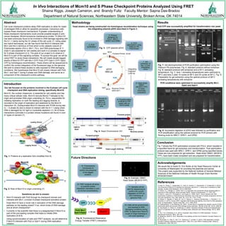
- 1. MCS Integrating Fragment 2,492 bp Not1 Not1 Forward Primer, 32-62 Reverse Primer, 2453-2471 MCS CFP KAN AMP pDH3 4882 pb References K Abstract Introduction Methodology Conclusion Cell cycle checkpoint proteins delay DNA replication to allow for repair of damaged DNA or allow for apoptotic processes. Cancerous cells bypass these checkpoint mechanisms. A greater understanding of these checkpoint mechanisms could provide possible targets in anti- cancer therapies. Minichromosome maintenance protein 10 (Mcm10) has been previously found to be involved in DNA damage signaling with the 9-1-1 clamp during the G1 phase of the cell cycle (1). Using yeast two hybrid techniques, our lab has found that Mcm10 interacts with Mrc1 and the C-terminus of Pol2 which is the catalytic subunit of Polymerase epsilon (Pol ε). Mrc1, Pol ε, and DNA polymerase B 11 (Dpb11) are essential for cell viability and work in a complex to signal for S phase checkpoint (2,3). The goal of our project is to observe if Mcm10 is part of this signaling complex. To pursue this goal, we will be using FRET to study these interactions. We will create double-tagged strains of Mcm10-YFP with Mrc1-CFP/ Pol2-CFP/ Dpb11-CFP/ Dpb2- CFP by homologous recombination. These strains will be sequenced to confirm the correct integration of the fluorescent tags on the genome. We wish to extend these studies to cells exposed to DNA damaging conditions. We hypothesize that Mcm10 will closely interact with Pol2, Mrc1, and Dpb11 during S phase and DNA damage, and serve as a component of the checkpoint control pathway. Fig. 1: Proteins at a replication fork (modified from 8) Fig. 6: Fluorescence Resonance Energy Transfer (FRET) Interaction Future Directions Acknowledgements We would like to thank Dr. Eric Muller at the Yeast Resource Center at University of Washington for providing the plasmids and advice. This project was supported by the National Institute of General Medical Sciences of the National Institutes of Health through Grant Number 8P20GM103447. Our lab focuses on the proteins involved in the S-phase cell cycle checkpoint and DNA replication timing, specifically Mcm10. Mcm10, the nuclear chaperone, is essential for cell viability and has many critical cellular roles. Mcm10 recruits Mcm2-7 helicase to the origin and promotes DNA origin unwinding (4). Pol α, responsible for initiating replication on both the leading and lagging strands, is recruited to the origin of replication and stabilized by the Mcm10 interaction (5). Diubiquinated Mcm10 interacts with PCNA during late G1 – S phase (6) and is shown to interact with the 9-1-1 clamp when DNA is damaged by UV light or nucleotide depletion (1). All of these interactions culminate in a protein whose mutations are found in over 41 types of cancers (7). Fig. 7 shows the PCR optimization process with POL2, which resulted in a specified band for gel extraction and transformation. That optimization protocol was used with MRC1, DPB11, and DPB2 giving specified bands. These are now prepared for gel extraction. Yeast strain DB001 (MCM10- YFP), have been made competent and are prepared for transformation. Department of Natural Sciences, Northeastern State University, Broken Arrow, OK 74014 Shaina Riggs, Joseph Cameron, and Brandy Fultz Faculty Mentor: Sapna Das-Bradoo In Vivo Interactions of Mcm10 and S Phase Checkpoint Proteins Analyzed Using FRET Results Fig. 8: Successful digestion of pDH3 was followed by purification and PCR amplification using the optimal protocol for PCR primers with flanking ends for MRC1, DPB11. and DPB2. Digested Plasmid Digested Plasmid MRC1 DPB2 DPB2 DPB2 Neg Control DPB11 DPB11 Neg Control DPB11 3 kb 2 kb Yeast strains are being constructed via homologous recombination technique using the integrating plasmid pDH3 described in Figure 3. Fig. 3: CFP plasmid pDH3 Fig. 2: Role of Mcm10 in origin unwinding (7) 5’ 5’ 3’ 3’ 1- Alver, R., Zhang, T., Josephrajan, A., Fultz, B., Hendrix, C., Das-Bradoo, S., & Bielinsky, A. (2014). The N-terminus of Mcm10 is important for interaction with the 9-1-1 clamp and in resistance to DNA damage. Nucleic Acids Research, 42(13), 8389-8404. 2- Osborn, A.J., Elledge S.J. (2003). Mrc1 is a replication fork component whose phosphorylation in response to DNA replication stress activates Rad53. Genes & Development. 17, 1755-1767. 3- Araki,H., Leem,S.H., Phongdara,A. and Sugino,A. (1995). Dpb11, which interacts with DNA polymerase II (epsilon) in Saccharomyces cerevisiae, has a dual role in S-phase progression and at a cell cycle checkpoint. Proc. Natl. Acad. Sci. U.S.A. 92, 11791–11795. 4- Fatoba, S., Tognetti, S., Berto, M., Leo, E., Mulvey, C., Godovac-Zimmermann, J., Pommier, Y., & Okorokov, A. (2013). Human SIRT1 regulates DNA binding and stability of the Mcm10 DNA replication factor via deacetylation. Nucleic Acids Res., 41(7), 4065–4079. 5- Chattopadhyay, S and Bielinsky AK. (2007). Human Mcm10 regulates the catalytic subunit of DNA polymerase-alpha and prevents DNA damage during replication. Mol Bio of the Cell. 18(10), 4085-95. 6-. Das-Bradoo, S., Ricke, R., & Bielinsky, A. (2006). Interaction between PCNA and Diubiquitinated Mcm10 Is Essential for Cell Growth in Budding Yeast. Molecular and Cellular Biology, 26(13), 4806- 4817. 7- Thu, Y. M., & Bielinsky, A.-K. (2013). Enigmatic roles of Mcm10 in DNA replication. Trends in Biochemical Sciences, 38(4), 184–194. 8- Sabatinos, Sarah A., et al. "Replication Fork Stalling and the Fork Protection Complex."Nature 3.9 (2010): 40. 9- Muramatsu, S., Hirai, K., Tak, Y.-S., Kamimura, Y., & Araki, H. (2010). CDK-dependent complex formation between replication proteins Dpb11, Sld2, Pol ɛ, and GINS in budding yeast. Genes & Development, 24(6), 602–612. Fig. 7: Gel electrophoresis of PCR purification optimization using the Platinum Pfx polymerase. Fig. A: standard protocol without enhancer. Fig. B: standard protocol with enhancer and a 55o C annealing temperature. Fig. C: lane 1 annealing temperatures were optimized to 58o C and lane 2 used 10 cycles at 58o C and 20 cycles at 64o C. Fig. D: Preparation for gel extraction using the optimal protocol of 58o C annealing temperatures with enhancer. 10. Fluorescence microscopy to confirm the presence of the tagged proteins. Fig. 5: Example: DB001 Yeast Strain (MCM10-YFP) 11. Samples will be sent for sequencing to confirm the correct integration of CFP on the C-terminus of genomic Pol2. 12. FRET analysis will allow us quantify protein interaction intensities in vivo at normal concentrations 13. Cells will be synchronized and exposed to DNA damaging conditions during FRET to determine if Mcm10 is part of Origin licensing or cell cycle signaling! Questions we aim to answer. • Mcm10 interacts with Pol2 through its checkpoint domain and interacts with Mrc1, a known S-phase checkpoint activation protein. Does Mcm10 have a novel role in activation of the DNA damage pathway on the leading strand? If so, which kinds of DNA damage and at which checkpoints? • Currently in the scientific field there is a disagreement if Mcm10 is part of the pre-loading complex that helps to initiate DNA replication.(6 & 9). Using synchronization of cells and FRET analysis, we will determine if Mcm10 interacts with Pol2 or Dpb11 during DNA replication initiation. Pol2-CFP was successfully amplified for transformation into yeast PCR conditions were optimized to successfully amplify Mrc1, Dpb2 and Dpb11
- 2. 5. Primers designed for POL2, MRC1, DPB11 and DPB2 6. PCR optimization using Platinum Pfx Polymerase 7. Gel extraction of the ~2.5 kb integrating vector 8. Yeast strain DB001 (MCM10- YFP) is made chemically competent 9. Transformation of MCM10-YFP yeast cells 1. Growth of plasmid pDH3 in E. coli on LB-amp 2. Miniprep plasmid extraction 3. Restriction Enzyme digestion with Not1 4. PCR purification 1. Growth of plasmid pDH3 in E. coli on LB-amp
- 3. MCS Integrating Fragment 2,492 bp Not1 Not1 Forward Primer, 32-62 Reverse Primer, 2453-2471 MCS Mcm10 YFP Pol2/ Mrc1 Dpb11/Dpb2 CFP < 10 nm 3 kb 2 kb Expected band POL2 MRC1 3 kb MRC1 Expected band of 2.5 kb is absent MRC1 Neg Control POL2MRC1 POL2 Neg Control POL2 2 kb POL2 58o C POL2 Dual Cycle 3 kb 2 kb 3 kb 2 kb Unspecified, Expected band POL2 MRC1A. B. POL2 2 kb 3 kbSpecified, Expected band C. D. 3 kb MRC1 Expected band of 2.5 kb is absent MRC1 Neg Control POL2MRC1 POL2 Neg Control POL2 2 kb POL2 58o C POL2 Dual Cycle 3 kb 2 kb 3 kb 2 kb Unspecified, Expected band POL2 MRC1A. B. POL2 2 kb 3 kb ~2.5 kb POL2 band for gel purification Specified, Expected band C. D. Digested Plasmid Digested Plasmid MRC1 DPB2 DPB2 DPB2 Neg Control DPB11 DPB11 Neg Control DPB11 3 kb 2 kb 3 kb MRC1 Expected band of 2.5 kb is absent MRC1 Neg Control POL2MRC1 POL2 Neg Control POL2 2 kb POL2 58o C POL2 Dual Cycle 3 kb 2 kb 3 kb 2 kb Unspecified, Expected band POL2 MRC1A. B. POL2 2 kb 3 kb ~2.5 kb POL2 band for gel purification Specified, Expected band C. D. 154880 Stop 148212 Start Fig. 4: Yeast Chromosome XIV Forward primer 3’ 5’ 5’ 3’ Reverse primer 40 149000 154000153000152000151000150000 156000 POL2 40 ORC5 154880 Forward primer 3’ 5’ 5’ 3’ 40 153000152000 154000 POL2 ORC5CFP 40 KAN 157319 Total Length 2439 bp Terminator