Hogarth_Cabrales_Strome Research Poster KAS 2014 Rev3 NCH
1. CHK1 MAD2
RAD9
MRC1
Future Directions
• We would like to determine if mutation of dpb11 also
causes increases in loss when combined with other
checkpoint gene deficiencies.
• The purpose is to explore other genetic interactions
that DPB11 might have to better characterize this gene.
• Based on the function of DPB11, we hypothesize that
the greatest instability will occur in strains having the
combination of the dpb11 and DNA damage checkpoint
knockout.
• Exploration of our results in the dpb11Δ/DPB11;
mad2Δ/mad2Δ strain regarding mechanism of the
observed genetic interaction.
Our project investigates the possible genetic interaction
between DPB11 and known checkpoint genes in
Saccharomyces cerevisiae. DPB11 is a gene that is
involved in DNA replication and transmitting signals to the
checkpoint “machinery.” We hypothesize that dpb11
mutations, when coupled with deficiencies in cellular
gatekeeping processes, will increase genome instability.
Additionally, we aim to couple dpb11 mutations with
strains deficient in other genes to gain a better picture of
DPB11’s genetic interactions in the cell. Genetic
instability of mutant strains is tested using fluctuation
analysis to measure whole chromosome loss rates.
Statistical analysis is performed to determine if changes
are seen due to combinations of dpb11 mutations and
checkpoint-deficiencies in the cells.
Investigation of dpb11 as a Genetic Modifier Contributing to
Genome Stability in Saccharomyces cerevisiae
Nathan C. Hogarth, Arianna E. Cabrales, and Erin D. Strome
Abstract
Introduction
Results
References
Acknowledgements
Materials and Methods Discussion
• PCR was performed to amplify a dpb11::KanMX
knockout cassette.
• A wildtype strain along with mrc1, chk1, rad9 and,
mad2-deficient strains were transformed with the
DPB11::KanMX cassette, to “knock out” one of the
two copies of the DPB11 gene.
• PCR was used to amplify DNA from the
transformed strains and gel electrophoresis was
used to verify cassette insertion site in these
transformants.
• Fluctuation analysis was used to measure whole
chromosome loss rates.
Figure 3: Lab strains are heterozygous for CAN1,
a gene that encodes the membrane transport
protein Can1p. Canavanine is toxic to yeast and
can easily pass through this membrane protein
and kill the cell. A yeast cell that loses its only
functioning copy of CAN1 will consequently
exhibit canavanine resistance. By counting the
number of colonies growing on canavanine-
containing plates, we can determine the rate of
chromosome loss for each strain.
Figure 2: Verification of insertion site of a KanMx cassette into
the DPB11 gene locus in mrc1 (300) and chk1 (314) strains.
DNA fragments of ~800bp indicate successful transformation.
Successful transformation of the chk1-deficient strain (314) with
the DPB11::KanMX cassette is shown here.
• Cancer is typically caused by random mutations in the
genome, but has also been shown to have a
hereditary factor.
• Many checkpoint genes, with yeast homologs, have
been identified as cancer susceptibility genes in
humans.
• DPB11 is an essential gene that codes for a protein
involved in the DNA polymerase II initiation complex.
Literature resources indicate that it is also involved in
the DNA damage checkpoint during S phase of mitosis
(Araki et al 1995).
• DPB11 plays a role in recognizing damaged DNA in
order to facilitate repair before mutations can be
passed on to a cell’s progeny.
• The protein encoded by the DPB11 gene plays a role
in halting DNA replication.
• We believe that strains with mutations in both dpb11
and mrc1, chk1, mad2, or rad9 may have a drastically
increased rate of genome instability.
• Strome E, Wu X, Kimmel M, and Plon S. Heterozygous Screen
in Saccharomyces cerevisiae Identifies Dosage-Sensitive
Genes That Affect Chromosome Stability. Genetics.178(3):
1193–1207
• Araki H, et al. (1995) Dpb11, which interacts with DNA
polymerase II(epsilon) in Saccharomyces cerevisiae, has a dual
role in S-phase progression and at a cell cycle checkpoint. Proc
Natl Acad Sci U S A 92(25): 11791-5
• Image in Figure 1 courtesy of the Max Planck Institute of
Biochemistry
http://www.biochem.mpg.de/en/rg/pfander/Research
• Strome E, Plon S. Utilizing Saccharomyces cerevisiae to
identify aneuploidy and cancer susceptibility genes. Methods
Mol Biol. 653:73-85
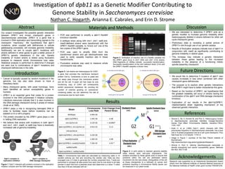
Figure 1: Dpb11 interacts with proteins involved in the replication
initiation complex and the DNA damage checkpoint.
Research was supported by an Institutional Development Award
(IDeA) from the National Institute of General Medical Sciences of
the National Institutes of Health under grant number P20GM1234.
• We are interested to determine if DPB11 acts as a
genetic modifier to increase genome instability when
heterozygously deleted in strains deficient for other cell
cycle checkpoint genes.
• Preliminary data is revealing a partial picture of
DPB11’s role through use of our genetic assays.
• Results of fluctuation analysis indicate loss of dpb11 in
conjunction with mad2 as significantly contributing to
genome instability in diploid yeast.
• This increase may indicate a genetic interaction
between these genes leading to the increased
instability in the absence of a functioning mitotic
spindle assembly checkpoint.
Strain
Chromosome
Loss Rate
Fold Change Over
Parent Strain
Wildtype 2.99E-06 -
dpb11Δ/DPB11 1.44E-06 0.48
mrc1Δ/mrc1Δ 3.12E-02 > 10,000*
dpb11Δ/DPB11; mrc1Δ/mrc1Δ 3.91E-03 0.13
chk1Δ/chk1Δ - -
dpb11Δ/DPB11; chk1Δ/chk1Δ 3.23E-06 -
mad2Δ/mad2Δ 1.49E-05 4.98
dpb11Δ/DPB11; mad2Δ/mad2Δ 7.34E-05 4.93 *
rad9Δ/rad9Δ - -
dpb11Δ/DPB11; rad9Δ/rad9Δ - - Figure 4: A cell’s ability to maintain genome stability
relies heavily upon cell-cycle checkpoints. The
proteins encoded by these genes work to ensure that
problems within the cell are addressed before
movement into the next phase. Deregulation of the cell
at these checkpoints is a hallmark of cancer and
problems resulting in chromosome fidelity issues can
be measured in our yeast model.
Table 1: Chromosome loss rates are calculated by measuring rates in 15
parallel cultures per trial and recording the median rate. Data are then
averaged over 2-3 trials to generate the number presented here. T-tests are
performed to compare all data points from each trial for one strain to all data
pointes in each trial from a parental strain and determine a p value. P-value
less than 0.05 are indicated with an *.