Atherosclerosis: New Insights on Foamy Macrophage Induction and PersistenceDr. Marian Laderoute
It is commonly assumed high cholesterol contributes to atherosclerosis which initiates with foamy macrophages. Here it is suggested the induction of foam in human macrophages relates instead to the induction of a newly described foamy virus, human endogenous retrovirus K102 (HERV-K102) in response to high cortisol and/or viruses, other intracellular pathogens, or tumors. HERV-K102 is a newly described human host protection mechanism and the particles are replication competent in vitro and in vivo. In healthy or younger adults, the foamy macrophages lyse on day 7 and release the protective HERV-K102 particles. When immunosenescence is evident (associated with low DHEA and high cortisol) the lysis and release of the HERV-K102 particles is blocked by alpha-fetoprotein (AFP). The dysfunctional and partly activated macrophages promote immunosenescence including atherosclerosis. See Laderoute MP, Discovery Medicine article published December 2015 for more details.
Approach for limited cell ChIP-Seq on a semiconductor-based sequencing platformThermo Fisher Scientific
Dendritic cell (DC) lineages coordinate immune system activity
through functional specialization.
• Irf4, a transcription factor(TF), is required for CD11b+ DC
lineage development from bone marrow stem cells and has
been implicated in multiple inflammatory diseases, eg. asthma.
• The epigenetic consequences of immune specialization in
CD11b+ DCs and relation to inflammatory diseases remain
largely unexplored partly due to the difficulty of using highly
purified, and typically, limited populations of cells in ChIP-seq
(chromatin immunoprecipitation then sequencing) assays.
• A robust, multiplexed ChIP-seq protocol – using an input
control, TF (CTCF) and histone modification marks (H3K9me3-
methylation, H3K27ac-acetylation) - was developed using
limited amounts of K562 cells, for the Ion ProtonTM system.
• Peak-calling analysis was performed using using MACS2.
• Significant data correlations were observed with ENCODE.
• The Ion ProtonTM results are based on chromatin derived from
1 million(M) cells, making it viable for generating data from a
limited number of primary cells. This is in contrast to the 10M
cells recommended by ENCODE.
• The developed methodology was used to compare Irf4 genomic
binding sites generated from flow-sorted populations of 1, 3, 5,
and 20M CD11b+ lineage murine DCs.
• Comparable Irf4 ChIP-seq results were obtained from 5M
versus 20M cells, indicating that as low as 5M flow-sorted cells
can be used to acquire high quality(FDR: 10-19) data.
• We identified genomic Irf4 binding sites proximal to genes,
whose activity is consistent with CD11b+ DC lineage activity
and/or known to contribute to inflammatory disease.
• We examined Irf4 functional regulation of the identified gene
targets via RNA-seq analysis with CD11b+ DCs and a related
lineage, CD103+ DCs. Integrating expression analysis with
ChIP-seq indicates a unique CD11b+ DC gene expression
program concordant with Irf4 loci association in comparison to
CD103+ DC (data not shown).
Cellular responses to ErbB-2 overexpression in human mammary luminal epitheli...RifathFarook
A review presentation of a paper published in British Journal of Cancer titled Cellular responses to ErbB-2 overexpression in human mammary luminal epithelial cells: comparison of mRNA and protein expression.
Current and Novel Immuno-Oncology Drug Evaluation Methods via Humanized Mouse...InsideScientific
Dr. Bin Xie discusses the current immuno-oncology drug development landscape, different humanized mouse models available for drug testing, and the investigation of potential mechanisms via imaging mass cytometry.
Since the first immune checkpoint blocker ipilimumab was approved by the US FDA in 2011, more drug companies have sought to develop their own immune therapy drugs. Humanized peripheral blood mononuclear cell (PBMC) reconstitution in immune deficient mice is becoming a valuable model for evaluating therapeutic antibodies, especially bispecific antibodies (BsAbs), which can mediate immune cells as well as target a tumor antigen.
However, this model has several drawbacks, including a limited dosing window due to graft-versus-host-disease and insufficient natural immune cell infiltration. This has hindered wide application of the model in the development of multiple immune checkpoint inhibitors or immune agonists.
To overcome these issues, LIDE has developed a unique human PBMC/cancer cell co-transfer model which can generate three-dimensional huPBMC-infiltrated tumor tissue for immunotherapy. This model has successfully been used to evaluate the biological function of several signaling proteins and biomarkers in multiple cancers, such as melanoma, breast cancer, and lung cancer.
In this webinar, Dr. Bin Xie discusses the current immuno-oncology drug development landscape, different humanized models available for drug testing, evaluates real-world case studies, and describes the investigation of potential mechanisms by imaging mass cytometry.
Key Topics Include:
- Introduction to immuno-oncology drug development and the importance of using humanized mouse models to address scientific questions
- Evaluation of current IO platforms and new methods from LIDE, including analysis of several case studies
- Understanding the spatiotemporal interaction between tissue-infiltrating immune cells and cancer cells via imaging mass cytometry
This ppt file represents a simple overview on what is antibody validation & how to validate an antibody before performing any research.
Used references are also included.
Western Blot Assessment of Polyclonal Anti-Host Cell Protein Antibody ProductionCovance
BEBPA HCP 2019 -- The most critical part of developing an ELISA for measuring Host Cell Protein (HCP) impurities in Biotherapeutics is generation of specific antibodies with appropriate recognition of the total population of HCPs potentially present in the product. The generation of the antiserum is dependent upon the sum of the individual biological responses (i.e., antibodies) of multiple animals to the antigen. Response is monitored throughout the program qualitatively by 1D (one dimension) Western Blot and adjustments are made to the antigen (immunogen) as needed to yield the broadest antigen recognition. Ultimately, the highest quality antisera from multiple animals are pooled. The quality of the final product is demonstrated through evaluation of coverage which is typically by 2D (two dimension) Western Blot or more recently by mass spectrometry. The BioCMC Group at Covance was tasked with development of a process capable of supporting this type of assessment. This capability will be illustrated through discussion of a typical antibody reagent production program.
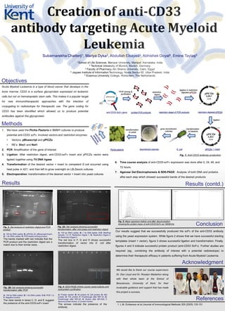
1. Creation of anti-CD33
antibody targeting Acute Myeloid
Leukemia
Subarnarekha Chatterji1, Mariya Dyka2, Abdullah Elsayed3, Abhishek Goyal4, Emine Taytaş5
1School of Life Sciences, Manipal University, Manipal, Karnataka, India
2 Technical University of Munich, Munich, Germany
3 Faculty of Pharmacy, Ain Shams University, Cairo, Egypt
4 Jaypee Institute of Information Technology, Noida Sector 62, Uttar Pradesh, India
5 Erasmus University College, Rotterdam, The Netherlands
Objectives
Methods
Results Results (contd.)
Conclusion
Acute Myeloid Leukemia is a type of blood cancer that develops in the
bone marrow. CD33 is a surface glycoprotein expressed on leukemic
cells but not on hematopoietic stem cells. This makes it a popular target
for new immunotherapeutic approaches with the intention of
conjugating to radioisotope for therapeutic use. The gene coding for
CD33 has been identified which allowed us to produce potential
antibodies against this glycoprotein.
1. We have used the Pichia Pastoris in BMMY cultures to produce
potential anti-CD33 scFv. Involved vectors and restriction enzymes:
• Vectors: pBluescript and pPICZα
• RE’s: Xho1 and Not1
2. PCR: Amplification of the gene of interest
3. Ligation: After restriction digest, anti-CD33-scFv insert and pPICZα vector were
ligated together using T4 DNA ligase
4. Transformation of the desired vector + insert to competent E.coli occurred using
heat pulse in 42℃ and then left to grow overnight on LB-Zeocin cultures
5. Electroporation: transformation of the desired vector + insert into yeast cultures
A: 100 bp DNA Ladder; B: 1 kb DNA ladder; C-E: MiniPrep
Sample 1-3; F: Restriction Digest 1; G: Restriction Digest 2;
H: Restriction Digest 3
The red box in F, G and H shows successful
transformation of vector into E. coli after
restriction digest.
A: 100 bp DNA Ladder; B: pPICZα; C: pBluescript+insert;
D: 1 kb DNA Ladder; E: PCR based cloning product
The bands marked with red indicate that the
PCR product and the restriction digest are a
match due to their similar sizes.
A: 100 bp DNA Ladder; B: 1 kb DNA Ladder; C-E: PCR 1-3;
F: Negative Control.
The clear bands in lanes C, D, and E suggest
the presence of the anti-CD33-scFv insert
Fig. 1: Anti-CD33 antibody production
Fig. 2: Gel analysis of restriction digest and PCR
product
Fig. 3A: Gel analysis showing successful
transformation after mini-preps and restriction digest
Fig. 3B: Gel analysis showing successful
transformation after PCR
6. Time course analysis of anti-CD33-scFv expression was done after 0, 24, 48, and
72 hours.
7. Agarose Gel Electrophoresis & SDS-PAGE: Analysis of both DNA and proteins
after each step which showed successful bands of the desired products
Acknowledgment
A: Protein marker; B: 0h protein; C: 24h protein; D: 48h
protein; E: 72h protein; F: Flowthrough after NPI-10; G:
Flowthrough after NPI-20; H: Elution after NPI-500; I:
Second protein elution; J: protein marker.
The arrows indicate the presence of the
antibody
Fig. 4: SDS-PAGE of time course yeast cultures and
post-protein purification
Our results suggest that we successfully produced the scFv of the anti-CD33 antibody
using the yeast expression system. While figure 2 shows that we have successful starting
templates (insert + vector), figure 3 shows successful ligation and transformation. Finally,
figures 4 and 5 indicate successful protein product (anti-CD33 ScFv). Further studies are
required (eg., combining the antibody of interest with a potential radioisotope) to
determine their therapeutic efficacy in patients suffering from Acute Myeloid Leukemia.
References
We would like to thank our course supervisors
Dr. Dan Lloyd and Dr. Rosalyn Masterton along
with their whole team at the School of
Biosciences, University of Kent, for their
invaluable guidance and support that has made
this project a success.
1. L.M. Emberson et al./Journal of Immunological Methods 305 (2005) 135-151
1000 bp
750 bp
Fig. 5: Mass spectrum before and after deconvolution
showing the protein mass of anti-CD33 ScFv as 19048 Da