Recommended

Recommended
More Related Content
What's hot
What's hot (20)
Similar to Next-Generation Sequencing Detects More BCR-ABL1 Mutations Associated with TKI Resistance
Similar to Next-Generation Sequencing Detects More BCR-ABL1 Mutations Associated with TKI Resistance (20)
Next-Generation Sequencing Detects More BCR-ABL1 Mutations Associated with TKI Resistance
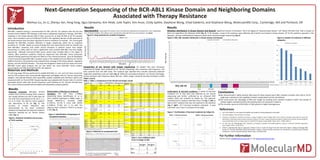
- 1. Next-Generation Sequencing of the BCR-ABL1 Kinase Domain and Neighboring Domains Associated with Therapy Resistance WeiHua Liu, Jin Li, Zhenyu Yan, Peng Fang, Agus Darwanto, Kim Pelak, Julie Toplin, Kim Anoe, Cindy Spittle, Stephane Wong, Chad Galderisi, and Stephane Wong, MolecularMD Corp., Cambridge, MA and Portland, OR Introduction BCR-ABL1 mutation testing is recommended for CML and Ph+ ALL patients who fail first line tyrosine kinase inhibitor (TKI) therapy or who have a suboptimal response to therapy. BCR-ABL1 mutations in the kinase domain (KD) of ABL1 account for at least 40-50% of all TKI resistant cases. Rare mutations such as E123Q and T212R in the regulatory domain of ABL upstream of the kinase domain have also been reported to lead to resistance to imatinib. The current gold standard for BCR-ABL1 mutation detection is Sanger sequencing, which has an analytical sensitivity of ~10-30%. Based on recent findings that mass spectrometry (ref) can identify low level BCR-ABL1 mutations that confer clinical resistance in patients sooner than Sanger sequencing, it is likely useful to have a significantly more sensitive BCR-ABL1 test than Sanger sequencing. Although commercial NGS cancer panels have included ABL1 in the region of interest, ABL1 resistance mutations should be sequenced from BCR-ABL1 fusion transcripts instead of being sequenced from genomic DNA as in the commercial panels. Here we developed a fusion transcript based BCR-ABL1 mutation assay on the scalable and cost-effective Ion Torrent platform that has 1-5% sensitivity and comprehensive coverage of the kinase domain, regulatory domain, and the SH2/SH3 domains. The assay was designed to detect both the major and minor BCR-ABL1 fusion gene products and can also detect the micro BCR-ABL1 fusion product accounting for over 99% of all CML and Ph+ ALL patients. Materials and Methods RT and long range PCR was performed to amplify BCR-ABL1 e1, e13, and e14 fusion transcripts and the PCR products were enzymatically fragmented and ligated with Ion Torrent sequencing adaptors. Size-selected libraries were quantified, pooled, amplified with OneTouch system and sequenced with Ion Torrent PGM. Sequencing data was analyzed with Torrent Suite 2.2 and the associated variant caller with variant frequency cutoff adjusted to 1%. Variants derived from Ts2.2 were further processed and annotated with proprietary analysis pipeline. Results Analytical sensitivity: BCR-ABL1 RT-PCR product from cell line carrying T315I mutation was serially diluted into that of wild type cell line with final concentration of 20, 10, 5, 2, 1, 0.5 % of T315I. The LOD for T315I mutation was determined to be 1% (Fig. 1). The accuracy of mutant frequency was demonstrated by the linear correlation between the expected and observed mutation frequency reported by Ion Torrent Variant caller (Fig. 1). y = 0.8858x + 0.1254 R² = 0.9998 0 2 4 6 8 10 12 14 16 18 20 22 0 2 4 6 8 10 12 14 16 18 20 22 ExpectedMutFreq(%) Mut Freq by Ion Torrent (%) Results Reproducibility: Long-range PCR products were enzymatically fragmented and sequenced in two independent runs. Among 21 samples, 39 mutations were reproducibly called with Mut Freq close between runs (Fig.3). Figure 3. Assay precision for mutation frequency Figure1. Analytical sensitivity and accuracy of T315I mutation 0 5 10 15 20 25 30 35 40 45 50 55 60 65 70 75 80 85 90 95 100 105 Mean CV MutFreq(%) Mutation Comparison of Ion Torrent with Sanger sequencing: 36 samples that were previously sequenced by Sanger were analyzed by Ion Torrent sequencing. Only mutations with frequency >1% were reported from this NGS study. The mutation type reported here includes commonly observed single base substitution and rare indel (Fig. 4). While the concordance between Ion Torrent and Sanger among mutations with frequency above 10% was 100%, Sanger missed 25 low-level mutations (<10%) (Table 1 and 2). Table 1. Table 1. Comparisons of Ion Torrent and Sanger results Results Mutation distribution in kinase domain and beyond: Significant fraction of mutations fall in the regions of “beyond kinase domain” and “below 10% Mut Freq” with a number of them not reported in the literature (ref3) (Fig. 5). In 36 samples, a total of 64 mutations were detected, 60 of which are located in kinase domain. Of 53 KD mutations reported in the literature, 18 have <10% Mutation Frequency which were missed by Sanger sequencing (Fig. 6). Figure 5. BCR_ABL mutation distribution in kinase domain and beyond MutFreq(%) Determination of phasing of compound mutations: The 520 flows (~200bp) sequencing allows identification of cis or trans mutation haplotype in 200bp range. In sample MMD-b2_6, mutation L248V (c.742C>G, 35.1%) is trans with G250E (c.749G>A, 47.3%) but is cis with the adjacent silent mutation c.741G>A as shown in IGV (Fig. 2). Figure 2. identification of haplotypes of compound mutations trans cis Figure 6. Number of mutations in different categories. Confirmation of low-level mutations: A subset of low-level mutations (n= 5) that were only detected by Ion Torrent sequencing was further confirmed by an enhanced RFLP method (ref4) and analyzed by Caliper Gx. This approach confirmed F317L mutation with Mut Freq 1.7% (Fig. 7, left) as well as K357 mutation that was not reported in the Literature (Fig. 7, right). Of 5 low-level mutations evaluated , 4 were confirmed and 1 was in conclusive at this stage. Figure 7. Confirmation of low-level mutations by Caliper Gx. WT digested WT digested F317L 1.7% by Ion Torrent MT digested MT digested K357E 7.6% by Ion Torrent Conclusions We demonstrated a highly sensitive NGS assay for deep sequencing of ABL1 resistant mutation with LOD at 1% for single base substitution and capability to detect complicated insertion and deletion. The study proves the advantage of NGS over Sanger by showing more resistant mutations within and outside of hotspot regions and determination the phasing status of compound mutations. The mutation spectrum of BCR-ABL1 of CML patients is highly heterogeneous. References 1. Life Technologies; Ion Ion Xpress Plus gDNA and Amplicon Library preparation user guide. 2. Life Technologies; Torrent Suite 2.2 manual 3. Soverini S, Hochhaus A, Nicolini FE, Gruber F, Lange T, Saglio G, Pane F, Müller MC, Ernst T, Rosti G, Porkka K, Baccarani M, Cross NC, Martinelli G. BCR-ABL kinase domain mutation analysis in chronic myeloid leukemia patients treated with tyrosine kinase inhibitors: recommendations from an expert panel on behalf of European LeukemiaNet. Blood. 2011; 118(5):1208-15 4. Liu WH, Kaur M, Makrigiorgos GM. Detection of hotspot mutations and polymorphisms using an enhanced PCR-RFLP approach. Hum. Mutat. 2003; 21(5):535-41 5. O'Hare T, Zabriskie MS, Eide CA, Agarwal A, Adrian LT, You H, Corbin AS, Yang F, Press RD, Rivera VM, Toplin J, Wang S, Deininger MW, Druker BJ. The BCR-ABL35INS insertion/truncation mutant is kinase-inactive and does not contribute to tyrosine kinase inhibitor resistance in chronic myeloid leukemia. Blood. 2011; 118:5250-5254 For Further Information Please contact info@molecularmd.com or visit www.molecularmd.com. Sanger- Sanger+ Ion- 7 0 Ion+ 3 26 Table 2. Cross-validation table of Ion vs Sanger (n=36) Sample b2_8 Figure 4. Detection of complicated indel Sample Name cds AA change NGS result (var freq) Sanger result (var freq) MMD-1 NA NA NA NA MMD-2 c.1348G>A E450K 96.80% 100% c.1423_1424insA CTTTGATAACCGT GAAGAAAGAACA AGATAGAAG Ins 475* 1.60% <20% MMD-3 c.763G>A E255K 97.40% 100% c.1069A>G K357E 7.60% NA c.1155C>T S385S 1.10% NA MMD-4 c.944C>T T315I 100% 100% MMD-5 c.944C>T T315I 32.00% 40% MMD-6 NA NA NA NA MMD-7 c.944C>T T315I 100% 100% MMD-8 c.1423_1424insA CTTTGATAACCGT GAAGAAAGAACA AGATAGAAG Ins 475* 2.80% Ins 475 detected MMD-9 c.749G>A G250E 79% 90% c.764A>T E255V 12.60% 10% c.1076T>G F359C 3.40% NA c.1423_1424insA CTTTGATAACCGT GAAGAAAGAACA AGATAGAAG Ins 475* 2.50% NA MMD-10 c.764A>T E255V 92% 100% c.951C>A F317L 1.70% NA c.1423_1424insA CTTTGATAACCGT GAAGAAAGAACA AGATAGAAG Ins 475* 2.10% NA MMD-11 NA NA NA NA MMD-15 NA NA NA NA MMD-16 c.1138G>A A380T 1.89% NA MMD-17 c.1577C>T P526L 8.40% NA MMD-18 NA NA NA NA MMD-19 c.944C>T T315I 100% 100% MMD-20 c.944C>T T315I 32% 40% Sample Name cds AA change NGS result (var freq) Sanger result (var freq) MMD-b2_1 c.763G>A E255K 21.09% 10% MMD-b2_2 c.756G>T Q252H 1.03% NA c.764A>T E255V 98.92% 100% c.944C>T T315I 21.02% 30% c.951C>A F317L 6.92% NA c.949T>C F317L 2% NA c.895G>T V299L 2.90% NA c.895G>C V299L 1.30% NA MMD-b2_3 c.835G>A E279K 18.60% 10% c.951C>G F317L 99.96% 100% c.1187A>G H396R 78.85% 80% c.841G>A E281K 1.20% NA MMD-b2_4 c.951C>G F317L 21.50% 20% MMD-b2_5 c.749G>A G250E 28.49% 20% c.1048G>T A350S 1.06% NA MMD-b2_6 c.742C>G L248V 35.95% 40% c.749G>A G250E 47.02% 50% c.944C>T T315I 14.31% 20% c.1052T>C M351T 4.23% NA MMD-b2_7 c.944C>T T315I 17.55% 20% MMD-b2_8 c.879- 881delinsGGGCG CGGGGGGCGG I293MGAGG;K294 G ~100% 100% c.944C>T T315I 99.97% 100% MMD-b2_9 c.951C>A F317L 94.99% 100% c.1075T>G F359V 3.47% NA c.1349A>G E450G 94.89% 90% MMD-b2_10 c.951C>G F317L 99.95% 100% c.1375G>A E459K 98.02% 100% MMD-b2_11 c.944C>T T315I 23.27% 40% c.951C>A F317L 69.36% 80% c.1076T>G F359C 97.77% 100% MMD-b2_12 c.740A>G K247R 99.82% 100% c.951C>G F317L 84.48% 80% MMD-b2_13 c.472C>T P158S 3.73% NA c.487A>G I163V 2.15% NA c.778G>T V260L 3.79% NA c.835G>T E279* 4.34% NA c.1245G>T K415N 3.05% NA MMD-b2_14 c.951C>G F317L 5.20% NA MMD-b2_15 NA NA NA NA MMD-b2_16 c.514T>C Y172H 1% NA c.1190C>T A397V 2.03% NA c.1350G>T E450D 1.93% NA del c.879-881 CAA insertion GGGCGCGGGGGGCGG c.944C>T AA change: I293MGAGG; K294G; T315I P loop SH3 contact SH2 contact A loop BCR_ABL KD 0 5 10 15 20 25 30 35 40 45 50 55 60 65 70 75 80 85 90 95 100 reported in literature not reported in literature all mutations Mutations in KD Mutations in literature Mut Freq <30% Mut Freq <20% Mut Freq <10% Mut Freq<5% Number of mutations 64 60 53 27 23 18 13 0 10 20 30 40 50 60 70 Number of mutations Data is not shown for MMD-12, 13 and 14 which are duplicate of MMD-18, 19 and 20 *This is a kinase-inactive mutation (see ref5).