1. Couzens 1
Chandra Couzens
AIP 197, F15
December 3, 2015
Rho A and the GEF-H1 Sec5 Pathway: cell motility implications in the metastasis of
human cancer.
The cytoskeleton is a key feature in the development, movement, and structure of
eukaryotic cells. Specifically, the cytoskeleton’s functions are threefold: to organize the
vesicles and organelles within the cell, to serve as a conduit between the cell and its
physical and chemical surroundings, and to allow the cell to move and reshape itself and
participate in basic cellular process (6). To understand the mechanisms of these
processes, it is necessary to understand the composition and function of the cytoskeleton.
The cytoskeleton is made up of actin filaments, microtubules, and intermediate filaments
(6). Polymerization and depolymerization of both actin and microtubules factor into
changes in cell shape, (6). Actin fibers elongate and are vital to cell movement, guiding
the cell along stress fibers powered by myosin, a sort of molecular motor. Intermediate
filaments are the remaining fibers that interact with the other two structures and are also
key in the restructuring of the cytoskeleton. Understanding the mechanisms of
cytoskeletal remodeling is vital in understanding cell movement, which in turn is key to
understanding basic cell process, particularly exocytosis, which shall be the focus of this
paper.
Cytoskeletal signaling mechanisms are particularly important due to their
implications for cell motility and the effects this has on mutant cells, such as in the case
of cancer. In particular, cytoskeletal remodeling of the exocyst, a protein complex located
near the cell membrane, is vital for driving cell movement and spread (6). The primary
2. Couzens 2
function of the exocyst has traditionally been seen in exocytosis, in which vesicle
contents are secreted out of the cell, a necessity for the maintenance of the cell. The
exocyst complex has also been found to have important implications for cell migration
through various other mechanisms outside of its primary function of exocytosis,
particularly regarding tumor cells and the metastasis of cancer (6).
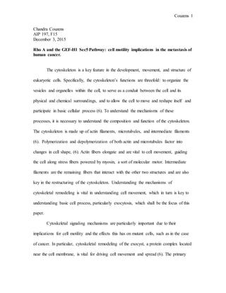
Figure 1: Diagram of
the exocyst complex and
its associated proteins in
the cell membrane (6).
The exocyst has a variety of surface properties that allow it to interact with
various other proteins, including Rho GTPases, which will be discussed in detail later. In
exocytosis, the exocyst takes part in vesicle trafficking, playing a key role in delivery of
substances necessary to membrane growth through polarization due to its position in the
plasma membrane (3). Current studies have focused on understanding the structure,
assembly and disassembly of the exocyst in order to better understand the mechanism of
exocystic pathways and their significance.
While its main function is seen as integrating information from different
molecules in order to regulate and ensure accurate spatial and temporal regulation of
exocytosis in cells, the exocyst also has implications in directional cell migration (6).
3. Couzens 3
Previous studies have focused on invadopodia, actin-rich protrusions that tumor cells
form, allowing them to invade into tissues (6). Overexpression of Exo70, an exocyst
protein, was found to cause more invasions, while knockdown was shown to suppress
tumor formation in a 2008 study (6), implying that the exocysts plays a key role in
signaling mechanisms that govern the metastasis of cancer cells.
Rho GTPases and the exocyst
In particular, the exocyst interacts with various different proteins within the
cytoskeleton that lead to cytoskeletal remodeling and thus migration and division of cells.
A major group of proteins that interacts with the exocyst are the Rho GTPases, which
regulate actomyosin structure and control movement (5). Rho GTPases like RhoA, Rac1,
and Cdc 42 have been shown to be key molecules involved with vesicle trafficking in
exocytic and endocytic pathways (7). GTPases also have associated regulatory proteins
like guanine nucleotide exchange factors (GEFs), GTPase activating proteins (GAPs),
and guanine nucleotide disassociation inhibitors (5). These proteins have been found to
have important implications in gene transcription, cell signaling and migration, and cell
adhesion, and cell cycle progression (5). A particular pathway of interest that has been
found as a key factor in driving migration is the RhoA pathway. Various studies have
studied RhoA and other similar exocyst proteins and their effects on cell motility and
exocyst function, whereas others have focused specifically on the effects of RhoA and
other pathways on cancer cell motility. As a type of GTPase, RhoA exchanges inactive
form GDP to GTP, as promoted by the protein GEF-H1, a microtubule associated Rho A
activator (7). These GTPases interact with various other exocyst proteins to perform a
4. Couzens 4
variety of different functions, especially with regards to regulating the actin cytoskeleton
to respond to stimuli (7).
Figure 2:Image of the GEF-H1 RhoA pathway and various other proteins within the cell (8).
The RhoA/ GEF-H1/Sec5 pathway
As shown in the figure above, the RhoA, GEF-H1, and Sec5 pathway regulates
the coordination of actin and microtubule cytoskeleton modulation, as well as vesicle
trafficking in migration and division (1). This pathway has been implicated in many
biological processes, including barrier permeability, cytokinesis, antigen presentation,
dendritic spine morphology, and cell mobility (6).
GEF-H1, one of the major molecules of interest, is also known as the RhoA
activating factor. Studies by Birkenfeld and Nablant have found that depletion of GEF-
H1 leads to a failure of cytokinesis, and defects in focal adhesion formation and actin
movement, reducing cell migration (8). GEF-H1 is part of a family of guanine exchange
factors, which promote the exchange of GDP for GTP and thus activate the GTPases (1).
GEF-H1 works to affect the way cells move by coupling microtubule movement to
5. Couzens 5
contraction of cells by stimulating nucleotide exchange by RhoA (7). GEF-H1 also
interacts with various other exocyst proteins to promote other functions besides the RhoA
pathway interacting with proteins such as Sec8 and Exo70 in the later stages of
membrane trafficking (8). GEF-H1 is an important factor in cell migration and
polarization, due to the importance of Rho GTPases regulating and coordinating cell
protrusion and interaction between microtubules and actin, as shown in the figure below
(1). RNAi depletion experiments have demonstrated that stress fiber formation when
microtubules are depolymerized is mediated by GEF-H1, making them key players in
how cells move (1). Issues with this GEF-H1 interaction have been found to be
associated with many different types of cancers, as shall be discussed later. However, the
primary function of the GEF-H1/RhoA function deals with its interaction with the protein
Sec5.
Figure 3: Illustration of how Rho helps the cell move (1).
GEF-H1 interacts with a protein called HA-Sec5, also shown in the diagram
(hereby referred to merely as Sec5), and this interaction affects the assembly and stability
of the exocyst complex by activating Rho A (1). The interaction between Sec5 and GEF-
H1is dependent on its interaction with RalA (1). These related Ral proteins also play a
6. Couzens 6
role in other RhoGTPase signaling pathways. Sec5 subunits bind to active Ral GTPases,
which regulate assembly of exocyst complex during vesicle tethering (7). The binding
between Sec5 and GEF-H1 has been found to be increased in cells overexpressing wild
type forms of RhoA that are constantly active, making the pathway much more active
and increasing the motility of the cells (7). GEF-H1 interacts directly with Sec5, which
acts as an exocyst intermediate and also allows GEF-H1 to interact with Exo70 in
addition to the GEF-H1/Sec 5/Rho A pathway, both of which have been found to be
crucial for the role of GEF-H1 (1). Sec5 was found to possibly even facilitate extraction
of GEF-H1 from microtubules, a possible mechanism for its interaction with RhoA (8).
Other related proteins, such as Exo70, have been found to also have important
implications. Exo70 guides other exocyst proteins to specific sites on the cell surface, and
also interacts with GEF-H1 (7). In studies, it was found that the loss of GEF-H1 in
knockout cells was found to impair the function of Exo70, suggesting that GEF-H1 is an
important regulator and component of the exocyst complex in other ways than its
interaction with Rho A (8).
Significance of Understanding the RhoA and related pathways
In a 2002 study, the various implications of the GEF-H1 pathway and vesicle
trafficking were determined (8). In this study, GEF-H1 depletion was performed in cells
in order to observe the effect it had on cellular processes. The depletion of GEF-H1, lead
to accumulation of heterogeneous vesicles, as well as defects in exocytosis and
endocytotic recycling (8). Thus it seems that the GEF-H1 Rho signaling pathway
combines cytoskeletal adjustment with vesicle trafficking (8). A proposed mechanism for
this is that RalA delivers GEF-H1 to certain sites where it activates RhoA, and then
7. Couzens 7
RhoA regulates the functions of the exocyst, thus making GEF-H1 vital for the
interaction between microtubules, vesicle trafficking, and actin dynamics in the
cytoskeleton (8). The significance of this pathway is observed directly when malfunctions
occur in these pathways, such as in the case of cancerous cells.
Invasive tumor cells often have uncontrolled spread, loss of cell polarity, different
interactions with surrounding cells, and increased migration abilities, and because of
RhoGTPases’ regulation of these processes, they have become a subject of interest in
understanding how cancer cells spread (4). Many studies have focused on the RhoA and
related Ras pathways as ways of understanding the mechanisms of cancer cell metastasis,
as mutations in these pathways, particularly in the form of overexpression, often lead to
tumor formation in individuals. As inappropriate migration characteristics are
commonplace in cancer cells, cell migration is of particular interest to scientists studying
the RhoA and related pathways, and is studied by observing polarity, actin movement,
microtubules, focal adhesions, and other features that are indicative of movement. Rho
A’s role in cell cycle regulation and cancer cell survival, as well as its crucial role in cell
motility processes and angiogenesis and branching of cells makes it a major contributor
to cancer when mutated (4).Overexpression of RhoA has been implicated in many types
of cancers, including ovarian cancer, lung cancer, progression of breast cancer, and
metastasis of bladder cancer (4).
GEF-H1 has also been found to be activated by mutant p53, a major tumor
suppressor gene observed to be mutated in a large percentage of cancers (1). Furthermore,
some oncogenes isolated with a transformation assay have been found to be activated forms
of Rho GTPase GEFs, suggesting that GEFs are directly involved in cancer (1).
8. Couzens 8
Knockdown of RhoA in both in vivo and in vitro models has found that reduced motility,
invasion, and growth rates of tumors that suggest that it is a viable target for drug
developments (5).
Examples of experimental procedures utilized in exocyst protein research.
In the DerMardirossian lab, various techniques were employed to investigate the
role of different segments of the genes coding for relevant exocyst proteins in different
cancer cell lines. The cell included 231 (breast cancer), U20S (bone osteosarcoma), 293
(human embryonic kidney cells), HT (B cell lymphoma), and HeLa (cervical cancer).
These cells were kept in adherent cell culture and continuously re-passaged to allow for
sufficient nutrients and controlled growth. The 231 cell line was separated into control
and peptide-treated cells, with the peptide being a GST-tagged competing peptide that
blocked the interaction between GEF-H1 and Sec 5. All cells were transfected with
various DNA fragments designed to express certain proteins (or not express them),
including various parts of the GEF-H1 gene, GFP domains, Exo 70, HA-Sec5, and
various other exocyst proteins.
PCR was performed on segments of the GEF-H1 domain, in order to isolate
particular parts of the genome, and these segments were isolated by purifying their place
on an agarose gel. Additional proteins, such as pRex and Exo70, were studied, but these
did not require the use of PCR before ligation into the pDisplay vector due to their larger
sizes. The PCR products and other proteins of interest were ligated into plasmids
(pDisplay and other vectors), which were was amplified collected by first transforming
bacteria with the appropriate DNA, and then isolating transformed colonies to purify the
9. Couzens 9
DNA using mini prep and maxi prep methods. After transfection of the mammalian cells
with the prepared DNA, the cells were mounted on fibronectin cover slips and prepared
for immunofluorescent microscopy with two antibodies. The slides were stained with
antibodies to detect various cytoskeletal and membrane features, including microtubule
protein, Sec8, GEF-H1, actin, and paxillin (for focal adhesions). The slides were then
imaged with an immunofluorescent microscope and images were observed to determine
how cytoskeletal structures were affected by each transformation by comparison with
cells transformed with empty vectors. Similar techniques have also been employed in
laboratories researching similar pathways.
Results of Research and Further Initiatives
The results of this lab and other studies have indicated that the GEF-H1 Sec5
pathway is a viable target for clinical treatments. Possible clinical applications of this
treatment have been looked into and studied in in vivo mice models in order to block
metastasis of cancer in human cells. For instance, overexpression of wild-type RhoA was
found to increase progression of tumors, but expression RhoA in conjunction with RhoB
and RhoC was also found to have tumor suppressor properties (4). Studies have theorized
that multiple targets for the GEF-H1/Sec5/RhoA pathway would be good candidates for
the development of anti-cancer drugs. For instance, the peptide described in the previous
section could potentially have future uses in drug development. GEF-H1 has been
proposed as a possible therapeutic target, as many pharmaceutical companies have begun
research on identifying proteins that inhibit the localization of Rho GTPases, such as
statins and biophosphonates that inhibit the activity of Rho GTPases like RhoA (1).
However, due to the fact that this would also cause issues with normal cells, other
10. Couzens 10
methods of treatment that have been considered are blocking GEF activity, especially
GEF-H1 for diseases that involve malfunctions of Rho A (1).
What also must be considered in the development of drugs for treatment is which
Rho GTPases are active in each type of cancer and if they affect invasion of the cancer
cells or accentuate proliferation of them. Once this is determined, specifically targeted
drugs for each relevant regulatory protein for the GTPase can be developed.
Furthermore, in order to effectively engineer drugs targeting these pathways, a
full understanding of the mechanisms of each pathway is necessary, and much future
research will also likely be devoted to this before proceeding with drug development.
Possible mechanisms of the pathway have been proposed and considered in development
thus far, but an understanding of how disrupting the interaction between RhoA, Sec5, and
GEF-H1 would affect surrounding proteins and all possible implications of how to
appropriately target only cancerous cells (7).
However there are issues in these methods, due to the fact that because of their
small size, the very low GTP concentration in cells, and very low binding affinity of Rho
GTPases for GDP and GTP, it is difficult to target Rho GTPases due to the very few and
small places available to target (5). Some bacterial toxins can inactivate these Rho
GTPases, but since they are non-specific, they cannot be used in clinical applications (5).
More promising targets are targeting the GEF-H1 and other GEFs in order to affect RhoA
or other Rho GTPases (5). One compound that could be used effectively to block the
GEF binding in Rho A is rhosin, which effectively inhibits Rho activation (5). Targeting
GAP proteins, another type of regulator, could also prove promising. Other targets
11. Couzens 11
include blocking effector activation or activity in other GTPases, for instance PAK
family members (5). By blocking downstream regulators of the Rho GTPase gene, drugs
could potentially be more targeted for specific cancerous cells or at least less damaging to
healthy cells. These targets for drug development are still in the early stages of research,
but eventually, drugs that target these cell pathways may become widespread in clinical
treatment of cancer.
Conclusion
Rho GTPases, especially RhoA, are important molecules in driving cell motility
and thus, when mutated, become important drivers of cancer cell invasion. Thus, with
further investigation, they can also become powerful targets for anti-cancer drug
development. By studying how exactly the RhoA and other Rho GTPase pathways work
and considering the implications for cytoskeletal structure, cell motility, and other
cellular processes, better, more effective drugs may be able to be developed to prevent
the metastasis of cancer and the scientific community will gain a better understanding of
how the exocyst and other cellular features work and develop.
12. Couzens 12
References
1) Birkenfeld J, Nalbant P, Yoon, SH and Bokoch, GM. Cellular functions of GEF-
H1, a microtubule-regulated Rho-GEF: is altered GEF-H1 activity a crucial
determinant of disease pathogenesis? Trends in Cell Biology (2008), p210-217.
2) Fletcher DA and Mullins RD. Cell Mechanics and the Cytoskeleton. Nature
(2010) 463, p485-492.
3) Heider MR, Munson M. Exorcising the Exocyst Complex. Traffic (2012), p898-
907
4) Karlsson R, Pedersen E.D, Wang Z, Brakebusch C. Rho GTPase function in
tumorigenesis. Biochimica et Biophysica Acta (2009), p91-98.
5) Lin Y and Zhen Y. Approaches of targeting Rho GTPases in cancer drug
discovery. Expert Opinion on Drug Discovery (2015), 10:9, 991-1010
6) Liu J and Guo W. The exocyst complex in exocytosis and cell migration.
Protoplasma (2012) 249:587-597.
7) Pathak R, DerMardirossian C. GEF-H1 :orchestrating the interplay between
cytoskeleton and vesicle trafficking. Small GTPases (2013), p174-9. PMCID:
PMC3976975
8) Pathak R, Delorme-Walker V, Howell MC, Anselmo AN, White MA, Bokoch,
GM and DerMardirossian C. The Microtubule-associated Rho Activating Factor
GEF-H1 interacts with Exocyst complex to regulate Vesicle Traffic.
Developmental Cell (2012), p397-411.