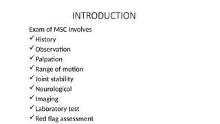
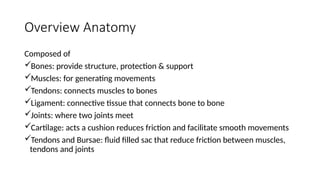
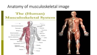
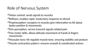
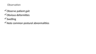
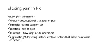
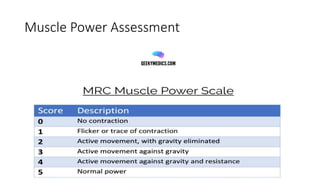
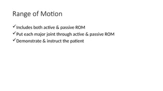
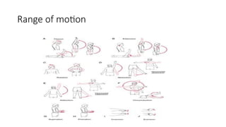
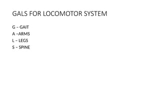
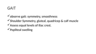
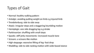
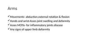
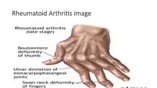
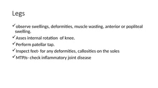
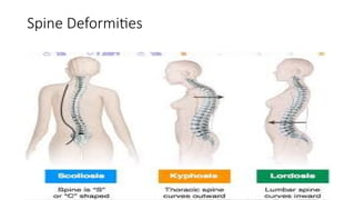
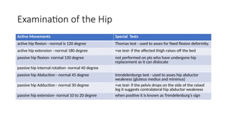
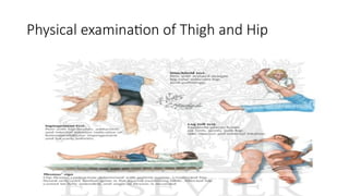
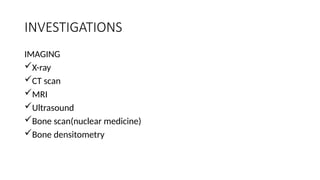
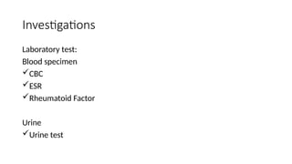
UNIVERSITY OF LUSAKA
PRESENTATION ON:
EXAMINATION OF THE MUSCULOSKELTAL
SYSTEM
By;
KENNEDY CHIBWE MBCHB2012069
SAMUEL CHOMBA SIMUCHENJE MBCH211483635
INTRODUCTION
Exam of MSC involves
๏ƒผHistory
๏ƒผObservation
๏ƒผPalpation
๏ƒผRange of motion
๏ƒผJoint stability
๏ƒผNeurological
๏ƒผImaging
๏ƒผLaboratory test
๏ƒผRed flag assessment
OBJECTIVES
๏ƒผmake an accurate diagnosis
๏ƒผdetermine severity of the condition
๏ƒผmanagement plan
Overview Anatomy
Composed of
๏ƒผBones: provide structure, protection & support
๏ƒผMuscles: for generating movements
๏ƒผTendons: connects muscles to bones
๏ƒผLigament: connective tissue that connects bone to bone
๏ƒผJoints: where two joints meet
๏ƒผCartilage: acts a cushion reduces friction and facilitate smooth movements
๏ƒผTendons and Bursae: fluid filled sac that reduce friction between muscles,
tendons and joints
Anatomy of musculoskeletal image
Role of Nervous System
๏ƒผMotor control: sends signals to muscles
๏ƒผReflexes: enables rapid, involuntary response to stimuli
๏ƒผProprioception: receptors in muscles give information to NS about
bodys position & movements
๏ƒผPain perception: nerves transmit signals related pain
๏ƒผFine motor skills: allows delicate movement of hands & fingers
movements
๏ƒผMuscular tone: NS regulate muscle tone, ensuring stability and posture
๏ƒผMuscle contraction pattern: ensures smooth & coordinated actions
History
๏ƒผSkin rash
๏ƒผH/o alcohol or caffeine use cigarette smoking
๏ƒผFamily H/o of osteoporosis
๏ƒผJoint/bone pains
๏ƒผJoint swellings
๏ƒผYellowing of eyes/palms
Observation
๏ƒผObserve patient gait
๏ƒผObvious deformities
๏ƒผSwelling
๏ƒผNote common postural abnormalities
Palpation
๏ƒผTenderness
๏ƒผEdema
๏ƒผResistance to pressure
๏ƒผDiscomforts during palpation
๏ƒผSwellings in the joint and muscle
๏ƒผMuscle should be firm
Eliciting pain in Hx
WILDA pain assessment
๏ƒผWords - description of character of pain
๏ƒผIntensity - rating scale 0 - 10
๏ƒผLocation - site of pain
๏ƒผDuration โ€“ how long, acute or chronic
๏ƒผAggravating/Alleviating factors- explore factors that make pain worse
or better.
Muscle Power Assessment
Range of Motion
๏ƒผIncludes both active & passive ROM
๏ƒผPut each major joint through active & passive ROM
๏ƒผDemonstrate & instruct the patient
Range of motion
GALS FOR LOCOMOTOR SYSTEM
G โ€“ GAIT
A โ€“ARMS
L โ€“ LEGS
S โ€“ SPINE
GAIT
๏ƒผobserve gait: symmetry, smoothness
๏ƒผShoulder Symmetry, gluteal, quadricep & calf muscle
๏ƒผAssess equal levels of iliac crest.
๏ƒผPopliteal swelling
Types of Gait
โ€ข Normal: healthy walking pattern
โ€ข Antalgic: avoiding putting weight on limb e.g injured limb
โ€ข Trendelenburg: side to side sway
โ€ข Ataxic: irregular steps and a staggering/stumbling motion
โ€ข Hemiplegic: one side dragging leg e.g stroke
โ€ข Parkinsonian: shuffling with small steps
โ€ข Spastic: stiff jerky movements- increased muscle tone
โ€ข Scissors: a scissors-like motion
โ€ข High steppage: excessive lifting of leg- foot drop
โ€ข Waddling: side to side rocking motion with wide-based stance
Arms
๏ƒผMovements- abduction,external rotation & flexion
๏ƒผHands and wrist-Asses joint swelling and deformity
๏ƒผAsses MCPJs- for inflammatory joints disease
๏ƒผAny signs of upper limb deformity
Rheumatoid Arthritis image
Legs
๏ƒผobserve swellings, deformities, muscle wasting, anterior or popliteal
swelling.
๏ƒผAsses internal rotation of knee.
๏ƒผPerform patellar tap.
๏ƒผInspect feet- for any deformities, callosities on the soles
๏ƒผMTPJs- check inflammatory joint disease
Spine
๏ƒผKyphosis
๏ƒผlordosis
๏ƒผscoliosis
๏ƒผAssess posture
Spine Deformities
Examination of the Hip
Active Movements Special Tests
active hip flexion - normal is 120 degree Thomas test - used to asses for fixed flexion deformity.
active hip extension - normal 180 degree +ve test- if the affected thigh raises off the bed
passive hip flexion- normal 120 degree not performed on pts who have undergone hip
replacement as it can dislocate
passive hip internal rotation- normal 40 degree
passive hip Abduction - normal 45 degree trendelenburgs test - used to asses hip abductor
weakness (gluteus medius and minimus)
passive hip Adduction - normal 30 degree +ve test- if the pelvis drops on the side of the raised
leg it suggests contralateral hip abductor weakness
passive hip extension- normal 10 to 20 degree when positive it is known as Trendelenburgโ€™s sign
Physical examination of Thigh and Hip
INVESTIGATIONS
IMAGING
๏ƒผX-ray
๏ƒผCT scan
๏ƒผMRI
๏ƒผUltrasound
๏ƒผBone scan(nuclear medicine)
๏ƒผBone densitometry
Investigations
Laboratory test:
Blood specimen
๏ƒผCBC
๏ƒผESR
๏ƒผRheumatoid Factor
Urine
๏ƒผUrine test
Red Flag Assessment
๏ƒผSevere pain
๏ƒผNight pain
๏ƒผUnexplained weight loss
๏ƒผFever
๏ƒผHx of cancer
๏ƒผTrauma or fall
๏ƒผUnexplained swelling or mass
๏ƒผLoss of joint function
๏ƒผSevere stiffness
REFERENCES
๏ƒผ http://geekymedics.com/muscle-power-assessment-mrc-scale/
visited on 18th September 2023.
๏ƒผHutchisonโ€™s clinical methods of medicine. An Integrated approach to
clinical practice. 23rd
Edition, Michael Glynn and William Drake.
๏ƒผStandring, S (2015), Then Anatomy Basic of Clinical Practice. 41st
ed.
London: Elsevier
THANK YOUโ€ฆ

Final Muscular skeletal EXAM presentation.pptx

  • 1.
    UNIVERSITY OF LUSAKA PRESENTATIONON: EXAMINATION OF THE MUSCULOSKELTAL SYSTEM By; KENNEDY CHIBWE MBCHB2012069 SAMUEL CHOMBA SIMUCHENJE MBCH211483635
  • 2.
    INTRODUCTION Exam of MSCinvolves ๏ƒผHistory ๏ƒผObservation ๏ƒผPalpation ๏ƒผRange of motion ๏ƒผJoint stability ๏ƒผNeurological ๏ƒผImaging ๏ƒผLaboratory test ๏ƒผRed flag assessment
  • 3.
    OBJECTIVES ๏ƒผmake an accuratediagnosis ๏ƒผdetermine severity of the condition ๏ƒผmanagement plan
  • 4.
    Overview Anatomy Composed of ๏ƒผBones:provide structure, protection & support ๏ƒผMuscles: for generating movements ๏ƒผTendons: connects muscles to bones ๏ƒผLigament: connective tissue that connects bone to bone ๏ƒผJoints: where two joints meet ๏ƒผCartilage: acts a cushion reduces friction and facilitate smooth movements ๏ƒผTendons and Bursae: fluid filled sac that reduce friction between muscles, tendons and joints
  • 5.
  • 6.
    Role of NervousSystem ๏ƒผMotor control: sends signals to muscles ๏ƒผReflexes: enables rapid, involuntary response to stimuli ๏ƒผProprioception: receptors in muscles give information to NS about bodys position & movements ๏ƒผPain perception: nerves transmit signals related pain ๏ƒผFine motor skills: allows delicate movement of hands & fingers movements ๏ƒผMuscular tone: NS regulate muscle tone, ensuring stability and posture ๏ƒผMuscle contraction pattern: ensures smooth & coordinated actions
  • 7.
    History ๏ƒผSkin rash ๏ƒผH/o alcoholor caffeine use cigarette smoking ๏ƒผFamily H/o of osteoporosis ๏ƒผJoint/bone pains ๏ƒผJoint swellings ๏ƒผYellowing of eyes/palms
  • 8.
    Observation ๏ƒผObserve patient gait ๏ƒผObviousdeformities ๏ƒผSwelling ๏ƒผNote common postural abnormalities
  • 9.
    Palpation ๏ƒผTenderness ๏ƒผEdema ๏ƒผResistance to pressure ๏ƒผDiscomfortsduring palpation ๏ƒผSwellings in the joint and muscle ๏ƒผMuscle should be firm
  • 10.
    Eliciting pain inHx WILDA pain assessment ๏ƒผWords - description of character of pain ๏ƒผIntensity - rating scale 0 - 10 ๏ƒผLocation - site of pain ๏ƒผDuration โ€“ how long, acute or chronic ๏ƒผAggravating/Alleviating factors- explore factors that make pain worse or better.
  • 11.
  • 12.
    Range of Motion ๏ƒผIncludesboth active & passive ROM ๏ƒผPut each major joint through active & passive ROM ๏ƒผDemonstrate & instruct the patient
  • 13.
  • 14.
    GALS FOR LOCOMOTORSYSTEM G โ€“ GAIT A โ€“ARMS L โ€“ LEGS S โ€“ SPINE
  • 15.
    GAIT ๏ƒผobserve gait: symmetry,smoothness ๏ƒผShoulder Symmetry, gluteal, quadricep & calf muscle ๏ƒผAssess equal levels of iliac crest. ๏ƒผPopliteal swelling
  • 16.
    Types of Gait โ€ขNormal: healthy walking pattern โ€ข Antalgic: avoiding putting weight on limb e.g injured limb โ€ข Trendelenburg: side to side sway โ€ข Ataxic: irregular steps and a staggering/stumbling motion โ€ข Hemiplegic: one side dragging leg e.g stroke โ€ข Parkinsonian: shuffling with small steps โ€ข Spastic: stiff jerky movements- increased muscle tone โ€ข Scissors: a scissors-like motion โ€ข High steppage: excessive lifting of leg- foot drop โ€ข Waddling: side to side rocking motion with wide-based stance
  • 17.
    Arms ๏ƒผMovements- abduction,external rotation& flexion ๏ƒผHands and wrist-Asses joint swelling and deformity ๏ƒผAsses MCPJs- for inflammatory joints disease ๏ƒผAny signs of upper limb deformity
  • 18.
  • 19.
    Legs ๏ƒผobserve swellings, deformities,muscle wasting, anterior or popliteal swelling. ๏ƒผAsses internal rotation of knee. ๏ƒผPerform patellar tap. ๏ƒผInspect feet- for any deformities, callosities on the soles ๏ƒผMTPJs- check inflammatory joint disease
  • 20.
  • 21.
  • 22.
    Examination of theHip Active Movements Special Tests active hip flexion - normal is 120 degree Thomas test - used to asses for fixed flexion deformity. active hip extension - normal 180 degree +ve test- if the affected thigh raises off the bed passive hip flexion- normal 120 degree not performed on pts who have undergone hip replacement as it can dislocate passive hip internal rotation- normal 40 degree passive hip Abduction - normal 45 degree trendelenburgs test - used to asses hip abductor weakness (gluteus medius and minimus) passive hip Adduction - normal 30 degree +ve test- if the pelvis drops on the side of the raised leg it suggests contralateral hip abductor weakness passive hip extension- normal 10 to 20 degree when positive it is known as Trendelenburgโ€™s sign
  • 23.
  • 24.
  • 25.
  • 26.
    Red Flag Assessment ๏ƒผSeverepain ๏ƒผNight pain ๏ƒผUnexplained weight loss ๏ƒผFever ๏ƒผHx of cancer ๏ƒผTrauma or fall ๏ƒผUnexplained swelling or mass ๏ƒผLoss of joint function ๏ƒผSevere stiffness
  • 27.
    REFERENCES ๏ƒผ http://geekymedics.com/muscle-power-assessment-mrc-scale/ visited on18th September 2023. ๏ƒผHutchisonโ€™s clinical methods of medicine. An Integrated approach to clinical practice. 23rd Edition, Michael Glynn and William Drake. ๏ƒผStandring, S (2015), Then Anatomy Basic of Clinical Practice. 41st ed. London: Elsevier
  • 28.