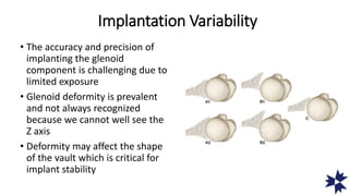
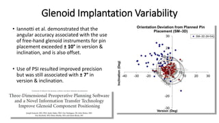
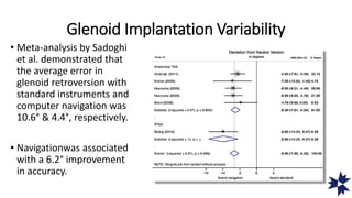
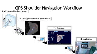
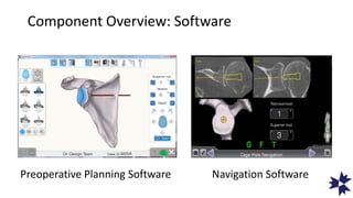
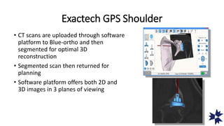
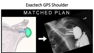
Rationale, Technique and
Tips
Moby Parsons, MD
Disclosure
Consulting and royalties
from EXACTECH© for the EQUINOXE℗ program
Implantation Variability
• The accuracy and precision of
implanting the glenoid
component is challenging due to
limited exposure
• Glenoid deformity is prevalent
and not always recognized
because we cannot well see the
Z axis
• Deformity may affect the shape
of the vault which is critical for
implant stability
Risk of too much glenoid retroversion?
• Studies have demonstrated that
forces in the cement, glenoid
prosthesis, and glenoid bone
increase when the prosthesis is
implanted in too much
retroversion
Increased Retroversion = Increased Glenoid
Micromotion
Increased Retroversion = Increased
Glenohumeral Translations
Reverse Prosthesis Biomechanics
• Position and orientation of the
glenosphere affect
• Impingement free range of motion
• Scapular notching
• Implant stability
Predictors of scapular notching in patients managed
with the Delta III reverse total shoulder replacement
Simovitch et al. JBJS 2007;89:588-600
Effects of glenosphere positioning on impingement-
free internal and external rotation after reverse total
shoulder arthroplasty Li et al. JSES 2013;22:807:813
Anatomic Reconstruction
• More accurate reproduction of
glenohumeral anatomy is the
primary design goal in shoulder
arthroplasty; this has recently
been associated with improved
aTSA clinical outcomes.
• With the technology available today, are our surgical
techniques as good as they can be?
• Have we set the bar too low?
• How often do we get it right?
Glenoid Implantation Variability
• Iannotti et al. demonstrated that the
angular accuracy associated with the use
of free-hand glenoid instruments for pin
placement exceeded ± 10° in version &
inclination, and is also offset.
• Use of PSI resulted improved precision
but was still associated with ± 7° in
version & inclination.
Glenoid Implantation Variability
• Meta-analysis by Sadoghi
et al. demonstrated that
the average error in
glenoid retroversion with
standard instruments and
computer navigation was
10.6° & 4.4°, respectively.
• Navigationwas associated
with a 6.2° improvement
in accuracy.
We don’t even know what we think we know!
Expert shoulder surgeons
freehanding baseplate
placement relative to
preoperative plan compared to
navigation
With the technology available today…..
• Advanced
computing/processing
power
• Ubiquitous chips and
sensors
• Artificial intelligence
• Machine Learning
Rationale for GPS
• Optimal position of implant to ensure best fixation in bone
• Optimal implant selection to restore glenoid version with minimal
glenoid reaming
• Reproducibility of good results particularly in complex cases
GPS Shoulder Navigation Workflow
1. CT data collection (1mm)
2. CT Segmentation  Blue Ortho
3. Planning
4. Navigation
Component Overview: Software
Preoperative Planning Software Navigation Software
Exactech GPS Shoulder
• CT scans are uploaded through software
platform to Blue-ortho and then
segmented for optimal 3D
reconstruction
• Segmented scan then returned for
planning
• Software platform offers both 2D and
3D images in 3 planes of viewing
Pre-operative Planning
• Multiple controls allow precise and
independent adjustment of…
• Depth
• Version
• Inclination
• Rotation
• AP/SI Position
Pre-operative Planning
• High resolution image can be freely rotated to view in any plane
• Both RTSA and ATSA can be planned allowing surgeons to choose
the best implant to deal with glenoid anatomy and any wear
complex wear patterns.
• User-controlled positioning permits selection of the ideal prosthesis
for that patient and enables the surgeon to position it to maximize
implant seating, minimize bone removal, and orient it in the desired
version, inclination, and overhang. And then set plan.
• Visualization is most useful in cases of glenoid deformation where
glenoid version may have gone unnoticed and/or small anatomies
which may have otherwise been non-ideally positioned in the vault
and resulted in cortical perforation.
Pre-operative Planning
• 2D slices can be very useful in
cases of small glenoids or glenoid
wear to determine optimal cage
position to maximize purchase in
vault and minimize cage
perforation.
Plan Transfer
Plan transferred by USB drive to GPS system allowing surgeons
to navigate starting point for drill, orientation of depth of
glenoid reaming, and reverse screw placement
Component Overview: Reusable
ExactechGPS® Station V2
with arm J00020
Camera/Screen
and Bed Mount
Probe and
Trackers
Coracoid
Tracker
Mount
Instrument
Navigation
Component Overview: Disposables
Batteries for Trackers
Drape Pack for Screen
Positioning
• Screen opposite surgeon and
between hip and foot of table so
patient head does not interfere
with line of site between camera
and trackers
Exactech GPS Shoulder
• Compact screen positioned in the sterile
field
• Screen position is adjustable for best
viewing and tracking
• Touch screen allows surgeon to control
execution of plan
Clean the Camera
• Note: the cameras are above
the screen
• Before each case make sure
camera surfaces are clean for
optimal tracking
• Also make sure thumb drive
with plan is in system before
draping
Insert Batteries in each tracker
Note: You can insert batteries whenever you want during instrument
preparation, even before switching on the GPS station.
No battery
inserted
When battery is inserted
the LED becomes
- Orange for 2-3 secs
- then blinks green
Tracker Preparation
“P” as Probe
- To register anatomical data
- To go forward and backward into the protocol
“T” as Tool
Tracker to reference
the drill instrument
“G” as Glenoid
Tracker to reference patient
glenoid/scapula bone
3 Different Trackers
Tracker Calibration
• All three (Probe, Tool, Glenoid)
need to be calibrated to camera
• Tool and Glenoid need to be
calibrated to Probe
Approach
• Do entire humeral preparation
and glenoid exposure first so that
dissection does not disrupt
tracker fixation
• Insert trial stem and protector
plate
Key Areas to Expose for Registration
• Upper and lower surface of coracoid
• Anterior glenoid neck
• Inferior glenoid neck and scapular
neck
• Posterior glenoid lip
• Remove any excess cartilage with a
cobb or ring curette so that stylus
registers boney anatomy
Glenoid Tracker Placement
• Place sharp Hohman behind coracoid
base to fully expose
• Use cautery to remove tissue from
superior surface
• Place tracker center/center
• Sequentially tighten screws
Exactech GPS Shoulder
• Acquisition of anatomic landmarks registers
the CT scan and plan to the patients anatomy.
• Registration is simple & takes ~2 minutes.
Data Point Acquisition
• Use two hands on probe for
optimal accuracy
• Keep probe on bone for
each acquisition step until
sufficient points captures
(beep)
• Do not move anterior
retractor or else risk tracker
loosening
Data Quality Determination
• Green points = good
• Yellow points = fair
• Red points = poor
• Can go back and recapture too
much yellow or red
Exactech GPS Shoulder
• Adjustment of the plan based on intra-op assessment
• Step-by-step guidance through the case, including images
of the appropriate instrument for the step
• Plan is indicated in blue.
• System constantly informs surgeon of the deviation from
plan, the Friedman axis for version, and also inclination.
Exactech GPS Shoulder
Advantage in Challenging Cases
• Case example of baseplate implantation in eroded glenoid:
Advantage in revision cases?
• CT reconstruction, preop plan, & navigation can occur w/ existing implant:
Accuracy & Precision
• Results of 9 cadavers demonstrate that the GPS
computer navigation system is accurate within 2°
version/inclination and 2mm in A/P and S/I
implant placement
Retroversion
(degrees)
Inclination
(degrees)
AP
Placement
(mm)
SI Placement
(mm)
Average (abs) 1.6 1.0 1.0 0.7
Average -0.3 0.5 0.8 0.3
Standard
Deviation
1.88 1.15 1.12 0.86
Effect of GPS on Implant Selection
• Augmented glenoids were selected more frequently: >60%
more for aTSA & >50% more for rTSA procedures
Cohort Standard Instruments Computer
Navigation
ATSA (n=24)
- Augmented <10% 67%
RTSA (n=56)
- 8° Post Aug
- 10° Sup Aug
18%
11%
46%
32%

Exactech gps presentation

  • 1.
  • 3.
    Disclosure Consulting and royalties fromEXACTECH© for the EQUINOXE℗ program
  • 4.
    Implantation Variability • Theaccuracy and precision of implanting the glenoid component is challenging due to limited exposure • Glenoid deformity is prevalent and not always recognized because we cannot well see the Z axis • Deformity may affect the shape of the vault which is critical for implant stability
  • 5.
    Risk of toomuch glenoid retroversion? • Studies have demonstrated that forces in the cement, glenoid prosthesis, and glenoid bone increase when the prosthesis is implanted in too much retroversion
  • 6.
    Increased Retroversion =Increased Glenoid Micromotion
  • 7.
    Increased Retroversion =Increased Glenohumeral Translations
  • 8.
    Reverse Prosthesis Biomechanics •Position and orientation of the glenosphere affect • Impingement free range of motion • Scapular notching • Implant stability Predictors of scapular notching in patients managed with the Delta III reverse total shoulder replacement Simovitch et al. JBJS 2007;89:588-600 Effects of glenosphere positioning on impingement- free internal and external rotation after reverse total shoulder arthroplasty Li et al. JSES 2013;22:807:813
  • 9.
    Anatomic Reconstruction • Moreaccurate reproduction of glenohumeral anatomy is the primary design goal in shoulder arthroplasty; this has recently been associated with improved aTSA clinical outcomes.
  • 10.
    • With thetechnology available today, are our surgical techniques as good as they can be? • Have we set the bar too low? • How often do we get it right?
  • 11.
    Glenoid Implantation Variability •Iannotti et al. demonstrated that the angular accuracy associated with the use of free-hand glenoid instruments for pin placement exceeded ± 10° in version & inclination, and is also offset. • Use of PSI resulted improved precision but was still associated with ± 7° in version & inclination.
  • 12.
    Glenoid Implantation Variability •Meta-analysis by Sadoghi et al. demonstrated that the average error in glenoid retroversion with standard instruments and computer navigation was 10.6° & 4.4°, respectively. • Navigationwas associated with a 6.2° improvement in accuracy.
  • 13.
    We don’t evenknow what we think we know! Expert shoulder surgeons freehanding baseplate placement relative to preoperative plan compared to navigation
  • 14.
    With the technologyavailable today….. • Advanced computing/processing power • Ubiquitous chips and sensors • Artificial intelligence • Machine Learning
  • 15.
    Rationale for GPS •Optimal position of implant to ensure best fixation in bone • Optimal implant selection to restore glenoid version with minimal glenoid reaming • Reproducibility of good results particularly in complex cases
  • 16.
    GPS Shoulder NavigationWorkflow 1. CT data collection (1mm) 2. CT Segmentation  Blue Ortho 3. Planning 4. Navigation
  • 17.
    Component Overview: Software PreoperativePlanning Software Navigation Software
  • 18.
    Exactech GPS Shoulder •CT scans are uploaded through software platform to Blue-ortho and then segmented for optimal 3D reconstruction • Segmented scan then returned for planning • Software platform offers both 2D and 3D images in 3 planes of viewing
  • 19.
    Pre-operative Planning • Multiplecontrols allow precise and independent adjustment of… • Depth • Version • Inclination • Rotation • AP/SI Position
  • 20.
    Pre-operative Planning • Highresolution image can be freely rotated to view in any plane • Both RTSA and ATSA can be planned allowing surgeons to choose the best implant to deal with glenoid anatomy and any wear complex wear patterns. • User-controlled positioning permits selection of the ideal prosthesis for that patient and enables the surgeon to position it to maximize implant seating, minimize bone removal, and orient it in the desired version, inclination, and overhang. And then set plan. • Visualization is most useful in cases of glenoid deformation where glenoid version may have gone unnoticed and/or small anatomies which may have otherwise been non-ideally positioned in the vault and resulted in cortical perforation.
  • 21.
    Pre-operative Planning • 2Dslices can be very useful in cases of small glenoids or glenoid wear to determine optimal cage position to maximize purchase in vault and minimize cage perforation.
  • 22.
    Plan Transfer Plan transferredby USB drive to GPS system allowing surgeons to navigate starting point for drill, orientation of depth of glenoid reaming, and reverse screw placement
  • 23.
    Component Overview: Reusable ExactechGPS®Station V2 with arm J00020 Camera/Screen and Bed Mount Probe and Trackers Coracoid Tracker Mount Instrument Navigation
  • 24.
    Component Overview: Disposables Batteriesfor Trackers Drape Pack for Screen
  • 25.
    Positioning • Screen oppositesurgeon and between hip and foot of table so patient head does not interfere with line of site between camera and trackers
  • 26.
    Exactech GPS Shoulder •Compact screen positioned in the sterile field • Screen position is adjustable for best viewing and tracking • Touch screen allows surgeon to control execution of plan
  • 27.
    Clean the Camera •Note: the cameras are above the screen • Before each case make sure camera surfaces are clean for optimal tracking • Also make sure thumb drive with plan is in system before draping
  • 28.
    Insert Batteries ineach tracker Note: You can insert batteries whenever you want during instrument preparation, even before switching on the GPS station. No battery inserted When battery is inserted the LED becomes - Orange for 2-3 secs - then blinks green Tracker Preparation
  • 29.
    “P” as Probe -To register anatomical data - To go forward and backward into the protocol “T” as Tool Tracker to reference the drill instrument “G” as Glenoid Tracker to reference patient glenoid/scapula bone 3 Different Trackers
  • 30.
    Tracker Calibration • Allthree (Probe, Tool, Glenoid) need to be calibrated to camera • Tool and Glenoid need to be calibrated to Probe
  • 31.
    Approach • Do entirehumeral preparation and glenoid exposure first so that dissection does not disrupt tracker fixation • Insert trial stem and protector plate
  • 32.
    Key Areas toExpose for Registration • Upper and lower surface of coracoid • Anterior glenoid neck • Inferior glenoid neck and scapular neck • Posterior glenoid lip • Remove any excess cartilage with a cobb or ring curette so that stylus registers boney anatomy
  • 33.
    Glenoid Tracker Placement •Place sharp Hohman behind coracoid base to fully expose • Use cautery to remove tissue from superior surface • Place tracker center/center • Sequentially tighten screws
  • 34.
    Exactech GPS Shoulder •Acquisition of anatomic landmarks registers the CT scan and plan to the patients anatomy. • Registration is simple & takes ~2 minutes.
  • 35.
    Data Point Acquisition •Use two hands on probe for optimal accuracy • Keep probe on bone for each acquisition step until sufficient points captures (beep) • Do not move anterior retractor or else risk tracker loosening
  • 36.
    Data Quality Determination •Green points = good • Yellow points = fair • Red points = poor • Can go back and recapture too much yellow or red
  • 37.
    Exactech GPS Shoulder •Adjustment of the plan based on intra-op assessment • Step-by-step guidance through the case, including images of the appropriate instrument for the step • Plan is indicated in blue. • System constantly informs surgeon of the deviation from plan, the Friedman axis for version, and also inclination.
  • 39.
  • 40.
    Advantage in ChallengingCases • Case example of baseplate implantation in eroded glenoid:
  • 41.
    Advantage in revisioncases? • CT reconstruction, preop plan, & navigation can occur w/ existing implant:
  • 42.
    Accuracy & Precision •Results of 9 cadavers demonstrate that the GPS computer navigation system is accurate within 2° version/inclination and 2mm in A/P and S/I implant placement Retroversion (degrees) Inclination (degrees) AP Placement (mm) SI Placement (mm) Average (abs) 1.6 1.0 1.0 0.7 Average -0.3 0.5 0.8 0.3 Standard Deviation 1.88 1.15 1.12 0.86
  • 43.
    Effect of GPSon Implant Selection • Augmented glenoids were selected more frequently: >60% more for aTSA & >50% more for rTSA procedures Cohort Standard Instruments Computer Navigation ATSA (n=24) - Augmented <10% 67% RTSA (n=56) - 8° Post Aug - 10° Sup Aug 18% 11% 46% 32%