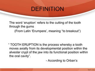
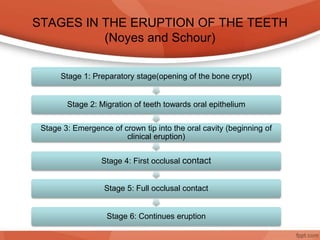
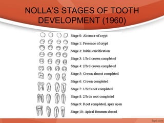
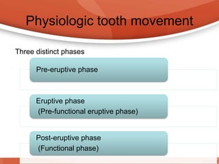
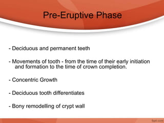
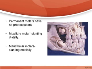
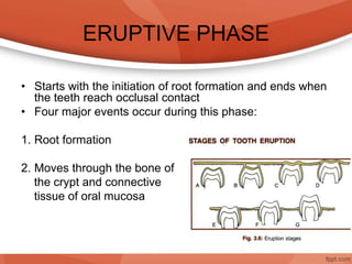
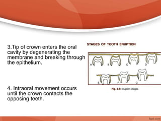
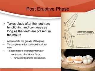
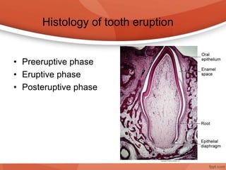
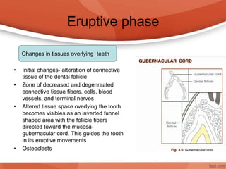
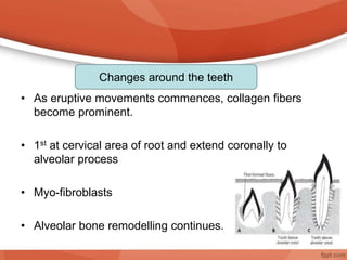
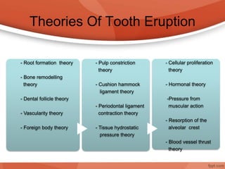
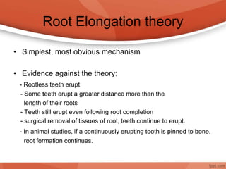
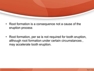
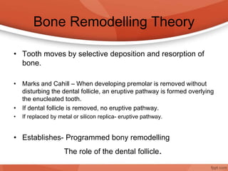
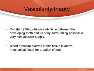
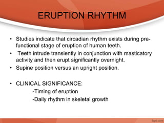
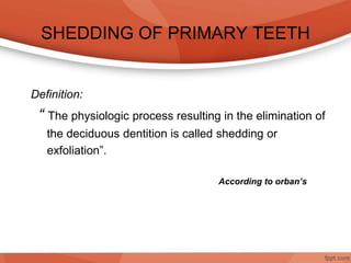
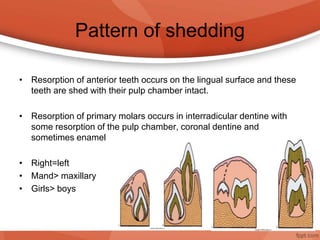
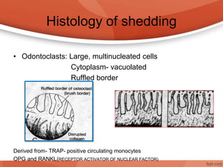
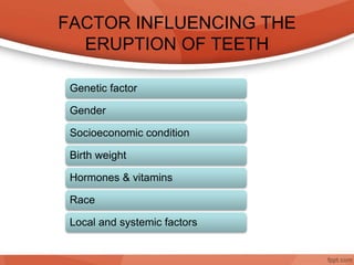
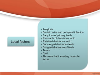
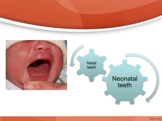
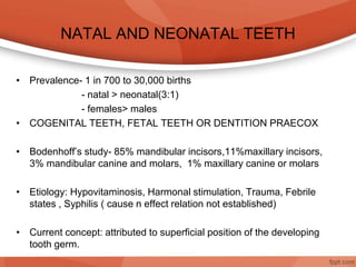
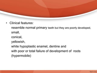
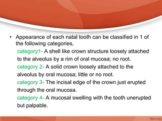
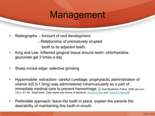
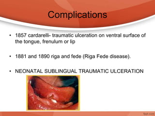
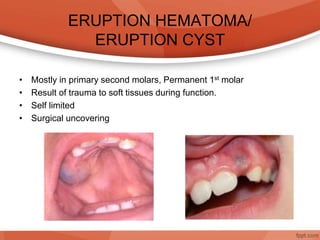
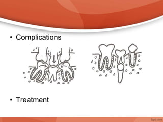
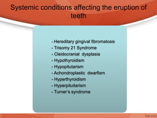
ERUPTION OF TEETH
DEFINITION
The word ‘eruption’ refers to the cutting of the tooth
through the gums
(From Latin ‘Erumpere’, meaning “to breakout”)
“ TOOTH ERUPTION is the process whereby a tooth
moves axially from its developmental position within the
alveolar crypt of the jaw into its functional position within
the oral cavity”.
- According to Orban’s
STAGES IN THE ERUPTION OF THE TEETH
(Noyes and Schour)
Stage 1: Preparatory stage(opening of the bone crypt)
Stage 2: Migration of teeth towards oral epithelium
Stage 3: Emergence of crown tip into the oral cavity (beginning of
clinical eruption)
Stage 4: First occlusal contact
Stage 5: Full occlusal contact
Stage 6: Continues eruption
NOLLA’S STAGES OF TOOTH
DEVELOPMENT (1960)
Physiologic tooth movement
Pre-eruptive phase
Eruptive phase
(Pre-functional eruptive phase)
Post-eruptive phase
(Functional phase)
Three distinct phases
Pre-Eruptive Phase
- Deciduous and permanent teeth
- Movements of tooth - from the time of their early initiation
and formation to the time of crown completion.
- Concentric Growth
- Deciduous tooth differentiates
- Bony remodelling of crypt wall
• Permanent molars have
no predecessors
• Maxillary molar- slanting
distally.
• Mandibular molars-
slanting mesially.
ERUPTIVE PHASE
• Starts with the initiation of root formation and ends when
the teeth reach occlusal contact
• Four major events occur during this phase:
1. Root formation
2. Moves through the bone of
the crypt and connective
tissue of oral mucosa
3.Tip of crown enters the oral
cavity by degenerating the
membrane and breaking through
the epithelium.
4. Intraoral movement occurs
until the crown contacts the
opposing teeth.
Post Eruptive Phase
• Takes place after the teeth are
functioning and continues as
long as the teeth are present in
the mouth
• Accomadate the growth of the jaws
• To compensate for continued occlusal
wear
• To accomadate interproximal wear
- Ant comp of occlusal force.
- Transeptal ligament contraction.
• Preeruptive phase
• Eruptive phase
• Posteruptive phase
Histology of tooth eruption
Eruptive phase
• Initial changes- alteration of connective
tissue of the dental follicle
• Zone of decreased and degenreated
connective tissue fibers, cells, blood
vessels, and terminal nerves
• Altered tissue space overlying the tooth
becomes visibles as an inverted funnel
shaped area with the follicle fibers
directed toward the mucosa-
gubernacular cord. This guides the tooth
in its eruptive movements
• Osteoclasts
Changes in tissues overlying teeth
• As eruptive movements commences, collagen fibers
become prominent.
• 1st at cervical area of root and extend coronally to
alveolar process
• Myo-fibroblasts
• Alveolar bone remodelling continues.
Changes around the teeth
• In soft tissues and fundic bone.
• As the tooth erupts, space is provided for the root to
lengthen,
• Primarily due to the crown moving occlusally and
increase in the height of the alveolar bone.
• Changes in the fundic region are thus, believed to be
largely compensatory to the lengthening of the roots.
Changes in tissues underlying teeth
Post eruptive phase
• Alveolar process increase in height and roots continue to
grow.
• Alveolar bone density increases
• Principle fibers of PDL establishes into separate groups.
• Diameter of fiber bundle increases.
Theories Of Tooth Eruption
- Root formation theory
- Bone remodelling
theory
- Dental follicle theory
- Vascularity theory
- Foreign body theory
- Pulp constriction
theory
- Cushion hammock
ligament theory
- Periodontal ligament
contraction theory
- Tissue hydrostatic
pressure theory
- Cellular proliferation
theory
- Hormonal theory
-Pressure from
muscular action
- Resorption of the
alveolar crest
- Blood vessel thrust
theory
Root Elongation theory
• Simplest, most obvious mechanism
• Evidence against the theory:
- Rootless teeth erupt
- Some teeth erupt a greater distance more than the
length of their roots
- Teeth still erupt even following root completion
- surgical removal of tissues of root, teeth continue to erupt.
- In animal studies, if a continuously erupting tooth is pinned to bone,
root formation continues.
• Root formation is a consequence not a cause of the
eruption process
• Root formation, per se is not required for tooth eruption,
although root formation under certain circumstances ,
may accelerate tooth eruption.
Bone Remodelling Theory
• Tooth moves by selective deposition and resorption of
bone.
• Marks and Cahill – When developing premolar is removed without
disturbing the dental follicle, an eruptive pathway is formed overlying
the enucleated tooth.
• If dental follicle is removed, no eruptive pathway.
• If replaced by metal or silicon replica- eruptive pathway.
• Establishes- Programmed bony remodelling
The role of the dental follicle.
Dental Follicle Theory
• Marks and Cahill (1980, 1984) demonstrated the role of
the dental follicle in tooth eruption with studies on dogs.
• Pattern of cellular activity - REE and the follicles.
• Intercellular signals.
• The REE also releases proteases.
Periodontal Ligament Traction
• Contractile element within the periodontal ligament,
collagen constriction and constriction due to fibroblasts
are responsible.
• Actual force required to move the tooth is linked to the
contractility of fibroblasts.
• Models:
Hydrostatic Pressure Theory
• Increase in hydrostatic pressure in the area around the
developing tooth creates the eruptive force.
• The hydrostatic theory was investigated by Hassel and
McMinn (1972) who demonstrated that the tissue
pressure apically was greater than occlusally
theoretically generating an eruptive force.
• No association was found between the rate of eruption
and the pressure gradient.
Hormonal Theory
• Sir Arthur keith- hormones of thyroids and pituitary
glands might govern the eruption of the teeth
• Doesn't explain the mechanism
• A study by Leache et al (1988), concluded that children
with delayed growth due to growth hormone deficiency
or low genetically determined height had delayed tooth
eruption. However those with delayed growth for other
reasons show normal dental development.
Pressure from muscular action
• Berten - Actions of muscle of cheeks and lips upon
alveolar peocess might serve to squeeze the crown of
the tooth into oral cavity.
• Fails to explain in cases of unilateral paralysis
Blood vessel thrust theory
• Eruption involves the blood supply to the tooth. The
blood generates the force by hydrodynamic and
hydrostatic force within the blood vessels.
Pulp constriction theory
• Growth of the root dentine and the subsequent
constriction of the pulp may cause sufficient pressure to
move the tooth occlusally.
• Neglected
Foreign body theory
• Gottlieb’s- Calcified body such as a tooth tends to be
exfoliated by the tissues just as it does to any foreign
body.
Resorption of alveolar crest
• Resorption of alveolar crest would serve to expose the
crown of the tooth out into the oral cavity.
• Not reliable since histological examination shows that
the alveolar crest is site where growth of the bone occurs
continuously.
Vascularity theory
• Constant (1896)- tissues which lie between the
developing tooth and its bony surrounding possess a
very rich vascular supply.
• Blood pressure exerted in this tissue is active
mechanical factor for eruption of teeth
ERUPTION RHYTHM
• Studies indicate that circadian rhythm exists during pre-
functional stage of eruption of human teeth.
• Teeth intrude transiently in conjunction with masticatory
activity and then erupt significantly overnight.
• Supine position versus an upright position.
• CLINICAL SIGNIFICANCE:
-Timing of eruption
-Daily rhythm in skeletal growth
SHEDDING OF PRIMARY TEETH
Definition:
“ The physiologic process resulting in the elimination of
the deciduous dentition is called shedding or
exfoliation”.
According to orban’s
Pattern of shedding
• Resorption of anterior teeth occurs on the lingual surface and these
teeth are shed with their pulp chamber intact.
• Resorption of primary molars occurs in interradicular dentine with
some resorption of the pulp chamber, coronal dentine and
sometimes enamel
• Right=left
• Mand> maxillary
• Girls> boys
Histology of shedding
• Odontoclasts: Large, multinucleated cells
Cytoplasm- vacuolated
Ruffled border
Derived from- TRAP- positive circulating monocytes
OPG and RANKL(RECEPTOR ACTIVATOR OF NUCLEAR FACTOR)
Mechanism of Resorption and
Shedding
Pressure: a key role because the odontoclasts
differentiates at predicted sites of pressure.
Force of mastication:
FACTOR INFLUENCING THE
ERUPTION OF TEETH
Genetic factor
Gender
Socioeconomic condition
Birth weight
Hormones & vitamins
Race
Local and systemic factors
- Ankylosis
- Dental caries and periapical infection
- Early loss of primary teeth
- Remnants of deciduous tooth
- Retained deciduous tooth
- Submerged deciduous teeth
- Congenital absence of teeth
- Tumor
- Cyst
- Abnormal habit exerting muscular
forces
Local factors
Teething and teething difficulties
• According to Macknin et al the teething period was defined as the 8-
day period, beginning 4 days before a tooth emergence and
extending 3 days after the event.
• Over half of babies have one or more problems during teething.
• Increased salivation
• In the past, a variety of physical disturbances such as croup,
diarrhea, fever, convulsions, primary herpetic gingivo-stomatitis, and
even death have been incorrectly attributed to eruption.
Clinical features of teething
Local signs
Hyperemia or Swelling of the mucosa
overlying the erupting tooth.
Patches of erythema on the cheeks
Flushed cheeks Systemic signs
General irritability and crying
Loss of appetitite
Sleeplessness restlessness
Increased salivation and drooling
Meningitis
Increased thirst
Circumoral rash
cough
Management
• Preventive measures:
Child’s oral and General body hygiene
Vitamins, Proteins, Minerals.
• General measures:
Rusks, Toasted bread, Hard fruits, Pacifiers,
Teething necklaces.
• Medical management:
Glycerin, Lignocaine Hydrochloride, Benzyl
Alcohol, Acetylsalicylic acid tablets, Hypnotic,
Homeopathy( chamomilla)
• Surgical treatment:
Disturbances In Eruption Of Teeth
Neonatal
teeth
Natal
teeth
NATAL AND NEONATAL TEETH
• Prevalence- 1 in 700 to 30,000 births
- natal > neonatal(3:1)
- females> males
• COGENITAL TEETH, FETAL TEETH OR DENTITION PRAECOX
• Bodenhoff’s study- 85% mandibular incisors,11%maxillary incisors,
3% mandibular canine and molars, 1% maxillary canine or molars
• Etiology: Hypovitaminosis, Harmonal stimulation, Trauma, Febrile
states , Syphilis ( cause n effect relation not established)
• Current concept: attributed to superficial position of the developing
tooth germ.
• The presence of such teeth may be a localized
manifestation of Environmental cause or an underlying
syndrome.
• Clinical features:
resemble normal primary teeth but they are poorly developed,
small,
conical,
yellowish,
white hypoplastic enamel, dentine and
with poor or total failure of development of roots
(hypermobile)
• Appearance of each natal tooth can be classified in 1 of
the following categories,
category1- A shell like crown structure loosely attached
to the alveolus by a rim of oral mucosa; no root.
category 2- A solid crown loosely attached to the
alveolus by oral mucosa; little or no root.
category 3- The incisal edge of the crown just erupted
through the oral mucosa.
category 4- A mucosal swelling with the tooth unerupted
but palpable.
Management
• Radiographs - Amount of root development.
- Relationship of prematurely erupted
tooth to its adjacent teeth.
• King and Lee- Inflamed gingival tissue around teeth- chlorhexidine
gluconate gel 3 times a day
• Sharp incisal edge- selective grinding
• Hypermobile- extraction- careful curettage, prophylactic administration of
vitamin k(0.5-1.0mg) was administered intramuscularly as a part of
immediate medical care to prevent hemorrhage. (J Oral Maxillofac Pathol. 2009 Jan-Jun;
13(1): 41–46. Natal teeth: Case report and review of literature Roopa S Rao and Sudha V Mathad)
• Preferable approach: leave the tooth in place, explain the parents the
desirability of maintaining this tooth in mouth.
Complications
• 1857 cardarelli- traumatic ulceration on ventral surface of
the tongue, frenulum or lip
• 1881 and 1890 riga and fede (Riga Fede disease).
• NEONATAL SUBLINGUAL TRAUMATIC ULCERATION
ERUPTION HEMATOMA/
ERUPTION CYST
• Mostly in primary second molars, Permanent 1st molar
• Result of trauma to soft tissues during function.
• Self limited
• Surgical uncovering
ERUPTION SEQUESTRUM
• Occasionally occurs at the time of eruption of 1st permanent molars.
• Starkey et al. (1963) described sequestra as a tiny spicule of non
viable bone overlying the crown of erupting molar just before or
immediately after emergence of the tips of the cusps through oral
mucosa.
• Watkins(1984)-composed of dentine and cementum
• Little or no significance
ECTOPIC ERUPTION
• Ectopic eruption is
defined as the abnormal
eruption of a permanent
tooth out of position and
causing the resorption of
a primary tooth in an
abnormal fashion.
• Arch length inadequacy
or variety of local factors
• Management
• (66%) correct
spontaneously
observation period of 2-3
month intervals
Ankylosis
• It is the aberration of tooth eruption in which continuity of
the periodontal ligament has been compromised and the
tooth is fused to the underlying bone.
• C/f:
• Common- mandibular primary molars.
anterior primary tooth- due to trauma
• Etiology: remains unknown
Extrinsic factors-
Local mechanical trauma,
Disturbed local metabolism,
Localized infection,
Chemical or thermal irritation
Tooth reimplantation.
Intrinsic factors;
Genetic or congenital gap in PDL
(Via WF1964 J Am Dent Assos :submerged deciduous molars: familial tendencies)
• Complications
• Treatment
Systemic conditions affecting the eruption of
teeth
- Hereditary gingival fibromatosis
- Trisomy 21 Syndrome
- Cleidocranial dysplasia
- Hypothyroidism
- Hypopitutarism
- Achondroplastic dwarfism
- Hyperthyroidism
- Hyperpituitarism
- Turner’s syndrome
ERUPTION OF TEETH.ppt

ERUPTION OF TEETH.ppt

  • 2.
  • 3.
    DEFINITION The word ‘eruption’refers to the cutting of the tooth through the gums (From Latin ‘Erumpere’, meaning “to breakout”) “ TOOTH ERUPTION is the process whereby a tooth moves axially from its developmental position within the alveolar crypt of the jaw into its functional position within the oral cavity”. - According to Orban’s
  • 4.
    STAGES IN THEERUPTION OF THE TEETH (Noyes and Schour) Stage 1: Preparatory stage(opening of the bone crypt) Stage 2: Migration of teeth towards oral epithelium Stage 3: Emergence of crown tip into the oral cavity (beginning of clinical eruption) Stage 4: First occlusal contact Stage 5: Full occlusal contact Stage 6: Continues eruption
  • 5.
    NOLLA’S STAGES OFTOOTH DEVELOPMENT (1960)
  • 6.
    Physiologic tooth movement Pre-eruptivephase Eruptive phase (Pre-functional eruptive phase) Post-eruptive phase (Functional phase) Three distinct phases
  • 7.
    Pre-Eruptive Phase - Deciduousand permanent teeth - Movements of tooth - from the time of their early initiation and formation to the time of crown completion. - Concentric Growth - Deciduous tooth differentiates - Bony remodelling of crypt wall
  • 10.
    • Permanent molarshave no predecessors • Maxillary molar- slanting distally. • Mandibular molars- slanting mesially.
  • 11.
    ERUPTIVE PHASE • Startswith the initiation of root formation and ends when the teeth reach occlusal contact • Four major events occur during this phase: 1. Root formation 2. Moves through the bone of the crypt and connective tissue of oral mucosa
  • 12.
    3.Tip of crownenters the oral cavity by degenerating the membrane and breaking through the epithelium. 4. Intraoral movement occurs until the crown contacts the opposing teeth.
  • 13.
    Post Eruptive Phase •Takes place after the teeth are functioning and continues as long as the teeth are present in the mouth • Accomadate the growth of the jaws • To compensate for continued occlusal wear • To accomadate interproximal wear - Ant comp of occlusal force. - Transeptal ligament contraction.
  • 14.
    • Preeruptive phase •Eruptive phase • Posteruptive phase Histology of tooth eruption
  • 15.
    Eruptive phase • Initialchanges- alteration of connective tissue of the dental follicle • Zone of decreased and degenreated connective tissue fibers, cells, blood vessels, and terminal nerves • Altered tissue space overlying the tooth becomes visibles as an inverted funnel shaped area with the follicle fibers directed toward the mucosa- gubernacular cord. This guides the tooth in its eruptive movements • Osteoclasts Changes in tissues overlying teeth
  • 16.
    • As eruptivemovements commences, collagen fibers become prominent. • 1st at cervical area of root and extend coronally to alveolar process • Myo-fibroblasts • Alveolar bone remodelling continues. Changes around the teeth
  • 17.
    • In softtissues and fundic bone. • As the tooth erupts, space is provided for the root to lengthen, • Primarily due to the crown moving occlusally and increase in the height of the alveolar bone. • Changes in the fundic region are thus, believed to be largely compensatory to the lengthening of the roots. Changes in tissues underlying teeth
  • 18.
    Post eruptive phase •Alveolar process increase in height and roots continue to grow. • Alveolar bone density increases • Principle fibers of PDL establishes into separate groups. • Diameter of fiber bundle increases.
  • 19.
    Theories Of ToothEruption - Root formation theory - Bone remodelling theory - Dental follicle theory - Vascularity theory - Foreign body theory - Pulp constriction theory - Cushion hammock ligament theory - Periodontal ligament contraction theory - Tissue hydrostatic pressure theory - Cellular proliferation theory - Hormonal theory -Pressure from muscular action - Resorption of the alveolar crest - Blood vessel thrust theory
  • 20.
    Root Elongation theory •Simplest, most obvious mechanism • Evidence against the theory: - Rootless teeth erupt - Some teeth erupt a greater distance more than the length of their roots - Teeth still erupt even following root completion - surgical removal of tissues of root, teeth continue to erupt. - In animal studies, if a continuously erupting tooth is pinned to bone, root formation continues.
  • 21.
    • Root formationis a consequence not a cause of the eruption process • Root formation, per se is not required for tooth eruption, although root formation under certain circumstances , may accelerate tooth eruption.
  • 22.
    Bone Remodelling Theory •Tooth moves by selective deposition and resorption of bone. • Marks and Cahill – When developing premolar is removed without disturbing the dental follicle, an eruptive pathway is formed overlying the enucleated tooth. • If dental follicle is removed, no eruptive pathway. • If replaced by metal or silicon replica- eruptive pathway. • Establishes- Programmed bony remodelling The role of the dental follicle.
  • 23.
    Dental Follicle Theory •Marks and Cahill (1980, 1984) demonstrated the role of the dental follicle in tooth eruption with studies on dogs. • Pattern of cellular activity - REE and the follicles. • Intercellular signals. • The REE also releases proteases.
  • 24.
    Periodontal Ligament Traction •Contractile element within the periodontal ligament, collagen constriction and constriction due to fibroblasts are responsible. • Actual force required to move the tooth is linked to the contractility of fibroblasts. • Models:
  • 25.
    Hydrostatic Pressure Theory •Increase in hydrostatic pressure in the area around the developing tooth creates the eruptive force. • The hydrostatic theory was investigated by Hassel and McMinn (1972) who demonstrated that the tissue pressure apically was greater than occlusally theoretically generating an eruptive force. • No association was found between the rate of eruption and the pressure gradient.
  • 26.
    Hormonal Theory • SirArthur keith- hormones of thyroids and pituitary glands might govern the eruption of the teeth • Doesn't explain the mechanism • A study by Leache et al (1988), concluded that children with delayed growth due to growth hormone deficiency or low genetically determined height had delayed tooth eruption. However those with delayed growth for other reasons show normal dental development.
  • 27.
    Pressure from muscularaction • Berten - Actions of muscle of cheeks and lips upon alveolar peocess might serve to squeeze the crown of the tooth into oral cavity. • Fails to explain in cases of unilateral paralysis
  • 28.
    Blood vessel thrusttheory • Eruption involves the blood supply to the tooth. The blood generates the force by hydrodynamic and hydrostatic force within the blood vessels.
  • 29.
    Pulp constriction theory •Growth of the root dentine and the subsequent constriction of the pulp may cause sufficient pressure to move the tooth occlusally. • Neglected
  • 30.
    Foreign body theory •Gottlieb’s- Calcified body such as a tooth tends to be exfoliated by the tissues just as it does to any foreign body.
  • 31.
    Resorption of alveolarcrest • Resorption of alveolar crest would serve to expose the crown of the tooth out into the oral cavity. • Not reliable since histological examination shows that the alveolar crest is site where growth of the bone occurs continuously.
  • 32.
    Vascularity theory • Constant(1896)- tissues which lie between the developing tooth and its bony surrounding possess a very rich vascular supply. • Blood pressure exerted in this tissue is active mechanical factor for eruption of teeth
  • 33.
    ERUPTION RHYTHM • Studiesindicate that circadian rhythm exists during pre- functional stage of eruption of human teeth. • Teeth intrude transiently in conjunction with masticatory activity and then erupt significantly overnight. • Supine position versus an upright position. • CLINICAL SIGNIFICANCE: -Timing of eruption -Daily rhythm in skeletal growth
  • 34.
    SHEDDING OF PRIMARYTEETH Definition: “ The physiologic process resulting in the elimination of the deciduous dentition is called shedding or exfoliation”. According to orban’s
  • 35.
    Pattern of shedding •Resorption of anterior teeth occurs on the lingual surface and these teeth are shed with their pulp chamber intact. • Resorption of primary molars occurs in interradicular dentine with some resorption of the pulp chamber, coronal dentine and sometimes enamel • Right=left • Mand> maxillary • Girls> boys
  • 36.
    Histology of shedding •Odontoclasts: Large, multinucleated cells Cytoplasm- vacuolated Ruffled border Derived from- TRAP- positive circulating monocytes OPG and RANKL(RECEPTOR ACTIVATOR OF NUCLEAR FACTOR)
  • 37.
    Mechanism of Resorptionand Shedding Pressure: a key role because the odontoclasts differentiates at predicted sites of pressure. Force of mastication:
  • 38.
    FACTOR INFLUENCING THE ERUPTIONOF TEETH Genetic factor Gender Socioeconomic condition Birth weight Hormones & vitamins Race Local and systemic factors
  • 39.
    - Ankylosis - Dentalcaries and periapical infection - Early loss of primary teeth - Remnants of deciduous tooth - Retained deciduous tooth - Submerged deciduous teeth - Congenital absence of teeth - Tumor - Cyst - Abnormal habit exerting muscular forces Local factors
  • 40.
    Teething and teethingdifficulties • According to Macknin et al the teething period was defined as the 8- day period, beginning 4 days before a tooth emergence and extending 3 days after the event. • Over half of babies have one or more problems during teething. • Increased salivation • In the past, a variety of physical disturbances such as croup, diarrhea, fever, convulsions, primary herpetic gingivo-stomatitis, and even death have been incorrectly attributed to eruption.
  • 41.
    Clinical features ofteething Local signs Hyperemia or Swelling of the mucosa overlying the erupting tooth. Patches of erythema on the cheeks Flushed cheeks Systemic signs General irritability and crying Loss of appetitite Sleeplessness restlessness Increased salivation and drooling Meningitis Increased thirst Circumoral rash cough
  • 42.
    Management • Preventive measures: Child’soral and General body hygiene Vitamins, Proteins, Minerals. • General measures: Rusks, Toasted bread, Hard fruits, Pacifiers, Teething necklaces. • Medical management: Glycerin, Lignocaine Hydrochloride, Benzyl Alcohol, Acetylsalicylic acid tablets, Hypnotic, Homeopathy( chamomilla) • Surgical treatment:
  • 43.
  • 44.
  • 45.
    NATAL AND NEONATALTEETH • Prevalence- 1 in 700 to 30,000 births - natal > neonatal(3:1) - females> males • COGENITAL TEETH, FETAL TEETH OR DENTITION PRAECOX • Bodenhoff’s study- 85% mandibular incisors,11%maxillary incisors, 3% mandibular canine and molars, 1% maxillary canine or molars • Etiology: Hypovitaminosis, Harmonal stimulation, Trauma, Febrile states , Syphilis ( cause n effect relation not established) • Current concept: attributed to superficial position of the developing tooth germ.
  • 46.
    • The presenceof such teeth may be a localized manifestation of Environmental cause or an underlying syndrome.
  • 47.
    • Clinical features: resemblenormal primary teeth but they are poorly developed, small, conical, yellowish, white hypoplastic enamel, dentine and with poor or total failure of development of roots (hypermobile)
  • 48.
    • Appearance ofeach natal tooth can be classified in 1 of the following categories, category1- A shell like crown structure loosely attached to the alveolus by a rim of oral mucosa; no root. category 2- A solid crown loosely attached to the alveolus by oral mucosa; little or no root. category 3- The incisal edge of the crown just erupted through the oral mucosa. category 4- A mucosal swelling with the tooth unerupted but palpable.
  • 49.
    Management • Radiographs -Amount of root development. - Relationship of prematurely erupted tooth to its adjacent teeth. • King and Lee- Inflamed gingival tissue around teeth- chlorhexidine gluconate gel 3 times a day • Sharp incisal edge- selective grinding • Hypermobile- extraction- careful curettage, prophylactic administration of vitamin k(0.5-1.0mg) was administered intramuscularly as a part of immediate medical care to prevent hemorrhage. (J Oral Maxillofac Pathol. 2009 Jan-Jun; 13(1): 41–46. Natal teeth: Case report and review of literature Roopa S Rao and Sudha V Mathad) • Preferable approach: leave the tooth in place, explain the parents the desirability of maintaining this tooth in mouth.
  • 50.
    Complications • 1857 cardarelli-traumatic ulceration on ventral surface of the tongue, frenulum or lip • 1881 and 1890 riga and fede (Riga Fede disease). • NEONATAL SUBLINGUAL TRAUMATIC ULCERATION
  • 51.
    ERUPTION HEMATOMA/ ERUPTION CYST •Mostly in primary second molars, Permanent 1st molar • Result of trauma to soft tissues during function. • Self limited • Surgical uncovering
  • 52.
    ERUPTION SEQUESTRUM • Occasionallyoccurs at the time of eruption of 1st permanent molars. • Starkey et al. (1963) described sequestra as a tiny spicule of non viable bone overlying the crown of erupting molar just before or immediately after emergence of the tips of the cusps through oral mucosa. • Watkins(1984)-composed of dentine and cementum • Little or no significance
  • 53.
    ECTOPIC ERUPTION • Ectopiceruption is defined as the abnormal eruption of a permanent tooth out of position and causing the resorption of a primary tooth in an abnormal fashion. • Arch length inadequacy or variety of local factors • Management • (66%) correct spontaneously observation period of 2-3 month intervals
  • 54.
    Ankylosis • It isthe aberration of tooth eruption in which continuity of the periodontal ligament has been compromised and the tooth is fused to the underlying bone. • C/f: • Common- mandibular primary molars. anterior primary tooth- due to trauma
  • 55.
    • Etiology: remainsunknown Extrinsic factors- Local mechanical trauma, Disturbed local metabolism, Localized infection, Chemical or thermal irritation Tooth reimplantation. Intrinsic factors; Genetic or congenital gap in PDL (Via WF1964 J Am Dent Assos :submerged deciduous molars: familial tendencies)
  • 56.
  • 57.
    Systemic conditions affectingthe eruption of teeth - Hereditary gingival fibromatosis - Trisomy 21 Syndrome - Cleidocranial dysplasia - Hypothyroidism - Hypopitutarism - Achondroplastic dwarfism - Hyperthyroidism - Hyperpituitarism - Turner’s syndrome

Editor's Notes

  • #6 Search for stages
  • #13 Tip of the crown enters the oral cavity breaking through the centre of epithelial cells .This is the beginning stage of clinical eruption The crown erupts further and lateral borders of oral mucosa become the dentino-gingival junction The reduced enamel epithelium now covering the crown like a cuff known as Junctional epithelium
  • #16 For successful tooth eruption there must be some resorption of the overlying bony crypts so that the tooth can erupt. Osteoclasts differentiates and resorb a portion of the bony crypt overlying the tooth eruption.. The eruption pathway, which is at 1 st small, increases in dimension thus allowing movement of the tooth.pg 121. nikhil marwah…
  • #17 Rate of eruption depends on the phase of movement. In the intraosseous phase the rate is 1-10µm per day. In soft tissues it is 75µm per day.
  • #20 THEORIES THAT MOST CONVINCINGLY EXPLAIN TOOTH ERUPTION IN MAN N OTHER MAMMELIAN SPECIES INITIAL THEORIES THAT DOESN’T EXPLAIN TOOTH ERUPTION SATISFACTORILY
  • #21 Clinical observation, experimental studies and histological analysis argue strongly against this theory: It was believed that root formation is an obvious cause of tooth eruption because it causes an overall increase in length of the tooth that must be accommodated by the growth of the root into the bone, an increase in jaw height or by the occlusal movement of the crown (eruption).
  • #23 Replace a tooth germ by a metal or silicon replica and the dental follicle is retained the replical will erupt, with the formation of eruptive pathway.
  • #24 Marks and Cahill (1980, 1984) demonstrated the role of the dental follicle in tooth eruption with studies on dogs. Investigators indicate a pattern of cellular activity involving the reduced enamel epithelium (REE) and the follicles associated with tooth eruption. Intercellular signals that recruit osteoclasts to the follicles thus allowing remodeling of bone that occurs with tooth movement The REE also releases proteases that assist in the breakdown of the connective tissue to make the pathway less resistant
  • #37 Tartarate resistant acid phophotase Trap positive cirulating monocytes RECEPTOR ACTIVATOR OF NUCLEAR FACTOR
  • #38 Initial removal of minerl then extracellular dissolution of organic matrix
  • #39 Racial differences Largest – Australians aborgines Factors Regulating & Affecting Eruption Heredity Race (White Americans erupt much later than Black Americans) Economic status Localized pathosis (periapical lesions, pulpitis) Pulpotomy Mechanical disturbance Time of extraction of primary tooth Exo of primary tooth when successor is in stage 6 – HASTEN Exo of primary tooth when successor is not yet in stage 6 – DELAY Smallest – Lapps
  • #40 Largest – Australians aborgines Factors Regulating & Affecting Eruption Heredity Race (White Americans erupt much later than Black Americans) Economic status Localized pathosis (periapical lesions, pulpitis) Pulpotomy Mechanical disturbance Time of extraction of primary tooth Exo of primary tooth when successor is in stage 6 – HASTEN Exo of primary tooth when successor is not yet in stage 6 – DELAY Smallest – Lapps
  • #41 Many of these are common in early childhood, and there is no evidence to support an association with dental eruption . It is now accepted that the localized symptoms of teething vary between individuals, however, severe systemic upsets are unrelated to teething .
  • #46 Nikhil marwah ..pg 135
  • #47 Ellis-van Creveld syndrome Hallermann-Streiff syndrome Pierre Robin syndrome Soto syndrome
  • #48 mostly hypermobile(limited root development) some teeth may be mobile to the extent that there is a danger of displacement of the tooth and possible aspiration.
  • #50 vitamin K (0.5=1.0mg) was administered intramuscularly as a part of immediate medical care to prevent hemorrhage; J Oral Maxillofac Pathol. 2009 Jan-Jun; 13(1): 41–46. doi:  10.4103/0973-029X.44574 PMCID: PMC3162856 Natal teeth: Case report and review of literature Roopa S Rao and Sudha V Mathad
  • #51 vitamin K (0.5=1.0mg) was administered intramuscularly as a part of immediate medical care to prevent hemorrhage;
  • #54 A significant number (66%) of ectopic eruption correct spontaneously thus if diagnosed early, an observation period of 2-3 month intervals is indicated. Treatment is required for the teeth that do not self correct
  • #55 tooth below the occlusal level, immobile, solid sound on percussion
  • #57 About half the time the growth of the  permanent tooth will be blocked by the ankylosed tooth because the roots  will not dissolve
  • #58 OTHER DISORERS - Albright hereditary osteodystropy - Chondroectodermal dysplasia - de lange syndrome - Frontometapyseal dysplasia - Gardner syndrome - Goltz syndrome - Hunter syndrome - Incontinentia pigmenti syndrome - Maroteaux-Lamy mucopolysaccharidosis - Miller-Dieker syndrome - Progeria syndrome - Familial hypophosphatemia