1. EPITHELIAL TISSUE CHART
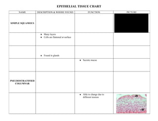
NAME DESCRIPTION & WHERE FOUND FUNCTION PICTURE
SIMPLE SQUAMOUS QuickTimeª and a
TIFF (Uncompressed) decompressor
are needed to see this picture.
♦ Many layers
♦ Cells are flattened at surface
♦ Found in glands
♦ Secrete mucus
PSEUDOSTRATIFIED
COLUMNAR
♦ Able to change due to
different tension
2. cube-shaped
found in glands rectangular
cells lines lower cells that conduct
Many layers, digestive tract nerve impulses these neuroglia
flat on top cells make the
myelin sheath
false layers __________________ Tissue __________________ Tissue
these cells nourish
-Tissue that lines surfaces & -tissue found in brain & spinal the neurons
1 flat, layer is used to protect. cord
Blood cells
__________________ Tissue __________________ Tissue
formed here
-Tissue that binds structures -tissue that accounts for involuntary
together, fills space, protects, movement in animals lines organs
osseus makes blood cells, stores fat
tissue
3 kinds - attached to
matrix is bone – many involuntary - in
semi-solid nuclei heart
Dermis of Tendons & Adipose
the skin ligaments tissue is
an example
Matrix is plasma