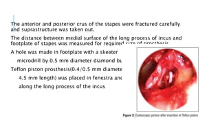
ENDOSCOPIC TRANSCANAL
STAPEDOTOMY:
EXPERIENCES AT A TERTIARY CARE
TEACHING HOSPITAL OF EASTERN INDIA
AUTHORS
Santosh Kumar Swain, Alok Das, Bulu Nahak, Jatindra Nath Mohanty
SOURCE
Department of Otorhinolaryngology, IMS and SUM Hospital, Siksha
“O” Anusandhan University, Bhubaneswar, Odisha, India
INTRODUCTION
Otosclerosis, more aptly called otospongiosis, is a primary
disease of the bony labyrinth.
In this, one or more foci of irregularly laid spongy bone
replace part of normally dense enchondral layer of bony otic
capsule
Stapedectomy/stapedotomy is the treatment of choice
Stapes surgery is done at a very fragile anatomical area where
there is a risk of injury to the chorda tympani nerve, incus,
facial nerve, and tympanic membrane.
There is a chance of sensorineural hearing loss due to
incorrect surgical procedure.
Surgeon often unable to observe the anterior crus of the
stapes via transcanal approach under a microscope which
may force the surgeon to do blindly fractures of such part
The possible complications, microscopic visual field, and
complex anatomy of the middle ear are causes of concern in
stapes surgery
The endoscope provides a wide-angle and high-resolution
image which allows improved visualization of the middle ear
cleft in comparison to the microscope.
The endoscope helps otolaryngologists to bring angled optics
and a high-contrast light to the middle ear, avoiding a
postaural approach and extended bony dissection
After introduction of 3 charge-coupled device camera,
high-definition video monitors and recently small diameter
fiberoptic and modern endoscopes provide high-resolution
images of the middle ear cleft structures
In this study they assess the use of rigid endoscope by transcanal
route for stapes surgery and find out the outcome and possible pros
and cons of this technique
AIM AND OBJECTIVE
To analyze the experiences of endoscopic transcanal stapedotomy
performed at a tertiary care teaching hospital of Eastern India
METHODOLOGY
This study involved a prospective analysis of patients with conductive
hearing loss those who were diagnosed as otosclerosis and who
underwent stapedotomy by endoscopic transcanal approach.
Sample size : 52 (28 female and 24 male between 29 to 48 years)
Duration: 2 years(March 2017–April 2019)
Place : tertiary care teaching hospital
INCLUSION CRITERIA
Patients who were diagnosed with otosclerosis based on clinical
history, otoscopic examinations, and audiological investigations
such as pure tone audiometry and tympanometry.
EXCLUSION CRITERIA
patients with narrow external auditory canal were avoided by
transcanal endoscopic surgery and excluded from this study
Pure tone audiometry was done in all cases preoperatively
and postoperatively with documentations of bone conduction
thresholds and air– bone gap (ABG) at the three speech
frequencies of 500 Hz, 1000 Hz, and 2000 Hz
Postoperative pure tone audiometry was performed at 1, 3, 6,
and 12 months following surgery
ABG closure within 10 dB was called as success, whereas
closure to within 20 dB called as improvement.
SURGICAL STEPS
surgeries were performed by senior authors under
local anesthesia
The patients were placed in same position as in
conventional ear surgeries
video monitor was placed in front of the surgeon
anterior and posterior wall were sutured in such
manner,to widen the meatus , and to make
accessibility to the middle ear easier
Trimming of the ear canal hair was done before starting
endoscopic surgery to reduce smudging of endoscope
3 mm diameter and 14 cm long rigid endoscope was used and
held in left hand
external auditory canal was injected with 1% lidocaine along
with 1:100,000 epinephrine
Posterior tympanomeatal flap was elevated transcanally with
the help of 0° endoscope and transposed anteriorly
All the procedures were performed with a 0° endoscope,
whereas a 30° endoscope was needed for better visualization
of oval window niche, anterior crura of the stapes, tympanic
segment of the facial nerve, and pyramidal eminence
The endoscopic picture of the middle ear was
excellent without drilling/curettage of the
scutum
Stapes fixation was assessed by gentle testing
of ossicular chain mobility.
Stapedius tendon was cut by curved microscissors
and stapes was dislocated from the incus at
incudostapedial joint
The anterior and posterior crus of the stapes were fractured carefully
and suprastructure was taken out.
The distance between medial surface of the long process of incus and
footplate of stapes was measured for required size of prosthesis
A hole was made in footplate with a skeeter
microdrill by 0.5 mm diameter diamond burr
Teflon piston prosthesis(0.4/0.5 mm diameter/
4.5 mm length) was placed in fenestra and fitted
along the long process of the incus
The ossicular chain mobility was ensured by palpating malleus
The footplate area was sealed with small pieces of dry gel-foam
placed by crocodile forceps
The tympanomeatal flap was repositioned and ear canal was filled
with gel-foam soaked with topical ear drops containing
antibiotics.
RESULTS
Stapedotomy was performed on the right ear in 34 patients and left
ear in 18 patients.
The mean age was 32.40 years.
The mean operation time was 38.43 min
The mean preoperative ABG was 34.84 dB, whereas the mean
postoperative ABG was 9.81 dB.
A 4.5 mm size diameter prosthesis was used in 48 cases and 4.75
size used in 4 cases
DISCUSSION
There are several difficulties encountered during stapes surgery and
these include exposure of the oval window niche, short chorda
tympani nerve, dehiscent facial canal with bulging toward oval
window area, and narrowing of the oval window niche by otosclerotic
focus.
Cutting of the stapedius tendon, incudostapedial joint dislocation,
and fracture of the crura of the stapes are easier with endoscope than
microscope
Excellent exposure of the stapes footplate without removing the bony
overhanging or touching the chorda tympani is usually possible with
endoscope
Under microscope, there is a need for partial curettage of the
medial bone segment on the posterior wall of the bony external
acoustic meatus which can damage chorda tympani
But this complication was avoided by using endoscope due to
direct visaulisation of the nerve
The main advantage of the endoscopic stapes surgery is
excellent vision
DEMERITS.
working with only one hand
the learning curve
Endoscope light can cause high temperature in middle and
inner ear which can easily pass to the cochlea by perilymph
and leads to sensorineural hearing loss
loss of depth perception which provide reluctance to some
otologists for adopting this technique.
CONCLUSION
Endoscopic stapedotomy has several benefits such as good
visualization, easy accessibility to the foot plate of stapes, oval
window niche, and facial nerve
In this study, all the stapedotomy surgeries were performed with
help of endoscope where all relevant anatomical structures were
visualized without any difficulty and minimal to no complications.
SIMILAR STUDIES
1)Bianconi L, Gazzini L, Laura E, De Rossi S, Conti A, Marchioni D.
Conducted a study :
Endoscopic stapedotomy: safety and audiological results in 150
patients between November 2014 and September 2018
The aim of the study was to describe the surgical steps in endoscopic
stapes surgery and to evaluate the audiologic and surgical outcomes.
There were no cases of major intraoperative complications.
They concluded that Endoscopic stapes surgery is a safe procedure
with a low risk of peri- or postoperative complications and is a
possible alternative to the traditional microscopic surgical procedure
in the treatment of otosclerosis.
2) Yang Q, Zhao Y, Hou ZH, Chen SJ, Yu YJ, Wang ZY,
conducted a study :
Evaluation of the safety and effect of the endoscopic stapes
surgery: a multi-
center study
Sample size: 137
Place: Fudan University from October 2015 to October 2017
Objective:To analyze the safety of artificial stapes surgery
under endoscope and compare its effectiveness with
microscopic surgery.
They concluded that Endoscopic artificial stapes surgery is
safe, and the audiological results obtained after surgery are
similar to microscopic surgery.
3) Rhona Sproat, Constantina Yiannakis, and Arunachalam Iyer
conducted a study :
Endoscopic Stapes Surgery: A Comparison With Microscopic
Surgery
Sample size: 81
Place :Department of Otolaryngology, Monklands Hospital,
Airdrie, Scotland, UK
Objective: : To investigate postoperative audiological
outcomes and complication rates for fully endoscopic and
microscopic stapes surgery carried out by a single surgeon in
one center.
Conclusion: : Air-bone gap closure and patient complications
did not vary significantly between endoscopic and
nonendoscopic groups
THANK YOU

ENDOSCOPIC TRANSCANAL STAPEDOTOMY.pptx

  • 1.
    ENDOSCOPIC TRANSCANAL STAPEDOTOMY: EXPERIENCES ATA TERTIARY CARE TEACHING HOSPITAL OF EASTERN INDIA
  • 2.
    AUTHORS Santosh Kumar Swain,Alok Das, Bulu Nahak, Jatindra Nath Mohanty SOURCE Department of Otorhinolaryngology, IMS and SUM Hospital, Siksha “O” Anusandhan University, Bhubaneswar, Odisha, India
  • 3.
    INTRODUCTION Otosclerosis, more aptlycalled otospongiosis, is a primary disease of the bony labyrinth. In this, one or more foci of irregularly laid spongy bone replace part of normally dense enchondral layer of bony otic capsule Stapedectomy/stapedotomy is the treatment of choice Stapes surgery is done at a very fragile anatomical area where there is a risk of injury to the chorda tympani nerve, incus, facial nerve, and tympanic membrane.
  • 4.
    There is achance of sensorineural hearing loss due to incorrect surgical procedure. Surgeon often unable to observe the anterior crus of the stapes via transcanal approach under a microscope which may force the surgeon to do blindly fractures of such part The possible complications, microscopic visual field, and complex anatomy of the middle ear are causes of concern in stapes surgery
  • 5.
    The endoscope providesa wide-angle and high-resolution image which allows improved visualization of the middle ear cleft in comparison to the microscope. The endoscope helps otolaryngologists to bring angled optics and a high-contrast light to the middle ear, avoiding a postaural approach and extended bony dissection After introduction of 3 charge-coupled device camera, high-definition video monitors and recently small diameter fiberoptic and modern endoscopes provide high-resolution images of the middle ear cleft structures
  • 6.
    In this studythey assess the use of rigid endoscope by transcanal route for stapes surgery and find out the outcome and possible pros and cons of this technique
  • 7.
    AIM AND OBJECTIVE Toanalyze the experiences of endoscopic transcanal stapedotomy performed at a tertiary care teaching hospital of Eastern India
  • 8.
    METHODOLOGY This study involveda prospective analysis of patients with conductive hearing loss those who were diagnosed as otosclerosis and who underwent stapedotomy by endoscopic transcanal approach. Sample size : 52 (28 female and 24 male between 29 to 48 years) Duration: 2 years(March 2017–April 2019) Place : tertiary care teaching hospital
  • 9.
    INCLUSION CRITERIA Patients whowere diagnosed with otosclerosis based on clinical history, otoscopic examinations, and audiological investigations such as pure tone audiometry and tympanometry. EXCLUSION CRITERIA patients with narrow external auditory canal were avoided by transcanal endoscopic surgery and excluded from this study
  • 10.
    Pure tone audiometrywas done in all cases preoperatively and postoperatively with documentations of bone conduction thresholds and air– bone gap (ABG) at the three speech frequencies of 500 Hz, 1000 Hz, and 2000 Hz Postoperative pure tone audiometry was performed at 1, 3, 6, and 12 months following surgery ABG closure within 10 dB was called as success, whereas closure to within 20 dB called as improvement.
  • 11.
    SURGICAL STEPS surgeries wereperformed by senior authors under local anesthesia The patients were placed in same position as in conventional ear surgeries video monitor was placed in front of the surgeon anterior and posterior wall were sutured in such manner,to widen the meatus , and to make accessibility to the middle ear easier
  • 12.
    Trimming of theear canal hair was done before starting endoscopic surgery to reduce smudging of endoscope 3 mm diameter and 14 cm long rigid endoscope was used and held in left hand external auditory canal was injected with 1% lidocaine along with 1:100,000 epinephrine Posterior tympanomeatal flap was elevated transcanally with the help of 0° endoscope and transposed anteriorly All the procedures were performed with a 0° endoscope, whereas a 30° endoscope was needed for better visualization of oval window niche, anterior crura of the stapes, tympanic segment of the facial nerve, and pyramidal eminence
  • 13.
    The endoscopic pictureof the middle ear was excellent without drilling/curettage of the scutum Stapes fixation was assessed by gentle testing of ossicular chain mobility. Stapedius tendon was cut by curved microscissors and stapes was dislocated from the incus at incudostapedial joint
  • 14.
    The anterior andposterior crus of the stapes were fractured carefully and suprastructure was taken out. The distance between medial surface of the long process of incus and footplate of stapes was measured for required size of prosthesis A hole was made in footplate with a skeeter microdrill by 0.5 mm diameter diamond burr Teflon piston prosthesis(0.4/0.5 mm diameter/ 4.5 mm length) was placed in fenestra and fitted along the long process of the incus
  • 15.
    The ossicular chainmobility was ensured by palpating malleus The footplate area was sealed with small pieces of dry gel-foam placed by crocodile forceps The tympanomeatal flap was repositioned and ear canal was filled with gel-foam soaked with topical ear drops containing antibiotics.
  • 16.
    RESULTS Stapedotomy was performedon the right ear in 34 patients and left ear in 18 patients. The mean age was 32.40 years. The mean operation time was 38.43 min The mean preoperative ABG was 34.84 dB, whereas the mean postoperative ABG was 9.81 dB. A 4.5 mm size diameter prosthesis was used in 48 cases and 4.75 size used in 4 cases
  • 19.
    DISCUSSION There are severaldifficulties encountered during stapes surgery and these include exposure of the oval window niche, short chorda tympani nerve, dehiscent facial canal with bulging toward oval window area, and narrowing of the oval window niche by otosclerotic focus. Cutting of the stapedius tendon, incudostapedial joint dislocation, and fracture of the crura of the stapes are easier with endoscope than microscope Excellent exposure of the stapes footplate without removing the bony overhanging or touching the chorda tympani is usually possible with endoscope
  • 20.
    Under microscope, thereis a need for partial curettage of the medial bone segment on the posterior wall of the bony external acoustic meatus which can damage chorda tympani But this complication was avoided by using endoscope due to direct visaulisation of the nerve The main advantage of the endoscopic stapes surgery is excellent vision
  • 21.
    DEMERITS. working with onlyone hand the learning curve Endoscope light can cause high temperature in middle and inner ear which can easily pass to the cochlea by perilymph and leads to sensorineural hearing loss loss of depth perception which provide reluctance to some otologists for adopting this technique.
  • 22.
    CONCLUSION Endoscopic stapedotomy hasseveral benefits such as good visualization, easy accessibility to the foot plate of stapes, oval window niche, and facial nerve In this study, all the stapedotomy surgeries were performed with help of endoscope where all relevant anatomical structures were visualized without any difficulty and minimal to no complications.
  • 23.
    SIMILAR STUDIES 1)Bianconi L,Gazzini L, Laura E, De Rossi S, Conti A, Marchioni D. Conducted a study : Endoscopic stapedotomy: safety and audiological results in 150 patients between November 2014 and September 2018 The aim of the study was to describe the surgical steps in endoscopic stapes surgery and to evaluate the audiologic and surgical outcomes. There were no cases of major intraoperative complications. They concluded that Endoscopic stapes surgery is a safe procedure with a low risk of peri- or postoperative complications and is a possible alternative to the traditional microscopic surgical procedure in the treatment of otosclerosis.
  • 24.
    2) Yang Q,Zhao Y, Hou ZH, Chen SJ, Yu YJ, Wang ZY, conducted a study : Evaluation of the safety and effect of the endoscopic stapes surgery: a multi- center study Sample size: 137 Place: Fudan University from October 2015 to October 2017 Objective:To analyze the safety of artificial stapes surgery under endoscope and compare its effectiveness with microscopic surgery. They concluded that Endoscopic artificial stapes surgery is safe, and the audiological results obtained after surgery are similar to microscopic surgery.
  • 25.
    3) Rhona Sproat,Constantina Yiannakis, and Arunachalam Iyer conducted a study : Endoscopic Stapes Surgery: A Comparison With Microscopic Surgery Sample size: 81 Place :Department of Otolaryngology, Monklands Hospital, Airdrie, Scotland, UK Objective: : To investigate postoperative audiological outcomes and complication rates for fully endoscopic and microscopic stapes surgery carried out by a single surgeon in one center. Conclusion: : Air-bone gap closure and patient complications did not vary significantly between endoscopic and nonendoscopic groups
  • 26.