Background
All thephysiological activities of the body are regulated by two major systems:
1. Nervous system
2. Endocrine system.
CELL-TO-CELL SIGNALING: transfer of information from one cell to another, intercellular
communication, through chemical substances called chemical messengers., may be the
hormones or hormone-like substances.
four types:
1. Endocrine messengers: classical hormones
2. Paracrine messengers: diffuse from the control cells to the target cells through the interstitial
fluid.
3. Autocrine messengers: control the source cells which secrete them
4. Neurocrine messengers: neurotransmitters and neurohormones
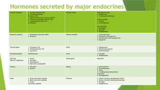
Hormones
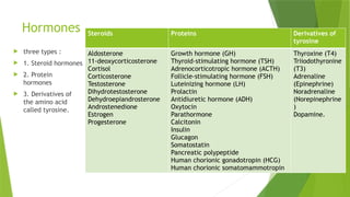
three types:
1. Steroid hormones
2. Protein
hormones
3. Derivatives of
the amino acid
called tyrosine.
Steroids Proteins Derivatives of
tyrosine
Aldosterone
11-deoxycorticosterone
Cortisol
Corticosterone
Testosterone
Dihydrotestosterone
Dehydroepiandrosterone
Androstenedione
Estrogen
Progesterone
Growth hormone (GH)
Thyroid-stimulating hormone (TSH)
Adrenocorticotropic hormone (ACTH)
Follicle-stimulating hormone (FSH)
Luteinizing hormone (LH)
Prolactin
Antidiuretic hormone (ADH)
Oxytocin
Parathormone
Calcitonin
Insulin
Glucagon
Somatostatin
Pancreatic polypeptide
Human chorionic gonadotropin (HCG)
Human chorionic somatomammotropin
Thyroxine (T4)
Triiodothyronine
(T3)
Adrenaline
(Epinephrine)
Noradrenaline
(Norepinephrine
)
Dopamine.
7.
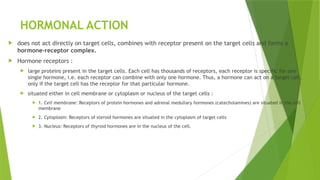
HORMONAL ACTION
doesnot act directly on target cells, combines with receptor present on the target cells and forms a
hormone-receptor complex.
Hormone receptors :
large proteins present in the target cells. Each cell has thousands of receptors, each receptor is specific for one
single hormone, i.e. each receptor can combine with only one hormone. Thus, a hormone can act on a target cell,
only if the target cell has the receptor for that particular hormone.
situated either in cell membrane or cytoplasm or nucleus of the target cells :
1. Cell membrane: Receptors of protein hormones and adrenal medullary hormones (catecholamines) are situated in the cell
membrane
2. Cytoplasm: Receptors of steroid hormones are situated in the cytoplasm of target cells
3. Nucleus: Receptors of thyroid hormones are in the nucleus of the cell.
8.
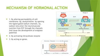
MECHANISM OF HORMONALACTION
1. By altering permeability of cell
membrane: eg. Acetylcholine by opening
the ligand-gated sodium channels. So,
sodium ions enter the neuromuscular
junction from ECF through the channels
and cause the development of endplate
potential
2. By activating intracellular enzyme
3. By acting on genes.
9.
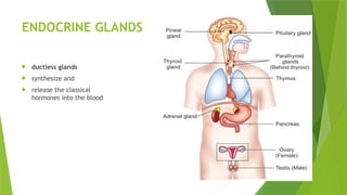
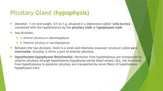
Pituitary Gland (hypophysis)
diameter 1 cm and weight 0.5 to 1 g, situated in a depression called ‘sella turcica’,
connected with the hypothalamus by the pituitary stalk or hypophyseal stalk.
two divisions:
1. Anterior pituitary or adenohypophysis
2. Posterior pituitary or neurohypophysis
Between the two divisions, there is a small and relatively avascular structure called pars
intermedia. Actually, it forms a part of anterior pituitary.
Hypothalamo-hypophyseal Relationship: Hormones from hypothalamus are transported to
anterior pituitary through hypothalamo-hypophysial portal blood vessels. But, the hormones
from hypothalamus to posterior pituitary are transported by nerve fibers of hypothalamo-
hypophyseal tract.
10.
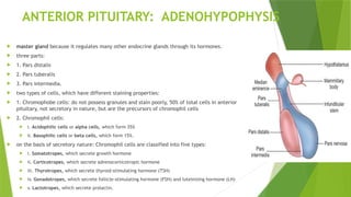
ANTERIOR PITUITARY: ADENOHYPOPHYSIS
master gland because it regulates many other endocrine glands through its hormones.
three parts:
1. Pars distalis
2. Pars tuberalis
3. Pars intermedia.
two types of cells, which have different staining properties:
1. Chromophobe cells: do not possess granules and stain poorly, 50% of total cells in anterior
pituitary, not secretory in nature, but are the precursors of chromophil cells
2. Chromophil cells:
i. Acidophilic cells or alpha cells, which form 35%
ii. Basophilic cells or beta cells, which form 15%.
on the basis of secretory nature: Chromophil cells are classified into five types:
i. Somatotropes, which secrete growth hormone
ii. Corticotropes, which secrete adrenocorticotropic hormone
iii. Thyrotropes, which secrete thyroid-stimulating hormone (TSH)
iv. Gonadotropes, which secrete follicle-stimulating hormone (FSH) and luteinizing hormone (LH)
v. Lactotropes, which secrete prolactin.
11.
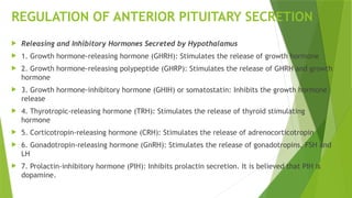
REGULATION OF ANTERIORPITUITARY SECRETION
Releasing and Inhibitory Hormones Secreted by Hypothalamus
1. Growth hormone-releasing hormone (GHRH): Stimulates the release of growth hormone
2. Growth hormone-releasing polypeptide (GHRP): Stimulates the release of GHRH and growth
hormone
3. Growth hormone-inhibitory hormone (GHIH) or somatostatin: Inhibits the growth hormone
release
4. Thyrotropic-releasing hormone (TRH): Stimulates the release of thyroid stimulating
hormone
5. Corticotropin-releasing hormone (CRH): Stimulates the release of adrenocorticotropin
6. Gonadotropin-releasing hormone (GnRH): Stimulates the release of gonadotropins, FSH and
LH
7. Prolactin-inhibitory hormone (PIH): Inhibits prolactin secretion. It is believed that PIH is
dopamine.
12.
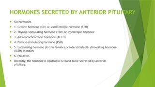
HORMONES SECRETED BYANTERIOR PITUITARY
Six hormones
1. Growth hormone (GH) or somatotropic hormone (STH)
2. Thyroid-stimulating hormone (TSH) or thyrotropic hormone
3. Adrenocorticotropic hormone (ACTH)
4. Follicle-stimulating hormone (FSH)
5. Luteinizing hormone (LH) in females or interstitialcell- stimulating hormone
(ICSH) in males
6. Prolactin.
Recently, the hormone β-lipotropin is found to be secreted by anterior
pituitary.
13.
GROWTH HORMONE
secretedby somatotropes which are the acidophilic cells of anterior pituitary.
protein in nature, having a single-chain polypeptide with 191 amino acids. molecular weight is 21,500.
Growth hormone is transported in blood by GH-binding proteins (GHBPs).
Half-life about 20 minutes. It is degraded in liver and kidney.
responsible for the general growth of the body. Hypersecretion causes enormous growth of the body,
leading to gigantism. Deficiency in children causes stunted growth, leading to dwarfism.
responsible for the growth of almost all tissues of the body, which are capable of growing. It increases the
size and number of cells by mitotic division. GH also causes specific differentiation of certain types of
cells like bone cells and muscle cells. GH also acts on the metabolism of all the three major types of
foodstuffs in the body, viz. proteins, lipids and carbohydrates
14.
On metabolism
GHincreases the synthesis of proteins, mobilization of lipids and conservation of carbohydrates.
a. GH accelerates the synthesis of proteins by:
i. Increasing amino acid transport through cell membrane:
ii. Increasing ribonucleic acid (RNA) translation
iii. Increasing transcription of DNA to RNA
iv. Decreasing catabolism of protein
v. Promoting anabolism of proteins indirectly: GH increases the release of insulin (from β-cells of islets in
pancreas), which has anabolic effect on proteins.
b. GH mobilizes fats from adipose tissue. So, the concentration of fatty acids increases in the body
fluids. These fatty acids are used for the production of energy by the cells. Thus, the proteins are
spared.
c. Major action of GH on carbohydrates is the conservation of glucose by:
i. Decrease in the peripheral utilization of glucose, for the production of energy,
ii. Increase in the deposition of glycogen in the cells,
iii. Decrease in the uptake of glucose by the cells,
iv. Diabetogenic effect of GH
15.
On bones
differentiationand development of bone cells.
GH increases the growth of the skeleton. It increases both the length as well as the thickness of the
bones.
GH increases:
i. Synthesis and deposition of proteins by chondrocytes and osteogenic cells
ii. Multiplication of chondrocytes and osteogenic cells by enhancing the intestinal calcium absorption
iii. Formation of new bones by converting chondrocytes into osteogenic cells
iv. Availability of calcium for mineralization of bone matrix.
GH increases the length of the bones, until epiphysis fuses with shaft, which occurs at the time of
puberty.
After the epiphyseal fusion, length of the bones cannot be increased. However, it stimulates the
osteoblasts strongly. So, the bone continues to grow in thickness throughout the life. Particularly,
the membranous bones
16.
Somatomedin
GH actson bones, growth and protein metabolism through somatomedin secreted by liver. GH stimulates
the liver to secrete somatomedin. Sometimes, in spite of normal secretion of GH, growth is arrested
(dwarfism) due to the absence or deficiency of somatomedin.
two types:
i. Insulin-like growth factor-I (IGF-I), which is also called somatomedin C
ii. Insulin-like growth factor-II.
Somatomedin C (IGF-I) acts on the bones and protein metabolism. Insulin-like growth factor-II plays an
important role in the growth of fetus.
Growth hormone receptor: GH receptor is called growth hormone secretagogue (GHS) receptor. It is a
transmembrane receptor, belonging to cytokine receptor family. GH binds with the receptor situated
mainly in liver cells and forms the hormonereceptor complex. Hormone-receptor complex induces various
intracellular enzyme pathways, resulting in somatomedin secretion. Somatomedin in turn, executes the
actions of growth hormone.
17.
Thyroid-stimulating Hormone (TSH)
TSH is necessary for the growth and secretory activity of the thyroid gland. It
has many actions on the thyroid gland.
Follicle-stimulating Hormone (FSH)
Follicle-stimulating hormone is a glycoprotein made up of one α-subunit and a β-subunit. The α-subunit
has 92 amino acids and β-subunit has 118 amino acids. The half-life of FSH is about 3 to 4 hours.
Actions of FSH
In males, FSH acts along with testosterone and accelerates the process of spermeogenesis
In females FSH:
1. Causes the development of graafian follicle from primordial follicle
2. Stimulates the theca cells of graafian follicle and causes secretion of estrogen
3. Promotes the aromatase activity in granulosa cells, resulting in conversion of androgens into estrogen.
20.
Luteinizing Hormone (LH)
LH is a glycoprotein made up of one α-subunit and one β-subunit. The α-subunit has 92
amino acids and β-subunit has 141 amino acids. The half-life of LH is about 60 minutes.
In males, LH is known as interstitial cell-stimulating hormone (ICSH) because it
stimulates the interstitial cells of Leydig in testes. This hormone is essential for the
secretion of testosterone from Leydig cells
In females, LH:
1. Causes maturation of vesicular follicle into graafian follicle along with follicle-
stimulating hormone
2. Induces synthesis of androgens from theca cells of growing follicle
3. Is responsible for ovulation
4. Is necessary for the formation of corpus luteum
5. Activates the secretory functions of corpus luteum.
21.
Prolactin
Prolactin isa single chain polypeptide with 199 amino acids. Its half-life is
about 20 minutes. Prolactin is necessary for the final preparation of mammary
glands for the production and secretion of milk.
Prolactin acts directly on the epithelial cells of mammary glands and causes
localized alveolar hyperplasia.
22.
β-lipotropin
β-lipotropin isa polypeptide hormone with 31 amino acids. It mobilizes fat
from adipose tissue and promotes lipolysis. It also forms the precursor of
endorphins. This hormone acts through the adenyl cyclase.
23.
POSTERIOR PITUITARY ORNEUROHYPOPHYSIS
three parts:
1. Pars nervosa or infundibular process
2. Neural stalk or infundibular stem
3. Median eminence.
HISTOLOGY: Posterior pituitary is made up of neural type of cells called pituicytes and unmyelinated
nerve fibers.
Pituicytes: are the fusiform cells derived from glial cells. These cells have several processes and brown
pigment granules. Pituicytes act as supporting cells and do not secrete any hormone.
Unmyelinated Nerve Fibers: Unmyelinated nerve fibers come from supraoptic and paraventricular nuclei of
the hypothalamus through the pituitary stalk.
Other Structures: Posterior pituitary also has numerous blood vessels, hyaline bodies, neuroglial cells and
mast cells.
24.
HORMONES OF POSTERIORPITUITARY
Posterior pituitary hormones are:
1. Antidiuretic hormone (ADH) or vasopressin
2. Oxytocin
Actually, the posterior pituitary does not secrete any hormone. ADH and oxytocin are synthesized in
the hypothalamus. From hypothalamus, these two hormones are transported to the posterior
pituitary through the nerve fibers of hypothalamo-hypophyseal tract , by means of axonic flow.
Proteins involved in transport of these hormones are called neurophysins
25.
ANTIDIURETIC HORMONE
(ADH) issecreted mainly by supraoptic nucleus of hypothalamus. It is also secreted by paraventricular
nucleus in small quantity. From here, this hormone is transported to posterior pituitary through the nerve
fibers of hypothalamo-hypophyseal tract, by means of axonic flow.
Antidiuretic hormone is a polypeptide containing 9 amino acids. Its half-life is 18 to 20 minutes.
Actions
1. Retention of water
2. Vasopressor action.
1. Retention of water Major function of ADH is retention of water by acting on kidneys. It increases the
facultative reabsorption of water from distal convoluted tubule and collecting duct in the kidneys. ADH
increases water reabsorption in tubular epithelial membrane by regulating the water channel proteins
called aquaporins through V2 receptors.
In large amount, ADH shows vasoconstrictor action. Particularly, causes constriction of the arteries in all
parts of the body. Due to vasoconstriction, the blood pressure increases. ADH acts on blood vessels
through V1A receptors. However, the amount of ADH required to cause the vasopressor effect is greater
than the amount required to cause the antidiuretic effect.
26.
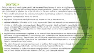
OXYTOCIN
Oxytocin is secretedmainly by paraventricular nucleus of hypothalamus. It is also secreted by supraoptic nucleus in small
quantity and it is transported from hypothalamus to posterior pituitary through the nerve fibers of hypothalamo-hypophyseal
tract. In the posterior pituitary, the oxytocin is stored in the nerve endings of hypothalamo-hypophyseal tract.
When suitable stimuli reach the posterior pituitary from hypothalamus, oxytocin is released into the blood.
Oxytocin is secreted in both males and females.
Oxytocin is a polypeptide having 9 amino acids. It has a half-life of about 6 minutes.
Actions in Females: In females, oxytocin acts on mammary glands and pregnant and non pregnant uterus.
ejection of milk from the mammary glands: . Ducts of the mammary glands are lined by myoepithelial cells. Oxytocin
causes contraction of the myoepithelial cells and flow of milk from alveoli of mammary glands to the exterior through duct
system and nipple. The process by which the milk is ejected from alveoli of mammary glands is called milk ejection reflex
or milk letdown reflex.
Oxytocin secretion increases during labor. At the onset of labor, the cervix dilates and the fetus descends through the
birth canal. During the movement of fetus through cervix, the receptors on the cervix are stimulated and start discharging
large number of impulses. These impulses are carried to the paraventricular and supraoptic nuclei of hypothalamus by the
somatic afferent nerve fibers. Now, these two hypothalamic nuclei secrete large quantity of oxytocin, which enhances
labor by causing contraction of uterus.
The action of oxytocin on non-pregnant uterus is to facilitate the transport of sperms through female genital tract up to
the fallopian tube, by producing the uterine contraction during sexual intercourse.
In males, the release of oxytocin increases during ejaculation. It facilitates release of sperm into urethra by causing
contraction of smooth muscle fibers in reproductive tract, particularly vas deferens
27.
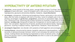
HYPERACTIVITY OF ANTERIOPITUITARY
Gigantism,: excess growth of the body, giants, average height of about 7 to 8 feet, before the fusion of
epiphysis of bone with shaft, tumor of acidophil cells in the anterior pituitary, General overgrowth,
hyperglycemia, glycosuria and pituitary diabetes, headache, visual disturbances
Acromegaly: enlargement, thickening and broadening of bones, particularly in the extremities of the
body, after the fusion of epiphysis with shaft of the bone, tumor of acidophil cells in the anterior
pituitary, Acromegalic or gorilla face, prognathism, protrusion of supraorbital ridges, broadening of
nose, thickening of lips, thickening and wrinkles formation on forehead, Enlargement of hands and feet,
Kyphosis, bulldog scalp, Overgrowth of body hair, Enlargement of visceral organs such as lungs,thymus,
heart, liver and spleen, Hyperactivity of thyroid, parathyroid and adrenal Glands, Hyperglycemia and
glucosuria, resulting in diabetes mellitus, Hypertension, Headache, Visual disturbance
Acromegalic Gigantism: rare disorder with symptoms of both gigantism and acromegaly,
Cushing Disease: characterized by obesity, basophilic adenoma of adenohypophysis, increases the
secretion of adrenocorticotropic hormone, which in turn stimulates the adrenal cortex to release
cortisol. Cushing disease also develops by hyperplasia or tumor of adrenal cortex. Usually, the disorder
due to the pituitary cause is called Cushing disease and when it is due to the adrenal cause, it is called
Cushing syndrome
28.
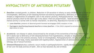
HYPOACTIVITY OF ANTERIORPITUITARY
Dwarfism: stunted growth, in children, Reduction in GH secretion in infancy or early childhood due to Tumor of
chromophobes, Deficiency of GH-releasing hormone secreted by hypothalamus, Deficiency of somatomedin C,
Atrophy or degeneration of acidophilic cells in the anterior pituitary, Panhypopituitarism, maximum height of
anterior pituitary dwarf at the adult age is only about 3 feet but proportionate , head becomes slightly larger,
mental activity is normal with no mental retardation, no deformity, Reproductive function is not affected.
Laron dwarfism: presence of abnormal growth hormone secretagogue (GHS) receptors in liver
Psychogenic dwarfism/psychosocial dwarfism or stress dwarfism.: if the child is exposed to extreme emotional
deprivation or stress
Acromicria: rare disease in adults characterized by the atrophy of the extremities of the body, Deficiency of
GH in adults due to Deficiency of GH-releasing hormone from hypothalamus, Atrophy or degeneration of
acidophilic cells in the anterior pituitary, Tumor of chromophobes, Panhypopituitarism, Atrophy and thinning of
extremities of the body, mostly associated with hypothyroidism, Hyposecretion of adrenocortical hormones,
lethargy, loss of sexual functions.
Simmond Disease/pituitary cachexia: occurs mostly in panhypopituitarism, rapidly developing senile decay, loss
of hair over the body and loss of teeth , Skin on face becomes dry and wrinkled.
29.
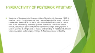
HYPERACTIVITY OF POSTERIORPITUITARY
Syndrome of Inappropriate Hypersecretion of Antidiuretic Hormone (SIADH):
cerebral tumors, lung tumors and lung cancers because the tumor cells and
cancer cells secrete ADH. in SIADH, secretion of ADH from tumor or cancer
cells is not inhibited by hypotonic plasma. So there is continuous loss of
sodium, resulting in persistent plasma hypotonicity. Signs and symptoms 1.
Loss of appetite 2. Weight loss 3. Nausea and vomiting 4. Headache 5. Muscle
weakness, spasm and cramps 6. Fatigue 7. Restlessness and irritability.
30.
HYPOACTIVITY OF POSTERIORPITUITARY
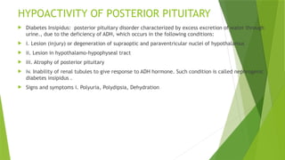
Diabetes Insipidus: posterior pituitary disorder characterized by excess excretion of water through
urine., due to the deficiency of ADH, which occurs in the following conditions:
i. Lesion (injury) or degeneration of supraoptic and paraventricular nuclei of hypothalamus
ii. Lesion in hypothalamo-hypophyseal tract
iii. Atrophy of posterior pituitary
iv. Inability of renal tubules to give response to ADH hormone. Such condition is called nephrogenic
diabetes insipidus .
Signs and symptoms i. Polyuria, Polydipsia, Dehydration
31.
HYPOACTIVITY OF ANTERIORAND POSTERIOR PITUITARY
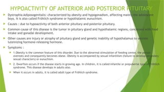
Dystrophia Adiposogenitalis: characterized by obesity and hypogonadism, affecting mainly the adolescent
boys. It is also called Fröhlich syndrome or hypothalamic eunuchism.
Causes : due to hypoactivity of both anterior pituitary and posterior pituitary.
Common cause of this disease is the tumor in pituitary gland and hypothalamic regions, concerned with food
intake and gonadal development.
Other causes are injury or atrophy of pituitary gland and genetic inability of hypothalamus to secrete
luteinizing hormone-releasing hormone.
Symptoms :
1.Obesity is the common feature of this disorder. Due to the abnormal stimulation of feeding center, the person
overeats and consequently becomes obese. Obesity is accompanied by sexual infantilism (failure to develop secondary
sexual characters) or eunuchism.
2. Dwarfism occurs if the disease starts in growing age. In children, it is called infantile or prepubertal type of Fröhlich
syndrome. This disease develops in adults also.
When it occurs in adults, it is called adult type of Fröhlich syndrome.
32.
Thyroid Gland
situatedat the root of the neck on either side of the trachea.
two lobes, which are connected in the middle by an isthmus .
It weighs about 20 to 40 g in adults.
Thyroid is larger in females than in males.
The structure and the function of the thyroid gland change in different stages of the sexual cycle in females. Its
function increases slightly during pregnancy and lactation and decreases during menopause.
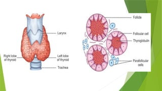
HISTOLOGY OF THYROID GLAND:
composed of large number of closed follicles. These follicles are lined with cuboidal epithelial cells, which are called the
follicular cells. Follicular cavity is filled with a colloidal substance known as thyroglobulin, which is secreted by the
follicular cells. Follicular cells also secrete tetraiodothyronine (T4 or thyroxine) and tri-iodothyronine (T3 ).
In between the follicles, the parafollicular cells are present . These cells secrete calcitonin.
HORMONES OF THYROID GLAND: Thyroid gland secretes three hormones:
1. Tetraiodothyronine or T4 (thyroxine)
2. Tri-iodothyronine or T3
3. Calcitonin. T4 is otherwise known as thyroxine and it forms about 90% of the total secretion, whereas T3 is only 9% to
10%.
34.
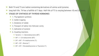
Both T4and T3 are iodine-containing derivatives of amino acid tyrosine.
long half-life. T4 has a halflife of 7 days. Half-life of T3 is varying between 10 and 24 hours.
STAGES OF SYNTHESIS OF THYROID HORMONES
1. Thyroglobulin synthesis
2. Iodide trapping
3. Oxidation of iodide
4. Transport of iodine into follicular cavity
5. Iodination of tyrosine
6. Coupling reactions.
Tyrosine + I = Monoiodotyrosine (MIT)
MIT + I = Di-iodotyrosine (DIT)
DIT + MIT = Tri-iodothyronine (T3)
MIT + DIT = Reverse T3
DIT + DIT = Tetraiodothyronine or Thyroxine (T4)
35.
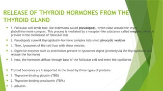
RELEASE OF THYROIDHORMONES FROM THE
THYROID GLAND
1. Follicular cell sends foot-like extensions called pseudopods, which close around the thyro
globulinhormone complex. This process is mediated by a receptor-like substance called megalin, which is
present in the membrane of follicular cell
2. Pseudopods convert thyroglobulin-hormone complex into small pinocytic vesicles
3. Then, lysosomes of the cell fuse with these vesicles
4. Digestive enzymes such as proteinases present in lysosomes digest (proteolysis) the thyroglobulin and
release the hormones
5. Now, the hormones diffuse through base of the follicular cell and enter the capillaries
Thyroid hormones are transported in the blood by three types of proteins:
1. Thyroxine-binding globulin (TBG)
2. Thyroxine-binding prealbumin (TBPA)
3. Albumin.
36.
FUNCTIONS OF THYROIDHORMONES
I. To increase basal metabolic rate
II. To stimulate growth in children.
37.
1. ACTION ONBASAL METABOLIC RATE
(BMR)
Thyroxine increases the metabolic activities in most of the body tissues,
except brain, retina, spleen, testes and lungs. calorigenic action.
In hyperthyroidism, BMR increases by about 60% to 100% above the normal
level and in hypothyroidism it falls by 20% to 40% below the normal level.
38.
2. ACTION ONPROTEIN METABOLISM
Thyroid hormone increases the synthesis of proteins in the cells. The protein synthesis is
accelerated by the following ways:
i. By Increasing the Translation of RNA
ii. By Increasing the Transcription of DNA to RNA
iii. By Increasing the Activity of Mitochondria
iv. By Increasing the Activity of Cellular Enzymes
39.
3. ACTION ONCARBOHYDRATE METABOLISM
Thyroxine stimulates almost all processes involved in the metabolism of carbohydrate.
Thyroxine:
i. Increases the absorption of glucose from GI tract
ii. Enhances the glucose uptake by the cells, by accelerating the transport of glucose through the
cell membrane
iii. Increases the breakdown of glycogen into glucose
iv. Accelerates gluconeogenesis.
40.
4. ACTION ONFAT METABOLISM
Thyroxine decreases the fat storage by mobilizing it from adipose tissues and
fat depots. The mobilized fat is converted into free fatty acid and transported
by blood. Thus, thyroxine increases the free fatty acid level in blood.
41.
5. ACTION ONPLASMA AND LIVER FATS
Even though there is an increase in the blood level of free fatty acids, thyroxine
specifically decreases the cholesterol, phospholipids and triglyceride levels in plasma. So,
in hyposecretion of thyroxine, the cholesterol level in plasma increases, resulting in
atherosclerosis.
Thyroxine also increases deposition of fats in the liver, leading to fatty liver. Thyroxine
decreases plasma cholesterol level by increasing its excretion from liver cells into bile.
Cholesterol enters the intestine through bile and then it is excreted through the feces.
42.
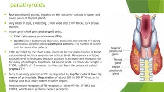
parathyroids
four parathyroidglands, situated on the posterior surface of upper and
lower poles of thyroid gland.
very small in size, 6 mm long, 3 mm wide and 2 mm thick, dark brown
colored.
made up of chief cells and oxyphil cells.
Chief cells secrete parathormone (PTH).
Oxyphil cells : degenerated chief cells, these cells may secrete PTH during
pathological condition called parathyroid adenoma. The number of oxyphil
cells increases after puberty.
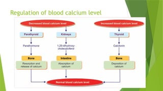
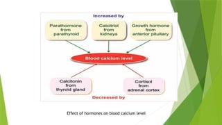
PTH: secreted by the chief cells, essential for the maintenance of blood
calcium level within a very narrow critical level. Maintenance of blood
calcium level is necessary because calcium is an important inorganic ion
for many physiological functions. 84 amino acids. Its molecular weight is
9,500, half-life of 10 minutes, synthesized from the precursor called
prepro-PTH
Sixty to seventy percent of PTH is degraded by Kupffer cells of liver, by
means of proteolysis. Degradation of about 20% to 30% PTH occurs in
kidneys and to a lesser extent in other organs.
Parathormone receptors (PTH receptors): three PTHR1, PTHR2 and
PTHR3, which are G protein coupled receptors
43.
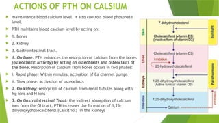
ACTIONS OF PTHON CALSIUM
maintenance blood calcium level. It also controls blood phosphate
level.
PTH maintains blood calcium level by acting on:
1. Bones
2. Kidney
3. Gastrointestinal tract.
1. On Bone: PTH enhances the resorption of calcium from the bones
(osteoclastic activity) by acting on osteoblasts and osteoclasts of
the bone. Resorption of calcium from bones occurs in two phases:
i. Rapid phase: Within minutes, activation of Ca channel pumps
ii. Slow phase: activation of osteoclasts
2. On kidney: resorption of calcium from renal tubules along with
Mg ions and H ions
3. On Gastrointestinal Tract: the indirect absorption of calcium
ions from the GI tract, PTH increases the formation of 1,25-
dihydroxycholecalciferol (Calcitriol) in the kidneys
44.
ACTIONS OF PTHON BLOOD PHOSPHATE LEVEL
PTH decreases blood level of phosphate by increasing its urinary excretion. It also acts on bone and GI
tract.
1. On Bone: Along with calcium resorption, PTH also increases phosphate absorption from the bones.
2. On Kidney
Phosphaturic action: It is the effect of PTH by which phosphate is excreted through urine. PTH increases
phosphate excretion by inhibiting reabsorption of phosphate from renal tubules. It acts mainly on PCT.
3. On GI Tract: PCT increases the absorption of phosphate from GI tract through calcitriol.
45.
REGULATION OF PTHSECRETION
Blood level of calcium is the main factor regulating the secretion of PTH. Blood phosphate level also
regulates PTH secretion.
Blood Level of Calcium: PTH secretion is inversely proportional to blood calcium level. Increase in blood
calcium level decreases PTH secretion.
Conditions when PTH secretion decreases are: 1. Excess quantities of calcium in the diet 2. Increased vitamin D in the
diet 3. Increased resorption of calcium from the bones, caused by some other factors such as bone diseases.
On the other hand, decrease in calcium ion concentration of blood increases PTH secretion, as in the case of rickets,
pregnancy and in lactation.
Blood Level of Phosphate
PTH secretion is directly proportional to blood phosphate level. Whenever the blood level of phosphate
increases, it combines with ionized calcium to form calcium hydrogen phosphate. This decreases ionized
calcium level in blood which stimulates PTH secretion.
46.
APPLIED PHYSIOLOGY –DISORDERS OF
PARATHYROID GLANDS
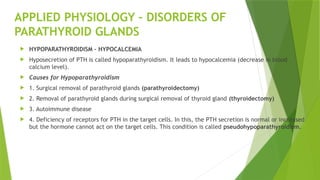
HYPOPARATHYROIDISM – HYPOCALCEMIA
Hyposecretion of PTH is called hypoparathyroidism. It leads to hypocalcemia (decrease in blood
calcium level).
Causes for Hypoparathyroidism
1. Surgical removal of parathyroid glands (parathyroidectomy)
2. Removal of parathyroid glands during surgical removal of thyroid gland (thyroidectomy)
3. Autoimmune disease
4. Deficiency of receptors for PTH in the target cells. In this, the PTH secretion is normal or increased
but the hormone cannot act on the target cells. This condition is called pseudohypoparathyroidism.
47.
Hypocalcemic Tetany
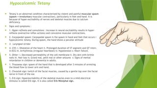
Tetanyis an abnormal condition characterized by violent and painful muscular spasm
(spasm = involuntary muscular contraction), particularly in feet and hand. It is
because of hyper-excitability of nerves and skeletal muscles due to calcium
deficiency.
Signs and symptoms :
1. Hyper-reflexia and convulsions : Increase in neural excitability results in hyper-
reflexia (overactive reflex actions) and convulsive muscular contractions.
2. Carpopedal spasm: Carpopedal spasm is the spasm in hand and feet that occurs in
hypocalcemic tetany. During spasm, the hand shows a peculiar attitude
3. Laryngeal stridor
4. CVS: i. Dilatation of the heart ii. Prolonged duration of ST segment and QT interval
in ECG iii. Arrhythmias (irregular heartbeat) iv. Hypotension v. Heart failure.
5. Other: i. Decreased permeability of the cell membrane ii. Dry skin with brittle
nails iii. Hair loss iv. Grand mal, petit mal or other seizures v. Signs of mental
retardation in children or dementia in adults.
1. Trousseau sign: spasm of the hand that is developed after 3 minutes of arresting
the blood flow to lower arm and hand.
2. Chvostek sign: twitch of the facial muscles, caused by a gentle tap over the facial
nerve in front of the ear.
3. Erb sign: Hyperexcitability of the skeletal muscles even to a mild electrical
stimulus is called Erb sign. It is also called Erb-Westphal sign.
48.
HYPERPARATHYROIDISM- HYPERCALCEMIA
Hypersecretionof PTH is called hyperparathyroidism. It results in hypercalcemia
3 types of hyperparathyroidism :
Primary hyperparathyroidism: due to the development of tumor in one or more parathyroid glands
Secondary hyperparathyroidism: due to the physiological compensatory hypertrophy of parathyroid glands,
in response to hypocalcemia which occurs due to other pathological conditions such as: i. Chronic renal
failure ii. Vitamin D deficiency iii. Rickets.
Tertiary hyperparathyroidism: due to hyperplasia (abnormal increase in the number of cells) of all the
parathyroid glands that develops due to chronic secondary hyperparathyroidism.
Hypercalcemia: is the increase in plasma calcium level. It occurs in hyperparathyroidism because of
increased resorption of calcium from bones.
Signs and symptoms of hypercalcemia: i. Depression of the nervous system ii. Sluggishness of reflex
activities iii. Reduced ST segment and QT interval in ECG iv. Lack of appetite v. Constipation. Other:
Development of bone diseases such as osteitis fibrosa cystica ii. Development of parathyroid poisoning, iii.
Deposition of calcium-phosphate crystals in renal tubules, thyroid gland, alveoli of lungs, gastric mucosa
and in the wall of the arteries, resulting in dysfunction of these organs iv.renal stones
49.
CALCITONIN
secreted bythe parafollicular cells or clear cells (C cells), 32 amino acids, molecular weight is about 3,400,
synthesized from procalcitonin, half life: 5 to 10 min.
ACTIONS OF CALCITONIN:
1. On Blood Calcium Level : plays an important role in controlling the blood
calcium level. It decreases the blood calcium level and counteracts PTH.
1. Calcitonin reduces the blood calcium level by acting on bones, kidneys and intestine.
i. On bones: stimulates osteoblastic activity and facilitates the deposition of calcium on bones, it suppresses the
activity of osteoclasts and inhibits the resorption of calcium from bones. It inhibits even the development of new
osteoclasts in bones.
ii. On kidney: Calcitonin increases excretion of calcium through urine, by inhibiting the reabsorption from the renal
tubules.
iii. On intestine: prevents the absorption of calcium from intestine into the blood.
2. On Blood Phosphate Level:With respect to calcium, calcitonin is an antagonist to PTH. But it has similar actions
of PTH, with respect to phosphate. It decreases the blood level of phosphate by acting on bones and kidneys.
i. On bones: inhibits the resorption of phosphate from bone and stimulates the deposition of phosphate on bones.
ii. On kidney: increases the excretion of phosphate through urine, by inhibiting the reabsorption from renal
tubules.
50.
REGULATION OF CALCITONISECRETION
High calcium content in plasma stimulates the calcitonin secretion through a
calcium receptor in parafollicular cells. Gastrin also is known to stimulate the
release of calcitonin.
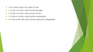
1 to2 million islets, four types of cells
1. A cells or α-cells, which secrete glucagon
2. B cells or β-cells, which secrete insulin
3. D cells or δ-cells, which secrete somatostatin
4. F cells or PP cells, which secrete pancreatic polypeptide.
55.
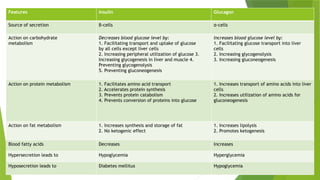
Features Insulin Glucagon
Sourceof secretion β-cells α-cells
Action on carbohydrate
metabolism
Decreases blood glucose level by:
1. Facilitating transport and uptake of glucose
by all cells except liver cells
2. Increasing peripheral utilization of glucose 3.
Increasing glycogenesis in liver and muscle 4.
Preventing glycogenolysis
5. Preventing gluconeogenesis
Increases blood glucose level by:
1. Facilitating glucose transport into liver
cells
2. Increasing glycogenolysis
3. Increasing gluconeogenesis
Action on protein metabolism 1. Facilitates amino acid transport
2. Accelerates protein synthesis
3. Prevents protein catabolism
4. Prevents conversion of proteins into glucose
1. Increases transport of amino acids into liver
cells
2. Increases utilization of amino acids for
gluconeogenesis
Action on fat metabolism 1. Increases synthesis and storage of fat
2. No ketogenic effect
1. Increases lipolysis
2. Promotes ketogenesis
Blood fatty acids Decreases Increases
Hypersecretion leads to Hypoglycemia Hyperglycemia
Hyposecretion leads to Diabetes mellitus Hypoglycemia
56.
SOMATOSTATIN
secreted from:
1. Hypothalamus
2. D cells (δ-cells) in islets of Langerhans of pancreas
3. D cells in stomach and upper part of small intestine.
57.
ACTIONS OF SOMATOSTATIN
1. Somatostatin acts within islets of Langerhans and, inhibits β and α
cells, i.e. it inhibits the secretion of both glucagon and insulin
2. It decreases the motility of stomach, duodenum and gallbladder
3. It reduces the secretion of gastrointestinal hormones gastrin, CCK, GIP
and VIP
4. Hypothalamic somatostatin inhibits the secretion of GH and TSH from
anterior pituitary. That is why, it is also called growth hormone-
inhibitory hormone (GHIH).
59.
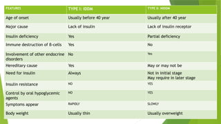
FEATURES TYPE I:IDDM TYPE II: NIDDM
Age of onset Usually before 40 year Usually after 40 year
Major cause Lack of insulin Lack of insulin receptor
Insulin deficiency Yes Partial deficiency
Immune destruction of β-cells Yes No
Involvement of other endocrine
disorders
No Yes
Hereditary cause Yes May or may not be
Need for insulin Always Not in initial stage
May require in later stage
Insulin resistance NO YES
Control by oral hypoglycemic
agents
NO YES
Symptoms appear RAPIDLY SLOWLY
Body weight Usually thin Usually overweight
60.
Signs and Symptomsof Diabetes Mellitus
1. Glucosuria
2. polyuria and polydipsia: Excess urine formation with increase in the frequency of voiding
urine is called polyuria. Increase in water intake is called polydipsia. Excess loss of water
decreases the water content and increases the salt content in the body. This stimulates the
thirst center in hypothalamus. Thirst center, in turn increases the intake of water.
4. Polyphagia : means the intake of excess food. It is very common in diabetes mellitus.
Asthenia:
Acidosis:
Acetone breathing: life-threatening condition
Kussmaul breathing: is the increase in rate and depth of respiration caused by severe acidosis
circulatory shock : Osmotic diuresis leads to dehydration
Coma
61.
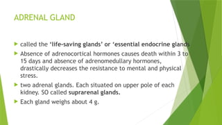
ADRENAL GLAND
calledthe ‘life-saving glands’ or ‘essential endocrine glands
Absence of adrenocortical hormones causes death within 3 to
15 days and absence of adrenomedullary hormones,
drastically decreases the resistance to mental and physical
stress.
two adrenal glands. Each situated on upper pole of each
kidney. SO called suprarenal glands.
Each gland weighs about 4 g.
62.
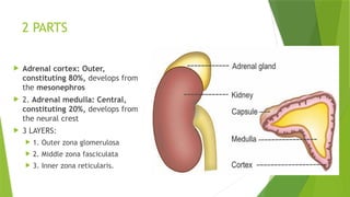
2 PARTS
Adrenalcortex: Outer,
constituting 80%, develops from
the mesonephros
2. Adrenal medulla: Central,
constituting 20%, develops from
the neural crest
3 LAYERS:
1. Outer zona glomerulosa
2. Middle zona fasciculata
3. Inner zona reticularis.
63.
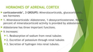
HORMONES OF ADRENALCORTEX
corticosteroids’, 3 GROUPS: Mineralocorticoids, glucocorticoids,
sex hormones
1. Mineralocorticoids: Aldosterone, 1-deoxycorticosterone. Ninety
percent of mineralocorticoid activity is provided by aldosterone.
Aldosterone has three important functions.
It increases:
1. Reabsorption of sodium from renal tubules
2. Excretion of potassium through renal tubules
3. Secretion of hydrogen into renal tubules.
64.
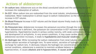
Actions of aldosterone
On sodium ions: Aldosterone acts on the distal convoluted tubule and the collecting duct and
increases the reabsorption of sodium.
On ECF: When sodium ions are reabsorbed from the renal tubules, simultaneously water is also
reabsorbed. Water reabsorption is almost equal to sodium reabsorption; so the net result is the
increase in ECF volume.
On Blood Pressure Increase in ECF volume and the blood volume finally leads to increase in
blood pressure.
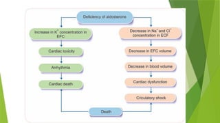
On Potassium Ions: Aldosterone increases the potassium excretion through the renal tubules.
When aldosterone is deficient, the potassium ion concentration in ECF increases leading to
hyperkalemia. Hyperkalemia results in serious cardiac toxicity, with weak contractions of heart
and development of arrhythmia. In very severe conditions, it may cause cardiac death. When
aldosterone secretion increases, it leads to hypokalemia and muscular weakness.
On Hydrogen Ion: While increasing the sodium reabsorption from renal tubules, aldosterone
causes tubular secretion of hydrogen ions. To some extent, secretion of hydrogen ions is in
exchange for sodium ions. It obviously reduces the hydrogen ion concentration in the ECF. In
normal conditions, aldosterone is essential to maintain acidbase balance in the body. In
hypersecretion, it causes alkalosis and in hyposecretion, it causes acidosis.
65.
On SweatGlands and Salivary Glands: Aldosterone has almost the
similar effect on sweat glands and salivary glands as it shows on
renal tubules. Sodium is reabsorbed from sweat glands under the
influence of aldosterone, thus the loss of sodium from the body is
prevented. Same effect is shown on saliva also. Thus, aldosterone
helps in the conservation of sodium in the body.
On Intestine Aldosterone increases sodium absorption from the
intestine, especially from colon and prevents loss of sodium through
feces. Aldosterone deficiency leads to diarrhea, with loss of sodium
and water.
Glucocorticoids
Glucocorticoids mainlyact on glucose metabolism:
1. Cortisol
2. Corticosterone
3. Cortisone
Cortisol or hydrocortisone is more potent and it has 95% of glucocorticoid activity.
Corticosterone is less potent.
cortisol is a life-protecting hormone because, it helps to withstand the stress and trauma in
life.
69.
Functions of glucocorticoids
On Carbohydrate Metabolism: increase the blood glucose level
On Protein Metabolism: promote the catabolism of proteins, leading to:
i. Decrease in cellular proteins
ii. Increase in plasma level of amino acids
iii. Increase in protein content in liver.
On Fat Metabolism: cause mobilization and redistribution of fats.
On Water Metabolism: play an important role in the maintenance of water balance, by accelerating excretion of water. The
adrenal insufficiency causes water retention and water intoxication after intake of large quantity of water.
On Mineral Metabolism: enhance the retention of sodium and to lesser extent, increase the excretion of potassium. Thus,
hypersecretion of glucocorticoids causes edema, hypertension, hypokalemia and muscular weakness. Glucocorticoids
decrease the blood calcium by inhibiting its absorption from intestine and increasing the excretion through urine.
On Bone: stimulate the bone resorption (osteoclastic activity) and inhibit bone formation and mineralization (osteoblastic
activity). So, in hypersecretion of glucocorticoids, osteoporosis occurs.
On Muscles: increase the catabolism of proteins in muscle. So, hypersecretion causes muscular weakness due to loss of
protein.
70.
On BloodCells: decrease the number of circulating eosinophils by increasing the destruction of eosinophils
in reticuloendothelial cells. These hormones also decrease the number of basophils and lymphocytes and
increase the number of circulating neutrophils, RBCs and platelets.
On Vascular Response Presence of glucocorticoids is essential for the constrictor action of adrenaline and
noradrenaline. In adrenal insufficiency, the blood vessels fail to respond to adrenaline and noradrenaline,
leading to vascular collapse.
On Central Nervous System: are essential for normal functioning of nervous system. Insufficiency of these
hormones causes personality changes like irritability and lack of concentration. Sensitivity to olfactory and
taste stimuli increases in adrenal insufficiency.
Exposure to any type of stress, either physical or mental, increases the secretion of adrenocorticotropic
hormone (ACTH), which in turn increases glucocorticoid secretion. The increase in glucocorticoid level is
very essential for survival during stress conditions, as it offers high resistance to the body against stress.
Anti-inflammatory Effects: Inflammation is defined as a localized protective response induced by injury
or destruction of tissues. When the tissue is injured by mechanical or chemical factors, some substances are
released from the affected area.
Anti-allergic Actions Corticosteroids prevent various reactions in allergic conditions as in the case of
inflammation.
Immunosuppressive Effects: suppress the immune system of the body by decreasing the number of
circulating T lymphocytes. It is done by suppressing proliferation of T cells and the lymphoid tissues (lymph
nodes and thymus). Glucocorticoids also prevent the release of interleukin-2 by T cells.
71.
ADRENAL SEX HORMONES
Androgens secreted by adrenal cortex:
1. Dehydroepiandrosterone: most active
2. Androstenedione
3. Testosterone.
small quantity of estrogen and progesterone are also secreted by adrenal cortex.
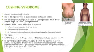
CUSHING SYNDROME
disordercharacterized by obesity.
due to the hypersecretion of glucocorticoids, particularly cortisol
If it is due to pituitary origin, it is known as Cushing disease. If it is due to
adrenal origin it is called Cushing syndrome.
Adrenal Origin: Cortisol secretion is increased by:
i. Tumor in zona fasciculata of adrenal cortex
ii. Carcinoma of adrenal cortex
iii. Prolonged treatment of chronic inflammatory diseases like rheumatoid arthritis
Two types:
i. ACTH-dependent Cushing syndrome which is due to hypersecretion of ACTH
ii. ACTH-independent Cushing syndrome in which the secretion of ACTH is
normal. The syndrome develops due to abnormal membrane receptors for some
peptides like interleukin-1, gonadotropin-releasing hormone and gastric inhibitory
polypeptide in the cells of zona fasciculata.
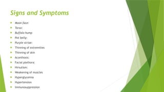
74.
Signs and Symptoms
Moon face:
Torso:
Buffalo hump
Pot belly:
Purple striae:
Thinning of extremities
Thinning of skin
Aconthosis:
Facial plethora:
Hirsutism:
Weakening of muscles
Hyperglycemia
Hypertension
Immunosuppression
75.
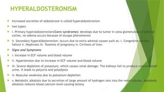
HYPERALDOSTERONISM
Increased secretionof aldosterone is called hyperaldosteronism
two types:
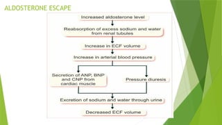
i. Primary hyperaldosteronism(Conn syndrome): develops due to tumor in zona glomerulosa of adrenal
cortex, no edema occurs because of escape phenomenon
ii. Secondary hyperaldosteronism: occurs due to extra adrenal causes such as: i. Congestive cardiac
failure ii. Nephrosis iii. Toxemia of pregnancy iv. Cirrhosis of liver.
Signs and Symptoms
i. Increase in ECF volume and blood volume
ii. Hypertension due to increase in ECF volume and blood volume
iii. Severe depletion of potassium, which causes renal damage. The kidneys fail to produce concentrated
urine. It leads to polyuria and polydipsia
iv. Muscular weakness due to potassium depletion
v. Metabolic alkalosis due to secretion of large amount of hydrogen ions into the renal tubules. Metabolic
alkalosis reduces blood calcium level causing tetany.
76.
ADRENOGENITAL SYNDROME
Secretionof abnormal quantities of adrenal androgens develops adrenogenital syndrome. Testosterone is
responsible for the androgenic activity in adrenogenital syndrome.
Causes: tumor of zona reticularis in adrenal cortex
characterized by the tendency for the development of secondary sexual character of opposite sex.
in females : development of male secondary sexual characters. The condition is called adrenal virilism.
Symptoms are: i. Masculinization due to increased muscular growth ii. Deepening of voice iii. Amenorrhea
iv. Enlargement of clitoris v. Male type of hair growth.
in males: Sometimes, the tumor of estrogen secreting cells produces more than normal quantity of
estrogens in males. It produces some symptoms such as: i. Feminization ii. Gynecomastia (enlargement of
breast) iii. Atrophy of testis iv. Loss of interest in women.
77.
ADDISON DISEASE ORCHRONIC ADRENAL
INSUFFICIENCY
failure of adrenal cortex to secrete corticosteroids.
3 Types
i. Primary Addison disease due to adrenal cause
ii. Secondary Addison disease due to failure of anterior pituitary to secrete ACTH
iii. Tertiary Addison disease due failure of hypothalamus to secret corticotropin-releasing factor (CRF).
Causes:
i. Atrophy of adrenal cortex due to autoimmune diseases
ii. Destruction of the gland because of tuberculosis
iii. Destruction of hormone-secreting cells in adrenal cortex by malignant tissues
iv. Congenital failure to secrete cortisol
v. Adrenalectomy and failure to take hormone therapy.
Common signs and symptom:
i. Pigmentation of skin and mucous membrane due to excess ACTH secretion, induced by cortisol deficiency. ACTH causes pigmentation by its melanocyte-
stimulating action
ii. Muscular weakness
iii. Dehydration with loss of sodium
iv. Hypotension
v. Decreased cardiac output and decreased workload of the heart, leading to decrease in size of the heart vi. Hypoglycemia
vii. Nausea, vomiting and diarrhea. Prolonged vomiting and diarrhea cause dehydration and loss of body weight viii. Susceptibility to any type of infection
ix. Inability to withstand any stress, resulting in Addisonian crisis
78.
Addisonian Crisis orAdrenal Crisis or Acute
Adrenal Insufficiency
Adrenal crisis is a common symptom of Addison disease, characterized by sudden collapse associated
with an increase in need for large quantities of glucocorticoids.
The condition becomes fatal if not treated in time.
Causes
i. Exposure to even mild stress
ii. Hypoglycemia due to fasting
iii. Trauma
iv. Surgical operation
v. Sudden withdrawal of glucocorticoid treatment.
79.
CONGENITAL ADRENAL HYPERPLASIA
Congenital adrenal hyperplasia is a congenital disorder, characterized by increase in size of adrenal cortex. Size increases due
to abnormal increase in the number of steroid-secreting cortical cells.
Causes
Even though the size of the gland increases, cortisol secretion decreases. It is because of the congenital deficiency of the
enzymes necessary for the synthesis of cortisol, particularly, 21-hydroxylase.
Lack of this enzyme reduces the synthesis of cortisol, resulting in ACTH secretion from pituitary by feedback mechanism.
ACTH stimulates the adrenal cortex causing hyperplasia, with accumulation of lipid droplets. Hence, it is also called
congenital lipid adrenal hyperplasia. Cortisol cannot be synthesized because of lack of 21-hydroxylase. Therefore, due to
the constant simulation of adrenal cortex by ACTH, the secretion of androgens increases. It results in sexual abnormalities
such as virilism.
Symptoms
In boys: Adrenal hyperplasia produces a condition known as macrogenitosomia praecox:
Features of macrogenitosomia praecox:
i. Precocious body growth, causing stocky appearance called infant Hercules
ii. Precocious sexual development with enlarged penis even at the age of 4 years.
In girls
In girls, adrenal hyperplasia produces masculinization. It is otherwise called virilism. In some cases of genetic disorders, the
female child is born with external geni talia of male type. This condition is called pseudohermaphroditism.
80.
Adrenal Medulla
Medullais the inner part of adrenal gland and it forms 20% of the mass of adrenal gland. It is
made up of interlacing cords of cells known as chromaffin cells.
Chromaffin cells are also called pheochrome cells or chromophil cells. These cells contain
fine granules which are stained brown by potassium dichromate.
Types of chromaffin cells
1. Adrenaline-secreting cells (90%)
2. Noradrenaline-secreting cells (10%).
HORMONES OF ADRENAL MEDULLA: amines derived from catechol and so these hormones are
called catecholamines.
Catecholamines secreted by adrenal medulla
1. Adrenaline or epinephrine
2. Noradrenaline or norepinephrine
3. Dopamine.
Half-life of catecholamines is about 2 minutes.
81.
ACTIONS OF ADRENALINEAND NORADRENALINE
stimulate the nervous system. Adrenaline has significant effects on metabolic functions and both
adrenaline and noradrenaline have significant effects on cardiovascular system.
Adrenergic receptors are of two types:
1. Alpha-adrenergic receptors, which are subdivided into alpha-1 and alpha-2 receptors
2. Beta-adrenergic receptors, which are subdivided into beta-1 and beta-2 receptors.
Effects of adrenaline and noradrenaline on various target organs depend upon the type of receptors
present in the cells of the organs. Adrenaline acts through both alpha and beta receptors equally.
Noradrenaline acts mainly through alpha receptors and occasionally through beta receptors.
82.
1. On Metabolism(via Alpha and Beta Receptors)
Adrenaline influences the metabolic functions more than noradrenaline.
i. General metabolism: Adrenaline increases oxygen consumption and carbon dioxide removal. It increases
basal metabolic rate. So, it is said to be a calorigenic hormone
ii. Carbohydrate metabolism: Adrenaline increases the blood glucose level by increasing the glycogenolysis in
liver and muscle. So, a large quantity of glucose enters the circulation
iii. Fat metabolism: Adrenaline causes mobilization of free fatty acids from adipose tissues.
Catecholamines need the presence of glucocorticoids for this action.
83.
On Blood (viaBeta Receptors)
Adrenaline decreases blood coagulation time.
It increases RBC count in blood by contracting smooth muscles of splenic
capsule and releasing RBCs from spleen into circulation.
84.
On Heart (viaBeta Receptors)
Adrenaline has stronger effects on heart than noradrenaline.
It increases overall activity of the heart, i.e.
i. Heart rate (chronotropic effect)
ii. Force of contraction (inotropic effect)
iii. Excitability of heart muscle (bathmotropic effect)
iv. Conductivity in heart muscle (dromotropic effect).
85.
On Blood Vessels(via Alpha and
Beta-2 Receptors)
Noradrenaline has strong effects on blood vessels.
It causes constriction of blood vessels throughout the body via alpha receptors. So it is
called ‘general vasoconstrictor’. Vasoconstrictor effect of noradrenaline increases total
peripheral resistance.
Adrenaline also causes constriction of blood vessels. However, it causes dilatation of blood
vessels in skeletal muscle, liver and heart through beta-2 receptors. So, the total
peripheral resistance is decreased by adrenaline.
Catecholamines need the presence of gluco corticoids, for these vascular effects.
86.
On Blood Pressure(via Alpha and
Beta Receptors)
Adrenaline increases systolic blood pressure by increasing the force of contraction of
the heart and cardiac output. But, it decreases diastolic blood pressure by reducing
the total peripheral resistance.
Noradrenaline increases diastolic pressure due to general vasoconstrictor effect by
increasing the total peripheral resistance. It also increases the systolic blood
pressure to a slight extent by its actions on heart.
The action of catecholamines on blood pressure needs the presence of
glucocorticoids.
Thus, hypersecretion of catecholamines leads to hypertension.
87.
On Respiration (viaBeta-2 Receptors)
Adrenaline increases rate and force of respiration.
Adrenaline injection produces apnea, which is known as adrenaline apnea. It
also causes bronchodilation.
88.
On Skin (viaAlpha and Beta-2 Receptors)
Adrenaline causes contraction of arrector pili. It also increases the secretion
of sweat.
89.
On Skeletal Muscle(via Alpha and
Beta-2 Receptors)
Adrenaline causes severe contraction and quick fatigue of skeletal muscle. It
increases glycogenolysis and release of glucose from muscle into blood. It also
causes vasodilatation in skeletal muscles.
90.
On Smooth Muscle(via Alpha and
Beta Receptors)
Catecholamines cause contraction of smooth muscles in the following organs:
i. Splenic capsule
ii. Sphincters of gastrointestinal (GI) tract
iii. Arrector pili of skin
iv. Gallbladder
v. Uterus
vi. Dilator pupillae of iris
vii. Nictitating membrane of cat.
Catecholamines cause relaxation of smooth muscles in the following organs:
i. Non-sphincteric part of GI tract (esophagus, stomach and intestine)
ii. Bronchioles
iii. Urinary bladder.
91.
On Central NervousSystem
(via Beta Receptors)
Adrenaline increases the activity of brain. Adrenaline secretion increases
during ‘fight or flight reactions’ after exposure to stress. It enhances the
cortical arousal and other facilitatory functions of central nervous system.
92.
Other Effects ofCatecholamines
i. On salivary glands (via alpha and beta-2 receptors): Cause vasoconstriction in
salivary gland, leading to mild increase in salivary secretion.
ii. On sweat glands (via beta-2 receptors): Increase the secretion of apocrine sweat
glands
iii. On lacrimal glands (via alpha receptors): Increase the secretion of tears
iv. On ACTH secretion (via alpha receptors): Adrenaline increases ACTH secretion
v. On nerve fibers (via alpha receptors): Adrenaline decreases the latency of action
potential in the nerve fibers, i.e. electrical activity is accelerated
vi. On renin secretion (via beta receptors): Increase the rennin secretion from
juxtaglomerular apparatus of the kidney.
93.
DOPAMINE
Dopamine issecreted by adrenal medulla. Type of cells secreting this hormone is not known.
Dopamine is also secreted by dopaminergic neurons in some areas of brain, particularly basal
ganglia. In brain, this hormone acts as a neurotransmitter.
Injected dopamine produces the following effects:
1. Vasoconstriction by releasing norepinephrine
2. Vasodilatation in mesentery
3. Increase in heart rate via beta receptors
4. Increase in systolic blood pressure.
Dopamine does not affect diastolic blood pressure. Deficiency of dopamine in basal ganglia
produces nervous disorder called parkinsonism
94.
APPLIED PHYSIOLOGY: PHEOCHROMOCYTOMA
condition characterized by hypersecretion of catecholamines.
Cause: tumor of chromophil cells in adrenal medulla. It is also caused rarely by tumor of sympathetic ganglia (extra-adrenal
pheochromocytoma).
Signs and Symptoms: Characteristic feature of pheochromocytoma is hypertension. This type of hypertension is known as endocrine or
secondary hypertension.
Other features:
1. Anxiety
2. Chest pain
3. Fever
4. Headache
5. Hyperglycemia
6. Metabolic disorders
7. Nausea and vomiting
8. Palpitation
9. Polyuria and glucosuria
10. Sweating and flushing
11. Tachycardia
12. Weight loss.