VIP Call Girl Sector 88 Gurgaon Delhi Just Call Me 9899900591
Dr. Sindhu Synopsis PPT.pptx
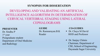
1. SYNOPSIS FOR DISSERTATION
DEVELOPING AND VALIDATING AN ARTIFICIAL
INTELLIGENCE ALGORITHM IN DETERMINATION OF
CERVICAL VERTEBRAL STAGING USING LATERAL
CEPHALOGRAMS
PRESENTER
Dr. Sindhu P
1st year
Postgraduate student
Department of Oral Medicine
and Radiology
GUIDE
Dr. Ramnarayan.B.K
Reader
CO-GUIDES
Dr. Chaya M David
HOD and Professor
Dr. Sanjay Chitnis
Chairman, Department of
CSE, School of Engineering,
Dayanada Sagar University
2. BRIEF RESUME OF THE INTENDED WORK
NEED FOR THE STUDY:
Skeletal maturation refers to the degree of ossification in bone.1
The developmental status of a growing child can be assessed by using various indicators,
which includes chronological age, dental development, secondary sexual characteristics, peak
height velocity, and skeletal maturation.1
Chronological age is unreliable for assessment of developmental status because of the wide
variation in timing and duration of the pubertal growth spurt and other developmental stages.1
3. Radiographic assessment of the hand-wrist bones, by evaluation of ossification stages, is a
reliable indicator of skeletal maturation and is found to be closely related to growth spurt.1
However its main drawback is that an additional radiographs is required causing an additional
radiographic exposure of the patients.1
Lateral Cephalograms have been traditionally used in diagnosis and treatment planning of
orthodontic patients. Lamparski was the first to assess skeletal maturation, which reduced both
radiation exposure and diagnostic cost to the patient. 2
4. He found that this method was a reliable and valid alternative to radiographic assessment of
hand-wrist bones for determination of skeletal age. 2
Lamperski in 1972 assessed the skeletal maturation using cervical vertebrae C2, C3 and C4
by dividing into 6 stages based on the morphological changes. They are Initiation,
Acceleration, Transition, Deceleration, Maturation and Completion. 2
Due to major advances in technology, computer programs that assist in the diagnosis,
treatment and prognosis are routinely used in the field of dentistry. 2
5. Currently, there are many studies utilizing artificial intelligence for the purposes of prediction,
classification and clustering of real-life problems. 2
Radiology is deemed to be the front door for Artificial intelligence(AI) into medicine as
digitally coded diagnostic images are more easily translated into computer language. 8
Machine learning is a key component of AI, and commonly applied to develop image-based AI
systems. 8
6. In the field of dental and maxillofacial radiology (DMFR), reports on AI models used for
diagnostic purposes and treatment planning cover a wide range of clinical applications,
including automated localization of craniofacial anatomical structures/pathological changes,
classification of maxillofacial cysts and/or tumors, and diagnosis of caries and periodontal
lesions.8
With this background, this study is being carried out to develop and validate the AI Algorithm
in determining cervical vertebral stages in lateral cephalograms, which will reflect the growth
and development of individuals.
7. PROBLEM STATEMENT
Traditionally CVS have been analysed by manually tracing lateral cephalograms, which is time
consuming and has the disadvantage of being subject to random and systematic error. Most errors
occur in landmark identification, which is based on observer experience, landmark definition, and
image density and sharpness. To overcome these problems a newer AI technologies have been
increasingly applied. It is proved to be a reliable and time saving tool in many aspects.
8. Giving the data as the foundation for well-construction of models, with high quality and
quantity of data, higher accuracy of predictive result and image interpretation can be achieved
through machine learning process. In the field of dentistry, a well-trained AI model can help
not only in landmark identification, but in all kinds of linear and angular measurements and
volumetric measurements as well. It can save tremendous time by fully automated AI
measurements so researchers will have more energy finding new insights within clinical
examinations.
9. RESEARCH QUESTION: Can Artificial intelligence be used in determination of cervical
vertebrae stages in Lateral Cephalogram ?
RESEARCH HYPOTHESIS (H1): Artificial intelligence can be used in determination of
cervical vertebrae stages in Lateral Cephalogram.
NULL HYPOTHESIS (H0): Artificial intelligence can not be used in determination of cervical
vertebrae stages in Lateral Cephalogram.
10. REVIEW OF LITERATURE:
A study was conducted on the validation of the cervical vertebral maturation(CVM) method
as an indicator of skeletal age in circum-pubertal period by correlating it to the hand-wrist
method(HWM). For this they used cephalometric radiographs of 400 chines with age group
of 10 to 15years for girls and 12 to 17years for boys. Skeletal ages were assessed according
to the CVM method and HWM method. It resulted as CVM was significantly correlated
with HWM skeletal age. Study concluded that CVM is valid indicator of skeletal growth
during the circumpubertal period.4
11. A study was conducted to evaluate and validate skeletal maturation by assessing 3rd and 4th
cervical vertebrae. For this they considered the lateral cephalograms of 50 patients in the age
group of 8 to 14years. Chronologically, they were divided into six groups, based on the age
consisting of a minimum of six to a maximum of 10 subjects. All patients were female. The
result suggested that cervical vertebral bone age calculated is reliable at estimation of bone
age in TW3 method on hand-wrist radiographs.The study concluded that ability to accurately
appraise skeletal maturity from cervical vertebral maturation, without the need for additional
radiographs has the potential to improve orthodontic diagnostic and therapeutic decisions.5
12. A study was conducted based on comparison of hand-wrist and cervical vertebral analysis.
For this they obtain hand wrist radiographs and lateral cephalograms of 72 subjects aged
between 7 and 16 years both male and female. The 9 stages were reduced to 5stages to
compare with cervical vertebral maturation study by Baccetti et al. the Bjork, grave and
brown stages were reduced to six intervals to compare with cervical vertebral maturation
index staging by Hassel and Farman. These measurements were then compared with the hand
wrist bone analysis, and the results were statistically analyzed using Mann-Whitney test.
This study concluded that vertebral analysis on lateral cephalogram is a valid as the hand-
wrist bone analysis with the advantage of reducing the radiation exposure of growing
subjects.1
13. A study was conducted to determine cervical vertebral stages by frequently used seven AI
classifiers and to compare the performance of these with each other. For this they used
cephalometric radiographs from 300 individuals aged between 8 and 17 years. Nineteen
reference points were defined on 2nd, 3rd and 4th cervical vertebrae, and 20 different linear
measurement were taken. The results were observed to have Artificial Neural Networks
(ANN) with second highest accuracy values in determining all stages. Hence study
concluded that ANN could be the preferred method for determining cervical vertebral
stages.3
14. A retrospective study was conducted towards the accuracy and efficiency of a novel software
version for automated bone age assessment in comparison to the Greulich-Pyle method. For
this they took radiographs of 514 patients. Total bone age was assessed independently by three
blinded radiologist applying the Greulich-Pyle method and by artificial intelligence software.
This study concluded that a novel artificial intelligence software enable highly accurate
automated bone age assessment. It may improve efficiency in clinical routine by reducing
reading times without compromising the accuracy compared with the Greulich-Pyle method.6
15. AIM OF THE STUDY:
To develop and validate an artificial intelligence algorithm in determination of cervical
vertebral staging using lateral cepholograms.
16. OBJECTIVES OF THE STUDY:
To determine cervical vertebrae stages in the selected lateral cephalograms obtained from
manual tracing of shape and size of the vertebre.
To derive an algorithm of artificial intelligence to determine cervical vertebral stages in the
selected lateral cephalograms using the data obtained from manual tracing.
To determine cervical vertebrae stages in the selected lateral cephalograms using artificial
intelligence.
To test and validate the developed algorithm in determining cervical vertebral stages in a
different sample of cephalograms
17. MATERIALS AND METHOD:
SOURCE OF DATA:
Lateral Cephalograms of subjects will be collected from Department of Oral Medicine and
Radiology, Dayananda Sagar College of Dental sciences, Bengaluru. which will be taken for
diagnosis and treatment planning of orthodontic purposes.
18. METHOD OF COLLECTION OF DATA:
Permission: Permission will be taken from Department of Oral Medicine and Radiology and
from the institutional review board of Dayananda Sagar College of Dental Sciences to
conduct study.
Sample size to train the AI machine: 420
Sample size to test the AI algorithm: 210
Sample size determination: The sample size for training in order to Derive an Algorithm
was calculated using data based on the study by Hatice kol, Ayse Merve Acilar and Mehmet
Said Izgi.1
19. The details are given below
N = 4 pq
L²
N = Sample size
P = Prevalence
q = (1-p)
L = Allowable error
N = 4×70×30
4.5×4.5
N = 8400
20
N = 420 training samples
Prevalence (p) 70%
q (1-p) 30%
Allowable error (L) 6.5% of prevalence
20. Sample Size Estimation for testing the AI algorithm
Roughly 50% of sample size which are taken for generating algorithm would be considered as a
sample size for testing purpose. And this sample used for testing purpose is selected from a new
set of population (new unlabelled lateral cephalograms)
As there are 6 stages ( 6 groups) under our study, we will select 35lateral cephalograms fulfilling
the criteria of each group.
So that the total sampling size for testing would be
35 × 6 = 210
21. SAMPLING METHOD: Criteria based sampling technique (sequential technique)
STUDY DESIGN: Descriptive study
CRITERIA FOR CASE SELECTION
INCLUSION CRITERIA:
Cephalometric Radiographs of subjects between 7 to 17 years showing adequate quality of
second (C2), third (C3) and fourth (C4) cervical vertebrae which are clearly observed will be
included.
22. EXCLUSION CRITERIA:
Individuals that were subjected to trauma and/or operation in head and neck region.
Individuals who have any disorder that could interfere with bone development.
Individuals with any systemic disease and/or growth and development retardation.
Individuals with congenital and/or acquired malformations in head and neck region.
23. STUDY METHOD
Materials required:
The demographic details such as age, gender will be recorded.
420 Cephalometric Radiographs of individuals between the range of 7 to 17 years which
have been taken for the reason other than current study will be taken from the department of
oral medicine and radiology, Dayananda Sagar College of Dental Sciences, Bengaluru.
Sidexis Software to mark and measure the points on the cervical vertebrae C2, C3 and C4.
Artificial Intelligence Algorithm will be developed in Department of CSE, School of
Engineering, Dayananda Sagar University, Bengaluru.
24. STAGES NUMBER OF SAMPLES
CVS 1 70
CVS 2 70
CVS 3 70
CVS 4 70
CVS 5 70
CVS 6 70
Study group:
Cephalometric Radiographs of 420 individuals in the age group between 7 to 17 years will
comprise of 6 study groups based on cervical vertebral maturation stages with 70 sample
each.
25. CEPHALOMETRIC RADIOGRAPHS:
For the purpose of standardization, Cephalometric Radiographs taken from Sinora
Orthophos XG Machine using Standardized Exposure Parameter having good contrast and
density and showing C2, C3 and C4 clearly will be considered.
Cephalometric Radiographs will be acquired in DICOM format and transferred to a personal
computer for further image analysis.
26. CALIBERATION OF THE EXAMINAERS:
There will be Two Oral and Maxillofacial Radiologist to interpret Cephalometric
Radiographs. For the assessment of reliability and accuracy, 100 Cephalometric
Radiograph will be chosen randomly and evaluated.
For intra-observer variability, the measurements will be repeated after 3 weeks.
For inter-observer variability, all the measurements will be done by two observers
independently.
27. ANALYSIS OF IMAGE:
For Determination of Growth and Maturation of individuals from Cephalometric Radiographs
the second (C2), third (C3) and fourth (C4) cervical vertebrae are evaluated as follows.
28. Identification of 19 references points on C2, C3 and C4(Table 1) along with a total of 20
different linear measurements will be performed as mentioned in table 1,3,4,5. These are
C3–C4 anterior-medial-posterior height, C3–C4 upper-medial-lower width, C2 lower
width, C3–C4 slope, and C2–C3–C4 depth measurements by manual method.
29. Marking will be made on C2, C3 and C4 at various points as mentioned in the table below;
NUMBER POINT DESCRIPTION
1 SVp The posterior point of the edge of the second vertebrae
2 SVd The deepest point of concavity at the lower edge of the second vertebra
3 SVa The anterior point at the lower edge of the second vertebra
4 TVup The posterior point of the upper edge of the third vertebra
5 TVum The center point of the upper edge of the third vertebra
6 TVua The anterior point of the upper edge of the third vertebra
7 TVpm The posterior point of the center edge of the third vertebra
8 TVam The anterior point of the center edge of the third vertebra
9 TVlp The posterior point of the lower edge of the third vertebra
30. 10 TVd The deepest point of the concavity at the lower edge of the third vertebra
11 TVla The anterior point of the lower edge of the third vertebra
12 FVup The posterior point of the upper edge of the fourth vertebra
13 FVum The center point of the upper edge of the fourth vertebra
14 FVua The anterior point of the upper edge of the fourth vertebra
15 FVpm The center point of the posterior edge of the fourth vertebra
16 FVam The center point of the anterior edge of the fourth vertebra
17 FVlp The posterior point of the lower edge of the fourth vertebra
18 FVd The deepest point of concavity at the lower edge of the fourth vertebra
19 FVla The anterior point of the lower edge of the fourth vertebra
31. Table 3: The 20 linear measurements
1) Horizontal and Vertical linear measurements
The horizontal linear measurements The vertical linear measurements
No Name Description No Name Description
A SVp-SVa Point 1 to point 3 A TVup-Tvlp Point 4 to point 9
B TVup-TVua Point 4 to point 6 B TVum-Tvd Point 5 to point 10
C TVpm-Tvam Point 7 to point 8 C TVua-TVla Point 6 to point 11
D TVlp-TVla Point 9 to point 11 D FVup-Fvlp Point 12 to point 17
E FVup-FVua Point 12 to point 14 E FVum-FVd Point 13 to point 18
F FVpm-Fvam Point 15 to point 16 F FVua-FVla Point 14 to point 19
G FVlp-Fvla Point 17 to point 19
32. 2) The anterior and posterior vertebral slope measurements
No. Name Description
G TVlp-TVup XY
H TVua-TVla XY
I FVlp-FVup XY
K FVua-FVla XY
The slope of the posterior edge of C3 vertebrae relative to
the x and y planes.
The slope of the anterior edge of C3 vertebrae relative to
the x and y planes
The slope of the posterior edge of C4 vertebrae relative to
the x and y planes
The slope of the anterior edge of C4 vertebrae relative to
the x and y planes
3) The vertebral depth measurements
No. Name Description
X SVD The perpendicular distance from “a” to deepest point of the inferior
border of the second vertebrae
Y TVD The perpendicular distance from “d” to deepest point of the inferior
border of thethird vertebrae
Z FVD The perpendicular distance from “g” to deepest point of the inferior
border of the forth vertebrae
33.
34. STAGES FEATURES DIAGRAMS
Initiation
(CVS 1)
The lower borders of all the three vertebrae are flat.
The body of C3 and C4 are trapezoidal in shape.
The peak in mandibular growth will occur on average 2 year after this stage.
Acceleration
(CVS 2)
The Concavity at the lower border of C2 is present.
C3 and C4 are trapezoidal in shape.
The peak in mandibular growth will occur on average 1 year after this stage.
Transition
(CVS 3)
The Concavity at the lower borders of C2 and C3 are present.
The bodies of C3 and C4 may be either trapezoid or rectangular in shape.
The peak in mandibular growth will occur during the year after this stage.
35. Deceleration
(CVS 4)
The Concavities at the lower borders of C2, C3 and C4 are present.
The bodies of C3 and C4 are rectangular horizontal in shape.
The peak in mandibular growth has occurred within 1 or 2 years before this
stage.
Maturation
(CVS 5)
The Concavities at the lower borders of C2, C3 and C4 are still present.
At least one of the bodies C3 and C4 is squared in shape. If not squared, the
body of the other cervical vertebra still is rectangular horizontal.
The peak in mandibular growth has occurred within 1 or 2 years before this
stage.
Completion
(CVS 6)
The concavities at the lower borders of C2, C3 and C4 are still present.
At least one of the bodies of C3 and C4 is rectangular vertical in shape. If
not rectangular vertical, the body of the cervical vertebra is squared.
The peak in mandibular growth has ended at least 2 years before this stage.
36. These measured data will be given as input to train the Artificial Intelligence Algorithms,
and the predicted CVS as a result of these Algorithms will be obtained. Then, the
predicted CVS will be compared with the actual CVS.
37. GENERATION OF AI ALGORITHM:
In this study we will be going to use the semi-supervised method. Where there will be set of
labelled data and set of unlabelled data.
The 420 Labelled Cephalometric Radiographs for generating Algorithm will be provided in
order to construct AI algorithm and obtain Deep Learning AI software.
Among the 420 labelled cephalometric radiographs, 50 radiographs will be randomly picked
and Reproducibility test will be conducted to validate the constructed Deep Learning AI
Algorithm.
38. The work flow of proposed algorithm:
IMAGE PREPROCESSING
SELECTION OF REGION OF INTREST
SEGMENTATION OF RADIOGRAPHS
EXTRACTION OF THE SELECTED POINTS IN THE SEGMENTED
RADIOGRAPHS
OUTPUT OF THE STAGING
INPUT IMAGE DATA
39. ALGORITHM VALIDATION:
The new set of unlabelled Cephalomatric radiographs of 210 images which are not used
for the generation of the AI algorithm will be taken to test and validate the accuracy of the
derived AI Algorithm.
STATISTICAL ANALYSIS:
Statistical Package for Social Sciences [SPSS] for Windows Version 22.0 Released 2013.
Armonk, NY: IBM Corp., will be used to perform statistical analyses.
40. Descriptive Statistics:
Descriptive analysis of all the explanatory parameters will be done using mean and
standard deviation for quantitative variables, frequency and proportions
for categorical variables.
Inferential Statistics:
McNemar’s test will be used to compare the distribution of CVM stages between Manual
and AI protocol. Intraclass correlation test will be used to assess for the concordance of
CVM stages between Manual and AI algorithm on random samples selected after the
Algorithm sequence generation. Sensitivity and Specificity analysis will be used to obtain
the accuracy levels of AI in determining the CVM stages on the lateral cephalogram. The
level of significance will be set at P<0.05. And any other relevant test, if found appropriate
during the time of data analysis will be dealt accordingly.
41. Does the study require any investigations or interventions to be conducted on patients or
other humans or animals? If so, describe briefly
NO. This study does not require any investigations or interventions to be conducted on patients
or other humans or animals. Cephalometric radiographs already acquired for reasons other than
our study will be collected. No patient will be subjected for Cephalometric exposure for our
study. However care will be taken to collect these Cephalometric radiographs from department
of oral medicine and radiology which follow the protocol of taking a consent for the
Cephalometric exposure procedure.
Has ethical clearance has been obtained from the institution?
Ethical clearance yet to be obtained.
42.
43.
44.
45.
46. LIST OF REFERENCES:
1. Pichai S, Rajesh M, Reddy N, Adusumilli G, Reddy J, Joshi B. A comparison of hand
wrist bone analysis with two different cervical vertebral analysis in measuring skeletal
maturation. J Int Oral Health. 2014;6(5):36-41.
2. Chandrasekar R, Chandrasekhar S, Sundari KS, Ravi P. Development and validation of
a formula for objective assessment of cervical vertebral bone age. Prog Orthod.
2020;21(1):1-8.
3. Kök H, Acilar AM, İzgi MS. Usage and comparison of artificial intelligence algorithms
for determination of growth and development by cervical vertebrae stages in
orthodontics. Prog orthod. 2019;20: 41-50.
4. Kunz F, Stellzig-Eisenhauer A, Zeman F, Boldt J. Artificial intelligence in orthodontics.
J Orofac Orthop. 2020;81(1):52-68.
47. 5. Prasad C, Reddy VN, Sreedevi G, Ponnada SR, Priya KP, Naik BR. Objective evaluation of
cervical vertebral bone age' its reliability in comparison with hand-wrist bone age: By TW3
method. J Contemp Dent Pract. 2013;14(5):806-13.
6. Booz C, Yel I, Wichmann JL, Boettger S, Al Kamali A, Albrecht MH, Martin SS, Lenga L,
Huizinga NA, D’Angelo T, Cavallaro M. Artificial intelligence in bone age assessment:
accuracy and efficiency of a novel fully automated algorithm compared to the Greulich-Pyle
method. Eur Radiol Exp. 2020;4(1):1-8.
7. Kunz F, Stellzig-Eisenhauer A, Zeman F, Boldt J. Artificial intelligence in orthodontics. J
Orofac Orthop. 2020;81(1):52-68.
48. 8. Hung K, Yeung AW, Tanaka R, Bornstein MM. Current Applications, Opportunities, and
Limitations of AI for 3D Imaging in Dental Research and Practice. Int J Environ Res Public
Health. 2020;17(12):4424.
9. Liu J, Qi J, Liu Z, Ning Q, Luo X. Automatic bone age assessment based on intelligent
algorithms and comparison with TW3 method. Comput Med Imaging Graph.
2008;32(8):678-84.
10. Alhadlaq AM, Al-Maflehi NS. New model for cervical vertebral bone age estimation in boys.
J King Saud Univ Sci. 2013;4(1):1-5.