Introduction
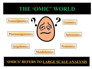
Emerging Field of‘Omics' Research
• Unbiased global survey of all low molecular-weight
molecules or metabolites in biofluid, cell, tissue,
organ, or organism
• Study of a range of metabolites in cells or organs &
ways they are altered in disease states and their
changes over time as a consequence of stimuli
(including biological perturbation such as diet,
disease, or intervention)
3.
• Metabolome refersto a complete set of
small-molecule metabolites in a biological
sample, such as a single organism.
Any organic molecule detectable in the body with MW
< 1000 Dalton with a concentration ≥ 1 µM
Includes peptides, oligonucleotides, sugars,
nucleotides, organic acids, ketones, aldehydes, amines,
amino acids, lipids, steroids, alkaloids and drugs
(xenobiotics)
Includes human & microbial products
4.
Introduction contd…
• Thename ‘metabolomics’ was coined in the late 1990s
– The first paper using the word was by Oliver, S. G., Winson,
M. K., Kell, D. B. & Baganz, F. (1998). Systematic functional
analysis of the yeast genome.Trends Biotechnol.1998
Sep;16(9):373-8.
• Study of metabolome, started decades ago with early
applications in field of toxicology, inborn metabolic errors &
Nutrition
• Original report to mention metabolomics approach in oncology
dates back 25 years ago when authors claimed that cancer could
be identified from nuclear magnetic resonance (NMR) spectra
generated from blood samples*
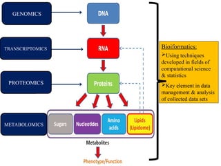
Bioiformatics:
Using techniques
developed infields of
computational science
& statistics
Key element in data
management & analysis
of collected data sets
GENOMICS
TRANSCRIPTOMICS
PROTEOMICS
METABOLOMICS
7.
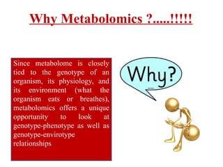
Why Metabolomics ?.....!!!!!
Sincemetabolome is closely
tied to the genotype of an
organism, its physiology, and
its environment (what the
organism eats or breathes),
metabolomics offers a unique
opportunity to look at
genotype-phenotype as well as
genotype-envirotype
relationships
8.
In Other Words……..
•Not all changes or abnormalities
detected in the genome or transcriptome
may be causing abnormality or disease
e.g. silent mutations
• Similarly not all enzymes & protein
products detected via proteomics are
functional
• Also they do not take into account
environmental influences occurring at a
later stage
• Can be used to monitor changes in the
genome or to measure the effects of
downregulation or upregulation of
specific gene transcript
• Metabolites are the ultimate result of
cellular pathways (taking into account
changes in genome, transcriptome,
proteome as well as metabolic
influences)
Direct correlation with abnormalities being caused
9.
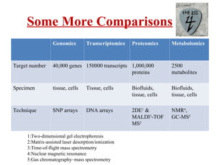
Some More Comparisons
GenomicsTranscriptomics Proteomics Metabolomics
Target number 40,000 genes 150000 transcripts 1,000,000
proteins
2500
metabolites
Specimen tissue, cells Tissue, cells Biofluids,
tissue, cells
Biofluids,
tissue, cells
Technique SNP arrays DNA arrays 2DE1
&
MALDI2
-TOF
MS3
NMR4
,
GC-MS5
1:Two-dimensional gel electrophoresis
2:Matrix-assisted laser desorption/ionization
3:Time-of-flight mass spectrometry
4:Nuclear magnetic resonance
5:Gas chromatography–mass spectrometry
10.
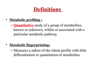
Definitions
• Metabolic profiling:
– Quantitative study of a group of metabolites,
known or unknown, within or associated with a
particular metabolic pathway
• Metabolic fingerprinting:
– Measures a subset of the whole profile with little
differentiation or quantitation of metabolites
11.
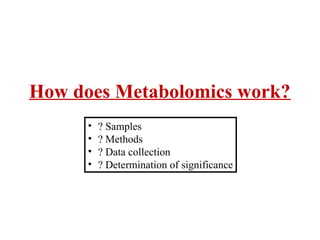
How does Metabolomicswork?
• ? Samples
• ? Methods
• ? Data collection
• ? Determination of significance
12.
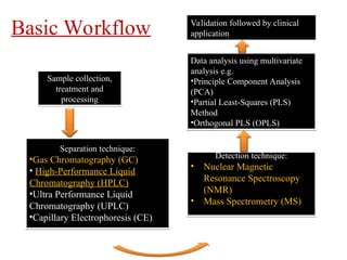
Sample collection,
treatment and
processing
Detectiontechnique:
• Nuclear Magnetic
Resonance Spectroscopy
(NMR)
• Mass Spectrometry (MS)
Separation technique:
•Gas Chromatography (GC)
• High-Performance Liquid
Chromatography (HPLC)
•Ultra Performance Liquid
Chromatography (UPLC)
•Capillary Electrophoresis (CE)
Data analysis using multivariate
analysis e.g.
•Principle Component Analysis
(PCA)
•Partial Least-Squares (PLS)
Method
•Orthogonal PLS (OPLS)
Basic Workflow Validation followed by clinical
application
13.
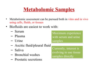
Metabolomic Samples
• Metabolomicassessment can be pursued both in vitro and in vivo
using cells, fluids, or tissues
• Biofluids are easiest to work with:
– Serum
– Plasma
– Urine
– Ascitic fluid/pleural fluid
– Saliva
– Bronchial washes
– Prostatic secretions
Maximum experience
with serum and urine
samples
Currently, interest is
evolving to use tissue
samples directly
14.
Sample Collection &Handling
• All biological samples collected for metabolic analysis require careful
sample handling, special requirements for diet, physical activities, &
other patient validation
• Due to the high susceptibility of metabolic pathways to exogenous
environments, maintaining low temperatures and consistent sample
extraction is essential
• For biofluids, standard sample volume: 0.1 to 0.5 mL
15.
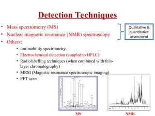
Detection Techniques
• Massspectrometry (MS)
• Nuclear magnetic resonance (NMR) spectroscopy
• Others:
• Ion-mobility spectrometry,
• Electrochemical detection (coupled to HPLC)
• Radiolabelling techniques (when combined with thin-
layer chromatography)
• MRSI (Magnetic resonance spectroscopic imaging)
• PET scan
Qualitative &
quantitative
assessment
MS NMR
16.
Gas Chromatography– &Liquid
Chromatography–Mass Spectrometry
(MS)
• Both approaches involve an initial chromatographic stage
followed by separation according to their mass-to-charge
ratio
• Current detection limits for MS-based approaches are 100
nM, allowing the detection of large no. of metabolites.
• However, not all metabolites can be ionized equally,
potentially biasing the information produced.
• Typical acquisition times of about 30 minutes
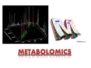
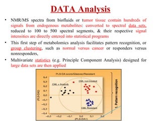
• NMR/MS spectrafrom biofluids or tumor tissue contain hundreds of
signals from endogenous metabolites: converted to spectral data sets,
reduced to 100 to 500 spectral segments, & their respective signal
intensities are directly entered into statistical programs
• This first step of metabolomics analysis facilitates pattern recognition, or
group clustering, such as normal versus cancer or responders versus
nonresponders,
• Multivariate statistics (e.g. Principle Component Analysis) designed for
large data sets are then applied
DATA Analysis
19.
Conclusion
• Metabolomics isa novel discipline encompassing comprehensive metabolite
evaluation, pattern recognition & statistical analyses
• May provide the ability to diagnose cancer in its curative state, determine the
aggressiveness of cancer to help direct prognosis, and therapy, & predict drug
efficacy
• Still in its infancy & has lagged behind other ‘omic’ sciences due to technical
limitations, database challenges
• It is a long path of discovery, confirmation, clinical trials, and approval to
establish test validity and utility
• Urgent need to establish spectral databases of metabolites, as well as cross-
validation of NMR- or MS-obtained metabolites & correlation with other
quantitative assays
• It is important to integrate it with other ‘omics’ technology so that the entire
spectrum of the malignant phenotype can be characterized
#1 Metabolomics is the solution to this problem. A comprehensive, systems biology conscious approach to understanding the Metabolome in its full scope. Metabolomics seeks to avoid reductionism and apply high throughput analysis methods on metabolic levels in the cell. It will revolutionize fields like metabolic engineering and increase our knowledge of biological function phenomenally.
#2 Context dependent
Metabolomics, one of the "omic" sciences in systems biology, is the global assessment and validation of endogenous small-molecule biochemicals (metabolites) within a biologic system.
Perhaps the best description of this approach was offered by Steve Oliver of University of Cambridge, who used the term ‘metabolomics’ to describe “the complete set of metabolites/low molecular weight intermediates, which are context dependent, varying according to the physiology, developmental or pathological state of the cell, tissue, organ or organism”.
Metabolomics, one of the "omic" sciences in systems biology, is the global assessment and validation of endogenous small-molecule biochemicals (metabolites) within a biologic system. Initially, putative quantitative metabolic biomarkers for cancer detection and/or assessment of efficacy of anticancer treatment are usually discovered in a preclinical setting (using animal and human cell cultures), followed by translational validation of these biomarkers in biofluid or tumor tissue. Based on the tumor origin, various biofluids, such as blood, urine, and expressed prostatic secretions, can be used for validating metabolic biomarkers noninvasively in cancer patients. Metabolite detection and quantification is usually carried out by nuclear magnetic resonance (NMR) spectroscopy, while mass spectrometry (MS) provides another highly sensitive metabolomics technology. Usually, sophisticated statistical analyses are carried out either on spectroscopic or on quantitative metabolic data sets to provide meaningful information about the metabolic makeup of the sample. Various metabolic biomarkers, related to glycolysis, mitochondrial citric cycle acid, choline and fatty acid metabolism, were recently reported to play important roles in cancer development and responsiveness to anticancer treatment using NMR-based metabolic profiling.Carefully designed and validated protocols for sample handling and sample extraction followed by appropriate NMR techniques and statistical analyses, which are required to establish quantitative (1)H-NMR-based metabolomics as a reliable analytical tool in the area of cancer biomarker discovery, are discussed in the present chapter.
emerging field of metabolomics is based on the premise that the identification and measurement of metabolic products will enhance our understanding of physiology and disease
Studies of tumour cell and tissue allow focused analysis on the tumour, whilst studies of biofluids have the appeal of concurrent assessment of tumour and host.
#4 The term metabolomics was first used in context of yeast in the late 90’s by mr.Oliver steve
Stephen Oliver is a Professor in the Department of Biochemistry at the University of Cambridge
Based on premise
Identification and measurement of metabolic products will enhance our understanding of physiology and disease
The first paper was titled, “Quantitative Analysis of Urine Vapor and Breath by Gas-Liquid Partition Chromatography”, by Robinson and Pauling in 1971.
Terminology relating to metabolomics has been controversial.
4
The term “metabolome” was first used by Olivier et al. in 1998
5
to describe the set of metabolites synthesized by an organism, in
a fashion analogous to that of the genome and proteome. This
definition has been limited
6
to “the quantitative complement of
all of the low molecular weight molecules present in cells in a
particular physiological or developmental state”. Metabolomics
was coined by Fiehn
7
and defined as a comprehensive analysis in
which all metabolites of a biological system were identified and
quantified
Many of the bioanalytical methods used for metabolomics have been adapted (or in some cases simply adopted) from existing biochemical techniques.
A sensitive and specific blood test for cancer has long been sought. The water-suppressed proton nuclear magnetic resonance (NMR) spectrum of plasma is dominated by the resonances of plasma lipoprotein lipids. We measured the mean line widths of the methyl and methylene resonances, which were found to be correlated with the presence or absence of malignant tumors. Values for the average line width were lower in patients with cancer. We analyzed plasma from 331 people (normal controls, patients with malignant and benign tumors, patients without tumors, and pregnant patients); NMR analysis and measurement of line widths were blinded to diagnosis or patient group. The mean line width for 44 normal controls (±SD) was 39.5±1.6 Hz. For 81 patients with untreated cancer, demonstrated by biopsy, the line width was 29.9±2.5 Hz. Patients with malignant tumors were reliably distinguished from normal controls by this method (P<0.0001), and differed from patients with diseases that did not involve tumors (line width, 36.1±2.6 Hz; P<0.0001). Patients with benign tumors (e.g., those of the breast, ovary, uterus, and colon) had line widths of 36.7±2.0 Hz and were different from those with malignant tumors (P<0.0001). However, pregnant patients and those with benign prostatic hyperplasia had line widths consistent with the presence of malignant tumors. The narrowing of lipoprotein-lipid resonances with cancer is consistent with the response of a host to tumor growth.
We conclude that these preliminary results demonstrate that water-suppressed proton NMR spectroscopy is a potentially valuable approach to the detection of cancer and the monitoring of therapy. (N Engl J Med 1986; 315:1369–76.)
#6 flux have a significant impact on metabolite concentra-
tions10–12
.This is because the control of the metabolic flux
of a pathway is spread across all the enzymes present
in the pathway, rather than being controlled by a rate-
determining step. Furthermore, there is not necessarily a
good quantitative relation between mRNA concentra-
tions and enzyme function, but as metabolites are down-
stream of both transcription and translation, they are
potentially a better indicator of enzyme activity13
.So,
metabolomics offers a particularly sensitive method to
monitor changes in a biological system, through observed
changes in the metabolic network.
#8 For example influences occurring at level of proteomes wont be picked up by genome or transcriptome
Metabolites are the ultimate result of cellular pathways (taking into account changes in genome, trancriptome, proteome as well as metabolic influences) hence more likely to
#9 Is metabolomics the greatest “omics” of all? Certainly, it has
been suggested that metabolomics may in fact provide the most
“functional” information of the omics technologies.
1
This reflects
the limitations associated with transcriptomics and proteomics;
for example, changes in the transcriptome and proteome do not
always result in altered biochemical phenotypes (the metabolome).
1,2
Furthermore, the metabolome represents the final “omic”
level in a biological system, and metabolites represent functional
entities, unlike messenger RNA molecules, which constitute the
transcriptome.
3 Metabolites thus have a clear function in the life
of the biological system and are also contextual,
3
reflecting the
surrounding environment. The metabolome can thus be thought
of as a looking glass, which if looked through can show information concerning the physiological, developmental, and pathological
status of a biological system
for the detection and prevention of adulteration.
Functional genomics, as the name implies, aims to decipher
gene function by establishing a better understanding of the
correlation between genes and the functional phenotype of an
organism.
28
Since the metabolome of a system represents the
amplification and integration of signals from other functional
genomic levels (e.g., transcriptome and proteome),
29
metabolomics
can be considered tool for functional genomics. Functional
genomics represents a way to do “smarter” genomics, rather than
simply gene mapping and sequencing, and motivation for this
research endeavor arises because of the large proportion of open
reading frames (typically 20-40%
30
) in a fully sequenced organism
that have no known function at the biochemical and phenotype
levels. Such genes are referred to as “silent” or “orphan” genes.
In the case of Saccharomyces cerevisiae, for example, around 6000
protein encoding genes exist; however, there are less than 600
low molecular weight intermediate metabolites (cited in ref 3)
Determining gene function can be achieved through metabolite
profiling of specific genetically altered organisms. These metabolite profiles may then be compared to that of a “control” organism
to yield information about the metabolic consequence of the
altered genome
31
and ultimately assign gene function. This
approach was first used by Roessner et al.,
Systems biology uses an approach similar to that of functional
genomics, but has significantly greater aims than the latter.
Systems biology represents the ultimate challenge in that is aims
to integrate genomics, transcriptomics, proteomics, and metabolomics
32
for a global understanding of biological systems. In
essence, systems biology looks at the big picture to obtain a better
understanding of how individual pathways or metabolic networks
are related. Systems biology does not investigate individual genes,
proteins, or metabolites one at a time, but rather investigates the
behavior and relationships of all the elements in a particular
biological system while it is functioning.
33
The general systems
biology approach is a perturbation of the system (biologically,
genetically, or chemically), followed by monitoring the impact of
the perturbation at the genomic, proteomic, and metabolomic
levels. These omic data can then be integrated and ultimately
modeled computationally for a complete understanding of system
functioning. The potential impact of systems biology is enormous,
ranging from metabolite engineering
1
to reshaping medicine
toward predictive, preventative, and personalized prevention of
cellular dysfunction and disease
One of the goals of systems biology is to define interacting cellular networks in the context
of a disease phenotype, tissue-specific functions or reaction to specific stimulus or
intervention. Systems biology as applied to cancer research encompasses the “omic”
sciences of genomics, transcriptomics, proteomics, and metabolomics. Metabolomics
(sometimes known as metabonomics) entails evaluation of the patterns and concentration of
low molecular weight metabolites over broad classes of compounds in a tissue or organ.
These metabolites are the small molecule intermediates and end products of the biochemical
reactions in a cell, and are represented by compounds with mass typically in the range of
80–1000 Daltons. Metabolomic studies range from targeted analysis of one or a small
number of metabolites associated with a specific biological pathway to the unbiased
profiling or fingerprinting of a large subset of metabolites associated with a specific
phenotype or stimulus. Although complementary to genomics, transcriptomics and
proteomics, metabolomics may have advantages for defining phenotypes because it is
downstream of changes in genes and proteins, and thus may be a better indicator of distinct
functional alterations in pathways affected by different pathological states. In this sense,
metabolomic profiles represent the integration of genetic regulation, enzyme activity and
metabolic reactions in a dynamic profile of the biological state of a tissue [8]. Furthermore,
because the total complement of metabolites is likely to be considerably smaller than the
number of genes, transcripts, or proteins, metabolomics may be able to more clearly
characterize altered cellular networks and activity associated with disease states.
#10
Most of the research today regarding metabolomics is based on characterizing metabolic profile
Aims at finding unique metabolic characteristics for a cell
Historical approaches to metabolite analysis include metabolite
profiling, metabolite fingerprinting, and target analysis. Metabolite
fingerprinting aims to rapidly classify numerous samples using
multivariate statistics, typically without differentiation of individual
metabolites or their quantitation. Target analysis is constrained
exclusively to the qualitative and quantitative analysis of a
particular metabolite or metabolites. As a result, only a very small
fraction of the metabolome is focused upon, signals from all other
components being ignored.
13 Metabolite profiling involves the
identification and quantitation by a particular analytical procedure
of a predefined set of metabolites of known or unknown identity
and belonging to a selected metabolic pathway.
7,10
By their nature,
these approaches provide a restrictive noncomprehensive view
of the metabolome. Nevertheless, metabolite profiling represents
the oldest and most established approach and can be considered
the precursor for metabolomics
Metabolic Fingerprinting: A mass profile of the sample of interest is generated and then compared in a large sample population to screen for differences between the samples. ‘Metabolic fingerprinting’refers to measuring a subclass of metabolites to create a ‘bar code’ of metabolism
In this approach, only a limited number of metabolites are quantified and used to distinguish between different samples, such as those of different disease or physiological states
Metabolic profiling : has been proposed as a means of measuring the total complement of individual metabolites in a given biological sample
Jeremy Nicholson to coin the word ‘metabonomics’.He
defines metabonomics as “the quantitative measure-
ment of the multivariate metabolic responses of multi-
cellular systems to pathophysiological stimuli or
genetic modification”27
.In addition to the terms
‘Metabolic pro-
filing’ has been proposed as a means of measuring the
total complement of individual metabolites in a given
biological sample, whereas ‘metabolic fingerprinting’
refers to measuring a subclass of metabolites to create a
‘bar code’ of metabolism23,24
.In this approach, only a
limited number of metabolites are quantified and used
to distinguish between different samples, such as those
of different disease or physiological states
#11 What r the samples where test can be performed..methods used….how is data collected…whether observed difference or abnormality is really significantand can we apply them in clinical field
#12 Data analysis followed by validation and clinical application
#13 Most experience to date is with serum and urine samples as a surrogate system for tumor biochemistry
Interest is evolving for metabolomic
studies directly using tumor tissue; however, such analyses require a more difficult and careful
tissue preparation due to tissue heterogeneity. Surrounding stromal and epithelial cells can
cause contamination of the resulting metabolic profile, thereby skewing results compared with
that obtained from a pure tumor tissue sample. Microdissection techniques could enhance
sample purity but also increase the required equipment and expertise.
#14 For NMR, minimal sample preparation is required for urine and
other low-molecular-weight metabolite-containing fluids, whereas blood, plasma, and serum
require extraction (using acid, acetonitrile, or two-phase methanol/chloroform protocols) or
NMR-weighted techniques to separate polar and lipophilic metabolites (see Table 1; refs. 23,
24). Intact tissue specimens (e.g., biopsies, fine needle aspirates) can be analyzed using high-
resolution magic angle spinning (HR-MAS). HR-MAS probes for solid state NMR, as well as
cryoprobes and microprobes for liquid NMR, permit quantitative metabolic analysis on
samples as small as 3 μL with improved signal-to-noise ratios and solvent suppression (5). MS
analysis requires more labor-intensive and destructive tissue preparation than NMR, but has
greater sensitivity for metabolite detection
MS analysis requires more labor-intensive and destructive tissue preparation than NMR
#15 Ion-mobility spectrometry, electrochemical detection (coupled to HPLC) and radiolabel (when combined with thin-layer chromatography)
Magnetic resonance spectroscopic imaging (MRSI) measures metabolite concentrations in
vivo, in an analogous fashion to the way conventional magnetic resonance imaging (MRI)
measures water. Because the concentration of water and lipids in soft tissues such as the
prostate is orders of magnitude greater than the concentration of metabolites, MRSI requires
higher field strength than conventional MRI, and water and lipid suppression techniques to
allow accurate resolution of metabolite spectra. Potential combined modality applications
include combining MRSI and dynamic contrast enhanced MRI for enhanced visualization of
suspicious prostate lesions or areas of recurrence, and overlaying MRSI images on
transrectal ultrasound images for guiding prostate biopsy [13]. Current limitations to the use
of MRSI include relatively high cost and limited availability of higher field strength (3 Tesla
or higher) platforms needed for better spectral resolution. Most applications of MRSI in
prostate cancer have focused on diagnostic imaging rather than metabolomic profiling of
cellular networks so MRSI will not be further discussed in this article; for an excellent
review see Sciarra et al. [14].
#16 Both approaches involve an initial chromatographic stage in which metabolites are separated either in the gas or solution
phase, respectively. Subsequently the metabolites are ionized and then separated according to their mass to charge ratio,
which can be used to identify the metabolites.MS-based approaches are more sensitive than NMR spectroscopy, and so
can potentially detect metabolites at a concentration two orders of magnitude below that of NMR.However, not all
metabolites can be ionized (converted to a positively or negatively charged species suitable for mass spectrometry) to an
equal extent, potentially biasing the information produced.This approach is the method of choice for plant
metabolomics23,24
where the challenge of profiling all the metabolites in a given tissue is even greater than that in
mammals and yeast. In spite of the fact that plant genomes typically contain 20,000–50,000 genes, 50,000 metabolites
have been identified in the plant kingdom with the number predicted to rise to about 200,000 (REF. 74),compared with
30–600 metabolites identified in mammalian cells.The current detection limits for MS-based approaches are of the order
of 100 nM, allowing the detection of about 1,000 metabolites,with typical acquisition times of about 30 minutes.
Mass spectrometry (MS) requires an initial separation of metabolites by gas or liquid
chromatography (GC, LC), followed by ionization of metabolites and resolution according
to mass-to-charge ratio. The advantage of MS methods over NMR is much higher sensitivity
and detection of metabolites at much lower concentrations, and it is more suitable for high
throughput methods. However, these advantages come at the cost of more extensive sample
preparation (particularly for GC-MS), and metabolite detection can be complicated by
differences in ionization efficiency, stability, extraction efficiency, and fragmentation
behavior. Derivatization is used to optimize these characteristics, but different reagents are
used depending on the purpose of the derivatization and where in the GC-MS or LC-MS
process it occurs, which can complicate comparisons across studies. Derivatization can also
result in metabolite degradation. Other sources of variation include metabolite pK, polarity,
processes of extraction and quenching, and type of instrument [8,12].
#18 Because 1 H-NMR or MS spectra from biofluids or tumor
tissue contain hundreds of signals from endogenous metabo-
lites and are highly redundant, spectral data sets, reduced to
100 to 500 spectral segments, and their respective signal
intensities are directly entered into statistical programs (5, 21,
29). This first step of metabolomics analysis facilitates pattern
recognition, or group clustering, such as normal versus cancer
or responders versus nonresponders, based on spectral pattern
differences. The interpretation of scores reveals information
about relationships between samples and illustrates trends,
groupings, and/or outliers. In the last 5 years, due to the
quantity and complexity of spectroscopic data from NMR and
MS studies, the majority of metabolic profiling studies have
used computer-aided statistical interpretation of the data. This
improves the refining and distilling of complex raw data.
Similar to gene array analyses, multivariate statistics have been
designed for large data sets, with two major types of pattern
recognition processes, unsupervised and supervised. Unsuper-
vised data analysis, such as hierarchical cluster analysis and
principal component analysis, measures the innate variation in
data sets, whereas the supervised approach, including principal
component regression and neural networks, uses prior infor-
mation to generate the clusters of patterns (30). Although
beyond the scope of this review, many other statistical
approaches exist, including cluster analysis, linear discriminant
analysis, Bayesian spectral decomposition, and several other
chemometric methods (31).
#19 Should be used for identifying multivariate biomarkers, including fingerprints, profiles, or patterns characterizing state of cancer
Metabolomics is a novel discipline encompassing comprehensive metabolite evaluation,
pattern recognition, and statistical analyses. Biomarkers are widely used in clinical medicine
for prognostic or predictive interpretation of disease status. Metabolomics should be used for
identifying multivariate biomarkers, including fingerprints, profiles, or signatures, the patterns
of which characterize a state of cancer. By using this technology, we might eventually be able
to diagnose cancer earlier when it is still amenable to cure, determine aggressiveness of cancer
to help direct prognosis and therapy, and predict drug efficacy. These signatures can be practical
and accurate although they also require sophisticated analytic techniques (70,71).