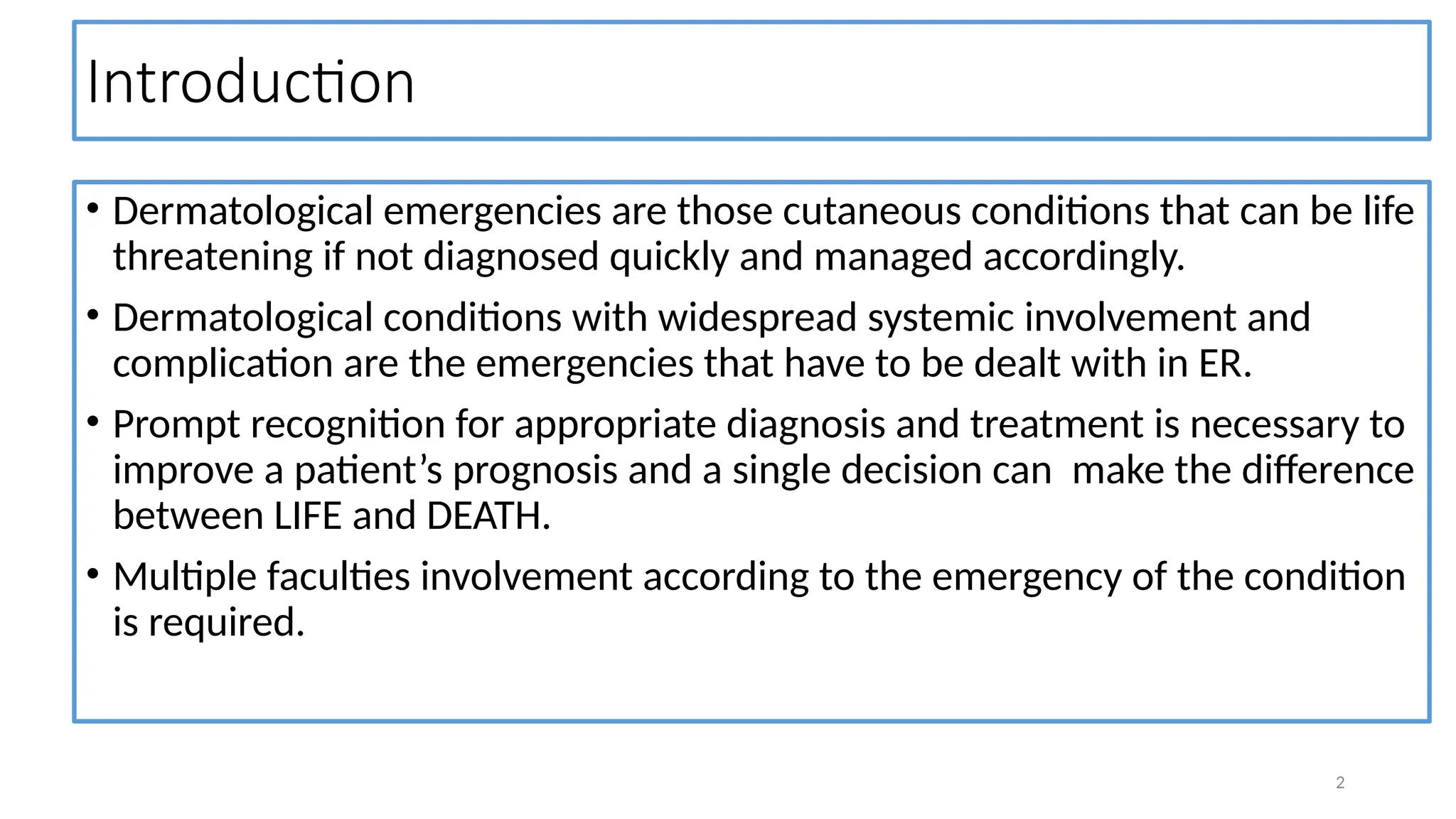
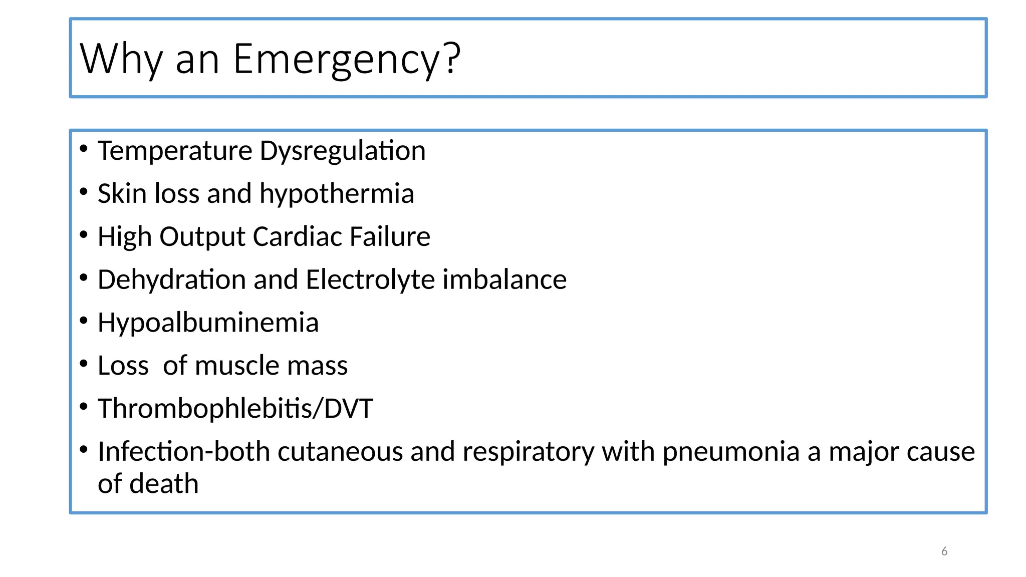
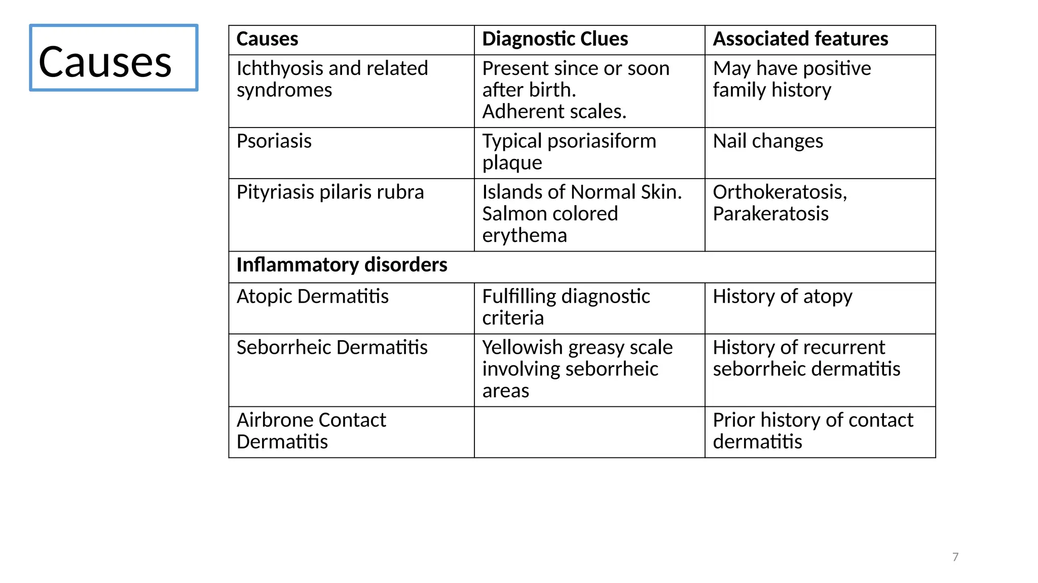
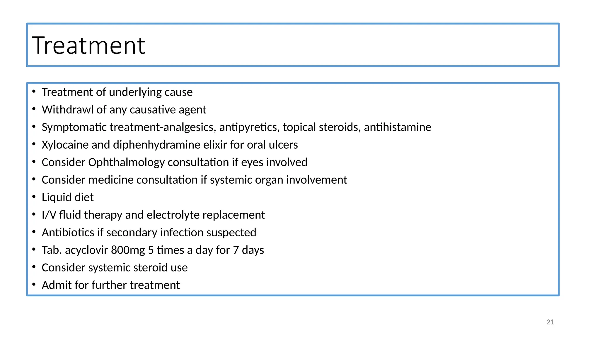
1
DERMATOLOGICAL
EMERGENCIES
Sunil Timilsina
1st
year PG Resident GP and EM
NAIHS
Outline
• Introduction
• Conditions resulting in dermatological emergencies
• References
1
Introduction
• Dermatological emergencies are those cutaneous conditions that can be life
threatening if not diagnosed quickly and managed accordingly.
• Dermatological conditions with widespread systemic involvement and
complication are the emergencies that have to be dealt with in ER.
• Prompt recognition for appropriate diagnosis and treatment is necessary to
improve a patient’s prognosis and a single decision can make the difference
between LIFE and DEATH.
• Multiple faculties involvement according to the emergency of the condition
is required.
2
Conditions
resulting in
Dermatological
Emergencies
1.Erythoderma 8. Bacterial Toxin Mediated Illnessess
- Staphylococcal Scalded Skin Syndrome
-Toxic Shock Syndrome
2. Anaphylaxis 9. Drugs induced Cutaneous Necrosis
Warfarin/Heparin/Vasopressors
3. Severe Cutaneous Drug Reactions 10. Vesiculobullous disorders
-Pemphigus Foliaceus
4. Psoriasis
- Psoriatic Erythroderma
- Acute generalized pustular Psoriasis
11. Vascular disorders
5. Leprosy Reactions 12. Acute Graft Vs Host Disease
6. Erythema Multiforme 13. Metabolic Disorders
-Pellagra
- Acrodermatitis Enteropathica
7. Infections
3
Erythroderma
• Erythroderma is an inflammatory skin disease with redness and scaling
that affects 90% or more of the patients skin.
• Also known as Exfoliative Dermatitis
• Extensive skin exfoliation : 20-30g/day
• 18-20% mortality
• Males affected more (4x)
4
Presentation
• Initial lesions
• Erythema – rapidly progressing later scaling
• Established Case
• Erythema, Scaling and Induration involving >90% BSA
• Ectropion, Eclabium, Everted nostrils and flexion contracture of joints
• Later
• Exagerrated skin folds and shedding of hair, eyebrows, eyelashes and nostrils
• Lymphadenopathy
• Association : Extreme pruritus, Enteropathy,
• Lesion of primary illness
5
Why an Emergency?
• Temperature Dysregulation
• Skin loss and hypothermia
• High Output Cardiac Failure
• Dehydration and Electrolyte imbalance
• Hypoalbuminemia
• Loss of muscle mass
• Thrombophlebitis/DVT
• Infection-both cutaneous and respiratory with pneumonia a major cause
of death
6
Causes Diagnostic Clues Associated features
Ichthyosis and related
syndromes
Present since or soon
after birth.
Adherent scales.
May have positive
family history
Psoriasis Typical psoriasiform
plaque
Nail changes
Pityriasis pilaris rubra Islands of Normal Skin.
Salmon colored
erythema
Orthokeratosis,
Parakeratosis
Inflammatory disorders
Atopic Dermatitis Fulfilling diagnostic
criteria
History of atopy
Seborrheic Dermatitis Yellowish greasy scale
involving seborrheic
areas
History of recurrent
seborrheic dermatitis
Airbrone Contact
Dermatitis
Prior history of contact
dermatitis
Causes
7
Causes Diagnostic Clues Associated features
Pemphigus Foliaceous Superfical erosions with
collarette of scales
+ve Nikolsky sign
Subcorneal split in HPE
SSSS Skin tenderness +ve Nikolsky sign
Norwegian Scabies Intense pruritus in
background of
immunocompromised
states
Mite under microscope
Drug Hypersensitivity
Syndrome
Systemic involvement History of drug intake
Sezary syndrome Leonine facies in long
standing cases
Sezary cells in PBS
Dermatomyositis Heliotrop rash,
Poikiloderma, Gottron’s
papules, proximal
muscle weakness
Raised muscle enzymes
Sarcoidosis Ichthyosiform
erythroderma
Sarcoidal granuloma in
HPE
Causes
8
Investigations
• CBC,RFT with electrolyte, LFT, blood culture, RBS
• Skin swab-microscopy and culture sensitivity
• Chest x-ray
• Skin biopsy-if cause uncertain
9
Reference: DOI:https://doi.org/10.1016/j.mpmed.2017.04.005 10
11
Condition Clinical picture Treatment
Anaphylaxis • Intense pruritus
• Flushing
• Stridor
• Hypotension Shock
• Adrenaline
• Respiratory Support
• Antihistaminic
• Steroids
Utricaria • Intense pruritus
• Localized wheals
• Triggerer avoidance
• Emollients
• Antihistaminic
• Leukotriene antagonists
• Steroids
• Immunotherapy
Angioedema • Swelling of
face/lips/peri orbital
region
12
Acute Generalized Pustular Psoriasis
• Occurs in patients with any variant of psoriasis
• Due to any provocating factors
• Most commonly after sudden withdrawal of topical or systemic
steroids.
13
Why an Emergency?
• Severe hypoalbuminemia
• Hypocalcemia and tetany
• Oligemia and Acute tubular Necrosis
• Malabsorption
• Deep vein Thrombosis and Pulmonary Embolism
14
Clinical Presentation
• Generalized pustular eruptions with “lake
of pus” formation
• Intense burning sensation, erythema
• Skin tenderness
• Fever
• Repeated crops of lesions
• May develop erythroderma
1
Investigations
• Basic laboratory profile
• Gram staining of smear from pustules  PMN cells without organism
• Skin biopsy
16
Management
• DICU admission
• General Measures
• Mild sedation
• Fluid and electrolytes
• High protein diet
• Systemic Therapy:
• Acitretin 1mg/kg/day to overcome acute stage and then 0.5 to 0.75mg/kg/day to
control
• PUVA therapy
• Acetretin + PUVA
• Methotrexate IV/IM 0.3mg/kg/week
• High dose cyclosporine (9-12 mg/kg/day)
• Parenteral corticosteroids
17
Erythema Multiforme
• Acute, usually mild, self-limited cutaneous
and/or mucocutaneous syndrome
• Types:
EM major: mucous membrane involved
EM minor: mucous membrane not
involved
• Causes:
Infection (mostly): HIV,HSV, Mycoplasma
Drugs: Uncommon
18
Clinical features
• Rapid onset of lesions within few days,
• Mild pruritic or painful papular or urticarial
lesion- Target lesion
• Oral mucosa: ulcers, erosion, difficulty
swallowing
Oesophageal stricture
• Eye-permanent eye damage
• Urethra-stricture and urinary retention
• Sepsis, cellulitis, permanent skin damage &
scarring
• Inflammation of internal organs-lungs, liver
19
Investigation
• CBC, CRP, RFT with electrolytes, LFT with enzymes
• Biopsy: If diagnosis uncertain
• Swabs: HSV PCR
• Imaging: Chest X-ray PA view
• Serology: Mycoplasma (if respiratory symptoms present)
20
Treatment
• Treatment of underlying cause
• Withdrawl of any causative agent
• Symptomatic treatment-analgesics, antipyretics, topical steroids, antihistamine
• Xylocaine and diphenhydramine elixir for oral ulcers
• Consider Ophthalmology consultation if eyes involved
• Consider medicine consultation if systemic organ involvement
• Liquid diet
• I/V fluid therapy and electrolyte replacement
• Antibiotics if secondary infection suspected
• Tab. acyclovir 800mg 5 times a day for 7 days
• Consider systemic steroid use
• Admit for further treatment
21
Severe Cutaneous Drug Reactions
• High mortality
• Common in extremities of age
• Women more susceptible than men
• Includes:
• SJS/SJS-TEN overlap/TEN
• Drug Reaction with eosinophilia and systemic symptoms
• Serum Sickness
• Acute generalized Exanthematous Pustulosis
22
23
Condition Mechanism Clinical Picture Treatment Remarks
SJS/SJS-
TEN/TEN
Type IV
hypersensitivity
reaction
• Initial Prodrome
• Painful erythematous
macules of targetoid
appearance
• +ve Nikolsky sign
• Drug
discontinuation
• Fluid
Resuscitation
• Wound care
• Topical steroids
• IV Ig 1-2
mg/kg/day x 3-
4 days
SJS: <10 % BSA
SJS-TEN: 10-30%
BSA
TEN: > 30%BSA
SCORTEN scoring
for severity
DRESS Not fully
understood
• Initial Prodrome
• Generalized morbilliform
pruritic rash with cephalo
caudal progression
• Systemic involvement
• Drug
discontinuation
• Severity
assessment
• Adequate
Hydration
• Wound care
• Topical and
systemic
steroids
• Immunosuppre
sants
2 forms:
• Non serious
- no organ
involvement or
stage 1 AKI or mild
liver injury
• Serious
- stage 2 AKI or
severe liver injury
or other system
involvement
SJS TEN
DRESS
DRESS
24
25
Condition Mechanism Clinical Picture Treatment Remarks
Serum Sickness Type III
hypersensitivity
reaction
Triad of
• Fever
• Arthralgia
• Skin rash
• Discontinuation of
drug
• Antihistamines
• NSAIDS
• Systemic Steroids
Occurs by 2
weeks after
exposure to
drugs
Acute
Generalized
Exanthematous
Pustulosis
T cell mediated
neutrophilic
inflammation
• rapid development
• dozens to hundreds
nonfollicular, sterile,
pinhead-sized
pustules on a
background of
edematous erythema
with flexural
accentuation
• Drug withdrawal
• Supportive care
Symptomatic
treatment
• Pruritus and skin
• inflammation:
Medium Potency
topical steroid
• Desquamation
phase: Emollients
Resolves
spontaneously in
1-2 weeks after
stopping drug
26
AGEP AGEP with flexural accentuation Serum Sickness
Drugs induced Cutaneous Necrosis
Drug Mechanism Clinical Picture Why an Emergency? Management
Warfarin transient hypercoagulable
state during initial
warfarin administration that in
turn leads to vascular occlusion
and tissue infarction followed
by extravasation of blood.
• demarcated
areas of purpura
and necrosis
• Starts within D1-
D10
• Symmetrical
Extensive area of
necrosis may lead to
acute skin failure
• Discontinuation
• Heparin
• Vit. K IV
• FFP or Protein C
concentrate
transfusion
• Supportive
Measures
Heparin • Antigen Antibody complex
formation
• Incorrect injection
technique
• Persistence on
subcutaneous tissue
• Manifests within
first two weeks
• Well demarcated
erythema,
edema, pain
• Bullae formation
and necrosis
Rapid progression • Discontinuation
• Switch to non-
heparin
anticoagulant
• 4t score
calculation and
management
27
28
Heparin Induced skin
necrosis
Necrosis along peripheral
line by nor-adrenaline
Drugs induced Cutaneous Necrosis
29
Drug Mechanism Clinical Picture Why an Emergency? Management
Vasopressor • Excessive vasoconstriction
with inadequate circulating
volume
• Immunological
Phenomenon
• Ecchymotic area
which soon
changes to black
along the line of
vasopressor
infusion
• Extensive tissue
loss
• Rapid
progression
• Discontinuation
• Increasing
preload
• Analgesics
• Topical
nitroglycerine
• Inj.
Phentolamine
5mg
• Surgical
debridement
Metabolic Disorders
Cause Pathophysiology Clinical Picture Management
Acrodermatitis
Enteropathica
• Intestinal absorption
defect due to
defective intestinal
zinc transporter
protein
• Autosomal Recessive
• erythematous and
vesiculobullous
dermatitis
• Alopecia
• Ophthalmic
manifestations
• Severe growth
retardation
• Recurrent infections
• To measure fasting
plasma zinc level
• < 60mcg/dl
• 3 mg/kg/day of
elemental zinc (13.2
mg/kg/day of zinc
sulfate or 10.1
mg/kg/day of zinc
acetate)
• Lifelong zinc
supplementation
30
Metabolic Disorders
31
Cause Pathophysiology Clinical Picture Management
Pellagra • Deficiency of Niacin • symmetric
hyperpigmented rash,
similar in color a
distribution to a
sunburn.
• present in the exposed
areas of skin.
• "Casal necklace“
appearance
• Diarrhea
• Neuropsychiatric
symptoms
• Food fortification
• Niacin
supplementation
• Supportive care
Norwegian Scabies
• Seen in immunocompromised individuals
• begins with poorly defined, erythematous patches that quickly develop
prominent scale
• usually spreads inexorably and may eventually involve the entire integument
• Scales become warty, especially over bony prominences.
• Crusts and fissures appear.
• The lesions are malodorous.
• Nails are often thickened, discolored, and dystrophic.
• Pruritus may be minimal or absent.
32
33
Why an Emergency?
• Associated fissures provides entry point for organism
• Secondary infection
• Septicemia
• Glomerulonephritis
• Aesthetic complications
34
Management
• United States Centers for Disease Control and Prevention's
combination regimen for the treatment of crusted scabies:
• Permethrin 5% cream applied every two to three days for one to two
weeks
• and
• Oral ivermectin (200 mcg/kg/dose) given for three, five, or seven
nonconsecutive days depending on severity of infestation
• Second generation antihistaminics for pruritus
35
Pemphigus Foliaceous
Epidemiology Autoantigen C/F HPE DIF IIF ELISA Variants
• Middle
aged
adults
• Endemic
form
common
in children
and young
adults
Desmoglein 1 • Fragile
blisters
• Shallow
erosions
• Erythema
tous crust
• Absent
mucosal
involvem
ent
• Positive
nikolsky
sign
Subcorneal
or granular
layer
acantholysi
s
Intercellular
IgG
deposition
Circulati
ng IgG
DSG 1
autoanti
bodies
• Fogo
sevalgem
• Senar Usher
syndrome
36
Treatment
37
No
Yes
Rituximab therapy
feasible?
To start Prednisolone 0.5-
1mg/kg/days
And Rituximab
To start Prednisolone 0.5-
1mg/kg/days
Add MMF or AZA
Disease control achieved
within 1 month?
38
Patient treated with rituximab
Disease control achieved
within 1 month?
Patient not treated with rituximab
Increment of
prednisolone upto
1.5mg/kg/day
• Increment of
prednisolone upto
1.5mg/kg/day
• Switching
immunosuppressant
• Rituximab to be
considered
Disease control
achieved
within 1
month?
No
MMF or AZA or
treat as refractory
PF
Tapering prednisolone after achieving stable disease control
If replase occurs:
• Rituximab administration if not given previously or last dose: 4-6 months
• MMF or AZA to be added if prednisolone or rituximab insufficient
• Switching immunosuppressant if already on it
Yes
Yes
No
No
Ref: Joly P, et.al. Updated S2K guidelines on the management of Pemphigus Vulgaris and Pemphigus Foliaceous by EADV. J Eur Acad
Dermatol Venerol 2020; 34:1900
Yes
Lepra Reactions
Type 1(reversal) Type 2(ENL) Lucio phenomenon
Mechanism Delayed
hypersensitivity
possibly immune-
complex deposition
Cutaneous vasculitis
probably due to
immune-complex
deposition
Diseases Borderline(BT,BL) Lepromatous
(BL,LL)
Cutaneous lesions Erythema, edema
and scaling of
previous lesions
Appearance of –
several tender,
evanescent
erythematous
nodules on face,
flexures, legs, which
sometimes
becomes pustular
Large, sharply
marginated,
ulcerative lesions
that appear in crops
over the lower
limbs and may recur
episodically
Neuritis Common and
severe
Common and
severe
Not common
39
Lepra
Reactions
Type 1(reversal) Type 2(ENL) Lucio
phenomenon
Systemic
Manifestations
Not common Fever, malaise,
Arthralgia, tender
lymphadenopath
y, orchitis, acute
uveitis,
glomerulonephri
tis
Not common
Treatment Mild: NSAIDS
Mod: NSAIDS and
or Prednisolone
Severe: NSAIDS
and Prednisolone
(for up-to 18
months
depending upon
severity)
Mild: NSAIDS
Mod: NSAIDS,
Prednisolone
Severe:
NSAIDS,Prednisol
one, High dose
Aspirin
Supportive
Prednisolone
40
41
Bacterial Toxins Mediated Illness
42
Staphylococcal scalded Skin
Syndrome
Toxic Shock Syndrome
Cause d/t exfoliative toxin A and B TSS-1 toxin and super antigens of Staph.
Aureus (MRSA)
GAS organism
Skin
involvement
Yes Yes
Mucous
involvement
No Yes
Multisystem
Involvement
No Yes
Prognosis Good Poor
Treatment Good skin care
Antibiotics (Iv preferred)
Anti-Shock therapy
Surgical debridement
Antibiotic therapy
43
SSSS
STSS
44
Condition Mechanism Clinical Picture Treatment Remarks
Kawasaki
Disease
Multifactorial • Fever
• Conjunctivits
• Skin rash
• Oral mucositis
• Polymorphous rash
• Cervical
lymphadenopathy
• Erythema, induration
& or desquamation of
extremities
• IV IG
• Aspirin
• Steroids
• Supportive
measures
6mo-5yrs
commonly
affected
Boys more
affected
Purpura
Fulminans
Congenital or
Acquired
deficiency of
Protein C/
protein S/ anti
thrombin III
• Ecchymoses
• Areas of Skin necrosis
• FFP
• Protein C
concentrate
• Anticoagulants
3 variants
a) Idiopathic
b) Neonatal
c) Acute
Infective
45
46
Purpura fulminans
Acute Graft Vs Host Disease
• due to inflammatory immune cell infiltrate involving T cells,
macrophages, monocytes and neutrophil granulocytes with
associated tissue destruction and apoptosis
• After allogeneic hematopoietic cell transplant (HCT)
47
48
Clinical Picture
• maculopapular rash
• Blanchable erythema
• Initially nape of neck, face and extremities later
becomes generalized
• Pruritic and or painful
• persistent nausea and/or emesis
• abdominal cramps with diarrhea (bloody)
• rising serum bilirubin concentration
49
Why an Emergency?
• Multisystem involvement
• Marked thrombocytopenia
• ~50% chance of mortality in severe form
50
Management
• Based on the severity
• General Measures plus
• Grade 1:
• Topical steroids
• Topical 0.1% Tacrolimus
• Oral Roxulitinib
• Gardes ≥2 : Systemic steroids
• Methylprednisolone 2mg/kg/day in divided doses
• Oral Roxulitinib 10mg BD
51
Resistant cases
Bacterial Infections
Disease Clinical Feature Investigations Why an Emergency?
Cellulitis • poorly defined lesion
with induration
• erythema, edema,
warmth, tenderness
• Basic lab profile • Rapid progression
• Septicemia
NSSTI • diffuse erythema,
edema
• crepitus, skin necrosis,
• Ecchymosis
• signs of systemic
toxicity.
• Basic lab profile
• CPK raised
• Deep tissue culture
• Rapid progression
• Limb Amputation
• Sepsis
• DIC
• Death
52
53
Cellulitis
NSSTI
54
Bacterial Infections
Disease Clinical Feature Investigations Why an Emergency?
Abscess • Painful, tender,
and fluctuant red
swelling
• May have surrounding
erythema and
induration
• Basic lab profile • Rapid progression
• Septicemia
Signs of skin and soft tissue
infection?
Signs of necrotizing infection?
Yes
1. Emergency surgical debridement
+ Tissue culture and sensitivity
2. Empiric IV antibiotics
• Daptomycin OR linezolid OR
Vancomycin plus
• Carbapenam OR
Piperacillin/tazobactam, OR
Fluoroquinolone+Metronidazole
OR Ceftriaxone + Metronidazole
No
Non-purulent infection
Cellultis
Purulent infections
Abscess
Severity determination
PE, labs ± Imaging
TREATMENT
55
Severity determination
PE, labs ± Imaging
Mild infections
Locally confined
Oral Antibiotics:
• Cephalexin
• Flucloxacillin
• Dicloxacillin
• Clindamycin
Incision and Drainage only
Topical Antibiotics to be considered
Moderate infection
systemic involvement
IV antibiotics
• Penicillin
• Ceftriaxone
• Cefazoline
• Clindamycin
Incision and Drainage with
Culture and sensitivity
Oral/IV antibiotics:
• TMP/SMX
• Doxycycline
• Flucloxacillin
Contd…
56
Severe infection
• Failed initial treatment
• SIRS
• Immunocompromised
• Features of necrosis
IV antibiotics:
• Vancomycin PLUS
• Piperacillin/tazobactam
OR Meropenam
Surgical Debridement
+ Culture and
sensitivity
Incision and
drainage with
culture and
sensitivity
IV antibiotics:
• Vancomycin
• Daptomycin
• Linezolid
• Telavancanin
• Ceftaroline
• Specific antibiotic therapy as per culture and sensitivity reports
• To evaluate and treat risk factors for recurrent infections
Contd..
57
58
Condition Mechanism Clinical Picture Treatment
Herpes Zoster
Ophthalmicus
reactivation of VZV in the
ophthalmic division of
the trigeminal nerve
• Prodrome
• Pain
• Vesicles formation
• Conjunctivitis/Uveitis/
Keratitis
Rest and Hydration
• Acyclovir 800ng IV or
PO 5 times/d x 1-3
weeks
• Cyclopegics
• Steroids
Herpes Zoster Oticus reactivation of VZV in the
geniculate ganglion,
affecting the seventh
(facial) and eighth
(vestibulocochlear) cranial
nerves
• Prodrome
• Vesicles in EAC
• Ipsilateral CN VII
paralysis
• Otalgia
• SNHL
• Vertiogo ±
• Rest and Hydration
• Acyclovir 800ng IV or
PO 5 times/d x 1-3
weeks
• Prednisolone 1mg/kg/d
1-3 wks
• Amitryptilline for
neuralgia
Viral Infection
59
Ramsay Hunt Syndrome
Herpes zoster ophthalmicus
References
• Textbook of Adult Emergency Medicine-5th
edition-Peter Cameron
• Critical Care in Dermatology 2nd
edition – Jaypee Publications
• Uptodate
1
Thank You
1

Dermatological Emergencies: What should be known

  • 1.
  • 2.
    Outline • Introduction • Conditionsresulting in dermatological emergencies • References 1
  • 3.
    Introduction • Dermatological emergenciesare those cutaneous conditions that can be life threatening if not diagnosed quickly and managed accordingly. • Dermatological conditions with widespread systemic involvement and complication are the emergencies that have to be dealt with in ER. • Prompt recognition for appropriate diagnosis and treatment is necessary to improve a patient’s prognosis and a single decision can make the difference between LIFE and DEATH. • Multiple faculties involvement according to the emergency of the condition is required. 2
  • 4.
    Conditions resulting in Dermatological Emergencies 1.Erythoderma 8.Bacterial Toxin Mediated Illnessess - Staphylococcal Scalded Skin Syndrome -Toxic Shock Syndrome 2. Anaphylaxis 9. Drugs induced Cutaneous Necrosis Warfarin/Heparin/Vasopressors 3. Severe Cutaneous Drug Reactions 10. Vesiculobullous disorders -Pemphigus Foliaceus 4. Psoriasis - Psoriatic Erythroderma - Acute generalized pustular Psoriasis 11. Vascular disorders 5. Leprosy Reactions 12. Acute Graft Vs Host Disease 6. Erythema Multiforme 13. Metabolic Disorders -Pellagra - Acrodermatitis Enteropathica 7. Infections 3
  • 5.
    Erythroderma • Erythroderma isan inflammatory skin disease with redness and scaling that affects 90% or more of the patients skin. • Also known as Exfoliative Dermatitis • Extensive skin exfoliation : 20-30g/day • 18-20% mortality • Males affected more (4x) 4
  • 6.
    Presentation • Initial lesions •Erythema – rapidly progressing later scaling • Established Case • Erythema, Scaling and Induration involving >90% BSA • Ectropion, Eclabium, Everted nostrils and flexion contracture of joints • Later • Exagerrated skin folds and shedding of hair, eyebrows, eyelashes and nostrils • Lymphadenopathy • Association : Extreme pruritus, Enteropathy, • Lesion of primary illness 5
  • 7.
    Why an Emergency? •Temperature Dysregulation • Skin loss and hypothermia • High Output Cardiac Failure • Dehydration and Electrolyte imbalance • Hypoalbuminemia • Loss of muscle mass • Thrombophlebitis/DVT • Infection-both cutaneous and respiratory with pneumonia a major cause of death 6
  • 8.
    Causes Diagnostic CluesAssociated features Ichthyosis and related syndromes Present since or soon after birth. Adherent scales. May have positive family history Psoriasis Typical psoriasiform plaque Nail changes Pityriasis pilaris rubra Islands of Normal Skin. Salmon colored erythema Orthokeratosis, Parakeratosis Inflammatory disorders Atopic Dermatitis Fulfilling diagnostic criteria History of atopy Seborrheic Dermatitis Yellowish greasy scale involving seborrheic areas History of recurrent seborrheic dermatitis Airbrone Contact Dermatitis Prior history of contact dermatitis Causes 7
  • 9.
    Causes Diagnostic CluesAssociated features Pemphigus Foliaceous Superfical erosions with collarette of scales +ve Nikolsky sign Subcorneal split in HPE SSSS Skin tenderness +ve Nikolsky sign Norwegian Scabies Intense pruritus in background of immunocompromised states Mite under microscope Drug Hypersensitivity Syndrome Systemic involvement History of drug intake Sezary syndrome Leonine facies in long standing cases Sezary cells in PBS Dermatomyositis Heliotrop rash, Poikiloderma, Gottron’s papules, proximal muscle weakness Raised muscle enzymes Sarcoidosis Ichthyosiform erythroderma Sarcoidal granuloma in HPE Causes 8
  • 10.
    Investigations • CBC,RFT withelectrolyte, LFT, blood culture, RBS • Skin swab-microscopy and culture sensitivity • Chest x-ray • Skin biopsy-if cause uncertain 9
  • 11.
  • 12.
    11 Condition Clinical pictureTreatment Anaphylaxis • Intense pruritus • Flushing • Stridor • Hypotension Shock • Adrenaline • Respiratory Support • Antihistaminic • Steroids Utricaria • Intense pruritus • Localized wheals • Triggerer avoidance • Emollients • Antihistaminic • Leukotriene antagonists • Steroids • Immunotherapy Angioedema • Swelling of face/lips/peri orbital region
  • 13.
  • 14.
    Acute Generalized PustularPsoriasis • Occurs in patients with any variant of psoriasis • Due to any provocating factors • Most commonly after sudden withdrawal of topical or systemic steroids. 13
  • 15.
    Why an Emergency? •Severe hypoalbuminemia • Hypocalcemia and tetany • Oligemia and Acute tubular Necrosis • Malabsorption • Deep vein Thrombosis and Pulmonary Embolism 14
  • 16.
    Clinical Presentation • Generalizedpustular eruptions with “lake of pus” formation • Intense burning sensation, erythema • Skin tenderness • Fever • Repeated crops of lesions • May develop erythroderma 1
  • 17.
    Investigations • Basic laboratoryprofile • Gram staining of smear from pustules  PMN cells without organism • Skin biopsy 16
  • 18.
    Management • DICU admission •General Measures • Mild sedation • Fluid and electrolytes • High protein diet • Systemic Therapy: • Acitretin 1mg/kg/day to overcome acute stage and then 0.5 to 0.75mg/kg/day to control • PUVA therapy • Acetretin + PUVA • Methotrexate IV/IM 0.3mg/kg/week • High dose cyclosporine (9-12 mg/kg/day) • Parenteral corticosteroids 17
  • 19.
    Erythema Multiforme • Acute,usually mild, self-limited cutaneous and/or mucocutaneous syndrome • Types: EM major: mucous membrane involved EM minor: mucous membrane not involved • Causes: Infection (mostly): HIV,HSV, Mycoplasma Drugs: Uncommon 18
  • 20.
    Clinical features • Rapidonset of lesions within few days, • Mild pruritic or painful papular or urticarial lesion- Target lesion • Oral mucosa: ulcers, erosion, difficulty swallowing Oesophageal stricture • Eye-permanent eye damage • Urethra-stricture and urinary retention • Sepsis, cellulitis, permanent skin damage & scarring • Inflammation of internal organs-lungs, liver 19
  • 21.
    Investigation • CBC, CRP,RFT with electrolytes, LFT with enzymes • Biopsy: If diagnosis uncertain • Swabs: HSV PCR • Imaging: Chest X-ray PA view • Serology: Mycoplasma (if respiratory symptoms present) 20
  • 22.
    Treatment • Treatment ofunderlying cause • Withdrawl of any causative agent • Symptomatic treatment-analgesics, antipyretics, topical steroids, antihistamine • Xylocaine and diphenhydramine elixir for oral ulcers • Consider Ophthalmology consultation if eyes involved • Consider medicine consultation if systemic organ involvement • Liquid diet • I/V fluid therapy and electrolyte replacement • Antibiotics if secondary infection suspected • Tab. acyclovir 800mg 5 times a day for 7 days • Consider systemic steroid use • Admit for further treatment 21
  • 23.
    Severe Cutaneous DrugReactions • High mortality • Common in extremities of age • Women more susceptible than men • Includes: • SJS/SJS-TEN overlap/TEN • Drug Reaction with eosinophilia and systemic symptoms • Serum Sickness • Acute generalized Exanthematous Pustulosis 22
  • 24.
    23 Condition Mechanism ClinicalPicture Treatment Remarks SJS/SJS- TEN/TEN Type IV hypersensitivity reaction • Initial Prodrome • Painful erythematous macules of targetoid appearance • +ve Nikolsky sign • Drug discontinuation • Fluid Resuscitation • Wound care • Topical steroids • IV Ig 1-2 mg/kg/day x 3- 4 days SJS: <10 % BSA SJS-TEN: 10-30% BSA TEN: > 30%BSA SCORTEN scoring for severity DRESS Not fully understood • Initial Prodrome • Generalized morbilliform pruritic rash with cephalo caudal progression • Systemic involvement • Drug discontinuation • Severity assessment • Adequate Hydration • Wound care • Topical and systemic steroids • Immunosuppre sants 2 forms: • Non serious - no organ involvement or stage 1 AKI or mild liver injury • Serious - stage 2 AKI or severe liver injury or other system involvement
  • 25.
  • 26.
    25 Condition Mechanism ClinicalPicture Treatment Remarks Serum Sickness Type III hypersensitivity reaction Triad of • Fever • Arthralgia • Skin rash • Discontinuation of drug • Antihistamines • NSAIDS • Systemic Steroids Occurs by 2 weeks after exposure to drugs Acute Generalized Exanthematous Pustulosis T cell mediated neutrophilic inflammation • rapid development • dozens to hundreds nonfollicular, sterile, pinhead-sized pustules on a background of edematous erythema with flexural accentuation • Drug withdrawal • Supportive care Symptomatic treatment • Pruritus and skin • inflammation: Medium Potency topical steroid • Desquamation phase: Emollients Resolves spontaneously in 1-2 weeks after stopping drug
  • 27.
    26 AGEP AGEP withflexural accentuation Serum Sickness
  • 28.
    Drugs induced CutaneousNecrosis Drug Mechanism Clinical Picture Why an Emergency? Management Warfarin transient hypercoagulable state during initial warfarin administration that in turn leads to vascular occlusion and tissue infarction followed by extravasation of blood. • demarcated areas of purpura and necrosis • Starts within D1- D10 • Symmetrical Extensive area of necrosis may lead to acute skin failure • Discontinuation • Heparin • Vit. K IV • FFP or Protein C concentrate transfusion • Supportive Measures Heparin • Antigen Antibody complex formation • Incorrect injection technique • Persistence on subcutaneous tissue • Manifests within first two weeks • Well demarcated erythema, edema, pain • Bullae formation and necrosis Rapid progression • Discontinuation • Switch to non- heparin anticoagulant • 4t score calculation and management 27
  • 29.
    28 Heparin Induced skin necrosis Necrosisalong peripheral line by nor-adrenaline
  • 30.
    Drugs induced CutaneousNecrosis 29 Drug Mechanism Clinical Picture Why an Emergency? Management Vasopressor • Excessive vasoconstriction with inadequate circulating volume • Immunological Phenomenon • Ecchymotic area which soon changes to black along the line of vasopressor infusion • Extensive tissue loss • Rapid progression • Discontinuation • Increasing preload • Analgesics • Topical nitroglycerine • Inj. Phentolamine 5mg • Surgical debridement
  • 31.
    Metabolic Disorders Cause PathophysiologyClinical Picture Management Acrodermatitis Enteropathica • Intestinal absorption defect due to defective intestinal zinc transporter protein • Autosomal Recessive • erythematous and vesiculobullous dermatitis • Alopecia • Ophthalmic manifestations • Severe growth retardation • Recurrent infections • To measure fasting plasma zinc level • < 60mcg/dl • 3 mg/kg/day of elemental zinc (13.2 mg/kg/day of zinc sulfate or 10.1 mg/kg/day of zinc acetate) • Lifelong zinc supplementation 30
  • 32.
    Metabolic Disorders 31 Cause PathophysiologyClinical Picture Management Pellagra • Deficiency of Niacin • symmetric hyperpigmented rash, similar in color a distribution to a sunburn. • present in the exposed areas of skin. • "Casal necklace“ appearance • Diarrhea • Neuropsychiatric symptoms • Food fortification • Niacin supplementation • Supportive care
  • 33.
    Norwegian Scabies • Seenin immunocompromised individuals • begins with poorly defined, erythematous patches that quickly develop prominent scale • usually spreads inexorably and may eventually involve the entire integument • Scales become warty, especially over bony prominences. • Crusts and fissures appear. • The lesions are malodorous. • Nails are often thickened, discolored, and dystrophic. • Pruritus may be minimal or absent. 32
  • 34.
  • 35.
    Why an Emergency? •Associated fissures provides entry point for organism • Secondary infection • Septicemia • Glomerulonephritis • Aesthetic complications 34
  • 36.
    Management • United StatesCenters for Disease Control and Prevention's combination regimen for the treatment of crusted scabies: • Permethrin 5% cream applied every two to three days for one to two weeks • and • Oral ivermectin (200 mcg/kg/dose) given for three, five, or seven nonconsecutive days depending on severity of infestation • Second generation antihistaminics for pruritus 35
  • 37.
    Pemphigus Foliaceous Epidemiology AutoantigenC/F HPE DIF IIF ELISA Variants • Middle aged adults • Endemic form common in children and young adults Desmoglein 1 • Fragile blisters • Shallow erosions • Erythema tous crust • Absent mucosal involvem ent • Positive nikolsky sign Subcorneal or granular layer acantholysi s Intercellular IgG deposition Circulati ng IgG DSG 1 autoanti bodies • Fogo sevalgem • Senar Usher syndrome 36
  • 38.
    Treatment 37 No Yes Rituximab therapy feasible? To startPrednisolone 0.5- 1mg/kg/days And Rituximab To start Prednisolone 0.5- 1mg/kg/days Add MMF or AZA Disease control achieved within 1 month?
  • 39.
    38 Patient treated withrituximab Disease control achieved within 1 month? Patient not treated with rituximab Increment of prednisolone upto 1.5mg/kg/day • Increment of prednisolone upto 1.5mg/kg/day • Switching immunosuppressant • Rituximab to be considered Disease control achieved within 1 month? No MMF or AZA or treat as refractory PF Tapering prednisolone after achieving stable disease control If replase occurs: • Rituximab administration if not given previously or last dose: 4-6 months • MMF or AZA to be added if prednisolone or rituximab insufficient • Switching immunosuppressant if already on it Yes Yes No No Ref: Joly P, et.al. Updated S2K guidelines on the management of Pemphigus Vulgaris and Pemphigus Foliaceous by EADV. J Eur Acad Dermatol Venerol 2020; 34:1900 Yes
  • 40.
    Lepra Reactions Type 1(reversal)Type 2(ENL) Lucio phenomenon Mechanism Delayed hypersensitivity possibly immune- complex deposition Cutaneous vasculitis probably due to immune-complex deposition Diseases Borderline(BT,BL) Lepromatous (BL,LL) Cutaneous lesions Erythema, edema and scaling of previous lesions Appearance of – several tender, evanescent erythematous nodules on face, flexures, legs, which sometimes becomes pustular Large, sharply marginated, ulcerative lesions that appear in crops over the lower limbs and may recur episodically Neuritis Common and severe Common and severe Not common 39
  • 41.
    Lepra Reactions Type 1(reversal) Type2(ENL) Lucio phenomenon Systemic Manifestations Not common Fever, malaise, Arthralgia, tender lymphadenopath y, orchitis, acute uveitis, glomerulonephri tis Not common Treatment Mild: NSAIDS Mod: NSAIDS and or Prednisolone Severe: NSAIDS and Prednisolone (for up-to 18 months depending upon severity) Mild: NSAIDS Mod: NSAIDS, Prednisolone Severe: NSAIDS,Prednisol one, High dose Aspirin Supportive Prednisolone 40
  • 42.
  • 43.
    Bacterial Toxins MediatedIllness 42 Staphylococcal scalded Skin Syndrome Toxic Shock Syndrome Cause d/t exfoliative toxin A and B TSS-1 toxin and super antigens of Staph. Aureus (MRSA) GAS organism Skin involvement Yes Yes Mucous involvement No Yes Multisystem Involvement No Yes Prognosis Good Poor Treatment Good skin care Antibiotics (Iv preferred) Anti-Shock therapy Surgical debridement Antibiotic therapy
  • 44.
  • 45.
    44 Condition Mechanism ClinicalPicture Treatment Remarks Kawasaki Disease Multifactorial • Fever • Conjunctivits • Skin rash • Oral mucositis • Polymorphous rash • Cervical lymphadenopathy • Erythema, induration & or desquamation of extremities • IV IG • Aspirin • Steroids • Supportive measures 6mo-5yrs commonly affected Boys more affected Purpura Fulminans Congenital or Acquired deficiency of Protein C/ protein S/ anti thrombin III • Ecchymoses • Areas of Skin necrosis • FFP • Protein C concentrate • Anticoagulants 3 variants a) Idiopathic b) Neonatal c) Acute Infective
  • 46.
  • 47.
  • 48.
    Acute Graft VsHost Disease • due to inflammatory immune cell infiltrate involving T cells, macrophages, monocytes and neutrophil granulocytes with associated tissue destruction and apoptosis • After allogeneic hematopoietic cell transplant (HCT) 47
  • 49.
  • 50.
    Clinical Picture • maculopapularrash • Blanchable erythema • Initially nape of neck, face and extremities later becomes generalized • Pruritic and or painful • persistent nausea and/or emesis • abdominal cramps with diarrhea (bloody) • rising serum bilirubin concentration 49
  • 51.
    Why an Emergency? •Multisystem involvement • Marked thrombocytopenia • ~50% chance of mortality in severe form 50
  • 52.
    Management • Based onthe severity • General Measures plus • Grade 1: • Topical steroids • Topical 0.1% Tacrolimus • Oral Roxulitinib • Gardes ≥2 : Systemic steroids • Methylprednisolone 2mg/kg/day in divided doses • Oral Roxulitinib 10mg BD 51 Resistant cases
  • 53.
    Bacterial Infections Disease ClinicalFeature Investigations Why an Emergency? Cellulitis • poorly defined lesion with induration • erythema, edema, warmth, tenderness • Basic lab profile • Rapid progression • Septicemia NSSTI • diffuse erythema, edema • crepitus, skin necrosis, • Ecchymosis • signs of systemic toxicity. • Basic lab profile • CPK raised • Deep tissue culture • Rapid progression • Limb Amputation • Sepsis • DIC • Death 52
  • 54.
  • 55.
    54 Bacterial Infections Disease ClinicalFeature Investigations Why an Emergency? Abscess • Painful, tender, and fluctuant red swelling • May have surrounding erythema and induration • Basic lab profile • Rapid progression • Septicemia
  • 56.
    Signs of skinand soft tissue infection? Signs of necrotizing infection? Yes 1. Emergency surgical debridement + Tissue culture and sensitivity 2. Empiric IV antibiotics • Daptomycin OR linezolid OR Vancomycin plus • Carbapenam OR Piperacillin/tazobactam, OR Fluoroquinolone+Metronidazole OR Ceftriaxone + Metronidazole No Non-purulent infection Cellultis Purulent infections Abscess Severity determination PE, labs ± Imaging TREATMENT 55
  • 57.
    Severity determination PE, labs± Imaging Mild infections Locally confined Oral Antibiotics: • Cephalexin • Flucloxacillin • Dicloxacillin • Clindamycin Incision and Drainage only Topical Antibiotics to be considered Moderate infection systemic involvement IV antibiotics • Penicillin • Ceftriaxone • Cefazoline • Clindamycin Incision and Drainage with Culture and sensitivity Oral/IV antibiotics: • TMP/SMX • Doxycycline • Flucloxacillin Contd… 56
  • 58.
    Severe infection • Failedinitial treatment • SIRS • Immunocompromised • Features of necrosis IV antibiotics: • Vancomycin PLUS • Piperacillin/tazobactam OR Meropenam Surgical Debridement + Culture and sensitivity Incision and drainage with culture and sensitivity IV antibiotics: • Vancomycin • Daptomycin • Linezolid • Telavancanin • Ceftaroline • Specific antibiotic therapy as per culture and sensitivity reports • To evaluate and treat risk factors for recurrent infections Contd.. 57
  • 59.
    58 Condition Mechanism ClinicalPicture Treatment Herpes Zoster Ophthalmicus reactivation of VZV in the ophthalmic division of the trigeminal nerve • Prodrome • Pain • Vesicles formation • Conjunctivitis/Uveitis/ Keratitis Rest and Hydration • Acyclovir 800ng IV or PO 5 times/d x 1-3 weeks • Cyclopegics • Steroids Herpes Zoster Oticus reactivation of VZV in the geniculate ganglion, affecting the seventh (facial) and eighth (vestibulocochlear) cranial nerves • Prodrome • Vesicles in EAC • Ipsilateral CN VII paralysis • Otalgia • SNHL • Vertiogo ± • Rest and Hydration • Acyclovir 800ng IV or PO 5 times/d x 1-3 weeks • Prednisolone 1mg/kg/d 1-3 wks • Amitryptilline for neuralgia Viral Infection
  • 60.
    59 Ramsay Hunt Syndrome Herpeszoster ophthalmicus
  • 61.
    References • Textbook ofAdult Emergency Medicine-5th edition-Peter Cameron • Critical Care in Dermatology 2nd edition – Jaypee Publications • Uptodate 1
  • 62.

Editor's Notes

  • #4 Acute Skin Failure-by Irvine(1991) loss of normal temperature control with inability to maintain core body temperature and failure to prevent percutaneous loss of fluid, protein with resulting imbalance, and failure of mechanical barrier to prevent p-enetretation of –foreign materials
  • #5 Normal : 0.3g/day
  • #6 Scaling after 2-6 days Over Flexure surface
  • #8 Alternating O and P oriented in horizontal and vertical directions
  • #19 Type IV
  • #24 Sjs begins within 1-3 weeks of drug intake DRESS onset 2-8 weeks DRESS: type IV hypersensitivity rxn or primary infection or reactivation of HHV_6
  • #28 Higher dose of warfarin= 10mg or more
  • #31 Corneal scarring, cataract, retinal detachment, and optic atrophy
  • #36 (approximately days 1, 2, and 8; approximately days 1, 2, 8, 9, and 15; or approximately days 1, 2, 8, 9, 15, 22, and 29)
  • #38 Initial dose: IV: 1 g once every 2 weeks for 2 doses in combination with a systemic glucocorticoid, followed by a maintenance dose Maintenance dose (if complete remission is achieved with initial therapy): IV: 500 mg once 12 months after initial therapy, then every 6 months thereafter or based on clinical evaluation, but no sooner than every 16 weeks
  • #39 Disease control : point of cessation of new lesion formation and established lesions begins to heal Prednisolone tapering started after disease has been stable for 2 weeks and 80% established lesions have started to heal
  • #50 Acute: within 100 days Hyperacute: within first 2 weeks Late onset: after 100 days
  • #52 Monitoring for cytopenias
  • #57 Oint.Mupirocin Oint. Retapamulin
  • #58 IDSA SIRS definition: HR>90;RR>24;temp>38 C; abnormal WBC Vanco:15mg/kg q 12hrs Dapto: 4mg/kg q 24hr Tela:10mg/kg OD Cefta: 600 mg BD Line:600 mg BD