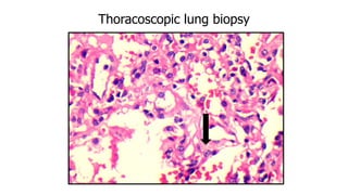
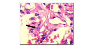
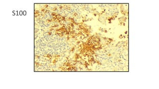
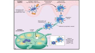
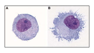
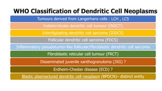
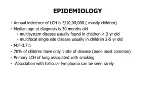
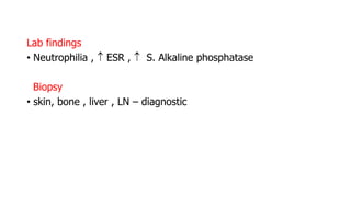
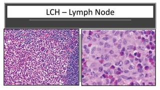
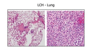
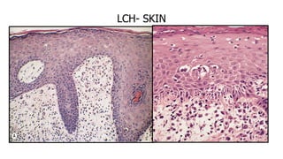
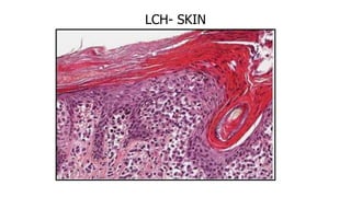
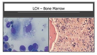
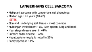
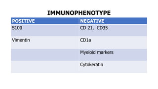
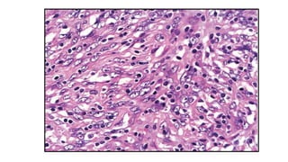
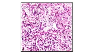
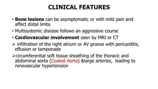
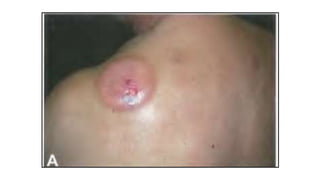
Dendritic cell tumors can arise from Langerhans cells or other dendritic cell subtypes. Langerhans cell histiocytosis (LCH) is characterized by proliferation of Langerhans cells and commonly involves bone, skin, lungs and lymph nodes. It ranges from localized eosinophilic granuloma to disseminated disease. Langerhans cell sarcoma is a rare malignant neoplasm that retains the immunophenotype of Langerhans cells. Indeterminate dendritic cell tumors have features intermediate between LCH and dendritic cell sarcoma.
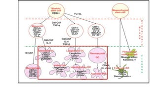
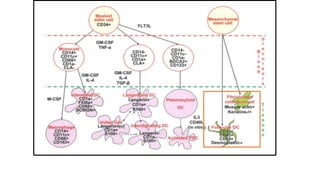


![HEMATOPOIETIC
STEM CELL (CD45+)
MESENCHYMAL
STEM CELL (CD45-)
PDC
FDC
FRC
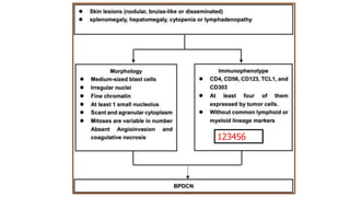
BPDCN
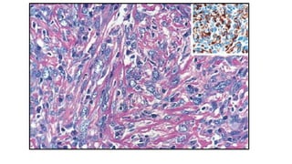
FRCT :
Indolent
to
aggressive
Spindle
cells with
collagen
fibres
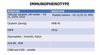
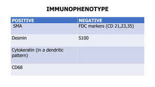
IHC: SMA,
Desmin;
CK, CD68
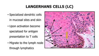
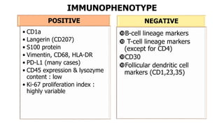
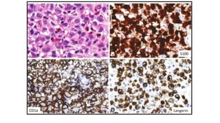
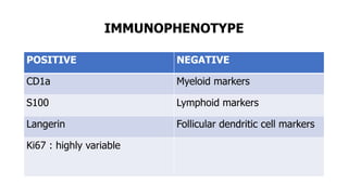
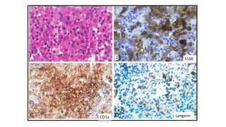
LCH:
Variable
clinical profile
LC cells
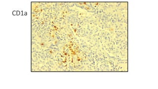
IHC: CD1a;
S100;Langerin
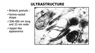
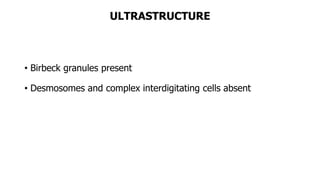
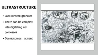
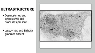
UM: Birbeck
granules
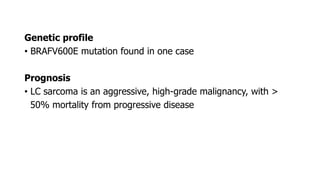
LCS:
aggressive
same IHC ;UM
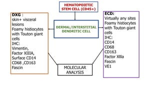
ECD
DXG
FDCS
:mostly LN,
mod
aggressive
Spindle cells
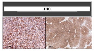
IHC:
CD21/35/23
Clusterin;
Fascin
IDDCS : Mostly
LN; very
aggressive
Spindle cells
IHC : S100;
Fascin
Absent :CD1a,
Langerin
INDCT:
mostly localized
Oval cells
IHC:
CD1a;S100
Fascin
Absent : Langerin
Birbeck granules
–]-](https://image.slidesharecdn.com/subjectseminardendriticcells-190225131558/85/DENDRITIC-CELL-TUMORS-PATHOLOGY-133-320.jpg)