The crystal structure of 4-amino-2,6-dichlorophenol is reported. The molecule adopts a planar conformation with the non-hydrogen atoms deviating from planarity by 0.020 Å. In the crystal structure, O—H···N hydrogen bonds form infinite chains along the [101] direction which are further linked by N—H···O hydrogen bonds to form (010) sheets through hydrogen bonding interactions. The title compound crystallizes in the monoclinic space group P21/n with unit cell parameters a = 4.6064 Å, b = 11.7569 Å, c = 13.2291 Å, and β = 96.760°.
![Crystal structure of 4-amino-2,6-di-
chlorophenol
Kyle J. McDonald,a
Vasumathi Desikan,a
James A. Golenb
and David R. Mankeb
*
a
Department of Science & Math, Massasoit Community College, 1 Massasoit
Boulevard, Brockton, MA 02302, USA, and b
Department of Chemistry and
Biochemistry, University of Massachusetts Dartmouth, 285 Old Westport Road,
North Dartmouth, MA 02747, USA. *Correspondence e-mail: dmanke@umassd.edu
Received 5 May 2015; accepted 13 May 2015
Edited by K. Fejfarova, Institute of Macromolecular Chemistry, AS CR, v.v.i, Czech
Republic
The title compound, C6H5Cl2NO, has a single planar molecule
in the asymmetric unit with the non-H atoms possessing a
mean deviation from planarity of 0.020 A˚ . In the crystal, O—
HÁ Á ÁN hydrogen bonds lead to the formation of infinite chains
along [101] which are further linked by N—HÁ Á ÁO hydrogen
bonds, forming (010) sheets.
Keywords: crystal structure; aminophenols; hydrogen bonding.
CCDC reference: 1400729
1. Related literature
For the crystal structure of the parent p-aminophenol, see:
Brown (1951). For other related structures, see: Ermer & Eling
(1994); Dey et al. (2005); Bacchi et al. (2009).
2. Experimental
2.1. Crystal data
C6H5Cl2NO
Mr = 178.02
Monoclinic, P21=n
a = 4.6064 (5) A˚
b = 11.7569 (12) A˚
c = 13.2291 (13) A˚](https://image.slidesharecdn.com/ee2e2235-c636-4108-921d-abe585860b58-160124192331/85/Crystal-structure-of-4-amino-2-6-dichlorophenol-1-320.jpg)
![Crystal structure of 4-amino-2,6-di-
chlorophenol
Kyle J. McDonald,a
Vasumathi Desikan,a
James A. Golenb
and David R. Mankeb
*
a
Department of Science & Math, Massasoit Community College, 1 Massasoit
Boulevard, Brockton, MA 02302, USA, and b
Department of Chemistry and
Biochemistry, University of Massachusetts Dartmouth, 285 Old Westport Road,
North Dartmouth, MA 02747, USA. *Correspondence e-mail: dmanke@umassd.edu
Received 5 May 2015; accepted 13 May 2015
Edited by K. Fejfarova, Institute of Macromolecular Chemistry, AS CR, v.v.i, Czech
Republic
The title compound, C6H5Cl2NO, has a single planar molecule
in the asymmetric unit with the non-H atoms possessing a
mean deviation from planarity of 0.020 A˚ . In the crystal, O—
HÁ Á ÁN hydrogen bonds lead to the formation of infinite chains
along [101] which are further linked by N—HÁ Á ÁO hydrogen
bonds, forming (010) sheets.
Keywords: crystal structure; aminophenols; hydrogen bonding.
CCDC reference: 1400729
1. Related literature
For the crystal structure of the parent p-aminophenol, see:
Brown (1951). For other related structures, see: Ermer & Eling
(1994); Dey et al. (2005); Bacchi et al. (2009).
2. Experimental
2.1. Crystal data
C6H5Cl2NO
Mr = 178.02
Monoclinic, P21=n
a = 4.6064 (5) A˚
b = 11.7569 (12) A˚
c = 13.2291 (13) A˚](https://image.slidesharecdn.com/ee2e2235-c636-4108-921d-abe585860b58-160124192331/75/Crystal-structure-of-4-amino-2-6-dichlorophenol-1-2048.jpg)
![= 96.760 (5)
V = 711.47 (13) A˚ 3
Z = 4
Cu K radiation
= 7.59 mmÀ1
T = 120 K
0.4 Â 0.2 Â 0.1 mm
2.2. Data collection
Bruker D8 Venture CMOS
diffractometer
Absorption correction: multi-scan
(SADABS; Bruker, 2014)
Tmin = 0.425, Tmax = 0.754
7481 measured reflections
1402 independent reflections
1273 reflections with I ! 2(I)
Rint = 0.043
2.3. Refinement
R[F2
2(F2
)] = 0.033
wR(F2
) = 0.091
S = 1.05
1402 reflections
99 parameters
2 restraints
H atoms treated by a mixture of
independent and constrained
refinement
Ámax = 0.33 e A˚ À3
Ámin = À0.35 e A˚ À3
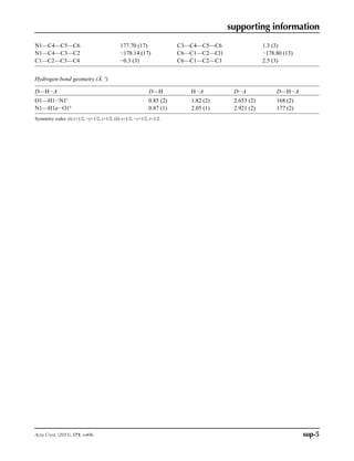
Table 1
Hydrogen-bond geometry (A˚ ,
).
D—HÁ Á ÁA D—H HÁ Á ÁA DÁ Á ÁA D—HÁ Á ÁA
O1—H1Á Á ÁN1i
0.85 (2) 1.82 (2) 2.653 (2) 168 (2)
N1—H1aÁ Á ÁO1ii
0.87 (1) 2.05 (1) 2.921 (2) 177 (2)
Symmetry codes: (i) x þ 1
2; Ày þ 1
2; z þ 1
2; (ii) x þ 1
2; Ày þ 1
2; z À 1
2.
Data collection: APEX2 (Bruker, 2014); cell refinement: SAINT
(Bruker, 2014); data reduction: SAINT; program(s) used to solve
structure: SHELXS97 (Sheldrick, 2008); program(s) used to refine
structure: SHELXL2014 (Sheldrick, 2015) and OLEX2.refine
(Bourhis et al., 2015); molecular graphics: OLEX2 (Dolomanov et al.,
2009); software used to prepare material for publication: OLEX2 and
publCIF (Westrip, 2010).
Acknowledgements
We greatly acknowledge support from the National Science
Foundation (CHE-1429086).
Supporting information for this paper is available from the IUCr
electronic archives (Reference: FF2137).
References
Bacchi, A., Carcelli, M., Chiodo, T., Cantoni, G., De Filippo, C. Pipolo, S.
(2009). CrystEngComm, 11, 1433–1441.
Bourhis, L. J., Dolomanov, O. V., Gildea, R. J., Howard, J. A. K. Puschmann,
H. (2015). Acta Cryst. A71, 59–75.
Brown, C. J. (1951). Acta Cryst. 4, 100–103.
Bruker (2014). APEX2, SAINT and SADABS. Bruker AXS Inc., Madison,
Wisconsin, USA.
Dey, A., Kirchner, M. T., Vangala, V. R., Desiraju, G. R., Mondal, R.
Howard, J. A. K. (2005). J. Am. Chem. Soc. 127, 10545–10559.
Dolomanov, O. V., Bourhis, L. J., Gildea, R. J., Howard, J. A. K. Puschmann,
H. (2009). J. Appl. Cryst. 42, 339–341.
Ermer, O. Eling, A. (1994). J. Chem. Soc. Perkin Trans. 2, pp. 925–944.
Sheldrick, G. M. (2008). Acta Cryst. A64, 112–122.
Sheldrick, G. M. (2015). Acta Cryst. C71, 3–8.
Westrip, S. P. (2010). J. Appl. Cryst. 43, 920–925.
data reports
o406 McDonald et al. doi:10.1107/S2056989015009172 Acta Cryst. (2015). E71, o406
ISSN 2056-9890](https://image.slidesharecdn.com/ee2e2235-c636-4108-921d-abe585860b58-160124192331/85/Crystal-structure-of-4-amino-2-6-dichlorophenol-2-320.jpg)
![supporting information
sup-1Acta Cryst. (2015). E71, o406
supporting information
Acta Cryst. (2015). E71, o406 [doi:10.1107/S2056989015009172]
Crystal structure of 4-amino-2,6-dichlorophenol
Kyle J. McDonald, Vasumathi Desikan, James A. Golen and David R. Manke
S1. Comment
The hydrogen bonding networks of aminophenols have been explored as hydroxy and amino groups are complementary
hydrogen bonding donors and acceptors. This is exemplified in p-aminophenol, which exhibits a supertetrahedral
hydrogen bonded architecture where all hydrogen bonding donors and acceptors are saturated (Brown, 1951; Ermer et al.,
1994). The mono-substitution in 4-amino-2-methylphenol and 4-amino-3-methylphenol yields a square motif structure
that again exhibits saturation among hydrogen bonding donors and acceptors (Dey et al., 2005). The more sterically
encumbered substitution of 4-amino-2,6-diphenylphenol prevents the saturation in hydrogen bonding, with only O–H···N
and N–H···aryl interactions observed (Bacchi et al., 2009). The 2,6-dichloro substitution of the title compound also
prevents saturation in its hydrogen bonding network.
The molecular structure of the title compound demonstrates a planar molecule with a mean deviation from the plane of
the non-hydrogen atoms of 0.020 Å. Intermolecular hydrogen bonding between O1–H1···N1 results in infinite chains
along [101] which combine with intermolecular hydrogen bonding between N1–H1a···O1 to give (010) sheets. The
packing for the title compound indicating hydrogen bonding is shown in Figure 2.
S2. Experimental
A commercial sample (Aldrich) was used for the crystallization. Crystals suitable for single crystal X-ray analysis were
grown by slow evaporation of a methanol solution.
S3. Refinement
All non-hydrogen atoms were refined anisotropically (Olex2) by full matrix least squares on F2
. Hydrogen atoms H1, H1a
and H1b were found from a Fourier difference map. H1 was allowed to refine freely with an isotropic displacement
parameter of 1.20 times Ueq of the parent O atom. H1a and H1b were refined with a fixed distance of 0.87 (0.005) Å and
isotropic displacement parameters of 1.20 times Ueq of the parent N atom. The two remaining hydrogen atoms were
placed in calculated positions and then refined with riding model with C–H lengths of 0.95 Å with isotropic displacement
parameters set to 1.20 times Ueq of the parent C atom.](https://image.slidesharecdn.com/ee2e2235-c636-4108-921d-abe585860b58-160124192331/85/Crystal-structure-of-4-amino-2-6-dichlorophenol-3-320.jpg)
![supporting information
sup-3Acta Cryst. (2015). E71, o406
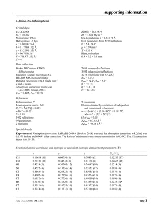
4-Amino-2,6-dichlorophenol
Crystal data
C6H5Cl2NO
Mr = 178.02
Monoclinic, P21/n
Hall symbol: -P 2yn
a = 4.6064 (5) Å
b = 11.7569 (12) Å
c = 13.2291 (13) Å
β = 96.760 (5)°
V = 711.47 (13) Å3
Z = 4
F(000) = 363.7579
Dx = 1.662 Mg m−3
Cu Kα radiation, λ = 1.54178 Å
Cell parameters from 5198 reflections
θ = 5.1–72.2°
µ = 7.59 mm−1
T = 120 K
Plate, colourless
0.4 × 0.2 × 0.1 mm
Data collection
Bruker D8 Venture CMOS
diffractometer
Radiation source: microfocus Cu
HELIOS MX monochromator
Detector resolution: 102.4 pixels mm-1
φ and ω scans
Absorption correction: multi-scan
(SADABS; Bruker, 2014)
Tmin = 0.425, Tmax = 0.754
7481 measured reflections
1402 independent reflections
1273 reflections with I ≥ 2σ(I)
Rint = 0.043
θmax = 72.2°, θmin = 5.1°
h = −5→5
k = −14→14
l = −12→16
Refinement
Refinement on F2
Least-squares matrix: full
R[F2
2σ(F2
)] = 0.033
wR(F2
) = 0.091
S = 1.05
1402 reflections
99 parameters
2 restraints
7 constraints
H atoms treated by a mixture of independent
and constrained refinement
w = 1/[σ2
(Fo
2
) + (0.0618P)2
+ 0.1912P]
where P = (Fo
2
+ 2Fc
2
)/3
(Δ/σ)max 0.001
Δρmax = 0.33 e Å−3
Δρmin = −0.35 e Å−3
Special details
Experimental. Absorption correction: SADABS-2014/4 (Bruker, 2014) was used for absorption correction. wR2(int) was
0.1370 before and 0.0641 after correction. The Ratio of minimum to maximum transmission is 0.5642. The λ/2 correction
factor is 0.00150.
Fractional atomic coordinates and isotropic or equivalent isotropic displacement parameters (Å2
)
x y z Uiso*/Ueq
Cl1 0.10618 (10) 0.09750 (4) 0.70434 (3) 0.02213 (17)
Cl2 0.79187 (11) 0.44532 (4) 0.61176 (4) 0.02666 (18)
O1 0.4519 (3) 0.30303 (12) 0.74687 (10) 0.0214 (3)
N1 0.4522 (4) 0.13356 (14) 0.35171 (12) 0.0199 (4)
C1 0.4563 (4) 0.26523 (16) 0.65055 (14) 0.0176 (4)
C4 0.4607 (4) 0.17796 (16) 0.45234 (13) 0.0176 (4)
C5 0.6112 (4) 0.27756 (16) 0.48060 (14) 0.0196 (4)
H5 0.7170 (4) 0.31620 (16) 0.43359 (14) 0.0235 (5)*
C2 0.3011 (4) 0.16753 (16) 0.61822 (14) 0.0171 (4)
C3 0.3014 (4) 0.12337 (16) 0.52110 (14) 0.0182 (4)](https://image.slidesharecdn.com/ee2e2235-c636-4108-921d-abe585860b58-160124192331/85/Crystal-structure-of-4-amino-2-6-dichlorophenol-5-320.jpg)