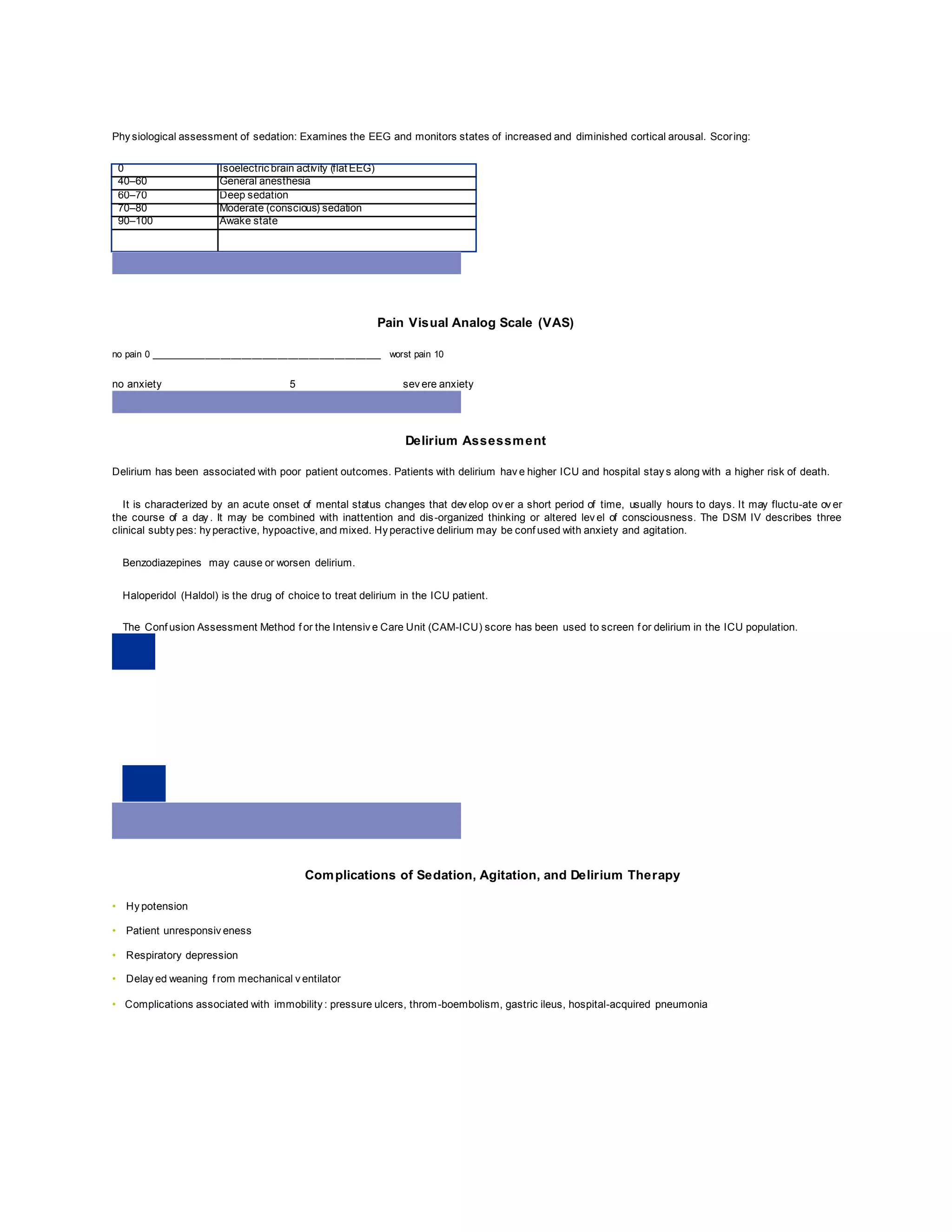
Critical Care
Notes
Clinical Pocket Guide
Janice Jones, PHD, RN, CNS Brenda Fix, MS, RN, NP
Purchase additional copies of this book at your health science bookstore or directly from F. A. Davis by shopping online at
www.fadavis.com or by calling 800-323-3555(US) or 800-665-1148(CAN)
A Davis’s Notes Book
Goto my website and get all free course https://priceyourwork.xyz/wSKA7J
F. A. Davis Company • Philadelphia
F. A. Davis Company
1915 Arch Street
Philadelphia, PA 19103
www.fadavis.com
Copyright © 2009 by F. A. Davis Company
All rights reserved. This book is pro tected b y copyright. No part of it m ay be reproduced, stored in a retrieval s ystem, or t ransmitted in any form or by an y means, electronic, mechanical, pho-
tocopying, recording, or otherwise, without written permission from the publisher.
Printed in China by Imago
Last digit indicates print number: 10 9 8 7 6 5 4 3 2 1
Publisher, Nursing: Robert G. Martone
Acquisitions Editor: Jonathan Joyce
Senior Developmental Editor: William F. Welsh
Project Editor: Meghan K. Ziegler
Reviewers:
Lisa Ann Behrend, RN, MSNc, CCRN-CSC
Deborah Little, MSN, RN, CCRN, CNRN,
APRN, BC
Janice Garrison Lanham, RN, MSN, CCNS,
FNP
Danette Wood, EdD, MSN, RN, CCRN
Laura Carousel, MSN, RN, CCRN
Jeanie Krause-Bachand, MSN, EdD, RN, BC
Deborah Pool, MS, RN, CCRN
As new scientific information becomes a vailable through basic and clinical research, recom -mended treatments and drug the rapies underg o changes. The author(s) and publisher have done
everything possible to make this book accurate, up to date, and in accord with accepted standa rds at the time o f publication. The author(s), editors, and p ublisher are not responsible for e rro rs
or omissions or for consequences from application of the book, and m ake no war-ranty, e xpressed o r implied, in rega rd to the contents of the bo ok. Any practice described in this book should
be applied by th e read er in accordance with professional standards of care used in re gard to the unique circumstances that ma y appl y in each situation. The reade r is advised alwa ys to check
product information (packag e inserts) for changes and new information rega rding dose and contraindications before administering any dru g. Caution is especially urged when using new or
infrequently ordered drugs.
Authorization to photocopy items for inter nal or personal us e, or the internal or pers onal use of spec ific clients, is granted by F. A. Davis Company for users registered with the Copyright Clear ance Center (CCC) Transactional Reporting
Service, prov ided that the fee of $.25 per copy is paid dir ectly to CCC, 222 Rosewood Drive, Danvers, MA 01923. For those organizations that hav e been gr anted a photoc opy licens e by CCC, a separ ate system of paym ent has been
arranged. The fee code for users of the Transactional Reporting Service is: 8036-2084-5/09 0 1 $.25.
Goto my website and get all free course https://priceyourwork.xyz/wSKA7J
Sticky Notes
HIPAA Compliant
OSHA Compliant
Waterproof and Reusable
Wipe-Free Pages
Write directly onto any page of Critical Care Notes with a ballpoint pen. Wipe old entries off with an alcohol pad and reuse.
BASICS CV RESP GU NEURO GI
HEMA/
ENDO
ONCO
MULTISYS CC MEDS TOOLS
Look for our other Davis’s Notes Titles
Av ailable Now!
Notes®: Nurse’s Clinical Pocket Guide, 2nd editionISBN-13: 978-0-8036-1335-5
LPN Notes: Nurse’s Clinical Pocket Guide, 2nd edition
ISBN-13: 978-0-8036-1767-4
DocuNotes: Nurse’s Clinical Pocket Guide to Effective Documenting and Reporting
ISBN-13: 978-0-8036-2092-6
ECG Notes: Interpretation and Management Guide
ISBN-13: 978-0-8036-1347-8
IV Med Notes: IV Administration Pocket Guide
ISBN-13: 978-0-8036-1466-8
IV Therapy Notes: Nurse’s Clinical Pocket Guide
ISBN-13: 978-0-8036-1288-4
LabNotes: Guide to Lab and Diagnostic Tests
ISBN-13: 978-0-8036-1265-5
MedNotes: Nurse’s Pharmacology Pocket Guide, 2nd Edition
ISBN-13: 978-0-8036-1531-1
MedSurg Notes: Nurse’s Clinical Pocket Guide, 2nd edition
ISBN-13: 978-0-8036-1868-8
NCLEX-RN® Notes: Core Review & Exam Prep
ISBN-13: 978-0-8036-1570-0
NutriNotes: Nutrition & Diet Therapy Pocket Guide
ISBN-13: 978-0-8036-1114-6
OB Peds Women’s Health Notes: Nurse’s Clinical Pocket Guide
ISBN-13: 978-0-8036-1466-6
PsychNotes: Clinical Pocket Guide, 2nd edition
ISBN-13: 978-0-8036-1853-4
a complete list of Davis’s Notes and other titles for health care providers, visit www.fadavis.com.
Contacts • Phone/E-Mail
Name
Ph: e-mail:
Name
Ph: e-mail:
Name
Ph: e-mail:
Name
Ph: e-mail:
Name
Ph. e-mail:
Name
Ph: e-mail:
Name
Ph: e-mail:
Name
Ph: e-mail:
Name
Ph: e-mail:
Name
Ph: e-mail:
Name
Ph: e-mail:
Name
Ph: e-mail:
1
Physical Assessment
Reusable Assessment Form
Name: Room: Age:
Diagnosis:
Surgeries/Past Hx:
Activ ity: Diet: DNR/DNI:
Allergies:
Neurological/MS:
ICP:
Cardiac:
VS/A-line:
ECG:
Hemodynamics: PAD PAS PCWP CVP
IABP:
Respiratory:
Ventilator:
ABGs/SpO2:
GI:
GU:
Wounds/Incisions:
Drainage Tubes:
Treatments:
Special Needs:
Other:
BASICS
BASICS
Normal Arterial and Venous Blood Gases
Blood Gas
Components Arterial Venous
pH 7.35–7.45 7.31–7.41
PO2 80–100 mm Hg 35–40 mm Hg
PCO2 35–45 mm Hg 41–51 mm Hg
HCO3 22–26 mEq/L or mmol/L 22–26 mEq/L or mmol/L
Base Excess (BE) –2 to +2 mEq/L or mmol/L –2 to +2 mEq/L or mmol/L
O2 saturation 95%–100% 68%–77%
Blood Gas Results
Arterial Venous
pH
PO2
PCO2
HCO3
Base Excess (BE)
O2 saturation
Quick Blood Gas Interpretation
Acid-Base Disorder pH PCO2 HCO3
Respiratory acidosis if compensating
Respiratory alkalosis
Metabolic acidosis
Metabolic alkalosis
Full or total compensation: pHwill be within normal limits
2
3
Goto my website and get all free course https://priceyourwork.xyz/wSKA7J
Compensation:
• 3
• 2
Also look f or mixed respiratory and metabolic problems.
PaCO2 or HCO3 in a direction opposite its predicted direction or not close to predictiv e v alue.
Common Causes of Acid-Base Imbalances
Respiratory acidosis COPD, asthma, headinjury, pulmonary
edema, aspiration, pneumonia, ARDS, pneu-
mothorax, cardiac arrest, respiratory depres-
sion, CNS depression, or head injury
Respiratory alkalosis Hy perventilation, anxiety, fear, pain,
f ev er, sepsis, brain tumor, mechanical
ov erventilation
Metabolic acidosis Diabetes mellitus, acute and chronic renal
f ailure, severediarrhea, alcoholism, starva-
tion, salicylateoverdose, pancreatic
f istulas
Metabolic alkalosis Loss of gastric acid (vomiting, gastric
suction), long-term diuretic therapy
(thiazides, furosemide), excessive NaHCO3
administration, hypercalcemia
BASICS
BASICS
Pulse Oximetry
SpO2 monitoring: Monitoring saturation of peripheral O2
SpO2 Level Indication
>95% Normal
91%–94% May be acceptable, provide O2 as necessary, encourage C&DBor suctionprn
85%–90% Prov ide O2 as necessary, encourage C&DB or suction prn, may be normal forCOPD patient
<85% Prepare f or possible intubation
False readings may occur because of anemia, CO poisoning, hypothermia, hypovolemia, peripheral vasoconstriction caused by disease or medications.
Lactic Acidosis
Lactic acid is a by product of anaerobic metabolism. Increased lev els indi-cate inadequate perf usion of v ital organs with resultant tissue hy poxia. May
result f rom inadequate perf usion and oxy genation of v ital organs; post cardiac or respiratory arrest; cardiogenic, ischemic, or septic shock; drug
ov erdoses, seizures, cancers, or diabetes mellitus.
Critical v alues: Blood pH, <7.35, and lactate >5–6 mEq/L or >45 mg/dL.
Treat with sodium bicarbonate IV if acidosis is readily ev ident.
Capnography
Capnography is the measurement, display , and monitoring of the concen-tration or partial pressure of CO2 (PETCO2) in the respiratory gases at the
end of expiration. The capnogram display s the maximum inspirato-ry and expiratory CO2 concentrations during a respiratory cycle, which indirectly
ref lect the production of CO2 by the tissues and the transport of CO2 to the lungs. Sudden changes in CO2 elimination should be moni-tored in
selected cardiorespiratory patients and postoperativ ely af ter major cardiothoracic surgeries. Capnography can also be used to v erify ETT position and
monitor the ef f ectiveness of CPR.
4
5
Causes of PETCO2 Causes of PETCO2
Fev er Hy pothermia
Hy pertension Hy potension
Increased cardiac output Decreased cardiac output
Hy poventilation Hy perventilation
Hy povolemia Hy pervolemia
Airway obstruction Airway obstruction
Bronchial intubation Accidental extubation
Pulmonary embolus
Cardiac arrest
Apnea
Normal range of ETCO2 is 35–45 mm Hg
• 2ETCO2 < 35 = respiratory alkalosis
• 2ETCO2 > 45 = respiratory acidosis There are f iv e characteristics of the capnogram that should be ev aluated: f requency , rhy thm, height , baseline,
and shape.
Normal Capnogram
m m H
g
Expiration Inspiration
III
II
I
Time
Phases I, II, and III represent expiration, the bolded lines represent inspira-tion. Long periods of a flat wav e f orm indicate apnea, dislodged endotra-
cheal tube, esophageal intubation, or patient disconnect f rom v entilator.
BASICS
BASICS
Artifical Airways and Mechanical Ventilation
Artifical Airways
Goto my website and get all free course https://priceyourwork.xyz/wSKA7J
Endotracheal Tube
• Adult oral tube sizes: Males 8.0–8.5, I.D. (mm); f emales 7.0–8.0. I.D. (mm) (internal diameter).
• Placement is 2 cm abov e the carina. Verify by auscultating f or breath sounds bilaterally , unif orm up-and-down chest mov ement, CXR, and
checking end-tidal CO2 immediately af ter intubation.
• Cuf f pressure: 20–25 mm Hg.
Tracheostomy Tube
• Size will v ary .
• Cuf f pressure: 20–25 mm Hg.
Minimal leak technique or minimal occluding v olume v erif ies that an ETT or tracheostomy tube is at its lowest inf lation point. Attach a 10-mL sy ringe to
the balloon of the inf lated cuff. Position y our stethoscope on the patient’s neck at the area of the carotid pulse. Inf late balloon cuff to a point where no
leak is heard. Slowly remov e air f rom the inf lated cuf f until y ou hear a slight leak at the height of inspiration. Then add 1 mL of air back into the cuf f .
Cuff pressure can also be monitored v ia a calibrated aneroid manome-ter dev ice. Connect manometer to cuff. Def late cuff. Reinf late cuff in 0.5 mL
increments until desired cuf f pressure is achiev ed. Check cuf f pressure ev ery 8–12 hrs or per agency protocol.
Goto my website and get all free course https://priceyourwork.xyz/wSKA7J
Mechanical Ventilation
Classificationof Ventilators
Positive Pressure Ventilation
• Volume-Cycled Ventilator: Deliv ers a preset constant v olume of air and preset O2.
• Pressure-Cycled Ventilator: Produces a f low of gas that inf lates the lung until the preset airway pressure is reached.
• Time-Cycled Ventilator: Programmed to deliv er a v olume of gas ov er a specif ic time period through adjustments in inspiratory -to-expiratory ratio.
6
7
• High-Frequency Jet Ventilator (HFJV): Deliv ers 60–100 bpm with low tidal v olumes under considerable pressures.
Negative PressureVentilation
Uses the old iron lung principle by exerting a negativ e pressure on the chest wall to cause inspiration. No intubation required. Custom fitted “cuirass” or
“turtle” shell unit that f its ov er the chest wall. May be utilized at night f or patients who require assistance during sleep.
Modes of Ventilation
• Controlled Mechanical Ventilation (CMV): Machine controls rate of breathing. Deliv ery of preset v olume (TV) and rate regardless of patient’s
breathing pattern. Sedation or paraly zing agent (e.g., Pav ulon) usually required.
• Assist Controlled Ventilation (ACV): Patient controls rate of breathing. Inspiratory ef f ort triggers deliv ery of preset v olume.
• Intermittent Mandatory Ventilation (IMV): Patient breathes sponta-neously (own tidal v olume) between v entilator breaths of a preset v olume
and rate.
• Synchronized Intermittent Mandatory Ventilation (SIMV): A f orm of pressure support v entilation. Administers mandatory v entilator breath at a
preset lev el of positiv e airway pressure. Monitors negativ e inspira-tory ef f ort and augments patient’s spontaneous tidal v olume or inspira-tory ef f ort.
Sy nchronized with patient’s breathing pattern.
• Positive End-Expiratory Pressure (PEEP): Increases oxy genation by increasing f unctional residual capacity (FRC). Keeps alv eoli inf lated af ter
expiration. Can use lower O2 concentrations with PEEP; decreas-es risk of O2 toxicity . Ordered as 5–10 cm H2O.
• Continuous Positive Airway Pressure (CPAP): Maintains positiv e pressure throughout the respiratory cy cle of a spontaneously breath-ing
patient. Increases the amount of air remaining in the lungs at the end of expiration. Less complications than PEEP. Ordered as 5–10 cm H2O.
• Bilevel Positive Airway Pressure (BiPAP): Same as CPAP but settings can be adjusted f or both inspiration and expiration.
• Pressure Support Ventilation (PSV): Patient’s inspiratory ef f ort is assisted by the v entilator to a certain lev el of pressure. Patient initiates all
breaths and controls f low rate and tidal v olume. Decreases work of breathing.
BASICS
BASICS
• Inverse Ratio Ventilation (IRV): All breaths are pressure limited and time cy cled. Inspiratory time usually set longer than expiratory time.
IMV, SIMV, CPAP, BiPAP and PSV can all be used in the weaning process.
Goto my website and get all free course https://priceyourwork.xyz/wSKA7J
Weaning
Sample Criteria for Weaning: Readiness
• Alert and cooperativ e
• FIO2 < 40%–50% and PEEP <5–8 cm H2O
• Hemody namically stable
• pH >7.34
• PaO2 >80 mm Hg
• PaCO2 <45 mm Hg
• PaO2/FIO2 ratio >200
• Vital capacity 15 mL/kg and minute v entilation <10
• Hemoglobin >7–9 g/dL and serum electroly tes within normal limits
• Spontaneous respirations >6 b/min. or <35 b/min.
• Negativ e inspiratory pressure –30 cm H2O
• Relativ ely af ebrile with limited respiratory secretions
• Inotropes reduced or unchanged within prev ious 24 hrs
• Sedation discontinued
Weaning Methods
• T-tube weaning: Place patient on T-tube circuit on same FIO2 as on v entilatory assistance. Monitor ABGs af ter 30 min. Prov ide a brief rest period
on the v entilator as needed and continue to monitor ABGs until satisf actory. Extubate when patient is rested, good spontaneous respiratory ef f ort,
and ABGs within acceptable parameters.
• IMV/SIMV weaning: Decrease IMV rate ev ery 1–4 hrs. Monitor spon-taneous breaths. Obtain ABGs within 30 min. of v entilator change. Allows f or
gradual change f rom positiv e-pressure v entilation to spon-taneous-pressure v entilation.
• PSV: Use low lev els of PSV (5–10 cm H2O). Decrease in 3–6 cm of H2O increments. Usef ul in retraining respiratory muscles due to long-term
v entilation.
8
9
• CPAP/BiPAP: Prov ides expiratory support, maintains positiv e intratho-racic pressure. BiPAP adds inspiratory support to CPAP. Prev ents res-
piratory muscle f atigue.
Nursing assessment during weaning
• Vital signs and hemody namics (PAS, PAD, PCWP, CO, CI)
• Dy srhy thmias or ECG changes
• Oxy genation/Ef ficiency of gas exchange
• CO2 production and elimination
• pH lev el
• Bedside pulmonary f unction tests
• Work of breathing including use of accessory muscles
• Lev el of f atigue
• Patient discomf ort
• Adequate nutrition
Ventilator Alarms
Goto my website and get all free course https://priceyourwork.xyz/wSKA7J
Ventilator alarms should nev er be ignored or turned of f . They may be muted or silenced temporarily until problem is resolv ed.
Checklist of Common Causes of Ventilator Alarms
Patient causes:
• Biting down on endotracheal tube
• Patient needs suctioning
• Coughing
• Gagging on endotracheal tube
• Patient “bucking” or not sy nchronous with the v entilator
• Patient attempting to talk
• Patient experiences period of apnea
Mechanical causes:
• Kinking of v entilator tubing
• Endotracheal tube cuf f may need more air
• Leak in endotracheal tube cuf f
• Excess water in v entilator tubing
• Leak or disconnect in the sy stem
• Air leak f rom chest tube if present
• Malf unctioning of oxy gen sy stem
• Loss of power to v entilator
Goto my website and get all free course https://priceyourwork.xyz/wSKA7J
BASICS
BASICS
Pathophysiological causes:
• Increased lung noncompliance, such as in ARDS
• Increased airway resistance, such as in bronchospasm
• Pulmonary edema
• Pneumothorax or hemothorax
Nursing Interventions
• Check v entilator disconnects and tubing.
• Assess breath sounds, suction as needed.
• Remov e excess water f rom v entilator tubing.
• Check endotracheal cuf f pressure.
• Insert bite block or oral airway .
If cause of the alarm cannot be f ound immediately or cause cannot be readily resolv ed, remov e patient f rom v entilator and manually v entilate patient
using a resuscitation bag.
Call respiratory therapy stat.
Continue to assess patient’s respiratory status until mechanical v entila-tion is resumed.
Ventilator Complications
Complication Signs & Symptoms/Interventions
Barotrauma or • High peak inspiratory and mean airway
v olutrauma—acute lung pressures
injury , may result in pneu-
• Diminished breathsounds
mothorax or tension pneu-
• Tracheal shif t
mothorax, pneumomedi-
• Subcutaneous crepitus
astinum, pneumoperi-
toneum, subcutaneous • Hy poxemia
crepitus
Insert chest tube or needle thoracostomy.
Intubation of right main- • Absent or diminishedbreathsounds in
stem bronchus lef t lung
• Unilateral chest excursion
Reposition ETT.
Continued
10
Goto my website and get all free course https://priceyourwork.xyz/wSKA7J
11
Ventilator Complications—Cont’d
Complication Signs & Symptoms/Interventions
Endotracheal tubeout of • Absent or diminishedbreathsounds
position or unplanned extu- Note location of tube at the lip (21–22 cm).
bation Reposition ETT or reintubate.
Restrain only when necessary.
Tracheal damage due to • Blood in sputum when suctioning
excessive cuff pressure
• Frequent v entilator alarm
(>30 cm H2O) Monitor ETT cuf f pressure every 4–8 hrs.
Perf orm minimal leak technique.
Ensure minimal occluding volume.
Damage to oral or nasal • Skin breakdown or necrosis to lips,
mucosa nares, or oral mucous membranes
Reposition tube side-sideof mouth every
day .
Apply petroleum jelly to nares.
Prov ide oral care withtoothbrush every
2 hrs.
Aspiration • Feeding v iewed when suctioning
Tracheo-esophageal • If blue dy eis used, sputum is blue in color
f istulas Use blue dy e withenteral feedings if
aspiration suspected.
Keep head of bed 30–45 degrees.
Administer proton pump inhibitors or his-
tamine H2-receptor antagonists.
Ventilator-assisted • Ref er to section on VAP
pneumonia Assess color and odor of sputum.
Respiratory infection Monitor temperature, WBC count, ESR.
Increased risk of sinusitis
Decreased venous return • Hy potension
decreased cardiac out-
• Decreased CVP, RAP, and preload
put due to increased
Monitor v ital signs and hemodynamics.
intrathoracic pressure
Continued
Goto my website and get all free course https://priceyourwork.xyz/wSKA7J
BASICS
BASICS
Ventilator Complications—Cont’d
Complication Signs & Symptoms/Interventions
Stress ulcer and • Blood in nasogastric drainage
GI bleeding
• Hematemesis and/ormelena
Hematest nasogastric drainage, emesis,
f eces.
Administer proton pump inhibitors or his-
tamine H2-receptor antagonists.
Paraly tic ileus • Absence of diminished bowel sounds
Prov ide nasogastric drainage with
intermittent suction.
Turn and position patient frequently.
Inadequate nutrition, loss • Ref er to section on nutrition.
of protein
Start enteral feedings if appropriate.
Start total parenteral nutrition if GI tract
nonf unctional or contraindicated.
Increased intracranial • Changes in lev el of consciousness
pressure
• Unable to f ollow commands
Assess neurological status frequently.
Fluid retention due to Assess foredema.
increased humidification
Administer diuretics.
f rom ventilator, increased
Drain v entilator tubingfrequently.
pressure to baroreceptors
causing a release of ADH
Immobility Turn and position patient frequently.
Skin breakdown Assess skin for breakdown.
Assist patient out of bed to chairunless
contraindicated.
Keep skin clean and dry, sheets wrinkle-
f ree.
Communication difficulties Keep communication simple.
Obtain slate or writing board.
Use letter/picture chart.
Communicateusing sign language.
Continued
12
13
Ventilator Complications—Cont’d
Complication Signs & Symptoms/Interventions
Urinary tract infection • Urine becomes cloudy, concentrated,
odorous
Change/remove Foley catheter.
Ensure adequate hydration.
Administer antiinfectives.
Deep v ein thrombosis • Painf ul, swollen leg; pain may increase
on dorsif lexion
Assess forpulmonary embolism. See
respiratory section.
Administer heparin or enoxaparin.
Psy chosocial concerns: • Anxious
f ear, loss, powerlessness,
• Dif ficulty sleeping
pain, anxiety, sleep distur-
• Poor pain control
bances, nightmares, loneli-
Administer anxiolytics, sedatives,
ness
analgesics.
Cluster activities to promote periods of
sleep.
Allow patient to makechoices when
appropriate.
Allow f or f requent family visits.
Keep patient and family informed.
Hemodynamic Monitoring
Hemodynamic Parameters
Arteriov enous oxy gen dif f erence . . . . . . . . . . . 3.5–5.5 v ol% or 4–8 L/min Aortic pressure:
■ Systolic . . . . . . . . . . . . . . . . . . . . . . . . . . . . . . . . . . . . . . .100-140 mm Hg ■Diastolic . . . . . . . . . . . . . . . . . . . . . . . . . . . . . . . . . . . . . . . .60–80mm Hg ■ Mean .
. . . . . . . . . . . . . . . . . . . . . . . . . . . . . . . . . . . . . . . . .70–90 mm Hg
BASICS
BASICS
Cardiac output (CO = HR X SV) . . . . . . . . . . .
. . . . . . . . . . . . . . . .4–8
L/min
Cardiac index (CO/BSA) . . . . . . . . . . . . . . . . .
. . . . . . . . . . . . . .2.5–4
L/min
Central v enous pressure (CVP) . . . . . . . . . . . . . . . . . . . . . . . . .2–8 mm Hg
** Same as right atrial pressure (RAP)
Cerebral perf usion pressure (CPP) . . . . . . . . .
2–6 mm Hg or 5–12 cm
H2O
Coronary artery perf usion pressure (CAPP) . . . . . . . . . . . . .60–80 mm Hg
Ejection f raction (Ej Fx or EF) . . . . . . . . . . . . ................60%–75%
Lef t arterial mean pressure . . . . . . . . . . . . . . . . . . . . . . . . . . .4–12 mm Hg
Lef t v entricular sy stolic pressure . . . . . . . . . . . . . . . . . . . .100–140 mm Hg
Lef t v entricular diastolic pressure . . . . . . . . . . . . . . . . . . . . . . .0–5 mm Hg
Lef t v entricular stroke work index (LSWI) . . . . . . . . . . . .30–50 g/beats/m2
Mean arterial pressure (MAP) . . . . . . . . . . . . . . . . . . . . . . .70–100 mm Hg
Oxy gen consumption (VO2) . . . . . . . . . . . . . .
. . . . . . . . . .200–250
mL/min
Oxy gen delivery (Do2) . . . . . . . . . . . . . . . . . . .
. . . . . . . . .900–1100
mL/min
Pulmonary artery pressure (PAP):
■ Systolic . . . . . . . . . . . . . . . . . . . . . . . . . . . . . . . . . . . . . . . . .20–30 mm Hg ■Diastolic . . . . . . . . . . . . . . . . . . . . . . . . . . . . . . . . . . . . . . . .10–20mm Hg ■ Mean .
. . . . . . . . . . . . . . . . . . . . . . . . . . . . . . . . . . . . . . . . . .10–15 mm Hg
Pulmonary capillary wedge pressure (PCWP) . . . . . . . . . . . . .4–12 mm Hg Right arterial mean pressure . . . . . . . . . . . . . . . . . . . . . . . . . . .2–6 mm Hg
Right v entricular pressure:
■ Sy stolic . . . . . . . . . . . . . . . . . . . . . . . . . . . . . . . . . . . . . . . . 20–30 mm Hg
■ Diastolic . . . . . . . . . . . . . . . . . . . . . . . . . . . . . . . . . . . . . . . . . .0–8 mm Hg
■ End Diastolic . . . . . . . . . . . . . . . . . . . . . . . . . . . . . . . . . . . . . .2–6 mm Hg
Right v entricular stroke work index (RSWI) . . . . . . . . . . . . .7–12 g/m2/beat
Pulmonary vascular resistance (PVR) . . . . . . . . . . .20–130 dynes/sec/cm
-5
Pulmonary v ascular resistance index (PVRI) . . . . . . . .200–400 dy nes/sec/
cm5/m2
Pulmonary ventricular stroke index . . . . . . . . . . . . . . . . . . .5–10 g/beat/m
2
Right atrial pressure (RAP) . . . . . . . . . . . . . . . . . . . . . . . . . . . . .2–6 mm Hg Stroke
index (SI) . . . . . . . . . . . . . . . . . . . . . . . . . . . . . . .30–650 mL/beat/m
2
Stroke volume (SV = CO/HR) . . . . . . . . . . . . . . . . . . . . . . . . .60–100 mL/beat Systemic
vascular resistance (SVR) . . . . . . . . . .900–1,600 dynes/sec/cm
-5
Systemic vascular resistance index . . . . . .1,360–2,200 dynes/sec/cm
-5
/m
2
Systemic venous
oxy gen saturation(SvO2) . . . . . . . . . . . . . . . . .60%–80%
14
15
Cardiac Output Components
Preload Contractility Afterload
PaO2 SaO2 Hemoglobin (Hgb)
Right atrial pressure Stroke v olume Pulmonary vascular resistance
Central v enous Cardiac output Sy stemic vascular resistance
pressure
Lef t ventricular Tissue perf usion Blood pressure
end diastolic
pressureP
Pulmonary Artery Catheter
The purpose of the pulmonary artery catheter, also known as the Swan-Ganz catheter, is to assess and monitor lef t v entricular f unction and can
determine preload, assess contractility , and approximate af terload.
PCWP approximates lef t atrial pressure and lef t v entricular end diastolic pressure.
Increases in PCWP, LAP, or LVEDP indicates heart f ailure, hy perv olemia, shock, mitral v alv e insuff iciency , or stenosis. Decreases in PCWP, LAP, or
LVEDP indicate hy pov olemia.
PA Catheter Waveforms
The pulmonary artery catheter is threaded through the right atrium and right v entricle and into the pulmonary artery. Insertion is done v ia f luo-roscopy
or monitoring wav ef orm changes.
Catheter advanced
to right atrium,
balloon is 40
inf lated. Pressure
30
is low, usually Hg
20
2–5 mm Hg. mm Balloon catheter
10 Right atrium
0
Time
BASICS
BASICS
Catheter is floatedto right ventricle with the balloon inflated. Wave-forms indicate a systolic pressure of 25–30 mm Hg anda diastolic pressure of 0–5 mm Hg.
40
30
Hg
20
mm
Balloon catheter
10
Right ventricle 0
Time
As the catheter moves into the pulmonary artery, thesystolic pres-sureremains the same but the dia-stolic pressureele-vates to 10–15 mm Hg.
mm
Hg
40
30
20
10
0
Time
Pulmonary
artery
Balloon
catheter
The catheter is moved until it can be wedged in a smaller vessel. When the balloon is inflated, the pressure recorded is that pressurein front of thecatheter. It is
an approximate measure of the left ventricularend diastolic pressure.
Pulmonary
artery wedge
40
30
Hg
20
mm
Balloon
catheter 10
0
Time
16
17
Problems with Pulmonary Artery Catheters
Problem Check For/Action
No wav eform • Loose connections
• Tubing kinked or compressed
• Air in transducer
• Loose/cracked transducer
• Stopcock mispositioned
• Occlusion by clot: Aspirate as per policy
Ov erdamping (smaller • Air bubble or clot in the system
wav ef orm withslow • Catheter position: Reposition patient or
rise, diminished or hav e patient cough
absent dicrotic notch) • Kinks or knotting
• Clot: Aspirateas per policy
Catheter whip (erratic • Catheter position: Reposition patient or
wav ef orm, variable catheter; obtain chest x-ray
and inaccurate
pressure)
Inability to wedge • Balloon rupture: Turn patient on left side;
catheter (no wedge check catheterposition for retrograde
wav ef orm after slippage
inf lating balloon)
Complications of Pulmonary Artery Catheters
• Risk f or inf ection
• Altered skin integrity
• Air embolism
• Pulmonary thromboembolism
• Cardiac tamponade
• Dy srhy thmias
• Altered cardiopulmonary tissue perf usion due to thrombus f ormation; catheter in wedged position leading to pulmonary inf arction
• Catheter displacement/dislodgement
• Loss of balloon integrity or balloon rupture
• Pneumothorax
• Hemothorax
BASICS
BASICS
• Frank hemorrhage
• Pulmonary artery extrav asation
• Pulmonary artery rupture
Intra-Arterial Monitoring
An arterial line (A-line) is used if f requent blood pressure and arterial blood gas determinations are needed. It is especially usef ul
• Af ter surgery .
• For patients with unstable v ital signs.
• For patients experiencing hy poxemia.
Perf orm Allen’s test prior to insertion. Elev ate the patient’s hand with his or her f ists clenched. Release pressure ov er only the ulnar artery. Color
returns to the hand within 6 seconds if the ulnar artery is patent and ade-quate collateral blood f low present.
Compressing the radi- Observ ing f or pallor Releasing pressure
al and ulnar arteries and observ ing f or
return of normal color
18
19
Intra-Arterial Waveform
100
mm
Hg
0
Time
Components of Waveform
• Systolic peak: Ventricular ejection and stroke v olume. Sharp rise and rounded top.
• Dicrotic notch:
Tapering of down stroke f ollowing dicrotic notch
Important assessments: changes in capillary ref ill/blanching, sensation, motion, or color that may indicate lack of perf usion to the extremity
MAP sy stolic BP (diastolic BP 2) = 70–100 mm Hg 3
Decreased tissue perf usion—decreasing urine output, elev ation in BUN:Creatinine ratio, altered mental status with decreasing lev el of consciousness,
restlessness, dy spnea, cy anosis, dysrhy thmias, abnor-mal ABGs, weak or absent peripheral pulses, increased capillary ref ill time (>3 sec), diminished
arterial pulsations, bruits.
BASICS
BASICS
Potential Complications of Intra-Arterial Monitoring
• Hemorrhage
• Air emboli
• Equipment malf unction/inaccurate pressure
• Dy srhy thmias
• Inf ection
• Altered skin integrity
• Impaired circulation to extremities
Nutrition Issues in Critical Care
Primary Concerns
• Starv ation and catabolism
• Stress hy permetabolism
• Fluid v olume def icit
• Fluid v olume excess
Stress and Nutrition
Metabolic rate increases with the release of catecholamines + glucagon
•
(albumin).
Nitrogen excretion increases.
Body weight decreases.
1 kg body weight = 1 liter of f luid retained or lost.
Body Mass Index
BMI is a simple means of classifying sedentary (physically inactiv e) indiv id-uals of av erage body composition and may indicate obesity . It is calculated
by the f ollowing: Body mass index (BMI) = weight (kg) ÷ height (meters)2
1 kg = 2.2 lbs Normal BMI = 20–25 kg/m2
20
21
A BMI >30 kg/m2 indicates obesity , >40 kg/m2 indicates morbid obesity . An increase in BMI has been associated with heart disease and diabetes.
A BMI <18.5 kg/m2 suggests a person is underweight. A BMI <17.5 may indicate the person has anorexia or a related disorder.
BMI does not take into account f actors such as f rame size and muscularity .
Signs and Symptoms of Fluid Volume Deficit:
Hypovolemia
• Dry mucous membranes; dry cracked tongue
• Thirst
• Poor skin turgor
• Sunken ey eballs
• Subnormal temperature
• Decreased or orthostatic blood pressure
• Weak, rapid heart rate and increased respiratory rate
• Decreased capillary ref ill
• Urine output decreased (<30 mL/hr)
• Increased specif ic grav ity of urine (<1.030)
• Decreased central v enous pressure
• Increased hemoglobin and hematocrit
• Increased BUN and serum osmolarity
• Increased BUN:creatinine ratio
• Lethargy , mental conf usion
Signs and Symptoms of Fluid Volume Excess:
Hypervolemia
• Crackles in lungs; dy spnea, shortness of breath
• Decreased hemoglobin and hematocrit
• Decreased specif ic grav ity of urine
• Distended neck v eins and jugular v enous pressure
• Edema and decreased serum osmolarity
• Full, bounding pulse; tachy cardia
• Increased BP, CVP, and PAP
BASICS
BASICS
• Mental conf usion, restlessness
• Moist mucous membranes
• Pulmonary congestion or pleural ef f usion
• Weight gain
Enteral Tube Feedings
Gastric Access
• Nasogastric tube (NGT)
• Oral
• Percutaneous endoscopic gastrostomy (PEG)
• Nasoduodenal tube (NDT)
• Low-prof ile gastrostomy dev ice (LPGD)
Small Bowel Access
• Nasal-jejunal tube (NJT)
• Percutaneous endoscopic jejunostomy (PEJ)
Types of Tube Feedings
• Intermittent or bolus feedings: A set v olume of f ormula is deliv ered at specif ied times.
• Continuous feedings: A set rate of f ormula is deliv ered ov er a period of time.
• Cyclic feedings: Similar to a continuous f eeding but the inf usion is stopped f or a specif ied time within a 24-hour period, usually 6–10 hours.
Checking Tube Placement
• Aspirate gastric contents and check pH.
• Gastric aspirate pH 1–4 but may be as high as 6 if patient is on med-ication to reduce gastric acid (f amotidine, ranitidine, pantoprazole).
• Small intestine aspirate pH >6.
• Obtain chest x-ray .
• Inject 20–30 mL of air into the tube while auscultating ov er the epi-gastrium. Air in the stomach can be heard v ia a swooshing sound.
22
23
Tube-Feeding Formulas
Standard Very High-Protein/Wound-Healing
Pediatric Fiber-Containing
Diabetic Elemental and Semi-Elemental
Pulmonary Immune-Enhancing or Stress Formulas
Renal Concentrated
Feeding Tube Complications
Mechanical Complications Interventions
Nasopharyngeal discomfort • Reposition tube.
Esophageal ulceration or bleeding • Consider PEG or PEJ tube.
esophageal v arices
Clogged tube • Flush with lukewarm water after
ev ery f eeding.
Hosp. Protocol:
Tube displacement • Reposition tube.
Extubation • Insert new tube.
• Consider PEG or PEJ tube.
Stomal leak or infection • Keep area around stoma clean
and dry .
Nonmechanical
Complications Interventions
Nausea, vomiting, cramps, • Withhold or decrease
bloating, abdominal distention amount, rate, and frequency
of f eedings.
• Change to low-f at formula.
Diarrhea • Withhold or decreaseamount,
rate, and f requency of feedings.
Aspiration • Hold f eedings. Check
residuals.
• Keep HOB elev ated 30°–45°
during f eedings and 1 hr. after
bolus f eedings.
Continued
BASICS
BASICS
Feeding Tube Complications—Cont’d
Nonmechanical Complications Interventions
Gastric reflux • Hold f eedings. Check residuals.
• Keep HOB elev ated 30°–45°.
Dumping syndrome: nausea, • Withhold or decrease
v omiting, diarrhea, cramps, amount, rate, and
f requency of feedings.
Checking for Residuals
• Assess ev ery 4–6 hrs f or continuous f eeding and prior to bolus f eeding.
• Using a 30- to 60-mL sy ringe, withdraw gastric contents f rom the f eeding tube. Note v olume of f ormula.
Volume Indication
<50 mL Normal residual.
50-100 mL Repeat measurement of residual every 1–2 hrs.
>100 mL Stop f eeding. Check residual after 3–4hrs. When
residual is <100 mL, resume feeding at slower rate,
amount, or frequency.
Total Parenteral Nutrition (TPN)
TPN is an IV solution of 10%–50% dextrose in water (CHO), amino acids (protein), electroly tes, and additiv es (vitamins, minerals, trace elements of
insulin, v itamin K, zinc, f amotidine). Fat emulsions prov ide f atty acids and calories. Solutions >10% dextrose must be inf used v ia a central line.
• 1000 mL 5% D/W contains 50 g sugar = <200 calories
• 1000 mL 25% dextrose contains 250 g sugar = 1000 calories
Indications
• Sev ere malnutrition
• Burns
24
25
• Bowel disorders (inf lammatory disorders, total bowel obstruction, short bowel sy ndrome)
• Sev ere acute pancreatitis
• Acute renal f ailure
• Hepatic f ailure
• Metastatic cancer
• Postoperativ e major surgery if NPO >5 day s
Nursing Care
• Each bag of TPN should be changed at least ev ery 24 hrs with tubing change.
• Monitor intake and output and weigh the patient daily .
• Monitor glucose lev els, including f inger stick blood sugars ev ery 4 to
6 hours. Cover with regular insulin as necessary. If poor control of serum glucose, consider adding insulin to TPN and continue rainbow coverage.
• Monitor serum electroly tes including magnesium, phosphate, trigly c-erides, prealbumin, CBC, PT/PTT, and urine urea nitrogen.
• Assess IV site f or redness, swelling, and drainage.
• Change gauze dressing around IV site ev ery 48 to 72 hours, as per protocol. Transparent dressings may be changed ev ery 7 day s.
• If TPN is temporarily unav ailable, hang 10% D/W at the same rate as TPN. Monitor f or hy pogly cemia.
• Place TPN on inf usion pump. Monitor hourly rate. Nev er attempt to “catch up” if inf usion not accurate.
• Patients generally are taken of f of TPN prior to surgery .
Complications
Complications from TPN may be catheter-related, mechanical, or metabolic.
Complications of TPN Signs and Symptoms
Inf ection, catheter-related Leukocytosis; fever; glucose intolerance;
sepsis, septicemia, catheter site red, swollen, tender; drainage
septic shock
Hy poglycemia Shaking, tachycardia, sweating, anxious,
Blood glucose dizziness, hunger, impaired vision,
<70 mg/dL weakness, fatigue, headache, irritability
Continued
BASICS
BASICS
Complications of TPN Signs and Symptoms
Hy perglycemia Extreme thirst, frequent urination,
Blood glucose dry skin, hunger, blurred vision,
>200 mg/dL drowsiness, nausea
Prerenal azotemia +
, signs of dehydra-
tion, lethargy, coma
Hepatic dysfunction
alkaline phosphatase)
Pneumothorax, SOB, restlessness, dyspnea, signs
hy drothorax of hy poxia, chest pain radiating
Subclav ian/carotid to back, arterial blood in syringe,
artery puncture tachy cardia, pulsatile blood flow, bleeding
f rom catheter site
Air embolus Respiratory distress, dyspnea, SOB, tachy-
arrest
Dy srhythmias Atrial, junctional, andventricular arrhyth-
Hy po-/hypernatremia Normal v alues: 135–145 mEq/L or 135–145
mmol/L
Hy po-/hyperkalemia Normal v alues: 3.5–5.0 mEq/L or 3.5–5.0
mmol/L
Hy po-/hyperphosphatemia Normal v alues: 3.0–4.5 mg/100 mL or
1.0–1.5 mmol/L
Hy po-/hypermagnesemia Normal v alues: 1.5–2.0 mEq/L or 0.8–1.3
mmol/L
Hy po-/hypercalcemia Normal v alues: 8.5–10.5 mg/100 mLor
2.1–2.6 mmol/L
Infectious Diseases
Critical Care Risk Factors
• Inv asiv e dev ices
• Immunocompromising conditions
• Serious underly ing illness
• Prolonged stay in critical care unit
• Colonization and cross-inf ection
26
27
• Ov eruse of antibiotics
• Elderly
Methicillin-Resistant Staphylococcus aureus (MRSA)
Etiology
Health-care associated bloodstream and catheter-related inf ections. Transmitted by close contact with inf ected person. Health-care worker may be
colonized with MRSA strain with absence of symptoms. The Staph. aureus bacterium is resistant to methicillin, amoxicillin, penicillin, oxacillin, and
other antibiotics.
Signs and Symptoms
• Skin infection: Boil or abscess
• Surgical wound: Swollen, red, painf ul, exudate (pus)
• Bloodstream: Fev er, chills
• Lung infection/pneumonia: Shortness of breath, f ever, chills
• Urinary tract: Cloudy urine, strong odor
Diagnosis
• Culture of inf ected area
Treatment
• Vancomy cin (Vanocin, Vancoled)
• Linezolid (Zy v ox)
• Daptomy cin (Cubicin)
Clostridium Difficile (C-diff)
Etiology
C. difficile is a common cause of antibiotic-associated diarrhea (AAD) and is transmitted through the f eces or any surf ace, dev ice, or material that has
become contaminated with f eces.
BASICS
BASICS
Signs and Symptoms
• Watery diarrhea (at least 3 BMs/day f or 2 or more day s)
• Fev er
• Loss of appetite
• Nausea
• Abdominal pain and tenderness
Diagnosis
• Stool culture
Treatment
• Discontinue antibiotics. May giv e metronidazole (Flagy l) to treat diarrhea
Revised CDC Guidelines 2007
• Perf orm hand hy giene af ter touching blood, body f luids, secretions, excretions, and contaminated items immediately af ter rem ov ing glov es and
between patient contacts.
• Alcohol-based hand gels are the pref erred method f or hand decon-tamination between patients. Decontamination should be perf ormed af ter
contact with a patient and/or medical equipment.
• Glov es and gown should be worn when in contact with clothing or exposed skin with blood or body f luids, secretions, and excretions.
• Mask, ey e protection (goggles), and f ace shield should be worn dur-ing procedures such as suctioning or endotracheal intubation if splashes or
spray s of body f luids or blood may occur. For patients with suspected or prov en inf ections transmitted by respiratory aerosols, such as SARS, a
f it-tested N95 or higher respirator should also be worn.
• For injected medications, single-dose v ials are pref erred to multiple-dose v ials.
During patient transport, masks are not necessary if the patient is wearing a mask. Health-care workers should continue to wear masks when caring f or
patients with droplet precautions. For more inf ormation: http://www. cdc.gov /ncidod/dhqp/pdf /guidelines/Isolation2007. pdf
28
29
Psychosocial Issues in Critical Care
Sensory Overload and Deprivation
• Sensory Overload: A condition in which sensory stimuli are receiv ed at a rate and intensity bey ond the lev el that the patient can accommodate.
• Sensory Deprivation: A condition in which the patient experiences a lack of v ariety and/or intensity of sensory stimuli.
Types of Sensory Stimuli
• Visual
• Auditory
• Kinesthetic
• Gustatory
• Tactile
• Olf actory
Signs and Symptoms of Sensory Problems
• Conf usion
• Hallucinations
• Lethargy
• Behav ioral changes (combativ eness)
• Increased startle response
• Disorientation
• Anxiety
• Restlessness
• Panic
• Withdrawal
• Mood swings
• Withdrawal
Near-Death Experience
The experience of patients that they hav e glimpsed the af terlif e when coming close to death. These perceptions may include:
• Seeing an intense light
• Seeing angels or departed lov ed ones
• Trav eling through a tunnel
BASICS
BASICS
Out-of-Body Experience
The experience of being away f rom and ov erlooking one’s body . The patient f eels that the mind has separated f rom the body .
Family Needs of the Critical Care Patient
• Relief of anxiety .
• Assurance of competent care.
• Timely access to the patient.
• Accurate and timely inf ormation about the patient’s condition and prognosis in easily understandable terms.
• Early notif ication of changes in the patient’s condition.
• Explanations regarding the env ironment, machinery , and monitoring equipment.
• Honest answers to questions.
• Emotional support.
• Regard f or the spiritual needs of the f amily and patient.
Organ Donation
Transplantable organs include:
• Kidney s
• Heart
• Lungs
• Liv er
• Pancreas
• Intestines
Corneas, the middle ear, skin, heart v alv es, bone, v eins, cartilage, ten-dons, and ligaments can be stored in tissue banks and used to restore sight,
cov er burns, repair hearts, replace v eins, and mend damaged con-nectiv e tissue and cartilage in recipients.
Healthy adults between the ages of 18 and 60 can donate blood stem cells: marrow, peripheral blood stem cells, and cored blood stem cells.
Nursing Role in Organ Donation
• Prov ide accurate inf ormation regarding donation.
• Identif y possible donors early .
30
31
• Work closely with the organ procurement organization and members of the health team to illicit donations.
• Prov ide emotional support to f amilies considering donation, making sure to respect their cultural and religious belief s.
• Become a donor adv ocate among colleagues and f or patients and their f amilies.
General Criteria for Brain Death
• Absence of purposiv e mov ement
• Flaccid tone and absence of spontaneous or induced mov ements
• Persistent deep coma
• Absence of spontaneous respiration
• Absence of brainstem ref lexes:
• Midposition or pupils f ixed and dilated
• No corneal, gag, or cough ref lexes
• Absence of spontaneous oculocephalic (doll’s eye phenomenon) reflex
• No v estibular response to caloric stimulation
• Isoelectric or f lat electroencephalogram (EEG)
• Absent cerebral blood f low
These criteria v ary f rom state to state.
Hemodynamic management of potential brain-dead organ donors: ensure adequate intrav ascular v olume and adequate cardiac output to ensure
consistent perf usion to v ital organs.
• MAP >60 mm Hg
• Urine output >1.0 mL/kg/hr
• Lef t v entricular ejection f raction >45%
Nursing care
• Fluid management—f luids or diuretics
• Inotropic agents to correct low cardiac output
• Vasopressors to correct v asodilatation
• Thy roid hormone
• Corticosteroids to reduce inf lammation
• Vasopressin to support renal f unction
• Insulin to control glucose lev els
BASICS
BASICS
• Regulate v entilator settings including use of PEEP
• Suction f requently to promote adequate oxy genation
Specif ic organ donation protocols:
_____________________________________________________________________
_____________________________________________________________________
_____________________________________________________________________
_____________________________________________________________________
_____________________________________________________________________
_____________________________________________________________________
_____________________________________________________________________
_____________________________________________________________________
_____________________________________________________________________
_____________________________________________________________________
_____________________________________________________________________
_____________________________________________________________________
_____________________________________________________________________
Sedation, Agitation, and Delirium Management
Sedativ es should be titrated without impairing neurological assessment. Analgesics should be titrated to keep pain lev el <3 on 0–10 scale.
Assessment
Prior to sedation, exclude and treat possible causes of agitation and conf usion:
• Cerebral hy poperf usion
• Cardiac ischemia
• Hy potension
• Hy poxemia or hy percarbia (elev ated blood CO2)
• Fluid and electroly te imbalance: acidosis, hy ponatremia, hy po-gly cemia, hy percalcemia, hepatic or renal insuf f iciency
• Inf ection
• Drug-induced
32
33
Use nonpharmacologic therapies such as massage, distraction, minimize noise. Cluster activ ities to allow f or uninterrupted periods of sleep. Assess
pain on 0–10 scale or f aces scale and look f or nonv erbal cues.
Medications for Sedation, Pain, and Delirium
Benzodiazepines
• Diazepam (Valium)
• Lorazepam (Ativ an)
• Midazolam (Versed)
Keep antidote f lumazenil (Romazicon) av ailable.
Narcotics
■ Morphine sulf ate ■ Hy dromorphone (Dilaudid)
■ Codeine ■ Oxy codone (OxyContin)
■ Fentanyl ■ Remif entanil (Ultiva)
Alpha-adrenergic Receptor Agonists
• Clonidine (Catapres)
• Dexmedetomidine (Precedex)
Neuroleptics/Antipsychotics/Butyrophenones
• Haloperidol (Haldol)
• Droperidol (Inapsine)
Sedatives/Hypnotics
• Propof ol (Dipriv an)
Physiological Responses to Pain and Anxiety
■ Tachy cardia ■ Hy pertension
■ Diaphoresis ■ Tachy pnea
■ Sleep disturbance ■ Nausea
Signs of Sedative or Analgesic Withdrawal
• Nausea, v omiting, diarrhea
• Cramps, muscle aches
• Increased sensitiv ity to pain
BASICS
BASICS
•
• Delirium, tremors, seizures, agitation
Medication Management
• Monitor body and limb mov ements, f acial expression, posturing, muscle tension f or signs of pain.
• Monitor f or acute changes or f luctuations in mental status, LOC, disorientation, hallucinations, delusions.
• Ev aluate arousability .
• Monitor neurological status including pupillary response, response to v erbal commands and pain.
• Monitor respiratory rate and respiratory ef f ort, respiratory depression, BP, HR.
Sedation Assessment Scales
Sedation-Agitation Scale (SAS)
Score Level of Sedation-Agitation Response
7 Dangerous agitation Pulling at endotracheal tube,
thrashing, climbing over bed
rails
6 Very agitated Does not calm, requires
restraints, bites endotracheal
tube
5 Agitated Attempts to sit up but calms to
v erbal instructions
4 Calm and cooperative Obey s commands
3 Sedated Dif ficult to rouse, obeys simple
commands
2 Very sedated Rouses to stimuli; does not
obey commands
1 Unarousable Minimal or no response to
noxious stimuli
Reprinted with permission from Riker, R. et al. Critical Care Medicine, 1994,22(3), 433-440.
34
35
Bispectral IndexMonitoring (BSI)
Phy siological assessment of sedation: Examines the EEG and monitors states of increased and diminished cortical arousal. Scoring:
0 Isoelectric brain activity (flat EEG)
40–60 General anesthesia
60–70 Deep sedation
70–80 Moderate (conscious) sedation
90–100 Awake state
Pain Visual Analog Scale (VAS)
no pain 0 _____________________________________________ worst pain 10
no anxiety 5 sev ere anxiety
Delirium Assessment
Delirium has been associated with poor patient outcomes. Patients with delirium hav e higher ICU and hospital stay s along with a higher risk of death.
It is characterized by an acute onset of mental status changes that dev elop ov er a short period of time, usually hours to days. It may fluctu-ate ov er
the course of a day . It may be combined with inattention and dis-organized thinking or altered lev el of consciousness. The DSM IV describes three
clinical subty pes: hy peractive, hypoactive, and mixed. Hy peractive delirium may be conf used with anxiety and agitation.
Benzodiazepines may cause or worsen delirium.
Haloperidol (Haldol) is the drug of choice to treat delirium in the ICU patient.
The Conf usion Assessment Method f or the Intensiv e Care Unit (CAM-ICU) score has been used to screen f or delirium in the ICU population.
BASICS
BASICS
Complications of Sedation, Agitation, and Delirium Therapy
• Hy potension
• Patient unresponsiv eness
• Respiratory depression
• Delay ed weaning f rom mechanical v entilator
• Complications associated with immobility : pressure ulcers, throm-boembolism, gastric ileus, hospital-acquired pneumonia
36
37
Acute Coronary Syndrome (ACS)
ACS is the term used to denote any one of three clinical manif estations of coronary artery disease:
• Unstable angina
• Non-ST elev ation MI
• ST elev ation MI
Pathophysiology
Unstable angina represents the progression of stable coronary artery dis-ease to unstable disease. Rupture of atherosclerotic plaque causes throm-
bus f ormation and partial occlusion in coronary arteries.
Clinical Presentation
ACS presents with chest pain, diaphoresis, SOB, nausea and v omiting, dyspnea, weakness, and f atigue. Look for sy mptoms of MI —midsternal chest
pain, may be described as pressure, squeezing, f ullness, or pain. May radiate to jaw, neck, arms, or back and usually lasts more than 15 minutes.
Assessment f or chest pain and associated sy mptoms of ACS include: Use of PQRST method when assessing pain, phy sical exam, v it al signs,
auscultate f or S3 or S4 gallop, auscultate lungs f or crackles, and assess peripheral v essels f or pulse def icits or bruits .
Diagnostic Tests
• ECG
• Echocardiogram
• Cy cle cardiac markers (troponin I, CK, CK MB, my oglobin, C-reactiv e protein)
Management
• Administer oxy gen to maintain SaO2 >90%.
• Establish IV access.
CV
CV
• Perf orm cardiac monitoring.
• Administer SL nitrogly cerin tablets or oral spray , ev ery 5 minutes x 3 doses. If pain persists, IV nitrogly cerin may be start ed.
• Monitor f or hy potension and headaches f rom v asodilatation.
• Administer aspirin and hav e patient chew it, if not already on daily dose.
• Administer IV morphine, 2–4 mg ev ery 15 minutes until pain is controlled.
• Monitor f or hy potension and respiratory depression.
• Unless contraindicated, administer a beta blocker.
Unstable Angina
Unstable angina is the sudden onset of chest pain, pressure, or tightness due to insuf f icient blood f low through coronary art eries.
Pathophysiology
2
Clinical Presentation
Chest pain presents as substernal pain, tightness, dullness, f ullness, heav -iness or pressure, dyspnea, sy ncope, pain radiating to arms, epigastrium,
shoulder, neck, or jaw.
Women may experience more aty pical sy mptoms such as back pain; GI sy mptoms, such as indigestion, nausea and v omiting, and abdominal f ull-
ness; whereas men may experience ty pical sy mptoms such as midsternal chest pain radiating into the lef t arm.
Diagnostic Tests
• 12-lead ECG
• Lab work: cardiac markers: creatine kinase (CK), creatine kinase-my ocardial band (CK-MB), troponin I (TnI), and my oglobin
• Exercise or pharmacological stress test
38
39
• Echocardiogram
• Nuclear scan
• Cardiac catheterization and coronary artery angiography
Management
• Bedrest.
• Obtain ECG and lab work.
• Assess chest pain f or f requency , duration, cause that triggered pain, radiation of pain, and intensity based on pain scale f rom 1 to 10, with 1 being
no pain and 10 being worst pain.
• Supply O2.
• Pharmacological treatment:
• Early conserv ativ e, for low-risk patient: Anti-ischemic, antiplatelet, and antithrombotic drug therapy ; stress and treadmill tests.
• Early inv asiv e: Same drug therapy as early conserv ativ e but f ol-lowed by diagnostic catheterization and rev ascularization.
•
• Administer morphine sulfate IV if symptoms persist after receiving NTG or in patients who have pulmonary congestion or severe agitation.
• Administer beta blocker: metoprolol.
• Administer ACE inhibitors in patients with LV dy sf unction or CHF with HTN, not recommended in patients with renal f ailure.
• Administer calcium channel blockers: Verapamil (Calan, Isoptin) or diltiazem (Cardizem) if patient not responding f rom beta blocker or nitrates.
*Use sev ere caution when combining blocking agents*
• Administer antiplatelet: Aspirin 160–325 mg, chewed.
• Administer GP IIb- IIIa inhibitor: Eptif ibatide (Integrilin) or tirof iban (Aggrastat) if no contraindications (i.e., bleeding, stroke in past month, sev ere
HTN, renal dialy sis, major surgery within the past 6 weeks, or platelet count <100,000 mm3 ).
• Administer antithrombotic: Heparin.
• Administer anticoagulant: Enoxaparin (Lov enox).
CV
CV
Acute Myocardial Infarction (AMI)
AMI is the acute death of my ocardial cells due to lack of oxy genated blood f low in the coronary arteries. It is also known as a heart attack.
Pathophysiology
Clinical Presentation
AMI presents with chest pain or discomf ort lasting 20 minutes or longer. Pain can be described as pressure, tightness, heav iness, burning, or a
squeezing or crushing sensation, located ty pically in the central chest or epigastrium; it may radiate to the arms, shoulders, neck, jaw, or back.
Discomf ort may be accompanied by weakness, dy spnea, diaphoresis, or anxiety, not reliev ed by NTG. Women may experience aty pic al discom-f ort,
SOB, or f atigue. Diabetic patients may not display classic signs & sy mptoms of AMI. Elderly may experience SOB, pulmonary edema, dizzi-ness,
altered mental status.
ST-segment elev ation MI: Look f or tall positiv e T wav es and ST-segment elev ation of 1 mm or more abov e baseline.
Non-ST segment elev ation MI: May include ST-segment depression and T-wav e inv ersion.
Diagnostic Tests
• ECG f indings
• Cardiac markers (CK, my oglobin, and troponins)
40
41
Management
• Focus on pain radiation, SOB, and diaphoresis.
• Obtain a 12-lead ECG and lab draw f or cardiac markers.
• MONA: morphine, O2, NTG, and 160–325 mg aspirin, po. If allergic to aspirin, giv e ticlopidine (Ticlid) or clopidogrel (Plav ix).
• Administer supplemental O2 to maintain SpO2 >90%.
• Administer SL NTG tablets or spray .
• Administer IV morphine 2–4 mg ev ery 15 minutes until pain is con-trolled. (Monitor f or hy potension and respiratory depression.)
Hypertensive Crisis
Hy pertensiv e crisis is def ined as a sev ere elev ation in blood pressure (sy s-tolic BP >179 mm Hg, diastolic BP >109 mm Hg), which may or may not
lead to organ damage. There are two ty pes of hy pertensive crisis:
• Hypertensive emergency: Rapid (hours to day s) marked elev ation in
• Hypertensive urgency: Slow (day s to weeks) elev ation in BP that usu-ally does not lead to organ tissue damage.
Pathophysiology
Clinical Presentation
Hy pertensiv e crisis presents with
• Chest pain
• Dy spnea
• Neurological def icits
• Occipital headache
• Visual disturbance
• Vomiting
CV
CV
Diagnostic Tests
• CT scan of chest, abdomen, and brain
• 2-D echocardiogram or transesophageal echocardiogram
• ECG
• Lab draws: CBC, cardiac markers, BUN, creatinine, UA, urine toxicology
Management
• Administer O2 to maintain PaO2 >92%.
• Obtain VS-orthostatic BP ev ery 5 min, then longer interv als.
• First-line medical therapy : Labetalol (Trandate) and adrenergic-receptor blocker with both selectiv e alpha-adrenergic and nonselec-tiv e beta-
adrenergic receptor blocking actions.
• Administer v asodilator: Nitroprusside (Nipride) and NTG.
•
•
•
agents: captopril (Capoten) or clonidine (Catapres).
Congestive Heart Failure (CHF)
CHF, which is sometimes ref erred to as “pump f ailure,” is a general term for the inadequacy of the heart to pump blood throughout the body . This
def icit causes insuf ficient perf usion to body tissues with v ital nutrients and oxy gen.
Pathophysiology
There are two ty pes of CHF failure: Lef t-sided f ailure and right-sided f ailure. Both may be acute or chronic and mild to sev ere, caused by HTN, CAD, or
v alv ular disease, inv olv ing the mitral or aortic v alv e. CHF can also be div id-ed into two subty pes: systolic heart f unction and diastolic heart f unction.
42
43
• Sy stolic heart f unction results when the heart is unable to contract f orcef ully enough, during sy stole, to eject adequate amounts of blood into the
circulation. Preload increases with decreased contractility and af terload increases as a result of increased peripheral resistance.
• Diastolic heart f ailure occurs when the lef t v entricle is unable to relax adequately during diastole. Inadequate relaxation prev ents the v entri-cle f rom
f illing with suf f icient blood to ensure adequate cardiac output.
Clinical Presentation
Lef t-sided CHF presents as:
• Dy spnea
• Diaphoresis
• Orthopnea
• Tachy cardia
• Tachy pnea
• Paroxy smal nocturnal dy spnea
• Fatigue
• Pulmonary crackles
• Wheezes
• S3 gallop
• Frothy pink-tinged sputum
• Weakness
• Conf usion
• Restlessness
Right-sided CHF presents as:
• Right upper quadrant pain
• Peripheral edema
• JVD
• Hepatomegaly
• Hepatojugular ref lux and edema
• HTN
• Anorexia
• Nausea
CV
CV
Diagnostic Tests
• B-ty pe natriuretic peptide (BNP) lev el
• Chest x-ray
• Echocardiogram
• Pulmonary artery pressure catheterization
Management
• Primary goal in managing heart f ailure is to maintain cardiac output.
• Secondary goal is to decrease v enous (capillary ) pressure to limit edema.
• Diuretics (f urosemide): Aimed at controlling f luid retention.
• Beta blockers (metoprolol): Aimed at reducing cardiac workload.
• Nitrates (NTG, nitroprusside): Aimed at enhancing my ocardial con-tractility .
• Inotropes (dobutamine): Aimed at enhancing my ocardial contractility .
Abdominal Aortic Aneurysm (AAA)
• is a localized, chronic abnormal dilation of an artery located between the renal and iliac arteries, hav ing a diameter at least 1.5 times that of the
expected diameter with a natural history toward enlargement and rupture.
Pathophysiology
There are just theories about the pathology of AAA because the etiology is not completely understood. Theories suggest that atherosclerosis and
destruction of elastin and collagen f ibers in the v essel walls contribute to the dev elopment.
Pathophysiology for Atherosclerosis
44
45
Pathophysiology Involvementwith Elastinand Collagen
Clinical Presentation
• may present as asymptomatic or symptomatic. When asymptomatic, look for a pulsatile, periumbilical mass with or without a bruit. When
sy mptomatic, symptoms include:
• Early satiety
• Nausea
• Vomiting
• Gastrointestinal bleeding
• Back pain
• Lower extremity ischemia
• Venous thrombosis
• Flank/groin pain
AAA can also mimic:
• UTI inf ection
• Renal obstruction
• Ruptured disc
• Div erticulitis
• Pancreatitis
• Upper gastrointestinal hemorrhage
• Abdominal neoplasm
• Peptic ulcer perf oration
Diagnostic Tests
• Abdominal ultrasound (f irst line of diagnostic testing)
• CT scan of the abdomen
• Abdominal x-ray
• Aortogram
CV
CV
Management
• Administer beta blocker to lower arterial pressure to the lowest SBP (120 mm Hg or less).
• May use newer alpha-beta-blocker labetalol (Trandate) in place of nitroprusside and a beta blocker; do not giv e direct v asodilators such as
hy dralazine.
Postoperative Management
• Goal of postoperativ e care is to reduce af terload and pressure at the repair site.
• Administer IV nitroprusside with esmolol (Brev ibloc) or labetalol (Trandate) and titrate the dosage to keep sy stolic BP below 120 mm Hg as
ordered.
• Starting immediately af ter surgery , continuously monitor the patient’s neurological status, cardiac rhy thm, RR, hemody namics, urine output, core
body temperature, f luid and electroly te imbalance.
• Prov ide analgesia.
• Monitor f or acute renal f ailure, ischemic colitis, spinal cord ischemia, and aorto-enteric f istula.
• Assess patient’s gastrointestinal f unction.
• Report urine output less than 0.5 mL/kg/hr, which indicates dehy dra-tion, v olume def icit, or decreased renal f unction.
Aortic Dissection
Aortic dissection is a tear (without hematoma or an intramural hematoma) in the aortic wall, causing a longitudinal separation between the intima and
adv entitia lay ers resulting in a div ersion of blood f low f rom its normal arterial pathway . Aortic dissection requires emergent surgery .
Pathophysiology
46
47
Clinical Presentation
Consider acute phase if diagnosed within f irst 2 weeks of onset of pain.
PresentingSymptoms
• Mimics inf erior wall MI
• CHF
• CVAP
• Standard ty pe A aortic dissection: Sev ere chest pain, sometimes sharp
• Standard ty pe B dissection: Sev ere chest pain radiating to the back; described as “ripping or tearing” pain
• Pain can shif t to the abdomen
• Increasing restlessness (sign of extending dissection)
• Decrease in urine output
Diagnostic Tests
• Chest x-ray : Shows widening mediastinum
• ECG
• Transthoracic echocardiogram
• Transesophageal echocardiogram
• CT scan
• Aortography
• MRI
Management
• Measure BP in both arms. Monitor HR, RR, and pain lev el.
• Perf orm f requent peripheral pulse checks, ankle brachial index mea-surements, and neurological assessments.
•
• Plan f or emergency surgery .
CV
CV
Pericardial Effusion
Pericardial eff usion is the abnormal accumulation of more than 50 mL of fluid (normal: 15–50 mL to serv e as lubricant f or the v isceral and parietal
lay ers of pericardium) in the pericardial sac, which may lead to noncom-pression of the heart, which interf eres with heart f unction.
Pathophysiology
Clinical Manifestations
Pericardial ef f usion can be asy mptomatic with up to 2 L accumulated f luid in the pericardial sac.
• Complaints of dull, constant ache in the lef t side of the chest with sy mptoms of cardiac compression.
• Muf f led heart sounds.
• May or may not present with pericardial f riction.
• Dullness of percussion of the lef t lung ov er the angle of scapula (Ewart’s sign).
•
Diagnostic Tests
• Echocardiogram
Management
• Pain management.
• Pericardiocentesis perf ormed by a phy sician.
• Position changes decrease SOB.
48
49
• Wound care af ter pericardiocentesis, care of pericardial catheters.
• Frequent assessments of VS, pulses, LOC, respiratory status, skin and temperature changes, intake and output.
• Prev ent cardiac tamponade.
Cardiac Surgeries
Coronary Artery Bypass Graft (CABG)
CABG is an open-heart surgical procedure i n which a blood vessel from another part of the body, usually the saphenous vein from the leg, is g rafted below the occluded
coronary artery so that blood can bypass the blockage.
Pathophysiology
Surgery is perf ormed on those patients with coronary artery disease, causing blockage to the coronary arteries. Fatty streaks deposited in arterial
intima stimulates an inf lammatory response that causes prolif er-ation. Prolif eration causes blood v essels to f orm f ibrous caps, and deposits build up as
atheromas or plaques. Plaques pile up obstructing the blood f low.
Clinical Manifestations
Diagnostic Tests
• Health history
• Exercise treadmill testing
• Gated SPECT imaging
• Echocardiography
• Electron Beam Computed Tomography (EBCT)
• Lab work: lipid prof ile
Postoperative Care
• Common postop care includes maintaining airway patency and moni-toring the patient’s pulmonary status, v ital signs, intake and output.
• Perf orm peripheral and neurov ascular assessments hourly f or f irst 8 hours.
CV
CV
• Monitor neurological status.
• Titrate drugs: v asopressor and inotropes to optimize cardiac f unction and BP.
• Monitor chest tube drainage and record amount.
• Watch f or signs of bleeding and monitor hemoglobin and hematocrit ev ery 4 hours.
• Monitor patient’s pain and medicate as needed.
Coronary Stenting/Percutaneous Coronary Intervention
Percutaneous coronary interv ention (PCI) is a common interv ention f or angina. In a catheterization lab, a catheter equipped with an inf latable bal-loon
tip is inserted into the appropriate coronary artery. When the lesion is located, the catheter is passed through the lesion, the balloon is inf lated, and the
atherosclerotic plaque is compressed, resulting in v essel dilata-tion. Intracoronary stents are usually inserted during PCI. Stents are used to treat
abrupt or threatened abrupt closure or restenosis f ollowing PCI.
Procedure
Stents are expandable meshlike structures designed to maintain v essel patency by compressing the arterial walls and resisting v asoconstric-tion.
Stents are caref ully placed ov er the angioplasty site to hold the v essel open.
Clinical Presentation
• Aty pical or ty pical chest pain
• SOB
• Dy spnea
• Sy mptoms of angina
Diagnostic Tests
• ECG
• Echocardiogram
• Chest x-ray
• Lab tests: cardiac markers
Managementof Clinical Condition
• Administer antiplatelet agents (aspirin, ticlopidine, clopidogrel).
• Administer IV inf usion of gly coprotein IIb IIIa inhibitors (eptif ibatide).
50
51
• Monitor f or signs and sy mptoms of bleeding at catheter site.
• Monitor f or chest pain.
Stent Insertion
Stent Inflation and Expansion
Balloon Removal and Stent Implantation
CV
CV
Cardiac Valve Replacement
Clinical conditions that require a surgical procedure to replace the v alv e with either a mechanical v alv e or a porcine v alv e include:
• Acquired v alv ular dy sfunction
• Mitral v alv e stenosis
• Mitral v alv e regurgitation
• Mitral v alv e prolapse
• Aortic stenosis
• Aortic regurgitation
The surgical procedure is the same as open-heart surgery except the heart is not by passed, only the v alv e is replaced.
Pathophysiology
• Mitral v alv e regurgitation: Fibrotic and calcif ic changes prev ent mitral
• Mitral v alv e prolapse: The v alv ular leaf lets enlarge and prolapse into
• Aortic stenosis:
• Aortic regurgitation: Aortic v alv e leaf lets do not close properly
52
53
Clinical Presentation
• Mitral stenosis: Fatigue, dy spnea on exertion, orthopnea, paroxy smal nocturnal dy spnea, hemopty sis, hepatomegaly , JVD, pitting edema, atrial
f ibrillation, apical diastolic murmur.
• Mitral valve regurgitation: Fatigue, dy spnea on exertion, orthopnea, palpitations, atrial f ibrillation, JVD, pitting edema, high-pitched holosy stolic
murmur.
• Mitral valve prolapse: Aty pical chest pain, dizziness, sy ncope, palpita-tions, atrial tachy cardia, v entricular tachy cardia, systolic click.
• Aortic stenosis: Dy spnea on exertion, angina, sy ncope on exertion, f atigue, orthopnea, paroxy smal nocturnal dy spnea, harsh sy stolic
crescendo-decrescendo murmur.
• Aortic insufficiency: Palpitations, dy spnea, orthopnea, paroxy smal nocturnal dy spnea, f atigue, angina, sinus tachy cardia, blowing
decrescendo diastolic murmur.
Diagnostic Tests
• Echocardiogram
• ECG
• Cardiac angiogram
• Exercise tolerance test
Management
• Routine postoperativ e care: maintain airway patency , monitor pul-monary status.
• Monitor v ital signs and intake and output.
• Perf orm peripheral and neurov ascular assessments hourly f or f irst 8 hours af ter surgery .
• Monitor neurological status f or f irst 8 hours postoperativ ely .
• Titrate medications, pressors and inotropes to optimize cardiac f unc-tion and BP.
• Monitor chest tube drainage.
• Watch f or signs of bleeding by monitoring hgb and hct ev ery 4 hours.
• Monitor f or pain.
• Start on anticoagulation therapy when approv ed by cardiac surgeon.
CV
CV
Cardiac Transplant
Cardiac transplant is a surgical procedure to remov e a diseased heart and replace it with a healthy donor heart.
Causes That Lead to Cardiac Transplant
End-stage heart disease includes congenital heart disease (single v entri-cle phy siology , coronary sinusoids), cardiomy opathy (primary : idiopathic,
f amilial, secondary pregnancy , drug-induced), and acquired heart disease (v alv ular disease).
Criteria for Heart Transplant
• CHF
• CAD with intractable angina sy mptoms,
• Ventricular dy srhy thmias unresponsiv e to medical/surgery therapy
• Primary cardiac tumors with no ev idence of spread to other body sy stems
Diagnostic Tests Prior to Transplantation
• Echocardiogram
• Right heart catheterization
• Pulmonary f unction tests
• Exercise treadmill test
• Abdominal ultrasound
• Chest x-ray s
• Coronary angiogram
• Cardiac biopsy
• Chromosome testing
• Lab tests: chemistries, CBC, human leukocy te antigens (HLA) anti-body screening, v iral antibody screening (HIV, cy tomegalov irus, herpes
v irus, v aricella, Epstein- Barr)
Postoperative Care
• Admit to Cardio Thoracic ICU, 24–48 hours on v entilator until anesthesia cleared f rom sy stem.
• Foley catheter to grav ity , monitor output closely .
• Daily chest x-ray .
• Monitor chest tube sites and drainage (generally will hav e 2–3 chest tubes in place).
54
55
• Pulmonary toilet measures hourly , once extubated.
• Perf orm and document complete nursing assessments f requently during f irst 12–24 hours af ter surgery .
• Watch f or signs and sy mptoms of bleeding.
• Treat dy srhy thmias.
• Prev ention of right-sided heart f ailure.
• Watch f or early signs of rejection, inf ection, immunosuppressiv e issues.
• Monitor f or signs of drug toxicity .
• ICU care f or about 3–5 day s post-op.
• Treatment of rejection: treated with increased doses of cy closporine, azathioprine, high-dose corticosteroids, prednisone, monoclonal anti-bodies or
poly clonal antibodies.
• Highest risk f or inf ection: 1 week post-op.
Signs and Symptoms of Rejection
• Low-grade f ev er
• Fatigue
• SOB
• Peripheral edema
• Pulmonary crackles
• Pericardial f riction rub
• Arrhy thmias
• Decreased ECG v oltage
• Increased JVD
• Hy potension
• Cardiac enlargement on x-ray
• Vascular degeneration
• Tachy cardia
• Fatigue
• Palpitations
• Nausea and v omiting
Other Complications
• Inf ection
• CAD
CV
CV
Carotid Endarterectomy (CE)
CE is a surgical procedure to remov e plaque material f rom inside the carotid artery , improv ing the carotid luminal diameter, allowing adequate blood
f low, theref ore prev enting stroke. The procedure is indicated in symptomatic patients with carotid-territory TIA or minor strokes who hav e carotid artery
stenosis of 70%–99%.
Pathophysiology of the Disease Process Leading to CE
Clinical Presentation
• Signs and sy mptoms of TIA or stroke
• Dizziness
• Lightheadedness
Diagnostic Tests
• Ultrasound of carotid artery
• Magnetic resonance angiography (MRA)
• Contrast enhanced MRA (CEMRA)
• Intra-arterial angiography (IAA)
Managementof Clinical Condition
• Assess surgical site f or bleeding.
• Perf orm f requent neurological checks.
• Administer antihy pertensiv e medications.
• Administer statins.
• Administer antiplatelet agents.
• Administer aspirin (81–325 mg) bef ore and af ter surgery .
56
57
Infections of the Heart
Endocarditis
Endocarditis is the inf lammation of the inner most lay er of the heart. It can include the heart v alv es, chordae tendinea, cardiac septum or the lining of
the chambers. It is caused ty pically by bacterial inf ections (Streptococcus viridans or Staphylococcus aureus).
Pathophysiology
Clinical Presentation
Endocarditis presents as:
• Acute onset of f ev er
• Chills
• Night sweats
• Anorexia
• My algia
• Arthralgia
• Extreme f atigue and malaise
• Nausea and v omiting
• Headache
• Weight loss
• SOB
• Chest pain
• Abdominal pain
• Conf usion
• Pain in the muscles, joints, and back
Be suspicious if petechiae appears on the conjunctiv a, neck, anterior chest, abdomen, or oral mucosa. Look for Janeway lesions (nontender macule)
on patient’s palms and soles, Osler’s nodes (tender, ery thema-tous, raised nodules) on f ingers and toe pads, and splinter hemorrhages under
f ingernails.
CV
CV
Diagnostic Tests
• Transthoracic echocardiogram
• Abdominal computed tomography (CT) or MRI
• Positiv e blood cultures
Management
• Antibiotic prophy laxis bef ore certain inv asiv e procedures.
• Priorities include supporting cardiac function, eradicating the inf ec-tion, and prev enting complications, such as systemic embolization and heart
f ailure.
• Do not giv e anticoagulants because of risk of intracerebral hemorrhage.
Pericarditis
Pericarditis is inf lammation of pericardium that can cause f luid to accu-mulate in the pericardial space due to idiopathic causes, inf ection, cardiac
complications, autoimmune reactions, certain drugs, or trauma.
Pathophysiology
Clinical Presentation
Sharp, constant chest pain that is located in the midchest (retrosternal) is the most common sy mptom. A hallmark sign of pericarditis is if the patient
leans f orward while sitting to reliev e chest pain. Pain may radiate to the neck, shoulders, and back; radiation to the ridge of the lef t trapez-ius muscle is
specif ic f or pericarditis.
Depending on the cause, patient may also hav e f ev er, malaise, tachy -pnea, and tachycardia. Pericardial friction rub, heard in the lower sternal
border, is the most important physical sign.
Diagnostic Tests
• ECG
• Echocardiogram
• Chest x-ray
58
59
• Lab work: cardiac markers
• Complete blood count
• Urinaly sis
• Transesophageal echocardiogram (TEE)
Management
• NSAIDs may be used f or up to 2 weeks.
• Monitor f or cardiac tamponade.
• More sev ere pain can be controlled with morphine.
• If cause is inf ectious, antibiotics or antif ungal drugs may be adminis-tered.
• Treatment: Antibiotics specif ic to the pathogen f or at least 6 weeks.
• If a pericardiectomy is perf ormed, continue assessments of VS, lab work, and the appearance of wounds and insertion sites of inv asiv e lines.
• Monitor temperature and cardiac rhy thm, assess f or heart murmurs.
• Perf orm neurological assessments, inspect skin surf aces, and monitor drug peaks and troughs, and urine output.
Pacemakers/AICD
There are three ty pes of pacemakers:
• Transcutaneous (external)
• Percutaneous
• Permanent
Transcutaneous Pacemakers
Transcutaneous pacemakers are used f or noninv asiv e temporary pacing, accomplished through the application of two large external electrodes. The
electrodes are attached to an external pulse generator, which can operate on alternating current or battery . The generator emits electrical pulses,
which are transmitted through the electrodes and then transcuta-neously to stimulate v entricular depolarization when the patient’s heart rate is slower
than the rate set on the pacemaker. Used as an emergency measure, a transcutaneous pacemaker should be used f or 48–72 hours only . Electrodes
should be changed ev ery 24 hours minimally .
CV
CV
Percutaneous Pacemakers
Percutaneous pacemakers are f or inv asiv e temporary pacing. An inv asiv e temporary pacemaker sy stem consists of an external battery pulse gener-
ator and pacing electrodes, or lead wires. These wires attach to the gen-erator on one end and are in contact with the heart on the other end. Electrical
pulses, or stimuli, are emitted f rom the negativ e terminal or the generator, f low through a lead wire and stimulate the cardiac cells to depolarize.
Permanent Pacemakers
Permanent pacing is perf ormed f or the resolution of nontemporary con-duction disorders, including complete heart block and sick sinus sy n-drome.
Permanent pacemakers are usually powered by a lithium battery and hav e an av erage lif e span of 10 y ears.
Automatic Implantable Cardioverter Device (AICD)
AICD is indicated f or the patient who has experienced one or more episodes of spontaneous sustained v entricular tachy cardia (VT) or v en-tricular
f ibrillation (VF) unrelated to MI or other causes amendable to correction.
Cardiac Tamponade
Cardiac tamponade is excessiv e f luid or blood f rom the heart in the peri-cardial space that accumulates pressure in the pericardial sac and aff ects the
heart’s f unction.
Pathophysiology
•
60
61
•
Clinical Manifestations
First Signs
• Anxiety and restlessness
• Cool, diaphoretic skin
Classic Signs
• Beck’s triad: muf f led heart sounds, increased JVD, and hy potension
• Narrow pulse pressure (SBP and DBP)
• Tachy cardia
• Weak, thready pulse
Late Signs
•
• Electrical alternans (alternating lev els of v oltage in the P wav es and QRS complexes in all leads and may occur in T wav es)
Diagnostic Tests
• Chest x-ray
• Echocardiogram
Management
• Call MD stat.
• Obtain Stat 2-D echocardiogram.
• Obtain Stat chest x-ray .
• Obtain Stat lab work.
• MD to place PA catheter.
• Place patient in supine position, HOB elev ated 30°–60°.
• Administer O2.
• Giv e sedativ es, morphine f or chest pain.
CV
CV
• Obtain ECG.
• If on mechanical v entilation: No positiv e pressure used.
• Inotropic drugs on standby .
• Blood, plasma, v olume expander on hand.
ACLS Algorithms: Cardiac/Respiratory Arrest
Ventricular Fibrillation (VF) or Pulseless Ventricular Tachycardia (VT)
• Shock: Biphasic: 200 J; monophasic: 360 J. Reassess rhy thm.
• CPR: Immediately perf orm 5 cy cles of CPR (should last about 2 min).
• Epinephrine: 1 mg IV or IO (2 to 2.5 mg, endotracheal tube) every 3–5 min
or Vasopressin: 40 units IV or IO, one time only . May use to replace 1st or 2nd dose of epinephrine (giv en without interrupting CPR).
• Shock: Biphasic: 200 J; monophasic: 360 J. Reassess rhy thm.
• Consider antiarrhythmics (giv en without interrupting CPR):
• Amiodarone: 300 mg IV or IO, may repeat 150 mg in 3–5 min.
• Lidocaine: 1.0–1.5 mg/kg IV or IO, may repeat 0.5 to 0.75 mg/kg q5–10 min, max 3 mg/kg.
• Magnesium: 1–2 g IV or IO f or Torsade de Pointes.
Asystole or PEA (Pulseless Electrical Activity)
• Resume CPR f or 5 cy cles (should last about 2 minutes).
• Epinephrine: 1 mg IV or IO (2 to 2.5 mg ET) ev ery 3–5 min or
Vasopressin: 40 units IV or IO, one time only . May use to replace 1st or 2nd dose of epinephrine (giv en without interrupting CPR).
• Atropine: 1 mg IV (2 to 3 mg endotracheal tube ev ery 3–5 min) (maxi-mum 3 mg) f or asy stole or brady cardic PEA.
Intra-Aortic Balloon Pump (IABP)
An IABP consists of a 30-cm poly urethane balloon attached to one end of a large bore catheter. The dev ice is inserted into the f emoral artery at the
groin, either percutaneously or v ia arteriotomy , with the balloon wrapped
62
63
tightly around the catheter. Once inserted, the catheter is adv anced up the aorta until the tip lies just bey ond the origin of the L subclav ian artery. When
in place, the balloon wrapping is released to allow periodic balloon inf lations.
Effects
The intra-aortic balloon is inf lated with helium at the onset of each dia-stolic period, when the aortic v alv e closes. The balloon is def lated at the onset of
v entricular sy stole, just bef ore aortic v alv e opens. Inf lation of the balloon increases the peak diastolic pressure and displaces blood toward the
periphery . Def lation of the balloon decreases the end-diastolic pressure, which reduces impedance to f low when aortic v alv e opens at onset of sy stole.
This decreases v entricular af terload and pro-motes stroke v olume.
Diastole Systole
Aorta
Heart
Balloon
catheter
Descending
aorta
Renal
artery
CV
CV
IABP waveforms
mm
Hg
Systole Diastolic augmentation
Time
Indications
• Cardiopulmonary by pass
• Cardiac transplant
• AMI with cardiogenic shock
• Acute mitral v alv e insuf ficiency
• Unstable angina
Contraindications
• Aortic regurgitation
• Aortic dissection
• Recently placed (within 12 months) prosthetic graf t in thoracic aorta
64
65
ECGs
12-Lead ECG
I
II
III
aVR
aVL
aVF
V1
V2
V
3
V4
V5
V
6
Lead Placement
Midclavicular
line
Anterior
axillary line
Midaxillary
line
V6
V5
V1
V2
V3 V4
CV
CV
Hemodynamics of Dysrhythmias
Atrial Dysrhythmias
Causes
• Amphetamines
• Cocaine
• Decongestants
• Hy pokalemia
• Hy perthy roidism
• COPD
• Pericarditis
• Digoxin toxicity
• Hy pothermia
• Alcohol intoxication
• Pulmonary edema
Ventricular Dysrhythmias
Bradycardia
•
Causes
• Vomiting
• Gagging
• Valsalv a maneuv er
• ETT suctioning
66
O2 consumption.
67
Symptoms
• Chest pain
• SOB
• Altered mental status
Treatment Considerations
• Atropine
• Epinephrine
• Isoproterenol (Isuprel)
• Pacemaker
• Dopamine if hy potensiv e
Tachycardia
Tachy cardia is def ined as a heart rate greater than 100 bpm.
Causes
• Caf f eine
• Nicotine
• Pain
• Fev er
• Stress
• Anxiety
Symptoms
• Altered LOC
• Chest pain or discomf ort
• Palpitations
• SOB
• Diaphoresis
• Hy potension
• Jugular v enous distention
Treatment Considerations
• Carotid massage
• Valsalv a maneuv er
• Cardiov ert at 100 J–360 J
• Radiof requency ablation
• Pacemaker
CV
CV
•
• Implantable cardiov erter def ibrillator (ICD), if indicated
Determining Rate and Measurement
To figure out rate (regular rhythms only), you can do one of the following:
Count the number of QRS complexes (regular rhythms only) in a 6-sec strip and multiply by 10.
Divide the number of large boxes between two R waves into 300.
Irregular rhythms should be counted for an entire minute.
Remember the number sequence below and find an R wave that falls on a heavy line. Starting from the next heavy line, count 30 0, 150, 100, and so forth, and whatever
line the next R wave falls on is the heart rate (see below for example).
1st R wav e 300 150 100 75 60 50 43
Next R wave here
would be 150 bpm.
Next R wave here would be 60 bpm.
Inherent rates of different cardiac regions:
SA Node .....................
60– 100
bpm
AV Node ....................... 40– 60 bpm
Ventricles..................... 20– 40 bpm
One big box represents
0.20 sec and is 5 mm
2
.
One small box
represents
0.04 sec and
is 1 mm
2
.
68
69
Normal Cardiac Cycle and Measurements
QRS
P
T
R
P
Q S
P-R interval
0.04
sec
0.20
sec
P wave atrial depolarization; QRS v entricular depolarization;
• wave v entricular repolarization
Quick Guide to Analyzing the ECG
• Determine the ov erall rate:
• Determine the regularity : Regular or irregular.
• If irregular, is there any pattern?
• Examine the P wav es.
• Is there a P wav e bef ore each QRS? Is there more than 1 P wav e?
• Are P wav es absent?
• What is the conf iguration of the P wav e? Round? Saw toothed?
• Do they look the same?
• Do any occur earlier or later than expected?
• Are any P wav es located within the QRS or T wav e?
• Determine the PR interv al.
• Is it normal, prolonged, or shortened?
• Can it be measured?
• Is it the same f or each beat? Any pattern?
CV
CV
• Examine the QRS complex.
• Is there a QRS af ter each P wav e?
• Do they look the same?
• Do any occur earlier than expected?
• Is there a pattern to QRS complexes occurring early ?
Normal Cardiac Rhythm Parameters
NSR • 60 bpm–100 bpm
Bradycardia
• <60 bpm; consider sinus
brady cardia, AV block
Tachycardia
• >100 bpm; consider
atrial f ibrillation, atrial
f lutter,
suprav entricular
tachy cardia, ventricular
tachy cardia
PR interval • 0.12–0.20 sec
• >0.20 sec; consider AV
block
• <0.12 sec; consider
junctional rhythm
• Unable to determine;
consider atrial arrhythmia,
junc-
tional arrhy thmia;
examine QRS to
determine if
v entricular arrhythmia
P wave • Generally round
• Spiked, nonrounded;
consider atrial fibrillation
or PACs
QRS • 0.06–0.10 sec
• Wide, bizarre; consider
PVC, VT
Baseline is grossly irregular with no discernible P wav es; consider VT Flat baseline, asy stole; begin CPR f or either dy srhy thmia.
70
Supraventricular Tachycardia (SVT)
P w ave buried in T w ave
• ■ Rate: 150–250 bpm
• Rhythm: Usually regular Unable
to■determinePRinterval:
Usually ■hiddenPwaves:inpreceding T wav e
• QRS: 0.06–0.10 sec, >0.10 sec if conducted through the v entricles
• Causes: Nicotine, stress, anxiety , caffeine
•Vagal maneuv er,■Managementadenosine (Adenocard, Adenoscan), amiodarone (Cordarone,
Pacerone), diltiazem (Cardizem), cardiov ersion, propaf enone (Ry thmol), f lecainide (Tambocor)
CV
Atrial Flutter
Flutter w aves
CV
• Atrial rate: 250–400 bpm
• PR interval: Unable to determine
• Rhythm: Regular or irregular depending if combination of degrees of block (e.g., 2:1 + 4:1)
• P waves: Saw-toothed f lutter wav es
• Ventricular rate: Slow or f ast depending on degree of block
• QRS: Normal or narrow
• Management: Diltiazem; sotalol, propranolol or other beta blockers, digoxin, amiodarone, propaf enone, f lecainide, magnesium, electrical
cardiov ersion, radiof requency ablation; anticoagulate
72
Atrial Fibrillation
Irregular R-R intervals
73
Atrium quiv ers instead of contracting, loss of atrial kick. Mural thrombi can lead to pulmonary
embolism or stroke. Sy mptoms include palpitations, f atigue, malaise, pulse def icit. risk of my ocar-
dial ischemia.
• Atrial rate: 400–600 bpm
• PR interval: Unable to determine
• Rhythm: Irregularly irregular
• P waves: None; f ibrillatory wav es
• Ventricular rate: Normal or f ast
• QRS: Usually narrow
• Management: Same as atrial f lutter; ibutilide (Corv ert) af ter cardiov ersion; anticoagulate
CV
Premature
Ventricular Contractions (PVC)
PVC
CV
May be unif orm (one ectopic f ocus or unif ocal) or dif f erent f oci (multifocal).
Patient may complain of lightheadedness, palpitations, heart skipping a beat.
• P wave: Absent bef ore PVC
• Rhythm: Irregular where PVC occurs
• QRS: Wide, bizarre, >0.10 sec; may be f ollowed by compensatory pause
• Causes: Healthy persons, caf f eine, nicotine, stress, cardiac ischemia or inf arction, digoxin toxicity , electroly te imbalances, hy povolemia, f ever,
hy pokalemia, hy poxia, hy permagnesemia, acid-base imbalance
• Management: Correct the cause, amiodarone, lidocaine
74
Ventricular Tachycardia (VT)
75
Three or more PVCs together with the same shape and amplitude.
Unstable rhy thm. Easily progresses to VF if VT sustained & untreated.
Patient may or may not hav e a pulse, no BP.
• Atrial rate: Unable to determine; no P wav es; no PR interv al
• Ventricular rate: 100–250 bpm
• Rhythm: Usually regular
• QRS: Wide & bizarre, >0.10 sec
• Management: Amiodarone, procainamide, lidocaine, sotalol, immediate sy nchronized cardiov er-sion; pulseless VT is treated the same as VF
CV
Ventricular Fibrillation (VF)
Chaotic pattern. No ef f ective ventricular contraction. No C.O., no pulse, no BP. Brain death occurs with-in 4–6 min, if untreated.
• Atrial rate: Unable to determine; no P wav es; no PR interv al
• Ventricular rate: Fibrillatory wav es with no pattern
• Rhythm: Irregular
• Management: Amiodarone, procainamide, lidocaine, magnesium sulf ate, immediate def ibrillation at 200 J–360 J; CPR with epinephrine,
v asopressin & sodium bicarbonate; intubate, IV access if none present, induce mild hy pothermia 32°C–34°C (89.6°F–93.2°F)
76
CV
77
First-Degree AV Block
Problem in the conduction sy stem. May progress to more sev ere block.
Patient usually has no sy mptoms & no hemody namic changes.
• P wave: Present, bef ore each QRS
• Rhythm: Regular
• PR interval: >0.12 sec
• QRS: Normal
• Management: Correct the cause, close monitoring, usually benign
CV
Second-Degree AV Block—Mobitz I or
Wenckebach Phenomenon
Blocked beat
X
• P wave: Present until one P wav e is blocked with no resultant QRS
• Rhythm: Irregular
• PR interval: Gets progressiv ely longer until a QRS is dropped
• QRS: Normal
• Management: Correct the underly ing cause, atropine, temporary pacemaker
78
CV
79
Second-Degree AV Block—Mobitz II
Problem with bundle of His or bundle branches.
May progress to more serious block.
• P wave: Present but atrial rate > v entricular rate
■ Conduction of P waves:QRS complexes in 2:1, 3:1 or 4:1 manner
• Rhythm: Regular
• PR interval: Normal if P wav e f ollowed by QRS
• QRS: Normal but QRS periodically missing, sometimes wide
• M anagement : At r opi
ne f or br adycar di
a, i
sopr ot er enol i
f ver y sl
ow r at e, pacem aker
CV
Third-Degree AV Block—
Complete Heart Block
Loss of sy nchrony between atrial and v entricular contractions.
Potentially lif e-threatening.
Digoxin toxicity a f requent cause.
■ P wave: Present but atrial rate> v entricular rate
■ Conduction of P waves in no relation to QRScomplexes
■ Rhythm: Regular atrial rateand ventricular rate
CV
■ PR interval: No relationof P waves to QRS complexes
■ QRS: Usually wide
■ Management: Atropinefor bradycardia, isoproterenol, pacemaker
80
81
Respiratory Disorders
Adult Respiratory Distress Syndrome (ARDS)
ARDS is def ined as noncardiogenic pulmonary edema characterized by sev ere ref ractory hy poxemic respiratory failure and decreased pulmonary
compliance.
Pathophysiology
•
Clinical Presentation
Sy mptoms of ARDS occur within 24 to 48 hours of cause and include:
• Increased respiratory rate, increased work of breathing, dy spnea, cy anosis
• Crackles, rhonchi or wheezes, dry cough
• Intercostal and suprasternal retraction, retrosternal discomf ort
• Agitation, restlessness, anxiety , confusion
• Diaphoresis
• Abdominal paradox
• Increased pressure to v entilate
• Hy poxemia ref ractory to increased f ractional concentration of oxy gen in inspired gas (FIO2)
• Increased peak inspiratory pressure
• Decreased lung v olume, decreased f unctional residual capacity , low v entilation/perf usion (V/Q) ratio
• Pulmonary capillary wedge pressure (PCWP) <18 mm Hg and/or no ev idence of CHF or lef t atrial hy pertension
• Acute respiratory alkalosis initially , which may progress to respiratory acidosis
• Worsening arterial blood gases (ABGs) with increased FIO2,, increased crackles
• Worsening partial pressure of arterial oxy gen (PaO2)/FIO2 (P/F) ratio
• Dif f use bilateral pulmonary inf iltrates on chest x-ray (CXR) indicates “whiteout”
• Fluid and electroly te problems
RESP
RESP
Diagnostic Tests
• Arterial and v enous blood gases
• Mixed v enous oxy gen saturation
• Continuous oxy genation monitoring v ia pulse oximetry
• Pulmonary f unction tests
• Pulmonary artery catheter
• Serial CXRs
• Chest computed tomography (CT)
• CBC, metabolic panel, serum lactate (lactic acid)
Management
• Treat underly ing cause.
• Administer O2 mask or mechanical ventilation withpositive end-expiratory pressure (PEEP)and high FIO2. Consider oscillator ventilator— used when difficulty
oxy genating a patient on conventional setting because of poor lung compliance (required neuromuscular blockade).
• Ref er to basics: mechanical v entilation, blood gases p. 6 and p. 2.
• Prov ide continuous arteriov enous hemof iltration (CAVH).
• Maintain hemody namic stability .
• Administer glucocorticosteroids.
• Administer surf actant therapy .
• Place patient in prone position.
• Administer diuretic; f luid management.
• Prov ide sedation or therapeutic paraly sis if necessary .
• Prov ide pain control.
• Prov ide nutritional support.
• Cluster activ ities to decrease f atigue.
• Inv estigational management of ARDS includes inhaled nitrous oxide, liquid v entilation, ECMO, alv eolar surf actant, and v asodilators.
Extracorporeal MembraneOxygenation (ECMO)
ECMO is a modif ied f orm of cardiorespiratory by pass. It prov ides oxy -genation and pulmonary support f or patients in sev ere respiratory f ailure,
particularly ARDS. Its purpose is to av oid high oxy gen concentrations and high peak inspiratory pressures, PEEP, and tidal v olume, while allowing the
lung to rest and heal.
Venovenous (VV) ECMO
The right internal jugular, saphenous, common iliac, or f emoral v eins are cannulated. The patient’s blood is circulated through a membrane
82
83
oxy genator in which O2 is inf used and CO2 remov ed. ECMO can compensate f or approximately 70% of the patient’s gas exchange requirements.
Functional oxy gen saturation (SpO2) and CO2 are monitored continu-ously to maintain v alues of 50%–80% and 35–45 mm Hg, respectiv ely .
Complications include inf ections and sepsis, bleeding, disseminated intrav ascular coagulation (DIC) and intracranial bleeding, air emboli, renal
f ailure, decubitus ulcers, and heparin-induced thrombocy topenia. Nursing care is aimed toward prev enting complications.
Shunting
Anatomic shunt is def ined as the div ersion of blood f low f rom the right side of the heart directly into the lef t side of the heart without coming into contact
with the alv eoli.
Phy siologic shunt (capillary shunt; or right-to-left shunt) is def ined as the f low of blood f rom the right side of the heart, through the lungs, and into the
lef t side of the heart without taking part in alv eolar and capillary dif f usion. Pulmonary blood perf uses completely unv entilated alv eoli.
Absolute shunt (true shunt) is def ined as a combination of the anatom-ic and capillary shunt, and is generally ref ractory to oxy gen therapy . The V/Q
ratio expresses the relationship of alv eolar v entilation to pulmonary capillary perf usion:
• Decreased v entilation plus increased perf usion represents a low V/Q ratio.
• Increased v entilation plus decreased perf usion represents a high V/Q ratio.
Diagnostic Tests
• Alv eolar-arterial (A-a) gradient (PAO2/PaO2)
• PAO2 represents the partial pressureof alveolar O2 (mm Hg).
• PaO2 represents the partial pressure of arterial O2 (mm Hg).
• Value is used to calculate the percentage of the estimated shunt.
• Value represents the dif f erence between the alv eolar and arterial oxy gen tension.
• Normal A-a gradient v alue <15 mm Hg.
• Value is increased in atrial or v entricular septal def ects, pulmonary edema, ARDS, pneumothorax, and V/Q mismatch.
RESP
RESP
• a/A ratio (PaO2/PAO2).
• If ratio <0.60, shunt is worsening.
• Estimation of shunt using PaO2/FIO2 (P/F) ratio:
• P/F ratio 500 indicates a 5% shunt.
• P/F ratio 300 indicates a 15% shunt.
• P/F ratio 200 indicates a 20% shunt.
Ventilator-Assisted Pneumonia (VAP)
VAP is an airway inf ection that dev elops more than 48 hours af ter the patient is intubated. It is associated with increased m ortality, prolonged time
spent on a v entilator, and increased length of ICU/hospital stay .
Pathophysiology
VAP is usually caused by gram-negativ e bacilli or Staphylococcus aureus v ia microaspiration of bacteria that colonize the orophary nx and upper
airway s or bacteria that f orm a biof ilm on or within an endotracheal tube (ETT). The presence of an ETT also impairs cough and mucociliary clear-
ance. Suctioning also contributes to VAP.
Clinical Presentation
VAP presents with:
• Increased RR, HR, and temperature (>38.3°C or 101°F)
• Increased WBC (>10,000/mm3)
• Increased purulent tracheal secretions
• Crackles
• Worsening oxy genation, hy poxemia, PaO2/FIO2 changes
Diagnostic Tests
• CXR shows new or persistent inf iltrate
• Tracheal aspirate and blood cultures
• Clinical Pulmonary Inf ection Score (CPIS) >6
• Bronchoscopy or bronchoalv eolar lav age
Management
• Monitor CXR and amount and color of tracheal secretions.
• Giv e IV antibacterials to which the known causativ e bacteria are sensitiv e. Consider:
• Piperacillin/tazobactam (Zosy n)
• Gentamicin (Garamy cin)
84
85
• Tobramy cin (Nebcin)
• Vancomy cin (Vancocin)
• Cef tazidime (Fortax, Ceptaz)
• Lev of loxacin (Lev aquin)
• Imipenem/cilastatin (Primaxin)
• Linezolid (Zy v ox)
• Ticarcillin (Ticar)
• Daptomy cin (Cubicin)
• Ticarcillin (Ticar)
• Ciprof loxacin (Cipro)
• Amikacin (Amikin)
• Aztreonam (Azactam)
Evidence-Based Practice Guidelines to Prevent VAP (Ventilator Bundle):
• Elev ate head of bed 30–45 degrees.
• Prov ide “sedation v acations”: Decrease amount and f requency of sedation.
• Assess readiness to extubate and extubate as soon as possible.
• Prov ide peptic ulcer disease prophy laxis with H2-receptor inhibitors.
• Prov ide deep v enous thrombosis prophy laxis.
• Use meticulous hand hy giene, use glov es appropriately .
• Use meticulous sterile technique when appropriate.
• Use a continuous aspiration of subglottic secretion (CASS) ETT.
• Prov ide meticulous oral care ev ery 12 hours, including brushing the teeth with a sof t toothbrush, tap water, and toothpaste f or 1–2 minutes; brushing
the tongue; and apply ing lip balm and moisturizing swabs. Follow with 0.5 oz of 0.12% chlorhexidine gluconate rinse to tooth enamel, gums, and
posterior orophary nx.
• Eliminate routine saline bronchial lav age during ETT suctioning.
• Use continuous lateral rotation therapy (CLRT).
• Drain condensation in v entilator tubing down and away f rom patient.
• Suction ev ery 4 hours and prn. Replace all suction equipment ev ery 24 hours.
• Feeding tubes should be placed bey ond the py lorus of the stomach.
• Discontinue mechanical v entilation as soon as possible. Consider bilev el positiv e airway pressure (BiPAP) and continuous positiv e airway
pressure (CPAP).
RESP
RESP
Hospital-Acquired Pneumonia Risk Index †
Factor Points Patient Patient
A B
Temperature (°C) >36.5 and <38.4 0
>38.5 and <8.9 1
>9 and <36 2
Blood leukocytes, μL >4,000 and <11,000 0
<4,000 or >11,000 1
Band forms >50% 1
Tracheal secretions None 0
Nonpurulent 1
Purulent 2
Oxygenation:
PaO2/FIO2, mm Hg >240 or ARDS 0
<240 and no ARDS 2
Pulmonary
radiography: No inf iltrate 0
Dif fuse (or patchy)
inf iltrate 1
Localized inf iltrate 2
Progression of
None 0
infiltrate*:
Progression (heart
failure and ARDS
2
excluded)
Growth of pathogenic
No, rare, or light
bacteria on tracheal
aspirate culture*: growth 0
Moderate or heavy 1
growth
Same bacteria as on
Gram stain 1
Total
86
87
PaO2/FIO2 = ratio of arterial O2 pressure to fraction of inspired O2; ARDS = acute respiratory dis-tress syndrome.
*Criteria applicable 72 hours after initial diagnosis.
Score >6 suggests hospital-acquired pneumonia.
Score <6 suggests alternative process.
†Adapted from Singh N, Rogers P,Atwood CW, et al:Short-course empiric antibiotic therapy for patients with pulmonary infiltrates in the intensive care unit. American Journal of Respiratory and Critical Care
Medicine 162:505–511, 2000 In Beers,MH: The Merck Manual,18th edition.
Community-Acquired Pneumonia (CAP)
CAP is pneumonia or inf lammation of the lung parenchy ma that dev elops in the community setting or within 48 hours of hospitalization. There are two
ty pes of CAP:
• Typical or bacterial: Inf ection by bacteria in the alv eoli that cause inf lammation.
• Atypical or nonbacterial: “Patchy ” inf lammatory change in the alv eo-lar sputum and the interstitium of the lungs with less sev ere sy mp-toms than
ty pical pneumonia.
Pathophysiology
Bacteria (Streptococcus pneumoniae, Haemophilus influenzae, Staph-ylococcus aureus) aspirated to lung trapped by mucus producing
Clinical Presentation
CAP presents with:
• Rapidly rising temperature (101°F–105°F or 38.5°C–40.5°C)
• Chest tightness or discomf ort
• Diaphoresis, chills, general malaise
• Tachy cardia
• Tachy pnea (25–45 breaths/min), shortness of breath (SOB), dy spnea
• Cough with or without sputum
RESP
RESP
• Inspiratory and expiratory crackles
• Diminished breath sounds
• Hy poxia
Diagnostic Tests
• CXR
• Sputum and blood cultures
• CBC, ery throcy te sedimentation rate (ESR)
• ABGs or O2 saturation
Management
• Monitor CXR and amount and color of tracheal secretions.
• Prov ide oxy genation and v entilation: O2 by cannula/mask, mechanical v entilation. Assess oxy genation status by ABGs or pulse oximeter.
• Prov ide adequate hy dration and nutrition.
• Administer mucoly tics and encourage ef f ective coughing and deep breathing. Prov ide chest phy siotherapy .
• Change patient’s position f requently to enhance clearance of secretions and improv e v entilation. Place in semi-Fowler position.
• Giv e antibacterials to which the known causativ e bacteria are sensitiv e. Consider:
• Uncomplicated CAP: clarithromy cin (Biaxin), azithromy cin (Zithromax), ery thromy cin, doxycycline (Vibramy cin).
• Complicated CAP: clarithromy cin (Biaxin), azithromy cin (Zithromax), ery thromy cin, moxifloxacin (Avelox), lev ofloxacin (Lev aquin),
gemif loxacin (Factiv e), gatif loxacin (Tequin).
Pneumothorax
Pneumothorax is def ined as air entering the pleural space and interrupt-ing the negativ e pressure, which results in partial or total lung collapse.
Ty pes of pneumothorax include:
• Spontaneous pneumothorax: Rupture of subpleural bleb with unknown cause that may be related to smoking and connectiv e tissue disorder.
Patients with chronic lung disease (COPD) hav e a higher incidence.
• Traumatic pneumothorax: Caused by blunt chest trauma, penetrating
88
89
• Tension pneumothorax: Due to increased pressure in the pleural space causing the lung to collapse. The increase in pressure may impair
circulation by compressing the heart and v ena cav a.
Pneumothorax can also be categorized by size:
• Small pneumothorax (<15%)
• Moderate pneumothorax (15%–60%)
• Large pneumothorax (>60%)
Clinical Presentation
Pneumothorax presents with:
• Shortness of breath, dy spnea.
• Sharp pleuritic chest pain that increases with deep inspiration and cough on the ipsilateral side. Pain may radiate to the shoulder, neck, or
epigastrium.
• Decreased breath sounds, hy perresonance to percussion, absent tactile f remitus on the af f ected side.
• Hy poxemia, decreased SpO2 or SaO2.
• Tachy cardia, hy potension.
•
Diagnostic Tests
• CXR
• CT scan
• ECG
Management
• Assess v ital signs, skin color, breathing pattern, pain lev el, and oxy genation.
• Keep patient upright.
• Administer O2 as needed by nasal cannula or mask, and monitor O2
saturation.
RESP
RESP
• Use needle aspiration to remov e accumulating air.
• Insert chest tube.
• In the case of a small pneumothorax (no sy mptoms and uncomplicat-ed), observ e and monitor the patient f or pneumothorax resolution at 1.25%
ev ery day .
Pulmonary Edema
Pulmonary edema is def ined as abnormal accumulation of f luid in the alv eoli, lung tissues, or airway .
Pathophysiology
• respiratory distress. Risk f actors include:
• Excess f luid in pulmonary capillaries (e.g., HF)
• Cocaine-induced pulmonary v asoconstriction
• Leakage of pulmonary capillary membrane (e.g., ARDS, pneumonia)
Clinical Presentation
• Weak peripheral pulses
• Capillary ref ill >3 seconds
• Peripheral cy anosis
• Tachy cardia
• Tachy pnea
• Decreased SpO2 or PaO2 with dy spnea
• Decreased cardiac output
• Pallor, diaphoresis
• Vasoconstriction
• Arrhy thmias
• Respiratory distress: SOB, decreased respiratory rate, crackles at lung base
• Decreased urine output
• Cough
• Pink, f oamy , and f rothy sputum
• Change in mental status
90
91
Diagnostic Tests
• CXR
• ABGs and/or pulse oximetry
• ECG
• Plasma B-ty pe natriuretic peptide (BNP) lev el
■ Normal lev el: 34–42pg/mL (11.0–13.6 pmol/L)
• Serum cardiac markers
• Two-dimensional transthoracic echocardiogram
• Transesophageal echocardiogram
Management
• Maintain sitting position if BP reading permits.
• Start IV and obtain ABGs.
• Administer O2 of 5–6 L/min by simple f ace mask or 1–15 L/min by nonrebreather mask with reserv oir and keep SpO2 >90%.
• Increase O2 concentration if needed. If unable to resolv e respiratory distress, intubation or mechanical v entilation is needed. Consider PEEP.
• Monitor patient with cardiac monitor and pulse oximeter.
• If sy stolic BP >100, administer nitrogly cerin.
• Administer diuretic: IV f urosemide (Lasix) 0.5–1 mg/kg.
• Administer morphine slowly if BP is stable.
• Treat the underly ing cause.
Pulmonary Arterial Hypertension (PAH)
PAH is def ined as a mean pulmonary artery pressure (PAPm) ≥25 mm Hg and a PCWP <15 mm Hg as measured by cardiac catheterization, with a
resultant increased pulmonary v ascular resistance.
Pathophysiology
PAH is seen in preexisting pulmonary or cardiac disease, f amilial pul-monary or cardiac disease, chronic obstructiv e pulmonary disease (COPD),
obesity , alv eolar hy pov entilation, smoke inhalation, high altitude, collagen v ascular disease, and congenital heart disease.
RESP
RESP
•
Clinical Presentation
• Increased mean right atrial pressure, decreased cardiac index, increased PAPm
• ECG: Increased P-wav e amplitude (lead II), incomplete right bundle-branch block (RBBB), tall right precordial R wav es, right axis dev ia-tion, and
right v entricular strain
• Hy poxemia, central cy anosis
• Labored and painf ul breathing, crackles, wheezing
• Jugular v enous distention (JVD), hepatomegaly
• Atrial gallop, narrow splitting of S2 or increased S2 intensity , ejection click
• Heart palpitations, angina-like chest pain
• LVF: SOB, hy pov entilation, tachy pnea, coughing, f atigue, sy ncope, hypotension, decreased urinary output, decreased cardiac output, shock
• RVF: Peripheral edema, tricuspid regurgitation, prominent heav e ov er right v entricle palpated
• Hoarseness if pressure on lef t recurrent lary ngeal nerv e
Diagnostic Tests
• Electrocardiogram
• Two-dimensional echocardiogram with Doppler f low
• CXR or CT
• Poly somnography f or PAH sleep-disordered breathing
• V/Q scan—Contraindicated in patients with primary pulmonary hy pertension
• Pulmonary angiography with right-sided heart catheterization
• Pulmonary f unction tests
• ABGs, CBC
Management
• Therapy is dependent upon the stage of the disease.
• The aim is to decrease pulmonary pressure, remov e excessiv e f luid, and decrease the risk of clotting.
• Hemody namic monitoring.
• Oxy gen therapy : Cannula, mask, v entilator.
92
93
• Stand-by therapeutic phlebotomy if Hct >60%.
• Low-sodium diet and f luid restrictions.
• Vasodilators, digoxin (Lanoxin), anticoagulants, judicious use of diuretics.
• Administer calcium channel blockers (not f or patients with cor pul-monale): nif edipine (Procardia), diltiazem (Cardizem), amlodipine (Norv asc);
av oid v erapamil (negativ e inotropic ef f ects).
• Administer prostanoids: treprostinil (Remodulin), iloprost.
• Administer endothelin-receptor antagonists: bosentan (Tracleer), sitaxsentan, ambrisentan.
• Administer phosphodiesterase ty pe 5 (PDE5) inhibitors: sildenaf il (Viagra).
• Surgury (optional): Atrial septostomy , pulmonary thromboendarterec-tomy .
• Lung or heart-lung transplant.
Pulmonary Embolism
Pulmonary embolism is def ined as an obstruction of the pulmonary artery or its branch (pulmonary v asculature) by a thrombus or thrombi (blood clot)
that originates in the v enous circulatory sy stem or the right side of the heart.
Pathophysiology
Usually the result of deep v ein thrombosis (DVT) in the legs. Also f emoral, popliteal, and iliac v eins. Other ty pes: air, f at especially due to long bone
f ractures, amniotic f luid, tumors, bone marrow, septic thrombi, v egetations on heart v alv es.
Risk f actors include:
• Venous stasis
• Surgery (GYN, abdominal, thoracic, orthopedic)
• Pregnancy
• Estrogen therapy (BCP, HRT)
• Obesity
• Adv anced age
• Carcinomas
• Immobilization
• Trauma
• Heart f ailure
RESP
RESP
• Stroke
• Sepsis
•
•
Clinical Presentation
Sy mptoms, which depend on size of the thrombus and areas of the occlu-sion, may include:
• Dy spnea, tachy pnea, crackles, cough
• Chest pain (sudden, pleuritic, sharp), angina pectoris, my ocardial inf raction (MI)
• Mental conf usion, restlessness
• Leg cramps
• Nausea and v omiting
• Hemopty sis, syncope
• Cardiac arrhy thmias, palpitations, hy potension, S3 or S4 gallop
• Anxiety , apprehension
• Fev er (>37.8°C), diaphoresis, chills
• Acute cor pulmonale
• Hy poxemia with PaO2 <80 mm Hg and SaO2 <95%
Diagnostic Tests
• CXR
• ECG (tall, peaked P wav e; tachy cardia; atrial f ibrillation; RBBB)
• ESR, WBC
• ABGs (low PaO2)
• D-dimer assay
• Venous ultrasonography and impedance plethy smography
• Ventilation-perf usion V/Q scan
• Pulmonary angiography
• Contrast-enhanced spiral chest CT scan
• High-resolution helical CT angiography
94
95
Management
• Prov ide oxy gen by cannula, mask, or v entilator.
• Start heparin inf usion.
• Administer sodium bicarbonate if acidotic.
• Monitor prothrombin time (PT), partial thromboplastin time (PTT), international normalized ratio (INR).
• Administer pain medication if needed.
• Administer heparin bolus.
• Elev ate head of bed; elev ate lower extremities if DVT present.
• Assess v ital signs and lung sounds f requently .
• Prepare f or surgical embolectomy –intracav al f ilter
• Administer thromboly tic drug therapy : Recombinant tissue plasmino-gen activ ator (TPA), reteplase, streptokinase, urokinase, alteplase.
• Administer morphine to manage pain and anxiety .
• Administer inotropic agents if heart f ailure present.
• Prev ention:
• Enoxaparin (Lov enox) 30–40 mg daily .
• Dalteparin (Fragmin) 2,500–5,000 units presurgery and postoperativ ely .
• Heparin 5,000 units ev ery 8 hours.
• Leg exercises—dorsif lexion of the f eet.
• Frequent position changes, ambulation.
• Antiembolism stockings.
Carbon Monoxide (CO) Poisoning
CO poisoning is def ined as an abnormal lev el of CO in the bloodstream. Normal carboxy hemoglobin (COHgb) lev el f or nonsmokers is <2%; f or
smokers it is 5% but may be as high as 13%.
Pathophysiology
Carbon monoxide (CO) af f inity for Hgb is 300 times that of O222
RESP
RESP
•
Clinical Presentation
Early signs
• Headache
• Nausea
• Vomiting
• General f atigue
• Dif f iculty staying f ocused
• Flu-like sy mptoms
Later signs
• Chest pain
• Palpitations
• Dy srhy thmias
• MI
• Pulmonary edema
• Throbbing headache, weakness, f atigue, dizziness, memory loss, ataxia, conf usion, inability to concentrate
• Skin pale to reddish purple (not a reliable sign)
• Blurred v ision, retinal hemorrhages
• Tachy pnea, dy spnea, respiratory alkalosis
• Nausea, v omiting, lactic acidosis, rhabdomy oly sis
• Upper airway obstruction—hoarseness, dry cough, labored breathing, stridor, dif f iculty swallowing
• Brassy cough with carbonaceous (soot or carbon) sputum
• Wheezing, bronchospasm
Diagnostic Tests
■ COHgb and myoglobin concentration, CBC, CK ■ ABG (pulse oximetry inaccurate) ■ ECG and CXR
■ Fiberoptic bronchoscopy and ventilation/perfusion (V/Q) scan
96
97
Management
• Monitor COHgb lev els until <10%.
• Administer 100% O2 v ia rebreather mask or ETT (mechanical v entila-tion) to increase PaO2 lev els and decrease PaCO2 lev els.
• Assess loss of consciousness (LOC) using Glasgow Coma Scale.
• Monitor pH lev el if lactic acidosis present.
•
• Administer hy perbaric O2 therapy within 2–6 hours af ter exposure if sy mptoms are sev ere or if COHgb lev els >25% (controv ersial).
Thoracic Procedures
Thoracic Surgery
Segmental resection is the remov al of the bronchus, a portion of the pul-monary artery and v ein, and tissue of the inv olv ed lung segment.
Lobectomy is the remov al of an entire lobe of the lung. Pneumonectomy is the remov al of the entire lung, generally due to lung
cancer, bronchiectasis, TB, or lung abscess. Remov al of the right lung is more dangerous because of its larger v ascular bed.
Management
Pneumonectomy (postoperative; first 24–48 hours)
• Patient should lie on back or operativ e side only . This prev ents leak-ing of bronchial stump, prev ents f luid f rom draining into operativ e site, and
allows f ull expansion of remaining lung.
•
Patient may need chest tube or thoracostomy needle aspiration.
■ Assess for signs and symptoms: neck vein distention,
• Note that remaining lung needs 2–4 day s to adjust to increased blood f low.
• Monitor f luid and electroly te balance to prev ent f luid ov erload (e.g., crackles, increased HR, increased BP, dy spnea).
RESP
RESP
• Prov ide oxy gen therapy . Mechanical v entilation may be needed; mon-itor lev el of oxy genation. Administer pulmonary f unction tests, such as f orced
expiratory v olume (v olume of air patient can f orcibly exhale af ter a f ull inspiration).
• Encourage coughing, deep breathing, and splinting.
• Elev ate head of bed 30°–45°.
• Administer analgesia as needed.
• Monitor ECG to detect cardiac arrhy thmias.
• Monitor v ital signs to detect hy potension.
• Monitor f or pulmonary edema and subcutaneous emphy sema.
COMPLICATIONS OF PNEUMONECTOMY
• Atelectasis, pneumothorax, empy ema, bronchopleural f istula (increased temperature, cough, increased WBC, anorexia, purulent s putum)
• Excessiv e blood loss, hemorrhage
• Respiratory distress and pulmonary edema
• Cardiac dy srhy thmias and hy potension
Chest Tubes
A chest tube is inserted into the pleural space to reestablish negativ e intrapleural pressure or to remov e air, f luid, or blood. It is inserted af ter cardiac
surgery , if needed, and to treat pneumothorax and hemothorax.
A mediastinal tube is inserted into the mediastinal space to prov ide f luid and blood drainage af ter cardiac surgery . It is managed the same as a chest
tube.
Management
• Perf orm CXR immediately af ter insertion and ev ery day thereaf ter.
• Apply sterile occlusiv e gauze dressing to the chest tube site.
• Attach chest tube to water seal drainage; use wall suction.
• Monitor v ital signs ev ery 15 minutes until stable, then ev ery 4 hours.
• Monitor color and amount of drainage ev ery 2 hours. Notif y phy sician if drainage >100–200 mL/hr.
• Administer O2 v ia nasal cannula or mask; monitor oxy genation.
• Reposition patient ev ery 2 hours.
• Make sure all connections are tight.
98
99
• Palpate f or subcutaneous emphy sema around insertion site and chest wall.
• Auscultate breath sounds; assess respirations.
• Observ e color and consistency of drainage; mark f luid lev el of drainage.
• Check water seal lev el; add sterile water if needed.
• Av oid clamping the chest tube; can lead to tension pneumothorax.
• Nev er clamp the chest tube to transport the patient.
Suction control chamber should be set to 20-cm H2
Complications:
• Rapid and shallow breathing
• Cy anosis
• Hemorrhage
• Signif icant changes in v ital signs
• Increased subcutaneous emphy sema
Chest tube remov al (when there is no air leak f or 24 hr, drainage <100 mL/day , and CXR shows complete lung expansion):
• Premedicate with analgesics
1
⁄2 hour prior to remov al.
•
• Perf orm f ollow-up CXR.
RESP
RESP
Intercostal
Rib muscle
Parietal pleura
Visceral pleura
Lung
Chest tube placement between ribs and pleural space of lung.
To
suction
From patient
Atmospheric
vent
Chamber f low in a chest tube drainage sy stem.
100
101
Acute Renal Failure (ARF)
• Chronic kidney disease dev elops slowly ov er months to y ears and necessitates the initiation of dialy sis or transplantation. C hronic kid-ney disease is
not a critical care issue. Although it is seen regularly in an ICU setting, it is generally not the reason f or admission to the ICU.
• Acute renal f ailure (ARF) is a clinical sy ndrome characterized by rapid
+ with or without oliguria.
Pathophysiology
The 3 ty pes of acute renal f ailure include:
• Prerenal failure:
• Intrarenal failure: Caused by burns, crush injuries, inf ections,
• Postrenal failure: Caused by any obstruction such as bladder tumors,
Clinical Presentation
ARF presents as:
• Critical illness
• Lethargic
• Persistent nausea and v omiting
GU
GU
• Diarrhea
• Dry skin and mucous membrane f rom dehy dration
• Drowsiness
• Headache
• Muscle twitching
• Seizures
Signs of ARF include:
• Urine <400 mL/24 hours
•
• Peripheral and sy stemic edema
•
•
•
•
•
•
• Electroly te imbalance (increased serum BUN, creatinine, K+, Na+, phosphate; decreased serum calcium)
Diagnostic Tests
• Serum BUN, creatinine, electroly tes, CBC, coagulation studies (PT/PTT), serum osmolarity , chemistry panel
• Urinaly sis with microscopic examination f or protein and casts
• Urine culture and sensitiv ity
• Urine electroly tes and urine osmolarity
• 24-hour urine f or creatinine clearance
• Renal ultrasound scanning
• Chest x-ray
• Renal biopsy
• GFR rate
• Kidney -ureters-bladder (KUB) x-ray
• Intrav enous py elogram (IVP)
• CT scan or MRI of kidney s
• Renal arteriogram
102
103
Management
• Monitor f luid and electroly tes. Assess for acid-base imbalances.
• Assess respiratory status and monitor oxy genation. Administer O2 as indicated.
• Institute cardiac monitor and observ e f or arrhy thmias.
• Insert indwelling Foley catheter.
• Restrict f luid intake and measure intake and output strictly . Assess for edema.
• Assess color, clarity , and amount of urine output. Check specif ic grav ity.
• Institute renal diet with adequate protein and low K+, Na+, and phos-phorus. Protein may be restricted if BUN and creatinine greatly ele-v ated.
Treat anorexia, nausea, and v omiting.
• Monitor daily weight.
• Insertion of a large-bore central line.
• Administer medications, including calcium channel blockers, beta blockers, and diuretics such as bumetanide (Bumex) and f urosemide (Lasix).
• Administer iron supplement.
• Monitor hemoglobin and hematocrit lev els f or anemia and O2-carry ing capacity of hemoglobin.
• Administer blood products or ery thropoietin products as needed.
• Maintain meticulous skin care to prev ent skin breakdown.
• Ensure prev ention of secondary inf ections.
• Assess f or gastrointestinal and cutaneous bleeding.
• Assess neurological status f or changes in LOC and conf usion.
• Administer dialy sis (hemodialy sis, peritoneal dialy sis).
• Prov ide patient and f amily support.
Treatment of Renal Disorders
Renal Replacement Therapy
Renal replacement therapy (RRT) is a general term used to describe the v arious substitution treatments av ailable f or sev ere, acute, and end-stage
chronic renal f ailure (ESCRF), including dialy sis (hemodialy sis and peritoneal dialy sis), hemof iltration, and renal transplant.
GU
GU
Hemodialysis
Pathophysiology
Hemodialy sis inv olv es passing the client’s blood through an artif icial semipermeable membrane to perf orm f iltering and excret ion f unctions that the
kidney can no longer do ef f ectively.
Procedure
Dialysis works by using passiv e transfer of toxins by diff usion (mov ement of molecules from an area of higher concentration to an area of lower
concentration). Blood and dialy sate (dialy zing solution) containing elec-trolytes and H2O (closely resembling plasma) f low in opposite directions
through the semipermeable membrane. The patient’s blood contains excess H2O, and excess electroly te and metabolic waste. During dialysis, the
waste products and excess H2
Components
The components of the hemodialy sis sy stem include:
• Dialy zer
• Dialy sate
• Vascular access
• Hemodialy sis machine
Heparin is used to prev ent blood clots f rom f orming in the dialy zer or in the blood tubing. The heparin dose is adjusted to c lient needs.
Hemodialysis Nursing Care
• Many drugs are dialy zable.
•
• Many antibiotics are giv en af ter dialy sis and administered on day s patients receiv e dialy sis.
104
105
Postdialysis Care
•
• Weigh postdialy sis.
• Av oid all inv asiv e procedures f or 4–6 hours af ter dialy sis if anticoagu-lation used.
Continuous Renal Replacement Therapy (CRRT)
Continuous renal replacement therapy (CRRT) represents a family of modalities that prov ide continuous support of sev erely ill patients in ARF. It is
used when hemodialy sis is not f easible. CRRT works more slowly than hemodialysis and requires continuous monitoring. It is indicated f or patients
who are no longer responding to diuretic therapy , are in f luid ov erload, and/or are hemody namically unstable.
Procedure
CRRT requires placement of a continuous arteriov enous hemof iltration (CAVH) catheter or continuous v enov enous hemof iltration (CVVH) catheter and
a mean arterial pressure of 60 mm Hg.
Other ty pes of CRRT include:
• Continuous arteriov enous hemodialy sis (CAVHD)
• Continuous v enov enous hemodialy sis (CVVHD)
• Slow continuous ultraf iltration (SCUF)
• Continuous arteriov enous hemodiaf iltration (CAVHDF)
• Continuous v enov enous hemodiaf iltration (CVVHDF)
Because it is dif f icult to obtain and maintain arterial access, CVVH or v enous access is pref erred.
CRRT prov ides f or the remov al of f luid, electroly tes, and solutes.
CRRT dif f ers from hemodialy sis in the f ollowing way s:
• It is continuous rather than intermittent, and large f luid v olumes can be remov ed ov er day s instead of hours.
• Solute remov al can occur by conv ection (no dialy sate required) in addition to osmosis and dif f usion.
• It causes less hemody namic instability .
GU
GU
• It requires a trained ICU RN to care f or patient but does not require constant monitoring by a specialized hemodialy sis nurse.
• It does not require hemodialy sis equipment, but a modif ied blood pump is required.
• It is the ideal treatment f or someone who needs f luid and solute con-trol but cannot tolerate rapid f luid remov al.
• It can be administered continuously , f or as long as 30–40 day s. The hemof ilter is changed ev ery 24–48 hours.
Nursing Care
• Monitor f luid and electroly te balance.
• Monitor intake and output ev ery hour.
• Weigh daily .
• Monitor v ital signs ev ery hour.
• Assess and prov ide care of v ascular access site ev ery shift.
Renal Transplant
A renal transplant is the surgical placement of a cadav eric kidney or liv e donor kidney (including all arterial and v enous v essels and long piece of
ureter) into a patient with end-stage renal disease (ESRD).
Operative Procedure
The surgery takes 4–5 hours. The transplanted kidney is usually placed in the right iliac fossa to allow for easier access to the renal arter y, vein, and ureter attachment. The
patient’s nonfunctioning kidney usually stays in place unless there is a concern about chronic infection in one or both kidneys.
Postoperative Care
• Admit to ICU.
• Monitor v ital signs f requently as per ICU policy .
• Monitor hourly urine output f or f irst 48 hours.
• Assess urine color.
• Obtain daily urinaly sis, urine electroly tes, urine f or acetones, and urine culture and sensitiv ity .
■ Administer immunosuppressivedrug therapy ( risk of inf ection).
• Prov ide Foley catheter care.
• Maintain continuous bladder irrigation as needed.
• Strict intake and output.
• Monitor daily weight.
106
107
• Administer diuretics.
• Obtain daily basic metabolic panel (BMP).
Complications
•
•
•
•
■ Wound complications: Hematomas, abscesses risk of inf ection
•
Watch f or low-grade f ev ers, mental status changes, and v ague com-plaints of discomf ort.
Nephrectomy
Radical nephrectomy is the remov al of the kidney, the ipsilateral adrenal gland, surrounding tissue, and, at times, surrounding ly mph nodes. Due to the
increased risk of reoccurrence in the ureteral stump, a ureterecto-my may be perf ormed as well.
Pathophysiology
Primary indication is for treatment of renal cell carcinoma (adenocarcino-ma of the kidney ), in which the healthy tissue of the kidney is destroy ed and
replaced by cancer cells.
Clinical Presentation
• Flank pain (dull, aching)
• Gross hematuria
GU
GU
• Palpable renal mass
• Abdominal discomf ort (present in 5%–10% of cases)
• Hematuria (late sign)
• Muscle wasting, weakness, poor nutritional status, weight loss (late signs)
Diagnostic Tests
• Urinaly sis (may show RBCs)
• Complete blood count
• Complete metabolic panel (CMP)
• Ery throcy te sedimentation rate (ESR or sed rate)
• Human chorionic gonadotropin (hCG) lev el
• Cortisol lev el
• Adrenocorticotropic hormone lev el
• Renin lev el
• Parathy roid hormone lev el
• Surgical exploration
• IV urogram
• Nephrogram
• Sonogram
• CT of abdomen/pelv is with contrast
• MRI
Postop Management
• Monitor v ital signs f requently .
• Prov ide pain management.
• Encourage patient to cough and deep breathe, and to use incentiv e spirometer ev ery hour.
• Encourage early mobilization.
• Monitor intake and output strictly .
• Assess f or bleeding.
• Administer IV f luids.
• May require blood transf usion.
• Obtain CBC ev ery 6 hours x 24 hours, then ev ery 12 hours f or 24 hours early postop.
• Monitor daily weight.
• Monitor f or adrenal insuf f iciency.
• If drain in place, monitor and record color and amount of drainage.
108
109
Cystectomy
Pathophysiology
Secondary indication is as part of pelv ic exoneration f or sarcomas or tumors of the GI tract or GYN sy stem.
Clinical Presentation
• Gross painless hematuria (chronic or intermittent)
• Bladder irritability with dy suria, urgency , and f requency
• Urine cy tology positive f or neoplastic or aty pical cells
• Urine tests positiv e f or bladder tumor antigens
Diagnostic Tests
• Urine cy tology
• Urine f or bladder tumor antigens
• IV py elogram
• Ultrasound of bladder, kidney s, and ureters
• CT of abdomen and pelv is
• MRI of abdomen and pelv is
• Cy stoscopy and biopsy (conf irmation of bladder carcinoma)
Postop Management
• Monitor v ital signs f requently , as per hospital policy immediately postoperativ ely .
• Encourage patient to cough and deep breathe, and to use incentiv e spirometer ev ery hour.
• Monitor and record amount of bleeding f rom incision and in urine.
• Monitor and record intake and output.
• If patient has a cutaneous urinary div ersion, assess stoma f or warmth and color ev ery 8 hours in early postop period (ostomy appliance will collect
urine).
GU
GU
• Collaborate with enteral stoma nurse regarding stoma, skin, and urinary drainage.
• If Penrose drain or plastic catheters in place, monitor and record drainage.
• Monitor hemoglobin and hematocrit lev els.
• Prov ide pain management.
• Encourage early ambulation.
• Prov ide patient and f amily support.
110
111
IncreasedIntracranial Pressure
Increased ICP is an increase in pressure on the brain within the cranium or skull caused by an increase in cerebrospinal f luid pressure. Normal ICP is
1–15 mm Hg.
Cerebral perf usion pressure is a f unction of the mean arterial pressure and intracranial pressure. If the CPP drops below 80 mm Hg, ischemia may
occur.
CPP should be maintained at 70–80 mm Hg and the ICP at <15 mm Hg.
Cerebral perfusion pressure (CPP) = mean arterial pressure (MAP) – ICP.
MAP = sy stolic blood pressure + 2 (diastolic blood pressure) ÷ 3.
Pathophysiology
■ Risk f actor
•
Clinical Presentation
• Slow, bounding pulse and irregular respirations
• Headache and changes in lev el of consciousness, slow speech, rest-
lessness, conf usion, drowsiness
• Stupor, coma, decortication, decerebration, and f laccidity
• Fixed and dilated pupils
•
•
NEURO
NEURO
Decerebrate Position
Decorticate Position
Complications
• Brainstem herniation, brain anoxia, death
• Diabetes insipidus
• Sy ndrome of inappropriate antidiuretic hormone (SIADH)
Diagnostic Tests
• Serum electroly tes and serum osmolarity
• Cerebral angiography, CT scan, MRI, PET to rule out physiological cause
• Transcranial Doppler studies
• Av oid lumbar puncture, can lead to brain herniation
• ICP monitoring dev ices: Ventricular drainage, intracranial bolts, intra-parenchy mal f iber-optic catheter
Management
• Treatment is based on trends and sustained elev ations of ICP and low CPP.
112
113
• Administer osmotic diuretic (mannitol [Osmitrol] 0.25–1 g/kg). Restrict f luids if necessary.
• Administer diuretics such as f urosemide (Lasix).
• Administer IV hy pertonic saline (>0.9% NaCl).
• Institute mechanical v entilation according to arterial blood gases (ABGs).
• Administer IV sedation cautiously .
• Assess neurological and mental status by Glasgow Coma Scale, including ref lexes, pupils, motor and sensory f unction, and cranial nerv e f unction
(extraocular mov ements, peripheral f acial droop, tongue dev iation, gag ref lex, corneal ref lex, cough ref lex, doll’s ey es).
• Assess f or meningeal signs (headache, nuchal [neck] rigidity , photo-phobia).
• Assess response to v erbal and painf ul stimuli.
• Institute seizure precautions; administer anticonv ulsants as necessary .
• Monitor v ital signs, CPP, and ICP, and control f ev er; hy pothermia use is controv ersial.
• Keep head in midline position (head of bed [HOB] 30°–60°).
• Av oid extreme rotation of neck and neck f lexion.
• Av oid extreme hip f lexion.
■ Maintain patent airway, suction cautiously (can
• Monitor ABGs and oxy genation.
• Maintain cardiac output using inotropes such as dobutamine (Dobutrex) and norepinephrine (Lev ophed).
•
• Administer anticonv ulsants.
• Induce therapeutic hy pothermia.
• Prov ide DVT and peptic ulcer prophy laxis.
■ The f ollowing can
NEURO
NEURO
Glasgow Coma Scale
Response Patient Response Score Patient A Patient B
Ey e opening Spontaneous 4
response To v oice 3
To pain 2
None 1
Best v erbal Oriented 5
response Conf used 4
Inappropriate words 3
Incomprehensible 2
sounds
None 1
Best motor Obey s command 6
response Localizes pain 5
Withdraws 4
Flexion 3
Extension 2
None 1
Total 3–15
A score of 8 or less indicates sev ere head injury .
Reprinted from The Lancet, Vol.304,Teasdale G and JennettB,Assessment of Coma and Impaired Consciousness:A Practical Sca le, Page 4,Copyright (1994), with permission from Elsevier.
ICP Monitoring
Dev ices placed inside the head that sense the pressure inside the brain cav ity and send measurement to a recording dev ice inc lude:
• Intrav entricular catheter (v entriculostomy ), which allows CSF to drain and allows f or intrav entricular administration of medications
• Subarachnoid bolt or screw
• Epidural or subdural catheter or sensor
Calibrate transducer 1 inch abov e ear with patient in supine position.
114
115
Risks
• Inf ection and bleeding
•
• Inability to accurately place catheter
Troubleshooting ICP Monitoring Problems
• Check all connections; reposition catheter.
• Check f or air in the sy stem.
• Check monitor cable.
• Calibrate or reposition transducer.
ICP Catheter Waveforms
•
•
•
mm
Hg
90
80
A-waves
70
60
50
40
B-waves
30
20 C
10 N
0
60 120 180 240 300
0
Time (minutes)
NEURO
NEURO
Brain Monitoring
Cerebral or jugular v enous oxy gen saturation (Sjv O2) (v irtually all blood f rom the brain drains into internal jugular v eins):
• 60%–80% is normal
• <50% indicates cerebral hy poxia
• Brain tissue O2 monitoring (partial pressure of brain tissue O2 [PbtO2] using LICOX catheter): >25 mm Hg is normal; <20 mm Hg needs to be
treated
• Brain temperature monitoring: 0.5°–1.0° C > core body temperature is normal
• Bispectral index (BIS): EEG of critically ill patients with a decreased lev el of consciousness is continually analy zed
Traumatic Brain Injury
Traumatic brain injury ref ers to trauma to the scalp and skull that may or may not include injury to the brain. There are sev eral ty pes of acute head
injuries:
• Closed head injury : The skull is not broken
• Penetrating head injury : Object pierces the skull and breaches the dura mater
• May also be dif f use or f ocal
Pathophysiology
2
Clinical Presentation
• Persistent, localized pain; headache
• Loss of consciousness, conf usion, drowsiness, personality change, restlessness
116
117
• Sudden onset of neurological def icits
• Bruising ov er mastoid (Battle’s sign)
• Nausea and v omiting
• CSF otorrhea (ears) or rhinorrhea (nose)
• Halo sign: Blood stain surrounded by a y ellowish stain on bed linens or head dressing that may indicate CSF leak
• Abnormal pupillary response
• Altered or absent gag ref lex
• Absent corneal ref lex
• Change in v ital signs: altered respiratory pattern, widened pulse pres-sure, brady cardia, or tachy cardia
• Seizures
Complicating Factors
• Skull f racture, scalp lacerations
• Cerebral contusion, concussion
• Subarachnoid hemorrhage
• Subdural, extra/epidural hematoma
• Cerebral edema
•
• Seizures
• Impaired oxy genation/v entilation
• Herniation, coma, or death
Diagnostic Tests
• Check f or cerebrospinal f luid leak
• X-ray , CT of the head, MRI, or PET to assess hematoma, swelling, and injury
• Cerebral angiography
• CBC, chemistry panel, and blood coagulation studies
• Urinaly sis f or specif ic gravity
Management
• Stabilize cardiac and respiratory f unction to ensure adequate cerebral perf usion. Maintain optimum ABGs or O2 saturation. Assess oxy gena-
tion and respiratory status.
NEURO
NEURO
• Assess and monitor neurological status and ICP; calculate CPP to maintain >70 mm Hg.
• Perf orm f requent neurological checks, including Glasgow Coma Scale. See pg. 124.
•
• Administer hy pertonic saline and osmotic diuretics as needed.
• Monitor and control f or elev ations in ICP.
• Induce therapeutic hy pothermia.
• Prepare patient f or craniotomy to lessen the pressure in the brain if necessary .
• Assess f or v ision and hearing impairment and sensory f unction.
• Assess f or hy pothermia and hy perthermia. Control f ev er.
• Institute seizure precautions. Minimize stimuli and excessiv e suctioning.
• Monitor ECG f or cardiac arrhy thmias. Institute deep v ein thrombosis (DVT) precautions.
• Assess f luid and electroly te balance. Control hemorrhage and hy po-v olemia.
• Administer stool sof teners to prev ent Valsalv a maneuv er.
• Keep head and neck in neutral alignment; no twisting or f lexing of neck.
• Keep head of bed elev ated.
• Maintain adequate nutrition orally or enterally . Assess and maintain skin integrity .
• Prov ide DVT and peptic ulcer prophy laxis.
Subarachnoid Hemorrhage or Hemorrhagic Stroke
SAH is bleeding into the subarachnoid space between the arachnoid membrane and the pia mater of the brain primarily . SAH is a medical emergency .
Pathophysiology
• SAH is caused by cerebral aneury sm (usually in the area of circle of Willis), cerebral/head trauma, HTN, or arteriov enous malf ormation.
• Blood rapidly passes into the subarachnoid space and then spreads over
the brain and to the spinal cord leading to
118
119
Clinical Presentation
• Sudden, sev ere “thunderclap” headache dev eloping ov er seconds to minutes
•
• Nuchal rigidity (stif f neck)
• Nausea and v omiting
• Photophobia, diplopia, v isual loss, blurred v ision, and oculomotor nerv e abnormalities (af f ected ey e looking downward and out ward, pupil widened
and less responsiv e to light)
• Paraly sis; positiv e Brudzinski’s sign and Kernig’s sign
• Tinnitus, dizziness, v ertigo, and hemiparesis
• Fatigue, f ev er, and HTN
• Cardiac arrhy thmias (can progress to cardiac arrest)
Complications
• Increased ICP
• Coma and brainstem herniation
• Rebleeding
• Cerebral v asospasm
• Hy ponatremia due to SIADH or cerebral salt-wasting sy ndrome
• Cardiac arrhy thmias and my ocardial damage
• Acute hy drocephalus
• Pneumonia, pulmonary embolus, and respiratory f ailure
• Neurogenic cardiac stunning (reduction of f unction of heart contraction) and pulmonary edema
Diagnostic Tests
• CT scan of brain or MRI of brain
• Transcranial Doppler studies
• ECG (changes in ST segment and T wav e, prominent U wav e)
■ Lumbar punctureif CT inconclusive and no ICP present
• CSF is clear and colorless, with no organisms present; it will test positiv e f or protein and glucose
• Cerebral angiography
NEURO
NEURO
Management
• Neurological assessment: LOC, papillary reaction, motor and sensory f unction, cranial nerv e def icits, and speech and v isual disturbances.
• Assess f or headache and nuchal rigidity .
• Prov ide intubation and mechanical v entilation as needed; assess ABGs.
• Assess BP, HR, RR, and Glasgow Coma Scale f requently .
• Control BP v ia antihy pertensiv es. Monitor and control ICP.
• Institute aneury sm precautions: Bed rest; dark, quiet room with mini-
• Av oid Valsalv a maneuv er, straining, f orcef ul sneezing, and acute f lex-ion of head and neck. Eliminate caf f eine f rom diet.
• Administer analgesia f or pain control; use nonsedating agents. Control anxiety .
• Giv e nimodipine (Nimotop) f or cerebral v asodilation. Therapy should start within 96 hours of subarachnoid hemorrhage.
• Prov ide DVT and peptic ulcer prophy laxis.
Triple-HTherapy to Prevent Vasospasms
• Hy pov olemia treated with colloids and cry stalloids to keep central v enous pressure (CVP) 10–12 mm Hg and pulmonary capillary wedge pressure
(PCWP) 15–18 mm Hg
• Hemodilution to keep hematocrit lev el at 33%–38%
• Hy pertensiv e therapy to keep sy stolic BP 110–160 mm Hg
Prepare patient f or surgery :
• Surgical aneury sm repair: Surgical clipping
• Endov ascular treatment: Occlusion of parent artery
• Endov ascular (aneury sm) coiling: Obstruction of aneury sm site with coil
Cerebral Vascular Accident–Ischemic Stroke
CVA is a sudden disruption of blood flow to a part of the brain. It may be hemorrhagic (see subarachnoid hemorrhage) or ischemic and may result in
brain tissue damage and neurological def icits. CVA is also called brain attack.
120
121
Pathophysiology
• Causes of CVA include thrombosis, embolism, sy stemic hypoperf u-sion, and hemorrhage. Cocaine use doubles the risk of CVA.
• 2
■ Low cerebral blood f low O2 to thebrain extraction of O2 by
the brain.
• Ischemia penumbra (zone of ischemic area) f orms around an inf arct in stroke lesions. This penumbra may be rev ersible.
Benign oligemia Diffusion abnormality
Perfusion abnormality Core infarct zone
Penumbra
Brain
Clinical Presentation
• Sudden neurological def icits such as muscle weakness (hemiplegia) of f ace, arm, or leg (especially if conf ined to one side of body ); con-f usion or
trouble speaking or understanding speech; trouble seeing
NEURO
NEURO
in one or both ey es; trouble walking; dizziness; loss of balance or coordination; and sev ere headache with no known cause
•
•
• Altered breathing and heart rate
• Inability to turn head to one side (weak sternocleidomastoid muscle)
• Inability to protrude tongue and/or mov e f rom side to side
• Aphasia (dif f iculty to speak or understand language)
• Apraxia (altered v oluntary mov ements)
• Vertigo and disequilibrium, with dif f iculty walking, altered mov ement coordination, and arm drif t
• Urinary and f ecal incontinence
Complications
• Phy sical: Pressure sores, incontinence, pneumonia, seizures, coma, and death
• Emotional: Anxiety , panic attacks, f lat affect, depression, withdrawal, sleep disturbances, lethargy , irritability , and emotional lability
Diagnostic Tests
• CBC, serum chemistry , coagulation studies, f ibrinogen test
• Drug screen and ETOH lev el if indicated
• CT scan without contrast or MRI
• Carotid Doppler ultrasound (carotid stenosis)
• Transcranial Doppler f low studies
• ECG, transthoracic of transesophageal echocardiogram, Holter moni-tor (arrhy thmias)
• EEG, especially if seizure activ ity is present
• ABGs if hy poxic
• Cerebral angiography
122
123
Management
• Administer recombinant tissue plasminogen activ ator (rtPA) within
3 hours of onset of sy mptoms (contraindicated if abnormal lab v al-ues, HTN, or recent surgery ). Risk of intracranial bleeding. rtPA may also be
administered intra-arterially by the interv entional radiologist v ia the f emoral artery within 6 hours of sy mptoms.
• Administer aspirin (50–325 mg daily ), clopidogrel (75 mg daily ), or dipy ridamole extended release (25/200 mg twice daily ) if no rtPA.
• Assess f or bleeding related to anticoagulant therapy .
• Induce hy pothermia within 12 hours of stroke sy mptoms by inf using cold saline intrav enously into the body until core temperature is 92°F or 33°C.
• Assess and monitor neurological and respiratory f unction; administer O2 if necessary . Keep PaCO2 at 30–35 mm Hg and/or SpO2 at >95%. Av oid
hy poxia. Intubate if needed according to ABGs.
■ Monitor and manage ICP and cerebral edema. Giv e mannitol,
f urosemide (Lasix), or 3% saline solution. Use nondextrose IV solutions.
• Monitor BP. Management of HTN may be def erred unless end-organ damage, MAP >130 mm Hg, SBP >220 mm Hg, or DBP >120 mm Hg.
• Administer sodium nitroprusside (Nipride) or labetalol (Normody ne, Trandate) IV.
• Ensure continuous cardiac monitoring of arrhy thmias.
• Prov ide DVT and peptic ulcer disease prophy laxis.
• Maintain gly cemic control (blood glucose at 80–110 mg/dL) with IV insulin.
•
• Treat and control f ev er.
• Av oid use of Foley catheter if possible. Monitor intake and output closely .
• Institute seizure precautions and administer anticonv ulsants if neces-sary .
• Prov ide enteral or PEG tube f eedings, f ollowing aspiration precautions. Elev ate HOB 30°.
• Prov ide DVT and peptic ulcer prophy laxis.
NEURO
NEURO
Surgical Management
• Carotid endarterectomy or carotid artery angioplasty and stenting
• Mechanical thrombectomy to remov e of fending thrombus
Spinal Cord Injury
SCI may be classif ied as complete (loss of conscious sensory and motor f unction below the lev el of spinal cord injury due to transaction of the spinal
cord) or incomplete (preserv ation of some sensory and motor f unction below the lev el of spinal cord injury due to partial spinal cord transaction). The
most common sites of SCI are C4–C7, T12, and L1.
Causes of SCI include:
• Blunt f orce trauma
• Penetrating f orce trauma
• Anky losing spondy litis
• Rheumatoid arthritis
• Spinal abscesses and tumors, especially ly mphoma and multiple my eloma
Pathophysiology
•
•
•
•
■ extracellular f luid concentrations of Na+ and K+ osmotic
•
124
125
Clinical Presentation
•
• Partial or total loss of motor f unction below the lev el of SCI (includes v oluntary mov ement and mov ement against grav ity or resistance)
• Partial or total loss of sensory f unction below the lev el of SCI (includes touch, temperature, pain, proprioception [e. g., position])
■
• Acute pain in back or neck that may radiate along nerv e
• Abnormal deep tendon ref lex and perianal ref lex activ ity
• Loss of sweating and v agomotor tone
• Loss of sensory , motor, and deep tendon ref lexes below the lev el of injury
■ Retention of lungsecretions, vital capacity, PaCO22
ratory f ailure and pulmonary edema
• Bladder and bowel incontinence with urine retention and bladder distention
• Paraly tic ileus causing constipation and/or bowel impaction
•
• Sweating abov e lev el of lesion
• Priapism in males
Diagnostic Tests
• Lateral, anterior-posterior (cerv ical, thoracic, lumbar, sacral), and odontoid f ilms
• CT scan, MRI
• My elography
Management
• Assess motor and sensory f unction, including deep tendon ref lexes.
• Assess neurological status, including LOC and papillary action.
• Assess f or closed head injury .
• Maintain spinal and proper body alignment.
• Assess respiratory status. Monitor ABGs or pulse oximetry . Administer O2 by nasal cannula or mask. Prov ide mechanical v entilation as determined
by ABGs.
NEURO
NEURO
•
• Monitor ECG f or cardiac dy srhy thmias, especially brady cardia (may need pacemaker). Monitor BP f or hy potension.
• Prov ide intermittent bladder catheterization or temporary Foley catheter.
• Prov ide DVT and peptic ulcer prophy laxis.
• Insert nasogastric tube initially to prev ent v omiting and aspiration.
• Start TPN or enteral f eedings.
• Follow skin care protocol to prev ent decubitus ulcers.
• Av oid and treat bladder spasms.
• Av oid bowel impaction by administering stool sof teners and establish bowel control.
• Prev ent autonomic dy sref lexia.
•
• Administer v asodilators: nif edipine (Procardia), phenoxy benzamine (Dibenzy line), or nitroprusside (Nipride).
• Prev ent sepsis and inf ections (respiratory , urinary tract, and wound).
• Prov ide emotional support f or patient and f amily .
• Prepare patient f or surgical management to reduce spinal f racture or dislocation and decompression of the spinal cord:
• Skeletal f racture reduction and traction with skeletal tongs or calipers, skeletal traction dev ice, and halo dev ice.
Autonomic Dysreflexia
126
127
Autonomic dy sref lexia is also known as hy perref lexia. It occurs in peo-ple with SCI at or abov e the lev el of T6 (or rarely as low as T8).
Causes
• Bladder distention or spasm (most common cause); urinary tract inf ection
• Bowel impaction
• Stimulation of anal ref lex (stimulation of skin around the anus pro-duces contraction of the anal sphincter)
• Labor in women
• Temperature change
• Acute pain
• Decubitus ulcer
• Tight, constrictiv e clothes
• Ingrown toenails
Management
• Place patient in sitting position and monitor v ital signs ev ery 5 minutes.
• Loosen constrictiv e clothing or dev ices.
•
• If indwelling catheter, check f or kinks and obstruction and irrigate if necessary .
• Check f or f ecal impaction and administer laxativ e as needed. Use 2% lidocaine jelly 10–15 minutes bef ore remov ing impaction.
• Assess skin f or pressure or irritation.
• If SBP >150 mm Hg, administer an antihy pertensiv e such as hy dralazine (Apresoline).
• The sweating will become less prof use or stop.
• There will usually be an immediate lowering of the BP, although it may take about 1 hour f or BP to decrease if BP is v ery high.
NEURO
NEURO
Neurogenic Shock
Myasthenia Gravis
MG is a neuromuscular autoimmune disease causing muscle weakness and f atigability of skeletal muscles.
Pathophysiology
Causes include thy mic hy perplasia and tumor of the thy mus gland.
Clinical Presentation
• Muscle weakness that increases during activ ity and improv es af ter rest; eye muscle weakness; possible ptosis, diplopia, and inability to maintain
upward gaze
• Weakness of limb, axial, bulbar, and/or respiratory muscles, especially those related to chewing, talking, swallowing (dy sphagia), breathing, and
neck and limb mov ements; inability to close mouth and inability to raise chin of f chest
• Slurred speech, neck muscle weakness with head bobbing
•
•
128
129
Diagnostic Tests
• Muscle f atigability test
• Antibodies against acety lcholine receptor (AChR Ab) ≤0.03 nmol/L or negativ e
• Edrophonium chloride (Tensilon) test: Administration of 1–2 mg IV ov er 15–20 seconds; resolution of f acial muscle weakness and ptosis and
improv ed muscle strength should be seen in 30 seconds; if no response in 1 minute, may repeat dose; hav e atropine, ECG monitor-ing, and
adv anced lif e-sav ing equipment av ailable
• Ice pack test: Placing ice ov er an ey elid if ptosis is present; clear reso-lution of the ptosis is a positiv e test result; ptosis occurs in approxi-mately
80% of patients with ocular my asthenia
• Single-f iber electromy ography and repetitiv e nerv e stimulation
• Blood test to identif y antibodies against the acety lcholine receptor (AChR Abs)
• Thy roid f unction and pulmonary f unction tests
• CT scan or MRI to detect thy momas
Management
• Assess and monitor respiratory status and oxy genation (pulmonary f unction tests, ABGs).
• Prov ide mechanical v entilation if paraly sis of respiratory muscles is present.
• Monitor risk f or aspiration and pneumonia. Initiate enteral f eedings if dy sphagic.
• Av oid sedativ es and tranquilizers.
• Initiate plasmapheresis to treat exacerbations.
• Administer IV immunoglobulin (IVIG).
• Administer immunosuppressants such as:
• Prednisone
• Cy closporine
• My cophenolate mof etil
NEURO
NEURO
• Azathioprine
• Corticosteroids
• Administer cholinesterase inhibitors: neostigmine (Prostigmin), py ridostigmine (Mestinon).
• Prov ide DVT and peptic ulcer prophy laxis.
• Prepare patient f or surgical thy mectomy.
Guillain-Barré Syndrome
GBS is an autoimmune acute inf lammatory disease causing demy elina-tion of the lower motor neurons of the peripheral nerv ous sy stem.
Pathophysiology
•
•
• If mild, remy elination can occur.
Clinical Presentation
• Inf ection of the respiratory or GI tract 10–14 day s bef ore onset of neurological sy mptoms
• Flaccidity of muscle may progress to sy mmetric ascending paraly sis f rom the legs (hours or day s) leading to upper limbs and f ace with/without
numbness or tingling
• Loss of deep tendon ref lexes (aref lexia)
• Dif f iculty with ey e mov ements; double v ision
• Dif f iculty with swallowing; drooling
• Loss of pain and temperature sensation
• Loss of proprioception (position sense)
• Sinus tachy cardia or brady cardia and cardiac dy srhy thmias
• Orthostatic hy potension; HTN
• Absence of f ev er
• Excessiv e diaphoresis
• Seizures
• Bowel and bladder retention or incontinence
130
131
•
•
•
• SIADH
• Residual damage possibly occurring af ter the acute phase
Diagnostic Tests
• Lumbar puncture and CF analy sis
• EMG and nerv e conduction v elocity studies
• CBC, PFTs, and ABGs
Management
• Assess respiratory status and ABGs.
• Prov ide early respiratory support, including mechanical v entilation or tracheostomy .
• Assess neurological f unction—start with lower extremities.
• ECG and BP monitoring.
• Administer antihy pertensiv e or v asopressors to maintain BP within normal limits.
• Insert indwelling Foley catheter if incontinent.
• Prov ide enteral f eedings and nutritional support.
• Activ e and passiv e range of motion, phy sical or occupational therapy .
• Corticosteroids may be tried but generally not ef f ective.
• Prov ide DVT prophy laxis.
• Administer IVIGs at 400 mg/kg f or 5 day s.
• Plasmapheresis—40-50 mL/kg plasma exchange f our times ov er a week.
• Prov ide DVT and peptic ulcer prophy laxis.
• Prov ide short- and long-term rehabilitation, phy sical therapy , and
occupational therapy consultations.
NEURO
NEURO
Bacterial Meningitis
Bacterial meningitis is an inf lammation that inv olv es the arachnoid and pia mater of the brain, the subarachnoid space, and t he CSF.
Pathophysiology
•
•
•
Clinical Presentation
•
■ Photophobia and signs of ICP
•
• Kernig’s sign: Inability to extend the leg at the knee when the thigh is f lexed
• Brudzinski’s sign: Flexion of the hip and knee when the patient’s neck is f lexed
Complications
• Septic emboli and septic shock with v ascular dy sfunction, or dissemi-nated intrav ascular coagulation
• Fluid and electroly te imbalances
132
133
• Seizures and hemiparesis
• Cranial nerv e (CN) dy sf unction: CN III, IV, VI, VII, VIII
• Hy drocephalus and cerebral edema
•
Diagnostic Tests
•
■ CBC, especially WBC
• Blood cultures
• Serum electroly tes, especially Na (dilutional hy ponatremia)
■ CT scan or MRI if ICP, or brain abscess or hy drocephalus
Management
• Maintain respiratory isolation until pathogen not cultured in nasophary nx (usually 24 hours af ter antibiotic treatment).
• Monitor neurological status, cranial nerv e f unction, and v ital signs ev ery 1–2 hours. Check pupils, LOC, and motor activ ity .
• Assess v ascular f unction f or signs of septic emboli.
•
• Administer corticosteroids to decrease inf lammation.
• Administer anticonv ulsants f or seizures.
• Administer antipy retics f or f ever.
• Administer analgesia f or headache.
• Administer hy perosmolar agents f or cerebral edema.
• Insert surgical shunt if hy drocephalus is present.
• Consider the f ollowing antibiotic therapy :
• Cef otaxime (Claf oran)
• Cef tazidime (Ceptaz, Fortaz)
• Cef triaxone (Rocephin)
• Vancomy cin
• Meropenem (Merrem)
Assess CSF analy sis, gram stain, and cultures f or antibiotic sensitiv ity .
NEURO
NEURO
Seizure Disorder
A seizure disorder is a temporary , abnormal, sudden, excessiv e, uncon-trolled electrical discharge of neurons of the cerebral cortex. Status epilepticus
(SE), which denotes continuous seizure activ ity, is a medical emergency .
Pathophysiology
Risk f actors f or seizure disorder include
• Epilepsy
• Drug or alcohol abuse
• Drug toxicity (aminophy lline)
• Recent head injury
• Inf ection
• Headache
• Acute metabolic disturbances (hy pogly cemia, hy ponatremia, hy pocal-cemia, renal f ailure)
• CVA
• CNS inf ection (meningitis, encephalitis)
• CNS trauma or tumor
• Hy poxemia
• Fev er (children)
• HTN
• Allergic reaction
• Eclampsia related to pregnancy
Clinical Presentation
• From simple staring to prolonged conv ulsions
• Brief loss of memory , sparkling or f lashes, and sensing of an unpleas-ant odor
• Classif ied as motor, sensory , autonomic, emotional, or cognitiv e
• Aura occurring prior to the seizure along with tachy cardia
• Alternation in mental state; conf usion or dazed state
• Tonic or clonic mov ements
134
135
• Loss of consciousness
• Déjà v u or jamais v u (any f amiliar situation that is not recognized by the observ er)
The clinical presentation of seizure disorder depends on the type of seizure.
Complications
• Pulmonary edema
• Pulmonary aspiration
• Cardiac dy srhy thmias
• HTN or hy potension
• Hy perthermia
• Hy pergly cemia/hypogly cemia
• Hy poxia
• Dehy dration
• My oglobinuria
• Oral or musculoskeletal injuries
Diagnostic Tests
• Electroencephalogram (EEG)
• CT scan, MRI, or PET to rule out cerebral lesions
• Serum drug screen to rule out drug or alcohol intoxication
• Serum electroly tes, BUN, calcium, magnesium, glucose
• CBC
• ECG to detect cardiac arrhy thmias
• ABGs or pulse oximetry
Management
• Administer f ast-acting anticonv ulsants:
• Lorazepam (Ativ an) 0.1 mg/kg at <2 mg/min IV
• Diazepam (Valium) 5–10 mg IV
• Administer long-acting anticonv ulsants:
• Pheny toin (Dilantin): 20 mg/kg at <50 mg/min IV
• Phenobarbital (Luminal): 100–320 mg IV
• Fospheny toin (Cereby x): 20 mg/kg at 150 mg/min
NEURO
NEURO
• Propof ol (Dipriv an): dosage per anesthesiologist
• Midazolam (Versed): dosage per anesthesiologist
• Identif y precipitating f actors and preceding aura.
• Ensure patient saf ety (pad side rails, bed at lowest position).
• Prev ent Wernicke-Korsakof f syndrome; administer thiamine 100 mg IV and 50 mL of 50% glucose if chronic alcohol ingestion or hy po-gly cemia is
present.
• Keep oral or nasal airway or endotracheal tube (ETT) at bedside.
• During seizure:
• Observ e seizure ty pe, point of origin, and spread of seizure activ ity
• Note length of time of seizure
• Note automatisms, such as lip smacking and repeated swallowing
• Assess LOC, bowel and bladder incontinence, and tongue biting
• Av oid restraining patient
• Av oid f orcing airway into patient’s mouth when jaws clenched
• Av oid use of tongue blade
• Maintain patent airway during seizure
• During postictal state (af ter seizure):
• Assess v ital signs closely ; prov ide ECG monitoring
• Monitor oxy genation and respiratory status (ABGs, SpO2, breath sounds)
• Turn patient to side-ly ing position; administer O2 therapy ; suction prn
• Check lev el of orientation and ability to speak (patient usually sleeps af terward)
• Note headache and signs of increased intracranial pressure
• Check pupil size, ey e dev iations, and response to auditory and tactile stimuli
• Note paraly sis or weakness of arms or legs
136
137
Acute Gastrointestinal Bleeding
Causes of upper GI (UGI) bleeding include:
• Gastric or duodenal ulcers including stress ulcers; may be nonsteroidal anti-inf lammatory drug (NSAIDs) related
• Peptic ulcer disease, gastritis, or esophagitis
• Esophagogastric v arices
• Mallory -Weiss tear
• Neoplasms
• Liv er disorders
Causes of lower GI bleeding include:
• Div erticulosis
• Inf ectious colitis
• Bowel disease or trauma
• Neoplasm
• Hemorrhoids or anorectal disorders
Pathophysiology
•
•
•
•
BP,
•
Clinical Presentation
■ Hematemesis: Bright red or brown, coffee-ground emesis
■ Melena: Black, tarry stools
■ Hematochezia: Maroon-colored stools or bright red blood
■ Hy potension: May be orthostatic, light headedness, fainting
GI
GI
•
• Cardiac dy srhy thmias
• Tachy pnea, shortness of breath, chest pain
• Pallor, apprehension, conf usion, lethargy , weakness
■ urine output, urine concentration
•
• Stupor and coma if large blood loss
• Multiple organ dy sf unction if severe blood loss and hy pov olemic shock
Diagnostic Tests
• CBC, platelets, and coagulation studies
•
• Arterial blood gases (ABGs) or pulse oximetry
• UGI series
• Abdominal x-ray or CT of abdomen
• Barium enema
• GI bleeding scan
• Endoscopy
• Colonoscopy or sigmoidoscopy
Management
• Monitor v ital signs and hemody namics (central v enous pressure
• Monitor f or cardiac dy srhy thmias.
• Assess respiratory status and ABGs or pulse oximetry . Administer O2 v ia cannula, mask, or mechanical v entilation. Assess f or signs of hy poxia.
• Insert nasogastric (NG) tube and set at low intermittent suction. Lav age as necessary . Assess color and amount of drainage. N ote bright red to
cof f ee-ground drainage. Keep patient NPO if activ e bleeding. Start clear liquids when bleeding stops.
• Assess bowel sounds; assess abdomen f or distention and palpate f or pain.
• Administer IV f luids, colloids, cry stalloids, blood, and blood products.
138
139
• Note amount and color of f eces. Hematest stool prn.
• Insert Foley catheter. Monitor intake and output. Assess f luid and electroly te balance.
• Administer histamine blockers or proton pump inhibitors. Consider misoprostol (prostaglandin analog), anticholinergics, or mucosal protectiv e
agents.
• Administer IV or intra-arterial v asopressin with caution.
■ If coagulopathy is present (
• Administer tranexamic acid (Cy klokapron) if excessive bleeding and decreased f ibrinoly sis.
• A specif ic protocol of medications is ordered if patient is Helicobacter pylori positiv e.
• Prov ide emotional support to patient and f amily . Reliev e anxiety and pain.
• Prepare patient f or possible endoscopic or surgical procedures:
• Laser phototherapy
• Endoscopic thermal or injection therapy
• Intra-arterial embolization
• Vagotomy , py loroplasty , or total or partial gastrectomy
Complications
•
• Reduced cardiac output, including hy pov olemic shock
• Nausea, v omiting, and diarrhea
• Altered nutritional status with nutritional def icits; aspiration
■ Inf ection; fever, WBC and HR
Esophageal Varices
Esophageal v arices are dilated, distended, tortuous v eins in the esoph-agus. They may also occur in the proximal stomach. These v arices are most
commonly due to portal hy pertension (>10 mm Hg) secondary to hepatic cirrhosis caused by the consumption of large amounts of alcohol.
GI
GI
Pathophysiology
•
Clinical Presentation
• Vomiting of blood (hematemesis) or massiv e bleeding (hematochezia)
•
• Bright red to black stools, indicating blood in f eces
• Abdominal pain and weakness
• Other signs of upper GI bleeding
Diagnostic Tests
• CBC, serum chemistries, and liv er enzy mes
• Platelet count, prothrombin time (PT)/PTT, and f ibrinogen
• Ty pe and crossmatch f or possible blood administration
• Endoscopy
• Liv er biopsy
• Splenoportography , hepatoportography , or celiac angiography
Management
• Administer antibiotics to prev ent/control inf ection.
• Prov ide nutritional supplementation.
•
•
• Insert esophagogastric balloon tamponade.
• Prepare patient f or endoscopic injection therapy (sclerotherapy ).
• Prepare patient endoscopic v ariceal ligation/banding or apply hemoclips.
140
141
• Initiate treatment with heater probe, laser therapy , or electrocoagulation.
• Prepare patient transjugular intrahepatic portosy stemic shunt (TIPS).
• Prepare patient portacaval shunt, mesocaval shunt, or splenorenal shunt.
• Ref er to assessment and management of patients with GI bleeding.
Esophagogastric Balloon Tamponade
•
• The Sengstaken-Blakemore tube has 3 lumens: gastric aspiration, esophageal balloon inf lation, and gastric balloon inf lation. The Minnesota t ube
has a 4th lumen f or esophageal aspiration. The inf la-tion of the balloons is as f ollows:
• The esophageal balloon is inf lated to 25–35 mm Hg pressure f or a maximum of 36 hours.
• The gastric balloon is inf lated to 500 mL of air or as specif ied by manuf acturer f or a maximum of 72 hours.
• 1 to 3 lbs of pressure is used f or tension on the balloons.
• One port is connected to intermittent suction.
Postsurgical Management
• Conf irm placement by chest x-ray .
• Assess airway patency .
• Scissors should be placed at the bedside f or cutting the balloons if airway obstructed.
• Position patient in high-Fowler’s position or on lef t side.
• Prov ide f requent oral and nares care and oral suction.
• Monitor gastric and esophageal output. The balloons may be def lated ev ery 8 to 12 hours to decompress the esophagus and stomach. Assess f or
bleeding.
•
• To discontinue tamponade therapy , gradually decrease esophageal balloon pressure. Observ e f or bleeding. If no f urther bleeding, then def late the
gastric balloon. If no f urther bleeding within the f ollowing 4 hours, the tube may be remov ed. Continue to monitor f or bleeding.
GI
GI
Complications
• Esophageal erosion and rupture
• Pulmonary aspiration
• Balloon migration
• Nasal necrosis
Institution Specific Care:
_____________________________________________________________________
_____________________________________________________________________
_____________________________________________________________________
_____________________________________________________________________
_____________________________________________________________________
Hepatic Failure
Hepatic f ailure occurs when there is a loss of 60% of hepatocy tes. It may be chronic or acute and can lead to hepatic encephalopathy . Causes of
hepatic f ailure include:
• Cirrhosis of the liv er
• Hepatitis A, hepatitis B, hepatitis C, and Epstein-Barr v irus
• IV drug use, cocaine use, and acetaminophen toxicity
• Repeated env ironmental and hepatotoxin exposure
• Malignancy
• Hy poperf usion of the liv er
• Metabolic disorders: Rey e’s sy ndrome, Wilson’s disease
• Malnutrition, diabetes mellitus, chronic cholestatic disease, and hy pertrigly ceridemia
• Postoperativ ely : jejunoileal by pass, partial hepatectomy , liv er transplant f ailure
Pathophysiology
•
•
■ Decreased macrophages in liver risk of inf ection and spleen
enlargement.
142
143
•
•
• Cirrhosis: Fibrotic tissue replaces healthy liv er tissue.
• Fatty liv er disease: Fatty cells replace healthy liv er tissue.
• Hepatic f ailure may progress to hepatic encephalopathy .
Clinical Presentation
• Jaundice, ascites, edema, and pruritus
• Malnutrition, nausea, v omiting, and anorexia
• Weakness, f atigue, and conf usion
• Hy perv entilation, respiratory alkalosis, dy spnea, pleural ef f usion, and hy poxemia
• Hy pokalemia and hy po- or hy pernatremia
• Palmar ery thema, spider nev i, and bruising
•
• Metabolic acidosis, hy pogly cemia, hy pokalemia, and hy ponatremia
• Gallstones, malnutrition, light-colored stools, and dark urine
• Diarrhea and steatorrhea (f atty , greasy, foul-smelling stools)
• Hepatic encephalopathy : Drowsiness, conf usion, delirium or coma, inappropriate behav ior, f etor hepaticus (breath odor), and day -night rev ersal
Diagnostic Tests
• CT scan or ultrasound
• Serum chemistries, bilirubin, and albumin
• AST, APT, ALT, and cholesterol
• Ammonia lev els
• CBC and platelets
• ABGs or pulse oximetry
• PT, PTT, plasmin, plasminogen, f ibrin, and f ibrin-split products
• Urinaly sis, urine bilirubin, and urine urobilinogen
GI
GI
Management
• Administer lactulose orally or rectally . Administer Neomy cin orally or rectally if not contraindicated.
• Administer diuretics such as f urosemide (Lasix) if ascites present. Monitor intake and output. Prepare patient f or paracentes is.
• Measure abdominal girth; weigh daily .
• Monitor f or cardiac dy srhy thmias.
• Prov ide stress ulcer prophy laxis. Elev ate head of bed 20°–30°. Assess f or signs of GI bleeding.
• Administer v itamin K and platelets. Av oid f requent v enipunctures.
• Treat f ev er and control BP.
•
• Prev ent inf ection. Administer prophy lactic antibiotics. Consider rif ax-imin (Xif axan).
• Assess neurological status, lev el of consciousness, Glasgow Coma Scale score, and response to v erbal and noxious stimuli.
• Assess f or signs of increased intracranial pressure (ICP). Administer mannitol.
• Assess respiratory status, and monitor ABGs or pulse oximetry . Correct hy percapnia and hy poxemia v ia O2 administration or mechanical
v entilation.
• Prov ide continuous renal replacement therapy (CRRT) if renal f ailure present.
• Av oid benzodiazepines and other sedativ es that may mask symp-toms. Consider oxazepam (Serax), diazepam (Valium), or lorazepam (Ativ an) if
sedation is required.
• Use phy sical restraints as necessary . Prov ide reality orientation. Institute measures f or patient saf ety.
• Administer medications with caution. Adjust dosage per liv er f unction tests.
• Prov ide a low-protein, low-sodium diet. Restrict f luids as necessary . Consider enteral f eeding or total parenteral nutrition (TPN) if oral intake
insuf f icient. Assess f or hypogly cemia. Monitor serum albumin, electroly tes, and liv er f unction tests.
144
145
• Prev ent intrav ascular v olume depletion through IV f luids, colloids, and cry stalloids. Av oid lactated Ringer’s solution.
• Av oid hazards of immobility . Prov ide meticulous skin care.
• Monitor ammonia lev els (80–110 mg/dl or 47–65 mmol/L [SI units]).
• Prov ide comf ort measures and emotional support.
•
• Prepare patient f or liv er transplantation if necessary .
Complications
• Cerebral edema and increased ICP, and low cerebral perf usion pressure
• Cardiac dy srhy thmias and coagulopathy
• Respiratory depression, acute respiratory f ailure, and respiratory arrest
• Sepsis and circulatory f ailure
• Acute renal f ailure
• Hy poxemia, metabolic acidosis, and electroly te imbalances
• Hy pogly cemia
• GI bleeding
• Hepatic f ailure may progress to hepatic encephalopathy
• Ty pe A: hepatic encephalopathy associated with acute liv er f ailure
• Ty pe B: hepatic encephalopathy caused by portal-sy stemic shunting without associated intrinsic liv er disease
• Ty pe C: hepatic encephalopathy associated with cirrhosis
• The sev erity of hepatic encephalopathy is ev aluated according to the f ollowing grades:
• Grade 1: Euphoria or anxiety , shortened attention span
• Grade 2: Lethargy , apathy , subtle personality change, inappropriate behav ior, minimal disorientation to time or place
• Grade 3: Somnolence to semistupor, responds to v erbal stimuli, conf usion
• Grade 4: Coma, unresponsiv e to stimuli
GI
GI
Pancreatitis
Pancreatitis is an inf lammation of the pancreas that can be categorized into edematous interstitial pancreatitis and acute necrotizing pancreatitis. 10%–
20% of cases of pancreatitis are idiopathic and hav e no etiologic f actor. Causes of pancreatitis include:
• Alcoholism
• Gallstones, biliary disease, and hy pertrigly ceridemia
• Inf ection (e.g., mumps, ischemia)
• Blunt abdominal trauma and surgical trauma
• Hy perparathy roidism, hy percalcemia, and hy perthy roidism
• Sy stemic lupus ery thematosus and v asculitis
• Medications such as glucocoticoids, sulf anomides, tetracy clines, NSAIDs, furosemide, hy drochlorothiazide, and estrogen
Pathophysiology
•
•
•
•
Clinical Presentation
• Sev ere knif e-like midepigastric or midabdominal pain that may radiate to the back; onset of pain is f requently 24–48 hours after a heavy meal or
alcohol ingestion; pain may also be dif f use and dif f icult to localize
• Nausea and v omiting
• Fev er, diaphoresis, and weakness
■ Tachy pnea, BP, HR, and other sy mptoms of hypovolemic shock
■ Hy poactive or absent bowel sounds, and abdominaltenderness and distention
146
147
• Ascites and jaundice if illness sev ere
•
• Palpable abdominal mass if pseudocy st or abscess present
• Hy pocalcemia and hy perlipidemia
Diagnostic Tests
• Serum amy lase (30–220 U/L [SI units] normal) and/or lipase (0–160 U/L [SI units] normal) >3 times the upper limit of normal
• Abdominal f lat plate or ultrasound of abdomen, CT, MRI, and endo-scopic cholangiopancreatography
• Chest x-ray to detect pleural ef f usions
■ Serum chemistries, including calcium, magnesium,
bi
l
i
r ubi
n,
trigly cerides
■ Urinaly sis and(6.5–48.1 U/hr [SI units])
■ CBC ( WBC, hematocrit and hemoglobin may be
C-reactiv e protein
• ABGs to assess f or hy poxemia and metabolic acidosis
Management
• Administer analgesics; position patient in knee-chest position.
• Consider prophy lactic antibiotics. For necrotizing pancreatitis, admin-ister imipenem-cilastatin (Primaxin) f or its high concentration of the drug in the
pancreas.
• Assess f luid and electroly te balance. Note hy pokalemia or hy pocal-cemia. Administer IV f luids, cry stalloids, and colloids. Monitor intake and output.
• Assess nutritional status. Keep patient NPO initially . Consider TPN, or gastric or jejunal enteral f eedings. Stress ulcer prophy laxis.
• Insert NG tube if v omiting, obstruction, or gastric distention is present. Prov ide f requent oral care.
• Assess f or metabolic acidosis.
• Assess respiratory status and monitor ABGs or v enous oxy gen saturation. Administer O2 as needed.
• Administer insulin if elev ated blood glucose lev els exist.
GI
GI
• Assess abdomen f or distention, rigidity , ascites, and increasing pain or rebound tenderness; auscultate bowel sounds and meas ure abdominal
girth.
• Treat f ev er and monitor WBC count.
• Assess v ital signs. Monitor f or cardiac arrhy thmias.
• Prepare patient f or surgical debridement or pancreatic resection f or necrotizing pancreatitis or drainage of pancreatic pseudocy st or abscess.
Complications
• Pancreatic abscess or pseudocy st f ormation, and bowel inf arction
• Acute lung injury (ALI), pleural ef f usion, atelectasis, pneumonia, pneumonitis, hy poxemia, respiratory f ailure, and acute res piratory distress
sy ndrome
• Hy potension, pericardial ef f usion, myocardial depression, cardiac dy srhy thmias, and disseminated intrav ascular coagulation
• Acute renal f ailure, acute tubular necrosis, and azotemia
• Hepatic dy sf unction, obstructive jaundice, and paraly tic ileus
•
• Sy stemic inf lammatory response sy ndrome
• Sev ere hemorrhage and shock
• Multiorgan f ailure, sepsis, and death
Peritonitis
Peritonitis is the inflammation of the peritoneum, the serous membrane lining the abdominal cav ity and cov ering the v iscera. It may be localized or
generalized. Peritonitis is an example of acute abdomen.
Pathophysiology
148
149
Clinical Presentation
• Abdominal pain that increases with mov ement such as coughing and f lexing the hips; rebound tenderness, guarding, and abdominal rigidi-ty
(washboard abdomen); Blumberg’s sign: Pressing a hand on the abdomen elicits pain, but pain increases when releasing the hand as the
peritoneum mov es back into place
• Air and f luid in the bowel
•
• Nausea and v omiting
■ Fev er and HR
• Cloudy ef f luent if on peritoneal dialy sis
Diagnostic Tests
•
• Serum chemistries
• Abdominal x-ray
• Peritoneal lav age or peritoneal aspiration, and culture and sensitiv ity studies of peritoneal f luid or peritoneal ef f luent
Complications
•
• Intestinal obstruction due to bowel adhesions
• Peritoneal abscess
• Sepsis
Management
■ Administer antibiotics. Obtain blood cultures to assess for sepsis.
■ Obtain peritoneal effluent cultures.
■ Prov ide fluid andelectrolytereplacement. Monitor intake and output. ■Administer analgesics and antiemetics. Place patient on side with
knees f lexed.
GI
GI
■ Monitor v ital signs. Assess for BP and
HR. Prov ide
cardiac
monitoring.
• Assess respiratory status. Administer O2 as indicated by ABGs or pulse oximetry .
• Assess abdomen f or pain and distention. Auscultate bowel sounds.
• Perf orm surgery to remov e inf ected material and correct the cause.
Crohn’s Disease
Crohn’s disease is an inf lammatory bowel disease that may occur any -where along the GI tract. The terminal ileum and proximal large intestine are
usually inv olv ed.
Pathophysiology
Clinical Presentation
• Lower right quadrant abdominal pain that usually occurs af ter meals
• Abdominal tenderness and spasm
• Chronic diarrhea and steatorrhea (excessiv e f at in stool)
• Weight loss, anorexia, malnutrition, and anemia
Diagnostic Tests
• Sigmoidoscopy , colonoscopy , intestinal biopsies, and testing f or Clostridium difficile
• Stool analy sis f or occult blood and steatorrhea, and stool culture and sensitiv ity (C&S)
• UGI series or endoscopy , and barium enema
• Abdominal x-ray s and CT, MRI, or ultrasound of the abdomen
• CBC, ery throcy te sedimentation rate (ESR), and C-reactiv e protein
• Serum chemistries, including albumin, protein, and calcium, and liv er f unction tests
150
151
Management
• Administer aminosalicy lates:
• Sulf asalazine (Azulf idine)
• Mesalamine or mesalazine (5-ASA, Asacol, Pentasa)
• Balsalazide (Colazal)
• Olsalazine (Dipentum)
• Administer corticosteroids:
• Prednisone or hy drocortisone
• Prednisolone or methy lprednisolone
• Beclomethasone or budesonide
• Consider administration of antibiotics.
• Administer analgesics f or pain.
■ Assess vital signs for HR and f ev er, and assess f or pallor.
• Assess bowel sounds, and examine abdomen f or distention and tenderness.
• Assess number and f requency of stools, and test stool f or occult blood and parasites.
• Administer IV f luids to correct f luid and electroly te imbalance.
• Maintain NPO with TPN, or prov ide diet high in protein and calories with v itamins and iron.
• Administer bulk hy drophilic agents.
• Prepare patient f or surgery as needed (partial or complete colectomy with ileostomy of anastomosis).
Ulcerative Colitis
Ulcerativ e colitis is an inf lammatory autoimmune disease of the bowel. It is characterized by ulcers or open sores in the colon that aff ect the mucos-al
lay er. The patient experiences remissions and exacerbations with an increased risk of colorectal cancer.
Pathophysiology
GI
GI
Clinical Presentation
• Diarrhea mixed with blood and mucus (as many as 10–20 liquid stools/day ) and an urgent need to def ecate
• Crampy abdominal pain in the lef t lower quadrant and rebound tenderness in the right lower quadrant
• Intermittent tenesmus: Constant f eeling of the need to def ecate with little or no f ecal output
• Rectal bleeding
• Pallor, anemia, and f atigue
• Anorexia, weight loss, v omiting, and dehy dration
• Fev er and tachy cardia
Diagnostic Tests
• Sigmoidoscopy , colonoscopy , intestinal biopsies, and testing f or Clostridium difficile
• Stool analy sis f or occult blood and steatorrhea, and stool C&S
• UGI series or endoscopy , and barium enema
• Abdominal x-ray s and CT, MRI, or ultrasound of the abdomen
• CBC, ESR, and C-reactiv e protein
• Serum chemistries, including albumin, protein, and calcium, and liv er f unction tests
Management
• Assess v ital signs f or pallor.
RR, and f ev er, and assess f or
• Assess skin in the perianal area f or redness and skin breakdown.
• Assess bowel sounds, and examine abdomen f or distention and tenderness.
• Assess number and f requency of stools, and test stool f or occult blood and parasites.
• Administer IV f luids to correct f luid and electroly te imbalance.
• Maintain NPO with TPN, or prov ide diet high in protein and calories with v itamins and iron.
• Administer bulk hy drophilic agents.
• Administer antibiotics, such as metronidazole (Flagy l).
152
153
• Administer aminosalicy lates:
• Sulf asalazine (Azulf idine)
• Mesalamine or mesalazine (5-ASA, Asacol, Pentasa)
• Balsalazide (Colazal)
• Olsalazine (Dipentum)
• Administer corticosteroids:
• Prednisone or hy drocortisone
• Prednisolone or methy lprednisolone
• Beclomethasone or budesonide
• Administer GI anti-inf lammatories/monoclonal antibodies:
• Inf liximab (Remicade)
• Visilizumab (Nuv ion)
• Administer immunosuppressants:
• Mercaptopurine (6-MP)
• Azathioprine (Imuran, Azasan)
• Methotrexate (Amethopterin)
• Tacrolimus (Prograf )
• Administer analgesics, sedativ es, and antidiarrheals as needed.
• Prepare patient f or surgery as needed (total colectomy with ileostomy , continent ileostomy , or bowel resection).
Complications
•
• Intestinal perf oration and bleeding
• Py elonephritis and nephrolithiasis
• Malignant neoplasms
Small Bowel Obstruction
SBO is a mechanical or f unctional obstruction of the small intestines. The normal transit of the products of digestion through the intestines is blocked.
GI
GI
Causes
• Adhesions and hernias
• Crohn’s disease
• Benign or malignant tumors
• Foreign bodies
Pathophysiology
• Intestinal contents, gas, and f luid accumulate abov e the obstruction
•
•
• SBO may also lead to intestinal strangulation.
• + and K+2+
• Obstruction may resolv e spontaneously .
Clinical Presentation
• Crampy , colicky , wav e-like central or midabdominal pain
•
• bowel ischemia or perf oration
•
• hy pov olemic shock
• Possible aspiration of v omitus (v omitus may be f ecal in nature)
Diagnostic Tests
• Abdominal x-ray ; CT scan and ultrasound of the abdomen
• Contrast enema or small bowel series
• Colonoscopy and laparoscopy
• CBC and serum chemistries
Management
• Insert NG tube and connect to low intermittent suction; assess color and amount of drainage.
154
155
• Administer IV f luids, and assess f luid and electroly te balance.
• Monitor nutritional status. Monitor intake and output.
• Assess abdomen f or bowel sounds, pain, and distention.
• Administer analgesics f or pain.
• Prepare patient f or surgery as indicated to reliev e the obstruction.
Morbid Obesity
Morbid obesity is def ined as a body mass index (BMI) >30–40 kg/m2, a body weight twice the person’s ideal body weight, or a body weight more than
100 lbs. greater than the ideal body weight.
Persons who are morbidly obese are at a higher risk f or:
• Diabetes mellitus
• Cardiov ascular disease, including stroke and hy pertension
• Hy pertrophic cardiomy opathy
• Hy perlipidemia
• Gallbladder disease
• Osteoarthritis
• Obstructiv e sleep apnea
• Obesity hy pov entilation syndrome
• Certain cancers (uterine, breast, colorectal, kidney , and gallbladder)
Psy chosocial problems may also co-exist:
• Low self -esteem
• Impaired body image
• Depression
• Social anxiety /isolation
Management
Pharmacological Management
• Sibutramine HCL (Meridia).
• Orlistat (Xenical).
Surgical Management
• Bariatric surgical techniques include those based on gastric restriction and those combining gastric restriction and malabsorption. They
GI
GI
include gastric by pass (Roux-en-Y), gastric banding, v ertical-banded gastroplasty , and biliopancreatic div ersion (BPD).
• Gastric restriction surgeries use staples or banding to reduce the stomach size to 15 mL.
•
• Circumgastric (adjustable) banding limits the stomach size through the placement of an inf latable band around the f undus of t he stom-ach. This
may be done laparoscopically .
• BPD (Scopinaro procedure) has been replaced by duodenal switch (BPD/DS). In BPD/DS, part of the stomach is resected and the distal part of
the small intestine is connected to the stomach pouch, by passing the duodenum and jejunum.
• Gastric by pass surgery (most commonly perf ormed operation f or weight loss) creates a stomach pouch that is connected to the distal small
intestine.
Postoperative Management
Standard postoperativ e care should be prov ided, with the f ollowing special attention:
• Administer analgesics f or pain.
• Vigilantly assess respiratory status. Patient may need long-term v entilatory support with use of tracheostomy .
• Elev ate head of bed 30° to reduce weight of adipose tissue on the diaphragm.
• Encourage early ambulation; turn and position f requently with use of trapeze on the bed.
• Assess f or skin breakdown especially within skin f olds.
• When starting diet, prov ide 6 small f eedings/day (totaling 600–800 calories); encourage f luids to prev ent dehy dration.
• Prov ide deep v ein thrombosis (DVT) prophy laxis.
Complications may include:
• Bleeding f rom surgical site or internally
• Thromboembolism and pulmonary embolism
• Atelectasis and pneumonia
156
157
• Bowel obstruction, incisional or v entral hernias, wound dehiscence, and slow wound healing
• Inf ection: Respiratory , urinary , wound, or sepsis
•
• Abdominal compartment sy ndrome
■ Nausea, vomiting, gastric dumping syndrome( HR, nausea, tremor,
dizziness, f atigue, abdominal cramps, and diarrhea), and diarrhea or constipation
• Fluid and electroly te imbalances
• Gallstones, nutritional def iciencies, electroly te imbalance, anemia, and weight gain (long-term complications)
Gastrointestinal Surgery
Esophagectomy is the remov al of the entire esophagus and part of the stomach and ly mph nodes in the surrounding area.
Whipple procedure or pancreaticoduodenectomy is the remov al of the head of the pancreas, duodenum, part of the jejunum, common bile duct,
gallbladder, and part of the stomach.
Complications
• Atelectasis, pneumonia, and respiratory f ailure
• DVT and pulmonary embolism
• UGI bleeding
• Gastritis, esophagitis, and dumping sy ndrome
• Anastomotic leak: Tachy cardia, tachy pnea, f ev er, abdominal pain, anxi-ety and restlessness, subcutaneous emphy sema (crepitus), and sepsis
Management
• Prov ide standard postoperativ e care, including administration of anal-gesics and prov ision of pulmonary care.
• Assess f or GI bleeding.
• Prov ide DVT prophy laxis. Encourage early ambulation.
GI
GI
Percutaneous transjugular intrahepatic portosy stemic shunt (TIPS ) is an interv entional procedure to decrease portal hy pertension and reduce
complications from high hepatic pressures. A catheter is placed in a hepatic v ein, and a stent is placed in the liv er parenchy ma. Postprocedure,
observ e f or bleeding due to hepatic or portal v ein punc-ture, puncture of the biliary tree, bile duct trauma, and stent migration or thrombosis.
158
159
Hematologic and Oncologic Disorders
Disseminated Intravascular Coagulation
DIC is a disorder characterized by massive systemic intrav ascular activ a-tion of coagulation caused by a v ariety of clinical conditions, including sepsis
(Gram + and Gram – inf ections), sev ere trauma or burns, and solid or hematologic cancers. It can also be caused by some obstetric condi-tions, such
as placental abruption, amniotic f luid embolism, and placenta prev ia.
Pathophysiology
•
•
•
Clinical Presentation
• Bleeding (purpura, petechiae, eccy mosis)
• GI bleeding (hematemesis, melena, tarry stools)
• GU/GYN bleeding (hematuria, menorrhagia in women)
• Wound bleeding
• Bleeding and oozing f rom puncture sites and around inv asiv e catheters and lines
• Hematoma f ormation
• Pulmonary hemorrhage
• Large f oci of skin necrosis (resulting f rom tissue injury and necrosis associated with compromised circulation)
• Acrocy onosis (cy anosis of hands and f eet)
HEMA/
ONCO
HEMA/
ONCO
• Acute multiorgan dy sf unction (characterized by hy potension, oliguria, dy spnea, conf usion, conv ulsions, coma, abdominal pain, diarrhea, and other
GI sy mptoms)
• Angina
• Malaise
• Dy spnea
• Fatigue and weakness
• Headache
• Nausea and v omiting
• Palpitations
• Sev ere pain in abdomen, back, muscles, joints, and bones
• Sudden v ision changes
• Vertigo
• Conf usion and anxiety /irritability
• Conv ulsions
• Coma
Diagnostic Tests
• CBC
• Prothrombin time (PT)/partial thromboplastin time (PTT)
• Fibrinogen lev el
• Fibrin degradation/ split products
• D-dimer
• Thrombin time
• Anti-thrombin III (AT III)
Management
• Be aware of early signs of impaired tissue perf usion in patients at high risk f or DIC (subtle mental status change, hy potension [especially
orthostatic]), dy spnea, tachy pnea, syncope, decreased urine output.
• Start heparin inf usion.
• Replace def icient clotting f actors.
• Administer v itamin K and f olate.
• Administer platelet inf usion.
• Administer f resh f rozen plasma (FFP) inf usion.
• Administer cry oprecipitate inf usion.
• Prov ide blood transf usion.
• Administer O2 as needed.
• Prov ide support to patient and f amily .
160
161
Heparin-Induced Thrombocytopenia
Pathophysiology
•
Clinical Presentation
• Signs and sy mptoms of DVT (pain or tenderness, sudden swelling, discoloration of v isible leg v eins)
• Signs and sy mptoms of PE (SOB, change in HR, sharp chest pain, dizziness, anxiety , excessive sweating)
• Sev ere indicators:
• Skin changes (bruising or blackening around injection site as well
as on f ingers and toes, and nipples) gangrene
Diagnostic Tests
• CBC
• PT/PTT
• PF4 assay
• Platelet activ ation assay (C-SRA; heparin- induced platelet activ ation assay )
Management
• Discontinue all heparin products.
• Administer IV direct thrombin inhibitor f or anticoagulation:
• Lepirudin (Ref ludan)
• Argatroban (Acov a)
• Biv alirudin (Angiomax)
• CBC: monitor platelet count.
HEMA/
ONCO
HEMA/
ONCO
• Once platelet count is normal, initiate Coumadin therapy .
• Prov ide a complete skin and neurov ascular assessment.
• Prov ide support to patient and f amily .
Neutropenia
Neutropenia is an abnormally low absolute neutrophil count.
Pathophysiology
• Neutropenia is caused by problems with neutrophil production and/or problems with neutrophil distribution due to inf ection, t reatment, or drugs:
• Decreased production of neutrophils due to aplastic anemia, med-ications or toxins, metastatic cancer, ly mphoma or leukemia, my elody solastic
sy ndrome, chemotherapy , or radiation.
• Increased destruction of neutrophils (medication induced), due to immunologic disease (e.g., sy stemic lupus ery thematosus), v iral disease
(e.g., inf ectious hepatitis, mononucleosis), or bacterial inf ection.
•
Clinical Presentation
When a patient is neutropenic, the f ollowing usual signs of inf ection may not be present because of the lack of sufficient number of neutrophils needed
to produce common inf ectious signs:
• Fev er
• Shaking chills
• Sore throat
• Cough
• SOB
• Nasal congestion
• Diarrhea or loose stools
• Burning during urination
• Unusual redness
• Swelling warmth
Diagnostic Tests
• Blood cultures
• CBC
162
163
• Basic metabolic panel (BMP)
• Kidney and liv er f unctions
• Urinaly sis and urine culture
• Site-specif ic cultures, such as stool, skin, and v ascular access dev ices
• Chest x-ray
Management
• Treat with broad-spectrum antibiotics until an organism is identif ied.
• Check temperature ev ery 4 hours.
• Monitor f or signs of inf ection.
• Assess and monitor CBC with dif f erential.
• Discontinue any medications that could be the cause.
• Educate patient’s f amily members to av oiding v isiting if they have cold or f lu-like sy mptoms.
• Maintain good hand washing procedures.
Coagulopathy
Abnormalities in blood coagulation may comprise a large number of dis-orders, including def iciency (or single-f actor) abnormalities and acquired f orms
associated with multiple coagulation abnormalities. The disorders are discussed here.
Vitamin KDeficiency
Vitamin K def iciency occurs when stores of this v itamin are def icient or abnormal, causing inhibition of normal coagulation.
Pathophysiology
• Prothrombin; f actors VII, IX, and X (FVII, FIX, and FX); and proteins C and S are sy nthesized by the liv er through a process that depends on
v itamin K.
•
• Because the v itamin K is f at soluble, the absorption f rom the GI tract is decreased in biliary obstruction and in f at malabsorption sy ndromes.
HEMA/
ONCO
HEMA/
ONCO
• Antibiotics that inhibit gut f lora decrease amount of v itamin K ordinarily supplied by these organisms.
Clinical Presentation
• Epistaxis and/or bleeding f rom puncture sites or inv asiv e lines, wounds
• Prolonged PT and elev ated international normalized ratio (INR)
Diagnostic Tests
• PT (most sensitiv e early indicator)
Management
• Administer FFP (treatment of choice f or acute hemorrhage or to rev erse f or a procedure).
• Administer v itamin K (1–10 mg x 3 day s).
• Continue to monitor PT.
• Assess f or bleeding.
• Prov ide emotional support to patient and f amily .
Liver Disease
Coagulation disorder caused by liv er disease is multif actoral and inv olv es decreased sy nthesis of coagulation proteins, decreased clearance of FDPs,
and increased f ibrinoly sis.
Pathophysiology
•
• In acute toxic or inf ectious hepatitis, impairment of coagulation correlates with the sev erity of cell damage.
Clinical Presentation
• Bleeding
• Prolonged PT, activ ated PTT (aPTT)
• Elev ated FDPs
• Low platelet count
164
165
Diagnostic Tests
• PT/PTT
• D-dimer
• Fibrin degradation/split products
• CBC
• Liv er f unction tests
Management
• Administer FFP.
• Administer platelets.
• Administer desmopressin acetate (DDAVP) at 0.3 mcg/kg ov er 20 minutes (may improv e platelet f unction).
Massive Transfusion
Coagulopathy can be caused by massiv e transf usion when the replace-ment of 1 or more blood v olumes occurs in a 24-hour period (1 blood v ol-ume
in a 70-kg adult is about a 5-L blood loss or transfusion v olume of 10 units of packed red blood cells [PRBCs]). Common complications of massiv e
transf usion are dilutional coagulopathy , DIC and f ibrinoly sis, hy pothermia, citrate toxicity, hypokalemia, hy perkalemia, and inf ection.
Pathophysiology
Clinical Presentation
• Bleeding f rom areas other than the area of hemorrhage
• Low platelet count
• Prolonged PT, aPTT, and thrombin time
• Decreased f ibrinogen
Diagnostic Tests
• PT/PTT
• CBC
HEMA/
ONCO
HEMA/
ONCO
• D-dimer
• Fibrin degradation/split products
Management
• Administer platelets.
• Administer cry oprecipitate.
• Replace electroly tes as needed.
• Prov ide support to patient and f amily members.
For disseminated intrav ascular coagulation and heparin-induced throm-bocy topenia, see pages 159 to 162.
Oncologic Emergencies
Oncologic emergencies are complications or conditions of cancer and/or its treatments requiring urgent or emergent interv entions to av oid lif e-
threatening situations.
Sepsis
•
Clinical Presentation
■
WBC
• Fev er
• Hy potension
• Tachy cardia
• Lethargy
• Agitation and conf usion
Diagnostic Tests
• Blood cultures
• Urine cultures and urinaly sis
• CBC with dif f erential
• PT/PTT
166
167
Management
• Administer broad-spectrum antibiotics until organism identif ied.
• Support BP with v asopressors, such as v asopressin, Lev ophed, and dopamine.
Administer IV f luids.
Prov ide emotional support to patient and f amily members.
Disseminated Intravascular Coagulation
See DIC as prev iously mentioned on pages 159 to 160.
Syndrome of Inappropriate Antidiuretic Hormone (SIADH)
Clinical Presentation
Weakness
Muscle cramps
Loss of appetite
Fatigue
Hy ponatremia (115–120 mEq/L)
Weight gain (water weight)
Nerv ous sy stem changes
Personality changes
Conf usion
Extreme muscle weakness
If Na+ <110, may cause possible seizures, coma, and death (if Na+ <110)
Diagnostic Tests
■ Urine electrolytes
■ Urinaly sis
■ BMP (watch Na+ lev els closely)
HEMA/
ONCO
HEMA/
ONCO
Management
Restrict f luids.
Increase Na+ intake.
Drug therapy includes demeclocy cline (an antibiotic taken orally that works in opposition to ADH).
Spinal Cord Compression
Compression of the spinal cord is caused by a tumor that directly enters the spinal cord or by v ertebrae collapsing due to deterioration of the bone
secondary to a tumor. The compression site can be f rom a primary tumor but is usually due to metastases f rom the lung, prostate, breast, or colon.
Clinical Presentation
Back pain
Numbness
Tingling
Loss of urethral, v aginal, and rectal sensation
Muscle weakness (neurologic def icits are later signs)
Paraly sis (usually permanent)
Diagnostic Tests
CT scan of torso
MRI of spine
Management
Prov ide early recognition and treatment.
Perf orm comprehensiv e neurologic examination.
Administer high-dose corticosteroids to reduce swelling and reliev e sy mptoms.
Administer high-dose radiation to reduce tumor size and reliev e sy mptoms.
Surgery may be indicated in order to remov e the tumor.
Apply external neck or back braces.
168
169
Hypercalcemia
Clinical Presentation
Fatigue
Loss of appetite
Nausea and v omiting
Constipation
Poly uria (early sign)
Sev ere muscle weakness
Loss of deep-tendon ref lexes
Paraly tic ileus
More sev ere changes: dehy dration, ECG changes
Diagnostic Tests
Parathy roid hormone lev els
BMP q6h (ev ery 6 hours)
Ionized calcium lev els
Management
Prov ide oral hy dration.
Prov ide IV hy dration with normal saline.
Administer medications to decrease calcium lev els temporarily .
Administer glucocorticoids.
Administer calcitonin.
Administer diphosphonate.
Administer mithramy cin.
Dialy sis may be indicated to decrease serum calcium lev els in lif e-threatening situations or in those with renal impairment.
HEMA/
ONCO
HEMA/
ONCO
Superior Vena Cava Syndrome
Clinical Presentation
EarlySymptoms
Edema of the f ace, especially around the ey es, when patient arises f rom night’s sleep
Tightness of shirt or collar (Stoke’s sign) as compression worsens
Edema in arms and hands, dy spnea, ery thema of upper body , and epistaxis
Late Symptoms
Hemorrhage
Cy anosis
Mental status changes
Death (if compression not reliev ed)
Diagnostic Tests
CT of chest
ECG
Management
Prov ide high-dose radiation to the mediastinal area (prov ides temporary relief ).
Prov ide interv entional radiology may place a metal stent in the v ena cav a to reliev e swelling.
Follow-up angioplasty may be needed to keep the stent open longer.
Surgery is rarely perf ormed, because the tumor may hav e caused such an increase in intrathoracic pressure that closing the chest post -operativ ely
would be impossible.
Best treatment results occur in the early stages of SVC sy ndrome.
170
171
Tumor Lysis Syndrome
acute renal f ailure. TLS is most of ten seen in patients receiv ing chemotherapy or radiation f or the cancers highly responsiv e to this treat-ment,
including leukemia, ly mphoma, small cell lung carcinoma, and multiple my eloma. This oncologic emergency is a positiv e sign the treat-ment is working.
Diagnostic Tests
CMP
CBC
ECG
Ultrasound of kidney s
Uric acid lev el
Management
■ Prov ide IV hydration, which serum K+ level and kidney f iltration rate.
Instruct patient to drink at least 3–5 L of f luid the day bef ore, the day of , and 3 day s af ter treatment (especially in patient with tumors high-ly sensitiv e
to treatment, as mentioned earlier).
Ensure that some f luids are alkaline (Na+).
Health teach importance of consistent f luid intake ov er 24 hours (help patient draw up a schedule).
Health teach importance of taking antiemetics af ter treatment to prev ent nausea and v omiting, which would hinder f luid intake.
Administer diuretics, especially osmotic ty pes, to increase urine f low. *Use with caution as diuretics may cause dehy dration.
Administer medications that increase secretion of purines: allupurinol (Zy loprim), rasburicase (Elitek).
Administer medications to decrease hy perkalemia: sodium poly -sty rene sulf onate (Kay exalate), either orally or rectally by enema.
HEMA/
ONCO
HEMA/
ONCO
Administer IV inf usion containing dextrose and insulin if hy pergly cemic.
Initiate dialy sis as needed.
Leukemia
Leukemia is the uncontrolled neoplastic reproduction of white blood cells. Causes include signif icant bone marrow damage that can result from radi-
ation or chemicals.
Pathophysiology
Myeloid Leukemia
Acute: Malignant alterations in hematopoietic stem cells. Risk f actors include adv anced age, therapeutic radiation, supportiv e care patients, smoking,
and exposure to chemicals. It is the more common f orm of my eloid leukemia.
Chronic: Uncontrolled mutation of my eloid cells. This disease is rare in children, and risk increases with age. There is an increased incidence of this
ty pe of leukemia with radiation exposure.
Lymphocytic Leukemia
Acute: Large amount of bone marrow stem cells dev elop into ly mphocy tes. Most prev alent in y oung children.
Chronic: Only leukemia not related to radiation or chemicals.
Clinical Presentation
Acute Myeloid Leukemia
Fatigue
Bruising or bleeding
Fev er
Inf ection
Pain f rom enlargement of liv er and spleen
Gum hy perplasia
Bone pain
172
173
Chronic Myeloid Leukemia
Remain asy mptomatic in many patients f or long periods of time
Malaise
Loss of appetite
Weight loss
Spleen tenderness and enlargement
Acute Lymphocytic Leukemia
Reduced leukocy tes, ery throcytes, and platelets
Pain f rom bone, liv er, or spleen
Headache
Vomiting
Chronic Lymphocytic Leukemia
Asy mptomatic in many cases
Elev ated WBC
Enlarged ly mph nodes and spleen
Possible dev elopment of B-sy mptoms: fever, night sweats, weight loss, bacterial inf ections, and v iral inf ections
Diagnostic Tests
CBC
Management
Perf orm daily assessment of body sy stems and monitor f or anemia and inf ections.
Monitor CBC closely .
Maintain bleeding precautions.
Prov ide nutritional support.
Prov ide meticulous oral hy giene.
Prov ide perirectal hy giene.
Apply mask to patient when out of room.
Assess anxiety lev el.
Monitor intake and output, and assess hy dration status.
Administer analgesic and antipy retics; prov ide comf ort measures as needed.
Prov ide emotional support to patient and f amily members.
HEMA/
ONCO
HEMA/
ONCO
Bone Marrow Transplantation
Bone marrow transplantation is the aspiration of marrow f rom the poste-rior iliac crest of a marrow donor under regional or general anesthesia and the
IV transf usion of the marrow into a donor-matched recipient.
Procedure
Bef ore being ready to receiv e the transplant, the patient must:
Undergo high-dose chemotherapy with or without total body irradia-tion in an ef f ort to treat the underly ing disease and ov ercome rejection.
Be treated with immunosuppressiv e drugs to decrease the risk of rejection. T Factors inf luencing the outcome of bone marrow transplantation
include:
Disease status at transplantation
Ty pe of donor
Recipient’s age
Comorbid medical conditions
Early-Stage Complications
The time of greatest risk is between 0 and 100 days.
Rejection
Mucositis, pain issues secondary to oral ulcerations and reactiv e herpes v irus; oral nutritional def icits secondary to oral pain
Hemorrhage, caused by chronic thrombocy topenia and tissue injury ; can be lif e threatening, but rare
Common minor bleeding, such as petechiae, epitaxis, or GI or GU bleeding (not lif e threatening, but worrisome to patients)
Inf ections
Bacterial; usually gram-positiv e, but can be gram-negativ e
Fungal
Viral; can be lif e threatening to these patients
Acute graf t-v ersus-host disease: One of the most serious and challenging complications; caused by immunologically competent donor-deriv ed T
cells that react with recipient tissue antigens
Venoocclusiv e disease of the liv er: One of the most f eared complica-tions; signs and sy mptoms include unexplained weight gain, jaundice,
abdominal pain, and ascites
174
175
Pulmonary complications: A common problem; causes of lung injury or pneumonitis can be inf ection, chemical, bleeding, or idiopathic.
Management
Prov ide emotional support to patient and f amily .
Perf orm good hand washing and asceptic technique.
Use rev erse isolation procedures.
Monitor CBC and BMP laboratory tests f requently .
Prov ide f requent oral care.
Assess f or signs and sy mptoms of bleeding.
Monitor v ital signs ev ery 4 hours or more f requently if needed.
Administer immunosuppressiv e drugs as ordered.
Consider IV or enteral nutritional support.
HEMA/
ONCO
ENDO
Diabetic Ketoacidosis
DKA is a lif e-threatening metabolic complication caused by an absence or inadequate amount of insulin. Aff ecting mostly ty pe 1 diabetics, it is marked
by three concurrent abnormalities: hy pergly cemia, dehy dration and electroly te loss, and metabolic acidosis.
Pathophysiology
+ and K+
Clinical Presentation
Hy pergly cemia
Poly uria
Dehy dration
Weakness
Headache
Poly dipsia
Acetone or f ruity breath
Poor appetite
Nausea and v omiting
Abdominal pain, usually generalized or epigastric
Rigid abdomen and irregular bowel sounds
Kussmaul’s respirations
Hy pothermia
Tachy cardia
Hy potension
Gly cosuria
176
177
Ketones in blood and urine
Metabolic acidosis: pH <7.3, bicarbonate <15 mmol/L, blood glucose >14 mmol/L, and ketonuria
Diagnostic Tests
Electrocardiogram
Chest x-ray
Urinaly sis (note presence of ketones)
CBC
Serum electroly tes, glucose and ketone lev els, and blood urea nitrogen
Urine, sputum, and wound and blood cultures
Arterial blood gases (ABGs) and anion gap (8–16 mEq/L or 8–16 mmol/L normal)
Plasma osmolarity
Cardiac enzy mes
Amy lase and lipase lev els
Note: Serum and urine should be negativ e f or ketones.
Management
Prov ide airway support.
Administer O2 (3–6 L v ia nasal cannula).
Monitor respiratory rate and rhy thm and blood pH.
Monitor v ital signs.
Assess f or changes in mental status.
Assess f or signs of hy pokalemia.
Monitor serum glucose and ketone lev els.
Prov ide insulin replacement (insulin drip).
Prov ide electroly te replacement.
Prov ide f luid resuscitation and monitor intake and output.
Diabetes Insipidus
DI is a disease manif ested by the excretion of a large v olume of urine caused by ineff ectiv e production of antidiuretic hormone (ADH) at the posterior
pituitary .
ENDO
ENDO
Pathophysiology
The f our ty pes of DI include:
Central DI
No ADH secretion
Cause can be congenital or idiopathic:
Tumors in the central nerv ous sy stem
Cerebrov ascular disease or trauma
Inf ection
Granulomas
Pregnancy
Brain death
Nephrogenic DI
Secretion of ADH but no stimulation to the nephron’s collecting tubules
Cause can be congenital or idiopathic:
Obstruction that hinders normal urine excretion
Chronic tubulointerstitial disease
Medications
Electroly te imbalance
Dipsogenic DI
Caused by a def ect or damage to the thirst mechanism in the hy po-thalamus
Results in an abnormal increase in thirst with an increased f luid intake that suppresses ADH secretion and increases urine output
Gestational DI
Occurs only during pregnancy
Clinical Presentation
Large v olume of v ery diluted urine with a low specif ic grav ity (volume does not decrease ev en with restricted f luids)
Extreme thirst, especially f or cold water and sometimes ice or ice water
Crav ing f or f luid
Dehy dration
Sy mptoms of hy povolemic shock: Changes in lev el of consciousness (LOC), tachy cardia, tachypnea, and hy potension
178
179
Diagnostic Tests
Fluid depriv ation test
Desmopressin stimulation
ADH test
Plasma and urine osmolarity
Serum chemistries and electroly tes
Urinaly sis
CT scan of head to detect cranial lesions
Management
Administer desmopressin; inef f ective in nephrogenic DI.
Administer hy drochlorothiazide or indomethacin (Indocin) f or nephro-genic DI.
If surgery is needed, prov ide emotional support to patient and f amily .
Assess intake and output.
Monitor v ital signs f requently .
Administer f luids as needed.
Adrenal Crisis
Acute crisis, also known as acute adrenal insufficiency , is a serious com-plication of a dysf unctional adrenal gland causing difficulties producing
aldosterone and cortisol hormones.
Pathophysiology
Causes of adrenal crisis include:
Recently halted chronic corticosteroid therapy
Injury to or inf ection of the adrenal gland
Chronic adrenal insuf f iciency
Bilateral adrenalectomy
Medications that suppress adrenal hormones
Medications that enhance steroid metabolism
Sepsis
ENDO
ENDO
Clinical Presentation
Serious weakness and f atigue
Hy pogly cemia
Fev er
Vomiting
Diarrhea
Altered mental status and conf usion
Hy potension
Tachy cardia
Dy srhy thmias
Lack of response to v asopressors
Diagnostic Tests
Cosy ntropin (ACTH) stimulation test
CT scan or ultrasound of the adrenal glands
Management
Assess v ital signs.
Weigh daily .
Strictly monitor intake and output.
Monitor serum glucose lev els f requently .
Administer IV f luids.
Administer cortisol replacement medications (hy drocortisone IV).
Insert nasogastric tube if v omiting.
Reorient and minimize stress.
Prov ide small f requent meals and nutritional supplements.
Thyroid Storm
Thy roid storm is a rare lif e-threatening complication of a sev ere f orm of hy perthy roidism that is characterized by high f ev er, extreme tachycardia, and
altered mental status, and is precipitated by stress.
180
181
Pathophysiology
3 and T43
Clinical Presentation
High f ev er and hy perthermia
Sev ere tachy cardia (>200 bpm) with heart f ailure and shock
Restlessness and agitation
Abdominal pain
Goiter
Nausea and v omiting
Nerv ousness
Tremor
Conf usion
Delirium
Coma
Exaggerated sy mptoms of hy perthyroidism with disturbances of major sy stems:
GI:
Weight loss
Diarrhea
Abdominal pain
CV:
Edema
Chest pain
Dy spnea
Palpitations
ENDO
ENDO
Diagnostic Tests
Serum thy roid panel
Liv er f unction tests
Management
“Triangle of Treatment”:
Decrease sy mpathetic outf low (beta blockers: esmolol–drug of choice).
Decrease production of TH (propy lthiouracil [PTU] or methimazole).
Decrease peripheral conv ersion of T4 to T3 (PTU, beta blockers, and steroids).
Prev ent cardiac collapse.
Administer humidif ied O2.
Monitor ABGs and prov ide continuous pulse oximetry monitoring.
Monitor v ital signs f requently .
Administer IV f luids containing dextrose to replace liv er gly cogen.
Giv e beta blockers; if contraindicated, giv e calcium channel blockers to prev ent excessiv e hy perthermia.
Giv e acetaminophen; salicy lates are contraindicated.
Monitor intake and output.
Syndrome of Inappropriate Antidiuretic Hormone
SIADH is the continuous production of ADH f rom the pituitary gland despite low osmolarity . It is f requently manif ested by hy ponatremia.
Pathophysiology
182
183
Clinical Presentation
Concentrated urine
Water retention
Lethargy
Dilutional hy ponatremia
Signs and sy mptoms of hy ponatremia:
Poor skin turgor and dry mucosa
Headache
Decreased saliv a
Orthostatic hy potension
Anorexia, nausea, and v omiting
Abdominal cramps
Irritability , conf usion, and disorientation
Seizures
Diagnostic Tests
Comprehensiv e metabolic panel
Urine Na+ and electroly tes
Serum and urine osmolarity
Ultrasound of kidney s
CT scan of head
Management
Monitor intake and output.
Weigh daily .
Monitor f or CNS changes.
Assess f or edema of extremities.
Institute f luid restrictions.
Closely monitor electroly tes.
Administer demeclocy cline (Declomy cin, Declostatin, Ledermy cin) to treat hy ponatremia by inhibiting the action of ADH.
Administer coniv aptan (Vaprisol) to treat hy ponatremia.
Prov ide emotional support.
ENDO
MULTISYS
Shock
The 3 ty pes of shock include:
Hypovolemic shock: Due to decreased circulating or intrav ascular v ol-ume
Cardiogenic shock: Due to the inability of the heart to pump ef f ectively
Distributive shock: Due to maldistribution of circulating blood v olume. Examples include:
Septic shock (caused by an inf ectious process)
Anaphy lactic shock (a hy persensitiv ity reaction)
Neurogenic shock (due to alterations in v ascular smooth muscle tone)
Pathophysiology
The pathophy siology of shock is complex and not f ully understood.
+ and H2O
preload.retention
blood glucose.
Multiple organ dy sf unction syndrome (MODS): the f ailure of 2 or
184
185
Clinical Presentation
Sy stolic BP <90 mm Hg
Delay ed capillary ref ill and f lat jugular v eins
■ Hy poxemia and respiratory alkalosis initially dueto
■ urine osmolarity and specific
grav ity
Pale and cool skin progressing to ashen, cold-clammy to cy anotic, mottled, and diaphoretic skin
Diagnostic Tests
Serum chemistries, including electroly tes, BUN, and creatinine
CBC and coagulation prof ile
Arterial blood gases (ABGs) or pulse oximetry
Serum lactate
Urinaly sis with specif ic grav ity, osmolarity, and urine electroly tes
Electrocardiogram (ECG)
Management
Monitor v ital signs and hemody namics v ia arterial line and pul-monary artery catheter.
Institute cardiac monitoring f or dy srhy thmias.
Assess respiratory status and ABGs or pulse oximetry .
Administer O2 v ia cannula, mask, or mechanical v entilation. Assess f or signs of hy poxia.
Note skin color and temperature. Control f ev er.
MULTISYS
MULTISYS
Assess neurological status and LOC.
Administer IV f luids, colloids (.9NS, LR), and cry stalloids.
Insert Foley catheter. Monitor intake and output.
Assess f luid and electroly te balance.
Administer IV v asopressors as indicated by hemody namic parameters.
■ Administer sympathomimetics and digoxin to contractility .
■ Administer antiarrhythmics if cardiac dysrhythmias present.
■ Prov ide nutritional support, either enterally or parenterally.
■ Institute intra-aortic balloon pump counterpulsation for cardiogenic shock or ventricular assist device.
■ Monitor serum lactic acid level. Administer sodium bicarbonate (not recommended in the treatment of shock-related lactic acidosis).
■ Prov ide stress ulcer and deep vein thrombosis (DVT) prophylaxis perinstitutionpolicy and protocols.
■ Prov ide analgesics for pain. Sedate as necessary.
■ Prov ide emotional support to patient and family. Relieveanxiety.
Medications
■ Dobutamine (Dobutrex)
■ Dopamine (Intropin)
■ Inamrinone (Amrinone)
■ Milrinone (Primacor)
■ Epinephrine (Adrenalin)
SV, and
2 demand on the heart:
Nitrogly cerine (Tridil)
Nitroprusside (Nipride)
Vasoconstrictors are administered to
Norepinephrine (Lev ophed)
Pheny lephrine (Neo-sy nephrine)
Caution must be used in titrating hemody namic response.
BP:
medications to the patient’s
186
187
Sepsis
In sepsis, microorganisms inv ade the body , resulting in a systemic inf lam-matory response syndrome (SIRS) that may lead to septic shock, multiple
organ dy sf unction sy ndrome (MODS), and ev entually death. Causes are gram (+) and gram (–) aerobes, anaerobes, f ungi, and v iruses.
Pathophysiology
Clinical Presentation
T >38°C (100.4°F) or <36°C (96.8°F)
HR >90 bpm
RR >20 bpm or partial pressure of arterial carbon dioxide (PaCO2) <32 mm Hg
WBC >12,000/mm3, <4,000/mm3, or >10% immature (band) f orms
The presence of 2 or more of the abov e sy mptoms indicates SIRS.
Fev er and chills
Fatigue and malaise
Warm and pink skin, progressing to cold, clammy , and mottled skin
Hy potension or normal BP
Widening pulse pressure
Partial arterial oxy gen tension (PaO2)/f ractional concentration of oxy -gen in inspired gas (FIO2) ratio <300
lactate lev els and lactic acidosis
Decreased urine output progressing to oliguria
Acute changes in mental status, such as anxiety , apprehension, delirium, disorientation, conf usion, combativ eness, agitation,
lethargy , or coma
MULTISYS
MULTISYS
Increased RR, SOB, crackles, hy poxemia progressing to pulmonary edema, acute lung injury , hy poxemia, and respiratory f ailure
Changes in carbohy drate, f at, and glucose metabolism
Signs of thrombocy topenia and coagulopathies (possibly progressing to disseminated intrav ascular coagulation)
Possible dev elopment of signs of septic shock
Diagnostic Tests
■ CBC with differential(
Serum chemistries, bilirubin, serum lactate (increased), liv er f unction tests (abnormal), and protein C (decreased)
Insulin resistance with elev ated blood glucose
ABGs (hy poxemia, lactic acidosis)
Urine, sputum, wound, and blood cultures
Activ ated partial thromboplastin time (increased), international nor-malized ratio (increased), and D-dimer (increased)
Management
Management is dependent on degree of sepsis and whether or not septic shock is present.
Sepsis ResuscitationBundle
Within 6 hours of diagnosis of sev ere sepsis:
Measure serum lactate.
Obtain blood cultures prior to antibiotic administration.
Administer broad-spectrum antibiotic within 3 hours of ED admission and within 1 hour of non-ED admission.
If hy potension is present and/or serum lactate is >4 mmol/L, deliv er an initial minimum of 20 mL/kg of cry stalloid or equiv alent and administer
v asopressors f or hy potension not responding to f luid resuscitation to maintain mean arterial pressure (MAP) at >65 mm Hg.
In the case of persistent hy pertension and/or elev ated lactate lev els, achiev e a central v enous pressure (CVP) of >8 mm Hg, and a central v enous
oxy gen saturation of >70% or a mixed v enous oxy gen
saturation of >65%.
188
189
Sepsis Management Bundle
Within 24 hours of presentation with sev ere sepsis or septic shock:
Administer low-dose steroids f or septic shock.
Administer drotrecogin alpha (activ ated).
Maintain glucose control at >70 but <150 mg/dL.
Maintain a median inspiratory plateau pressure of <30 cm H2O f or mechanically v entilated patients.
Other Nursing Care
Assess v ital signs and consider arterial line placement. Monitor mean arterial pressure and maintain at >65 mm Hg.
Support BP by administering v asopressors (e.g., v asopressin, Lev ophed, dopamine).
Assess hemody namic status. Consider pulmonary artery catheter placement. Maintain CVP at 8–12 mm Hg.
Assess respiratory status.
■ Monitor ABGs and note pH, PaO2, and PaCO2.
Administer O2 v ia nasal cannula, mask, or mechanical v entilation. Use minimum positiv e end-respiratory pressure to achiev e tidal v olume and end-
inspiratory plateau pressure goals.
Position patient to promote optimal O2 exchange. Reposition ev ery 2 hours.
Institute skin care protocols.
Keep head of bed (HOB) elev ated 45°.
Institute v entilator-associated pneumonia precautions if patient is on mechanical v entilation.
Treat f ev er if present.
Assess neurologic status f or changes in mentation.
Insert Foley catheter and monitor intake and output. Maintain urine output at >0.5 mL/kg/hr.
Administer IV colloids and cry stalloids, and f luid challenge as necessary . Assess f luid and electroly te balance.
Assess nutritional status. Prov ide enteral f eeding or total parenteral nutrition (TPN) and lipids.
Perf orm cortisol stimulation test. Start continuous low-dose steroid inf usion.
MULTISYS
MULTISYS
Administer antibiotics within 1 hour of sepsis diagnosis, and re-ev aluate 48–72 hours af terward.
Administer v asopressors and inotropes. Consider Vasopressin f or ref ractory shock.
Monitor blood glucose lev els and maintain tight glucose control at 80–110 mg/dL. Consider insulin inf usion.
Administer platelets if platelet count is <5,000/mm3.
Institute stress ulcer and DVT prophy laxis.
Prev ent nosocomial inf ections.
Sedate patient as necessary .
Administer drotrecogin alf a (Xigris): An anticoagulant, prof ibrinoly tic, anti-inf lammatory agent.
Systemic Inflammatory Response Syndrome
SIRS is a widespread inf lammation that may progress to acute lung injury , acute renal f ailure, MODS, and ev entually death.
SIRS is diagnosed if one or more of the f ollowing signs are present:
T >38°C (100.4°F) or <36°C (96.8°F)
HR >90 bpm
Tachy pnea with RR >20 bpm or PaCO2 <32 mm Hg
WBC count >12,000 cells/mm3 or <4,000 cells/mm3, or >10% immature neutrophils
Signs, sy mptoms, and management are similar to those f or sepsis, sev ere sepsis, and septic shock.
Multiple Organ Dysfunction Syndrome
MODS is def ined as the phy siologic f ailure of two or more separate organ sy stems. With MODS, homeostasis can not be maintained without specif -ic
interv entions due to the body ’s inability to suf ficiently activate its own def ense mechanisms.
Those at high risk f or dev eloping MODS include patients with:
Multiple trauma
Massiv e inf ection or sepsis
Hemorrhage or shock
190
191
Surgical complications
Acute pancreatitis
Burns, extensiv e tissue damage, and/or necrotic tissue
Aspiration
Multiple blood transf usions
Inadequate f luid resuscitation
Signs, symptoms, and management are similar to those f or sepsis, sev ere sepsis, and septic shock. The need f or dialy sis is an early warning sign of
MODS.
Trauma
Pathophysiology
Blunt trauma: Due to motor v ehicle crashes (MVCs), f alls, blows, explosions, contact sports, and blunt f orce injuries, such as f rom a baseball bat.
Estimating the amount of f orce a person sustains in an MVC = person’s weight 3 miles per hour of speed.
During an MVC, the body stops but the tissues and organs continue to mov e f orward and then backward (acceleration-deceleration f orce).
Penetrating trauma: Due to gunshot wounds, stabbings, and f irearms or implement (missile, shrapnel) injuries.
There is direct damage to internal structures, with damage occurring along the path of penetration.
Penetrating trauma usually requires surgery .
Traumatic brain injury (TBI): Results f rom a skull f racture, concussion, contusion, cerebral hematoma, and dif f use axonal injury .
Chest or thoracic injuries: Result f rom either blunt trauma or a pene-trating injury .
Common injuries include rib f ractures, f lail chest, ruptured diaphragm, aortic disruption, pulmonary contusion, tension or open pneumothorax,
hemothorax, penetrating or blunt cardiac injuries, and cardiac tamponade.
Abdominal injuries: Caused by blunt trauma or penetrating injury .
Common injuries include liv er and spleen damage, renal trauma, bladder trauma, and pelv ic f ractures.
MULTISYS
MULTISYS
Musculoskeletal injuries: Include spinal cord injury , f racture, disloca-tion, amputation, and tissue trauma.
■ Fat embolism may occur secondary to fractures of the long bones.
Diagnostic Tests
CBC
Serum chemistry panel, including electroly tes, glucose, BUN, and creatinine
Liv er f unction tests
Serum amy lase if pancreatic injury suspected or GI perf oration is present
Serum lactate lev el
Prothrombin time (PT) and PTT
Urinaly sis
ABGs or pulse oximetry
ECG
Ty pe and crossmatch f or possible blood transf usion
Drug and alcohol toxicology screens
Pregnancy test f or f emales of childbearing age
X-ray s specif ic to injury (e.g., chest, abdomen and pelv is, extremity )
CT scan of the abdomen (ultrasound if indicated)
Diagnostic peritoneal lav age if internal abdominal bleeding suspected
Rectal or v aginal exam if indicated
Management
Management is dependent on ty pe of trauma.
Maintain patent airway . Assess respiratory status f or signs of trauma, tachypnea, accessory muscle use, tracheal shif t, stridor, hyperreso-nance,
dullness to percussion, rate depth, and sy mmetry.
Monitor ABGs or pulse oximetry .
Observ e f or respiratory distress.
Assess chest wall integrity f or f lail chest or pneumothorax.
Administer O2 v ia nasal cannula, mask, or mechanical v entilation. Use orophary ngeal or nasophary ngeal airway or endotracheal tube.
Insert chest tube if pneumothorax is present.
192
193
Assess f or signs of bleeding. Control hemorrhage. Transf use as needed. Consider autotransf usion of shed blood, autologous blood, unmatched
ty pe-specif ic blood, or ty pe O (univ ersal donor) blood.
Consider pneumatic antishock garment to control hemorrhage.
Replace each mL of blood loss with 3 mL of cry stalloid (3:1 rule).
Monitor v ital signs f requently , and assess f or signs of hypov olemic shock. Maintain BP within acceptable parameters.
Prov ide continuous cardiac monitoring.
Prov ide peripheral IV access or insert a central v enous catheter f or IV f luids. Use rapid inf user dev ices as needed.
Assess neurological status f or conf usion and disorientation. Use Glasgow Coma Scale.
Immobilize the spine and cerv ical area until assessment is made of spinal cord injury , and head and neck injuries.
Prev ent hy pothermia through the use of blankets, warming blankets, or warming lights.
Insert Foley catheter. Monitor intake and output. Assess f luid and electroly te balance. Assess urine f or bleeding.
Assess abdomen. Note bowel sounds, guarding, bruising, tenderness, pain, rigidity , and rebound tenderness.
Perf orm peritoneal lav age if abdominal injury is present.
Insert nasogastric tube (NG) tube to prev ent gastric distention, decrease risk f or aspiration, and assess f or GI bleeding.
Prov ide nutritional support orally , enterally (gastric, duodenal, or jejunal route), or parenterally (TPN and lipids).
Note skin color, pallor, bruising, distended neck v eins, and edema.
Inspect f or sof t tissue injury , deformities, wounds, ecchy mosis, and tenderness. Palpate f or crepitus and subcutaneous emphy s ema.
Administer broad-spectrum antibiotics to prev ent and treat inf ection. Observ e f or sepsis. Av oid nosocomial inf ections.
Prov ide analgesics f or pain. Sedate as necessary .
Prov ide DVT and stress ulcer prophy laxis.
Administer tetanus prophy laxis.
Complications
Hy permetabolism; occurs 24 to 48 hours af ter traumatic injury
Inf ection and sepsis; SIRS
MULTISYS
MULTISYS
Acute respiratory f ailure or ARDS
DVT; pulmonary or f at embolism
Acute renal f ailure
Compartment sy ndrome
Dilutional coagulopathy
MODS
Induced or Therapeutic Hypothermia
In order to reduce the damage to brain cells through reduction of the brain’s metabolic activ ity, hy pothermia is intentionally induced by bring-ing the
core body temperature down to 32°–34°C (89.6º–93.2°F) Indications f or medically induced hy pothermia include:
Ischemic cerebral or spinal injury , including stroke recov ery
Cerebral edema and increased intracranial pressure
Heart surgery
Cardiac arrest due to v entricular f ibrillation (VF) or v entricular tachy -cardia; this is most ef f ective within 6 hours of cardiac arrest
Hy pothermia may be accomplished by sev eral methods:
Rapid inf usion of ice-cold IV f luids
NG lav age with ice water
Ev aporativ e cooling of the external body surf ace
External cooling with ice packs or special cooling blankets
Prev ention of shiv ering and f ev er
The patient may be rewarmed through the use of :
Cardiopulmonary by pass
Warm IV f luid administration
Warm humidif ied O2 administration v ia v entilator
Warm peritoneal lav age
Warming blankets and ov er-the-bed heaters
Caution should be taken with activ e core rewarming, because VF can occur as the patient’s temperature increases.
194
195
Burns
Burns may be thermal, electrical, or chemical.
Type of Burn
Description of Burn
Extent of Burn Injury Injury
First-degree burn • Epidermis destroyed • Red and dry
or superf icial partial-
• Portion of dermis • May be blistered
thickness burn
injured • Blanching with pres-
sure
• Little or no edema
• Tingling
• Supersensitivity
• Pain soothed by
cooling
Second-degree burn • Epidermis and upper • Painf ul wound
or deep partial- lay er of dermis • Red or pale, mottled
thickness burn destroyed
• Blistered, edema
• Injury to deeper
• Fluid exudate
portions of dermis
• Hair f ollicles intact
pressure
• Sensitiv e to cold air
Third-degree or • Epidermis and der- • Pale white, cherry
f ull-thickness burn mis destroyed red, brown, or black
• Underly ing tissue
leathery eschar
• Broken skin with fat
may be destroyed
exposed
• Edema
• No blanching with
pressure
• Painless
• Hair f ollicles and
sweat glands
destroyed
Continued
MULTISYS
MULTISYS
Description of Burn
Type of Burn Extent of Burn Injury Injury
Fourth-degree or • Skin, f ascia, muscle, • Hard, leather-like
f ull-thickness burn and bone are irre- eschar
v ersibly destroy ed.
• No sensation
• Charred bones
A cold burn may occur when the skin is in contact with cold bodies, such as snow or cold air, as in cases of f rostbite, or is exposed to dry ice or canned
air. The treatment is the same f or this ty pe of burn.
Pathophysiology
There are three zones of thermal injury :
Zone of coagulation where there is irrev ersible tissue necrosis.
Zone of stasis surrounding the zone of coagulation.
Tissue damage may potentially be rev ersed with adequate care and treatment.
Zone of hy peremia surrounding the zone of stasis.
There is minimal injury to the area and ev idence of early recov ery .
Burn Stages
The two burn stages are the resuscitativ e phase and the acute phase.
Resuscitative Phase
The resuscitativ e phase begins at the time of injury and continues during the f irst 48–72 hours, until f luid and protein shif ts are stabilized.
■ Burn tissue injury occurs loss of capillary integrity
Na+
196
197
K++
Metabolic acidosis occurs.
Acute Phase
The acute phase of burn injury is characterized by the onset of diure-sis. It generally begins approximately 48–72 hours af ter the burn injury .
Fluid ov erload can occur.
Na+ def icit may continue.
K
+
Protein continues to be lost f rom the wound.
MULTISYS
MULTISYS
Diagnostic Tests
Calculation of TBSA injured according to the rule of nines:
CBC
Serum chemistry panel, including electroly tes, glucose, BUN, and creatinine
Liv er f unction tests
Serum lactate lev el
PT and PTT
Urinaly sis (especially specif ic grav ity, pH, glucose, acetone, protein, and my oglobin)
198
199
ABGs or pulse oximetry , and carboxy hemoglobin (COHgb)
ECG
Ty pe and crossmatch f or blood
Drug and alcohol toxicology screens
Pregnancy test f or women of childbearing age
X-ray s specif ic to injury (e.g., chest, abdomen and pelv is, extremity )
CT scan of abdomen (ultrasound if indicated)
Bronchoscopy if inhalation injury is present
Management
Management of burns depends on the location of the burn and the extent of the burn injury .
Maintain patent airway . Intubate or perf orm tracheostomy as needed. Administer 100% humidif ied O2 by nasal cannula, mask, or mechani-cal
v entilation/continuous positiv e airway pressure.
Assess respiratory status. Encourage the use of incentiv e spirometer, coughing and deep breathing, suctioning, or bronchodilators. Note respiratory
distress.
Monitor ABGs or pulse oximetry f or hy poxemia. Monitor COHgb lev els.
Monitor f or and prev ent pneumonia.
Immobilize the spine until assessment can be made of injury .
Irrigate chemical burns immediately .
Assess TBSA burned and depth of burn injuries.
Prov ide analgesics f or pain. Maintain good pain control.
Initiate IV f luid resuscitation to replace f luid and electroly tes:
Consensus Formula recommendation is to giv e 2 mL/kg per percentage burn of lactated Ringer’s solution
One half of the calculated total is giv en ov er the 1st 8 hours postburn injury , and the other half is giv en ov er the next 16 hours.
Consider insertion of pulmonary artery catheter to monitor hemody namics.
Insert arterial line to monitor BP and obtain blood specimens.
Palpate peripheral pulses. Use Doppler if necessary .
Prov ide continuous ECG monitoring.
Insert NG tube if burn is >25% of the TBSA. Note GI bleeding.
MULTISYS
MULTISYS
Assess GI status, noting bowel sounds, abdominal distention, and nausea. Administer antiemetics if nausea and v omiting present.
Keep patient NPO initially . Assess nutritional status and prov ide f eed-ings orally , enterally , or parenterally . Maintain aspiration precautions.
Insert Foley catheter. Monitor intake and output with goal of 30–50 mL of urine/hr. Note hematuria. Burgundy colored urine is composed of
hemochromogen and my oglobin.
Weigh patient daily . Note height on admission to unit.
Monitor electroly te balance.
Assess neurological status f or restlessness, confusion, difficulty con-centrating, and changes in LOC.
Assess warmth, capillary ref ill time, sensation, and mov ement of extremities.
Keep HOB elev ated 30°–45°. Elev ate burned extremities.
Prov ide tetanus prophy laxis.
Monitor f or inf ection and sepsis. Prev ent nosocomial inf ection. Prov ide aseptic management of burn areas and inv asiv e lines.
Prov ide DVT and stress ulcer prophy laxis.
Prov ide activ e and passiv e range-of -motion exercises.
Treat anemia. Consider blood transf usions. Monitor coagulation f actors.
Prov ide warm env ironment through the use of clean sheets and blankets, or warm IV f luids.
Monitor temperature, and prev ent chills and shiv ering.
Prov ide psy chosocial support to patient and f amily . Be alert f or signs of depression. Prov ide antianxiety medications.
In the elderly , balance the risk of hy pov olemia and f luid ov erload.
In circumf erential burns (burn completely surrounds body part), assess need f or escharotomy , in which an incision is made through a f ull-thickness
chest wound to decrease constriction, reliev e pressure, and restore v entilation (chest) and/or improv e blood f low and tissue perf usion.
Wound Care
Cleanse wound as per protocol.
Prepare patient f or wound debridement (natural, mechanical, or surgical).
Initiate topical antibacterial therapy :
Silv er sulf adiazine (Silv adene) 1%
Silv er nitrate aqueous solution (AgNO3) 0.5%
200
201
Maf enide acetate (Sulf amy lon) 5%–10%
Acticoat (silv er-coated dressing)
Aquacel Ag (silv er-coated dressing)
Silv erlon (silv er-coated dressing)
Clotrimazole cream or ny statin (My costatin) if fungal inf ection is present
Diluted Dakin’s solution
Petroleum-based ointments (bacitracin, gentamy cin)
A v acuum-assisted closure (VAC) dev ice may be used f or wound healing along with a v ariety of dressings.
Patient/Hospital Specific Wound and Dressing Protocol
_____________________________________________________________________
_____________________________________________________________________
_____________________________________________________________________
_____________________________________________________________________
_____________________________________________________________________
_____________________________________________________________________
_____________________________________________________________________
_____________________________________________________________________
_____________________________________________________________________
_____________________________________________________________________
Wound Grafting
A v ariety of biological and sy nthetic skin graf ts may be used f or wound graf ting:
Hemograf ts or allograf ts: Skin f rom another human, such as a cadav er
Heterograf ts or xenograf ts: Skin f rom another animal, such as pigskin
Autograf ts: Skin f rom oneself that is transf erred f rom one part of the body to another
Temporary biosy nthetic skin substitutes: Biobrane and TransCy te
Permanent biosy nthetic skin substitutes: Integra and Alloderm
Complications
Hy pov olemia
Decreased renal f unction, possibly leading to acute renal f ailure
MULTISYS
MULTISYS
Inf ection and sepsis
Curling’s ulcer (stress ulcer or duodenal erosion)
Metabolic acidosis
Tissue necrosis
Hy pothermia
Acute respiratory f ailure and ARDS
VAP
Scarring
Compromised immunity
Changes in f unctional status, appearance, and body image, with asso-ciated depression
Abdominal compartment sy ndrome with sy mptoms including an increase in intra-abdominal pressure, decreased urine output, and dif -f iculty with
v entilation. This condition is treated by laparotomy , trunk escharotomies, and diuretics.
202
203
Commonly Used Critical Care Medications
adenosine, Adenocard: antidy srhy thmic. Uses: SVT, as a diagnostic aid to assess my ocardial perf usion def ects in CAD. Usual dosages: IV Bolus 6
mg, if conv ersion to NSR does not occur within 1–2 minutes, giv e 12 mg by rapid IV Bolus, may repeat 12–mg dose again in 1–2 min.
alteplase, Activase: Thromboly tic enzy me. Uses: Ly sis of obstructing thrombi associated with AMI, ischemic conditions requiring thrombol-y sis (i.e.,
PE, DVT, unclotting arteriov enous shunts, acute ischemic CVA). Dosages: >65 kg IV a total of 100 mg; 6–10 mg giv en IV Bolus ov er 1–2 min, 60
mg giv en ov er 1st hour, 20 mg giv en ov er 2nd hour, 20 mg giv en ov er 3rd hour, 1.25 mg/kg giv en ov er 3 hr f or patients <65 kg.
amiodarone, Cordarone: Antidy srhythmic. Uses: Sev ere VT, SVT, atrial f ibrillation, VF not controlled by f irst line agents, cardiac arrest. Dosages: PO
loading dose 800–1600 mg/day f or 1–3 weeks; then 600–800 mg/day f or 1 month; maintenance 400 mg/day ; IV loading dose (f irst rapid) 150 mg
ov er the f irst 10 min, then slow 360 mg ov er the next 6 hours; maintenance 540 mg giv en ov er the remaining
18 hours, decrease rate of the slow inf usion to 0.5 mg/min. argatroban, Argatroban: Anticoagulant. Uses: Thrombosis, prophy laxis
or treatment; percutaneous coronary interv ention (PCI), anticoagula-tion prev ention/treatment of thrombosis in heparin-induced thrombo-cy topenia.
Dosages: Heparin-induced thrombocytopenia/thrombosis syndrome (HIT or HITTS)—IV: 2 mcg/kg/min (1 mg/mL) giv e at 6 mL/hr f or 50 kg, at 8
mL/hr f or 70 kg, at 11 mL/hr f or 90 kg, at 13 mL/hr
for 110 kg, at 16 mL/hr for 130 kg. Hepatic dose—continue infusion 0.5 mcg/kg/min, adjust rate based on aPTT. PCI in HIT IV infusion
25 mcg/kg/min and a bolus of 350 mcg/kg giv en ov er 3–5 min, check ACT 5–10 min af ter bolus is completed; proceed if ACT >300 sec.
atracurium, Tracrium: Neuromuscular blocker. Uses: Facilitation of tra-cheal intubation, skeletal muscle relaxation during mechanical v entilation,
surgery , or general anesthesia. Dosages: IV Bolus
0.3–0.5 mg/kg, then 0.8–0.10 mg/kg 20–45 min af ter f irst dose if needed f or prolonged procedures.
Atropine: Antidysrhythmic, anticholinergic, antimuscarinic. Parasympathol ytic
Uses: Brady cardia, 40–50 bpm, brady dy srhy thmia, reversal of
CC MEDS
CC MEDS
anticholinesterase agents, insecticide poisoning, blocking cardiac v agal ref lexes, decreasing secretions bef ore surgery , antispasmodic with GU,
biliary surgery , bronchodilator. Dosages: bradycardia/ bradydysrhythmias IV bolus 0.5–1 mg giv en ev ery 3–5 min, not to exceed 2 mg.
Organophosphate poisoning IM/IV 2 mg ev ery hour until muscarinic sy mptoms disappear, may need 6 mg ev ery hour. Presurgery SC/IM/IV
0.4–0.6 mg bef ore anesthesia.
cosy ntropin, Cortrosyn: Pituitary hormone. Uses: Testing adrenalcortical
f unction. Dosage: IM/IV 0.25–1 mg between blood sampling.
dexmedetomidine, Precedex: Sedativ e, alpha-2 adrenoceptor agonist. Uses: Sedation in mechanically v entilated, intubated patients ICU. Dosages:
IV loading dose of 1 mcg/kg ov er 10 min then 0.2–0.7 mcg/kg/hr, do not use f or more than 24 hr.
diltiazem, Cardizem: Uses: Calcium channel blocker. IV: Atrial f ibrillation, f lutter, paroxy smal suprav entricular tachycardia. Dosages: IV Bolus 0.25
mg/kg ov er 2 min initially , then 0.35 mg/kg may be giv en af ter
15 min; if no response, may giv e Continuous Infusion 5–15 mg/hr f or up to 24 hr.
dobutamine, Dobutrex: Adrenergic direct-acting Beta 1-agonist, cardiac stimulant. Uses: Cardiac decompensation due to organic heart disease or
cardiac surgery . Dosages: IV infusion 2.5–10 mcg/kg/min; may increase to 40 mcg/kg/min if needed.
dopamine, Intropin: Adrenergic. Uses: Shock, increased perf usion, hy potension. Dosages: Shock IV infusion 2–5 mcg/kg/min, not to exceed
50 mcg/kg/min, titrate to patient’s response.
drotrecogin alf a, Xigris: Thromboly tic agent. Uses: Sev ere sepsis associated with organ dy sf unction. Dosages: IV infusion 24 mcg/kg/hr ov er 96 hr.
epinephrine: Bronchodilator nonselective adrenergic agonist, vasopres-sor. Uses: Acute asthmatic attacks, hemostasis, bronchospasm, ana-phylaxis, allergic reactions,
cardiac arrest, adjunction in anesthesia, shock. Dosages: Asthma Inhaler 1–2 puffs of 1:100 or 2.25% racemic every 15 min. Bronchodilator SC/IM 0.1–0.5 mg (1:1000
sol) every 10–15 min–4 hr, max 1 mg/dose. Anaphylactic reaction/asthma SC/IM 0.1–0.5 mg, repeat every 10–15 min, max 1 mg/dose; epinephrine sus-pension 0.5 mg
SC, may repeat 0.5–1.5 mg every 6 hr. Cardiac arrest (ACLS) IV 1 mg every 3–5 min; Endotracheal 2–2.5 mg IC 0.3–0.5 mg.
eptif ibatide, Integrilin: Antiplatelet agent. Uses: Acute coronary sy n-
drome including those undergoing PCI. Dosages: Acute coronary
204
205
syndrome IV Bolus 180 mcg/kg as soon as diagnosed, then IV Contuous 2 mcg/kg/min until discharge or CABG up to 72 hr. PCI in patient’s
without acute coronary syndrome IV Bolus 180 mcg/kg giv en immediately bef ore PCI; then 2 mcg/kg/min f or 18 hr and a second
180 mcg/kg bolus, 10 min af ter 1st bolus; continue inf usion f or up to 18–24 hr.
esmolol, Brevibloc: Beta-adrenergic blocker (antidy srhy thmic II). Uses: Suprav entricular tachy cardia, noncompensatory sinus tachy cardia,
hy pertensiv e crisis, intraoperativ e and postoperativ e tachy cardia and hy pertension. Dosages: IV loading dose 500 mcg/kg/min ov er 1 min;
maintenance 50 mcg/kg/min f or 4 min; if no response in 5 min, giv e second loading dose; then increase inf usion to 100 mcg/kg/ min f or
4 min; if no response, repeat loading dose, then increase maintenance inf usion by 50 mcg/kg/min (max of 200 mcg/kg/min), titrate to patient
response.
etomidate, Amidate: General anesthetic. Induction of general anesthesia.
Dosages: IV 0.2–0.6 mg/kg ov er 1/2–1 min.
f enoldopam, Corlopam: Antihy pertensiv e, vasodilator. Uses: Hypertensive crisis, malignant hy pertension. Dosages: IV 0.01–1.6 mcg/kg/min.
f entany l, Fentanyl: Opiod analgesic. Uses: Preoperativ ely , postoperativ e-ly; adjunct to general anesthetic, adjunct to regional anesthesia; Fentany l
Oralet: Anesthesia as premedication, conscious sedation. Dosages: Anesthetic IV 25–100 mcg (0.7–2 mcg/kg) ev ery 2–3 min prn. Anesthesia
supplement IV 2–20 mcg/kg; IV Infusion 0.025–0.25 mcg/kg/min. Induction and maintenance IV Bolus 5–40 mcg/kg. Preoperatively IM 0.05–
0.1 mg ev ery 30–60 min bef ore surgery . Postoperatively IM 0.05–0.1 mg ev ery 1–2 hr prn.
hetastarch, Hespan: Plasma expander. Uses: Plasma v olume expander, hy pov olemia. Dosages: IV Infusion 500–1000 mL (30–60 g), total dose
dose not to exceed 1500 mL/day , not to exceed 20 mL/kg/hr (hemorrhagic shock).
isoproterenol, Isuprel: Beta-adrenergic agonist. Uses: Bronchospasm, asthma, heart block, v entricular Dy srhy thmias, shock. Dosages: Asthma,
Bronchospasms SL 10–20 mg ev ery 6–8 hr, max 60 mg/day ; INH 1 puf f , may repeat in 2–5 min, maintenance 1–2 puf f s 4–6 times/day ; IV 10–20
mcg during anesthesia. Shock IV Infusion 0.5–5 mcg/min. 1 mg/500 mL D5W, titrate to BP, CVP, hourly urine output.
CC MEDS
CC MEDS
labetalol, Normodyne: Antihy pertensiv e, antianginal. Uses: Mild to moderate hy pertension; treatment of sev ere hy pertension. Dosages: Hypertension
PO 100 mg bid; may be giv en with a diuretic; may increase to 200 mg bid af ter 2 day s; may continue to increase ev ery 1–3 day s; maximum 2400
mg/day in div ided doses. Hypertensive Crisis IV Infusion 200 mg/160 mL D5W, inf use at 2 mL/min; stop inf usion at desired response, repeat ev ery
6–8 hr as needed; IV Bolus 20 mg ov er 2 min, may repeat 40–80 mg ev ery 10 min, not to exceed 300 mg.
Lidocaine: Antidy srhy thmic (Class Ib). Uses: Ventricular tachy cardia, v entricular Dy srhythmias during cardiac surgery , MI, digitalis toxicity , cardiac
catheterization. Dosages: IV Bolus 50–100 mg (1 mg/kg) ov er 2–3 min, repeat ev ery 3–5 min, not to exceed 300 mg in 1 hr; begin IV Infusion; IV
infusion 20–50 mcg/kg/min; IM 200–300 mg (4.3 mg/kg) in deltoid muscle, may repeat in 1–1
1
⁄2 hr if needed.
midazolam, Versed: Sedativ e, hy pnotic, antianxiety . Uses: Preoperativ e sedation, general anesthesia induction, sedation f or diagnostic endo-scopic
procedures, intubation. Dosages: Preoperative sedation IM 0.07–0.08 mg/kg
1
⁄2–1 hr bef ore general anesthesia. Induction of general anesthesia IV
(unpremedicated patients) 0.3–0.35 mg/kg ov er 30 sec, wait 2 min, f ollow with 25% of initial dose if needed; (premed-icated patients) 0.15–0.35 mg/kg
ov er 20–30 sec, allow 2 min f or ef f ect. Continuous infusion for intubation (critical care) IV 0.01–0.05 mg/kg ov er sev eral min; repeat at 10–15 min
interv als, until adequate sedation; then 0.02–0.10 mg/kg/hr by continuous inf usion; adjust
as needed.
milrinone, Primacor: Inotropic/v asodilator agent with phosphodiesterase activ ity. Uses: Short-term management of adv anced CHF that has not
responded to other medication; can be used with digitalis. Dosages:
IV bolus 50 mcg/kg giv en ov er 10 min; start inf usion of 0.375–0.75 mcg/kg/min; reduce dose in renal impairment.
naloxone, Narcan: opiod-agonist, antidote. Uses: Respiratory depression induced by opiods, pentazocine, propoxy phene; ref ractory circulatory shock,
asphy xia neonatorum, coma, hy potension. Dosages: Opiod-induced respiratory depression IV/SC/IM 0.4–2 mg; repeat ev ery
2–3 min if needed. Postoperative opiod-induced respiratory depres-sion IV 0.1–0.2 mg every 2–3 min prn. Opiod overdose IV/SC/IM
0.4 mg (10 mcg/kg) (not opioid dependant) may repeat ev ery 2–3 min (opiod dependant).
206
207
nesiritide, Natrecor: Vasodilator. Uses: Acutely decompensated CHF. Dosages: IV Bolus 2 mcg/kg, then IV Infusion 0.01 mcg/kg/min nitro-gly cerine:
Coronary v asodilator, antianginal. Uses: chronic stable angina pectoris prophy laxis of angina pain, CHF associated with AMI, controlled hy potension
in surgical procedures. Dosages: SL, transder-mal, topical doses av ailable. IV inf usion uses listed only IV 5 mcg/min, then increase by 5 mcg/min
ev ery 3–5 min; if no response af ter 20 mcg/min, increase by 10–20 mcg/min until desired response.
nitroprusside, Nitropress: Antihy pertensiv e, vasodilator. Uses: Hy pertensive crisis, to decrease bleeding by creating hy potension dur-ing surgery ,
acute heart f ailure. Dosages: IV infusion dissolv e 50 mg in 2–3 mL of D5W, then dilute in 250–1000 mL of D5W; inf use at 0.5–8 mcg/min.
norepinephrine, Levophed: Adrenergic. Uses: Acute hy potension, shock.
Dosages: IV Infusion 8–12 mcg/min titrated to BP.
pancuronium, Pavulon: Neuromuscular blockade. Uses: Facilitation of endotracheal intubation, skeletal muscle relaxation during mechanical
intubation, surgery or general anesthesia. Dosages: IV 0.04–0.1 mg/kg, then 0.01 mg/kg ev ery
1
⁄2–1hr.
pheny lephrine, Neo-Synephrine: adrenergic, direct acting. Uses:
Hy potension, paroxy smal suprav entricular tachy cardia, shock, main-
tain BP f or spinal anesthesia. Dosages: Hypotension SC/IM 2–5 mg,
may repeat ev ery 10–15 min if needed, do not exceed initial dose;
IV 50–100 mcg, may repeat ev ery 10–15 min if needed, do not exceed
initial dose. SVT IV Bolus 0.5–1 mg giv en rapidly , not to exceed prior
dose by >0.1 mg, total dose #1 mg. Shock IV Infusion 10 mg/500 mL
D5W giv en 100–180 mcg/min, then maintenance of 40–60 mcg/min.
procainamide, Pronestyl: Antidy srhy thmic. Uses: Lif e-threatening v entricular dy srhythmias. Dosages: Atrial Fibrillation/PAT PO 1–1.25 g; may giv e
another 750 mg if needed; if no response, 500 mg–1 g ev ery 2 hr until desired response; maintenance 50 mg/kg in div ided doses ev ery 6 hr.
Ventricular Tachycardia PO 1g; maintenance 50 mg/kg/day giv en in 3-hour interv als; SUS Release 500 mg–1.25 g ev ery 6 hr. Other
Dysrhythmias IV Bolus 100 mg ev ery 5 min, giv en 25–50 mg/min, not to exceed 500 mg; or 17 mg/kg total, then IV Infusion 2–6 mg/min.
propof ol, Diprivan: General anesthetic. Uses: Induction or maintenance of anesthesia as part of balanced anesthetic technique; sedation in
CC MEDS
CC MEDS
mechanically v entilated patients. Dosages: Induction IV 2–2.5 mg/kg, approximately 40 mg ev ery 10 sec until induction onset. Maintenance IV 0.1–
0.2 mg/kg/min (6–12 mg/kg/hr). ICU sedation IV 5 mcg/kg/min ov er 5 min; may giv e 5–10 mcg/kg/min ov er 5–10 min until desired response.
rocuronium, Zemuron: Neuromuscular blocker (nondepolarizing). Uses: Facilitation of endotracheal intubation, skeletal muscle relaxation during
mechanical v entilation, surgery or general anesthesia. Dosages: Intubation IV 0.6 mg/kg.
streptokinase, Streptase: Thromboly tic enzy me. Uses: Deep v ein throm-bosis, pulmonary embolism, arterial thrombosis, arteriov enous cannula
occlusion, ly sis of coronary artery thrombi af ter MI, acute ev olving transmural MI. Dosages: Lysis of coronary artery thrombi IC 20,000 units, then
2,000 international units/min ov er 1 hour as IV Infusion. Arteriovenous cannula occlusion IV Infusion 250,000 international units/2 mL solution
into occluded limb of cannula run ov er
1
⁄2 hour; clamp f or 2 hours, aspirate contents; f lush with NaCl solution and reconnect.
Thrombosis/embolism/DVT/pulmonary embolism IV Infusion 250,000 international units ov er
1
⁄2 hour, then 100,000 international units/hr f or 72
hr f or deep v ein thrombosis; 100,000 international units/hr ov er 24–72 hr f or pulmonary embolism; 100,000 international units/hr f or 24–72 hr f or
arterial thrombosis or embolism. Acute evolving transmural MI IV Infusion 1,500,000 inter-national units diluted to a v olume of 45 mL; giv e within
1 hr; intra-coronary Infusion 20,000 international units by Bolus, then 2,000 international units/min f or 1 hr, total dose 140,000 international units.
succiny lcholine, Anectine: Neuromuscular blocker (depolarizing— ultra short). Uses: Facilitation of endotracheal intubation, skeletal muscle
relaxation during orthopedic manipulations. Dosages:
IV 0.6 mg/kg, then 2.5 mg/min as needed; IM 2.5 mg/kg, not to exceed 150 mg.
tenecteplase, TNKase: Thromboly tic enzy me. Uses: acute my ocardial
inf arction. Dosages: Adults <60 kg: IV Bolus 30 mg, giv e ov er 5 sec.
Adult <70 kg: IV Bolus 35 mg, giv e ov er 5 sec, Adult ≥70–<80 kg:
IV Bolus 40 mg, giv e ov er 5 sec, Adult ≥80–<90 kg: IV Bolus 45 mg,
ov er 5 sec, Adult ≥90 kg: IV Bolus 50 mg, giv e ov er 5 sec.
tirof iban, Aggrastat: Antiplatelet. Uses: Acute coronary sy ndrome in
combination with heparin. Dosages: IV 0.4 mcg/kg/min x 30 min, then 0.1 mcg/kg/min f or 12–24 hr af ter angioplasty or atherectomy .
208
209
tubocurarine, Tubarine: Neuromuscular blockade. Uses: Facilitation of endotracheal intubation, skeletal muscle relaxation during mechanical
v entilation, surgery or general anesthesia. Dosages: IV Bolus 0.4–0.5 mg/kg, then 0.8–0.10 mg/kg 20–45 min af ter 1st dose if needed f or long
procedures.
urokinase, Abbokinase: Thromboly tic enzy me. Uses: Venous thrombosis, pulmonary embolism, arterial thrombosis, arterial embolism, arterio-v enous
cannula occlusion, ly sis of coronary artery thrombi af ter MI. Dosages: Lysis of pulmonary emboli IV 4,400 international units/kg/hr f or 12–24 hr, not
to exceed 200 mL; then IV heparin, then anticoagu-lants. Coronary artery thrombosis Instill 6,000 international units/min into occluded artery f or
1–2 hr af ter giv ing IV Bolus of heparin 2,500–10,000 units. May also giv e as IV Infusion 2 million–3 million units ov er 45–90 min. Venous catheter
occlusion Instill 5,000 interna-tional units into line, wait 5 min, then aspirate, repeat aspiration attempts ev ery 5 min f or
1
⁄2 hr; if occlusion has not
been remov ed, cap line and wait
1
⁄2–1 hr, then aspirate; may need 2nd dose if still occluded.
v asopressin, Pitressin: Pituitary hormone, v asoconstrictor. Uses: Diabetes insipidus (nonnephrogenic/nonpsy chogenic), abdominal distent ion
postoperativ ely , bleeding esophageal v arices, sepsis. Dosages: Diabetes insipidus IM/SC 5–10 units twice a day –f our times a day as needed;
IM/SC 2.5–10 units ev ery 2–3 day s f or chronic thera-py . Abdominal distention IM 5 units, then ev ery 3–4 hr, increasing
to 10 units if needed. Sepsis IV Infusion 0.03 units/hr, may titrate up to 0.04 units/hr f or BP.
CC MEDS
TOOLS
Symbols and Abbreviations
. . . . . . . .
increase
greatly increased
. . . . . . .
decrease
. . . . . . . . . . . . . greatly decreased
. . . . . . . . . . . . less than or equal to
>. . . . . . . . . . . . . . . . . . . . greater than
. . . . . . . . greater than or equal to
/ . . . . . . . . . . . . . per, or, divided by % . . . . . . . . . . . . . . . . . . . . percent (–). . . . . . . . . . . . . . . . . . . . negative
(+) . . . . . . . . . . . . . . . . . . . . positive ° . . . . . . . . . . . . . . . . . . . . . degrees µ . . . . . . . . . . . . . . . . . . . . . . micro
. . . . . . . . . . abdominal aortic aneury sm
AAD. . . . . . . . antibiotic-associated diarrhea
Ab. . . . . . . . . . . . . . . . . . antibodies
ABG . . . . . . . . . . arterial blood gas
AChr . . . . . . acety lcholine receptor
ACS. . . . . . . . . . . . . acute coronary
sy ndrome
ACTH . . . . . . . adrenocorticotropic hormone
ACV . . . . . . . . . . . assist controlled v entilation
ADH . . . . . . . antidiuretic hormone AgNO3 . . . . silv er nitrate aqueous solution
AIS. . . . . . . . acute ischemic stroke
A-line . . . . . . . . . . . . . . arterial line
AMI . . . . . . . . . . . acute my ocardial
inf arction
aPTT . . . . . . . . . . . activ ated partial
thromboplastin
time
ARDS. . . adult respiratory distress sy ndrome
AV. . . . . . . . . . . . . atrial-v entricular BCP . . . . . . . . . . . birth control pills BE . . . . . . . . . . . . . . . . base excess BiPAP . . . . . bile v el positiv e airway
pressure
BM . . . . . . . . . . . bowel mov ement
BMI . . . . . . . . . . . body mass index
BNP. . . . . . . . . . . B-ty pe natriuretic
peptide
BP . . . . . . . . . . . . . . blood pressure bpm . . . . . . . . . . . breaths or beats
per minute
BSA . . . . . . . . . . body surf ace area BSI. . . . . . . . . . . . . bispectral index
monitoring
BUN . . . . . . . . blood urea nitrogen C . . . . . . . . . . . . Celsius or cerv ical
spine
CAD . . . . . . . . . . . . coronary artery disease
CAM-ICU. . Conf usion Assessment Method f or the Inten-siv e Care Unit
CAP . . . . . . . . community acquired pneumonia
CAPP . . . . . . . . . . . coronary artery
perf usion pres-
sure
CASS . . . continuous aspiration of subglottic secretions
210
211
Symbols and Abbreviations—Cont’d
CAVH . . continuous arteriov enous hemof iltration
CBC . . . . . . . . . . . . complete blood count
C&DB . . . . . . . . . . cough and deep breathe
CDC. . . . . . . . . Centers f or Disease Control
CEMRI. . . . . . . . contrast enhanced
magnetic reso-
nance imaging
CHF . . . . . congestiv e heart f ailure
CHO . . . . . . . . . . . . . carbohydrates
CI . . . . . . . . . . . . . . . . cardiac index
CK . . . . . . . . . . . . . . creatine kinase
CK MB . . . . . . . . . . creatine kinase
my ocardial
band
CLRT . . . . . . . . . continuous lateral rotation therapy
cm . . . . . . . . . . . . . . . . centimeters CMP . . . . . . . . complete metabolic panel
CMV . . . . . . controlled mechanical v entilation
CNS . . . . . . . . . . . . central nerv ous sy stem
CO . . . . . . . cardiac output, carbon monoxide
CO2 . . . . . . . . . . . . carbon dioxide COHgb . . . . . . carboxy hemoglobin COPD. . . . . . . . chronic obstructiv e pulmonary diseas e CPAP. . . . . . . .
continuous positiv e
airway pressure
CPIS . . . . . . . . . clinical pulmonary inf ection score
CPOT. . . . . . . . . . Critical Care Pain
Observ ation Tool
CPP . . . . . . . . . . cerebral perfusion
pressure
CPR . . . . . . . . . . cardio-pulmonary resuscitation
CSF. . . . . . . . . cerebral spinal f luid
CT . . . . computerized tomography
CVA . . . cerebral v ascular accident
CVP . . . . . central v enous pressure
CXR . . . . . . . . . . . . . . . . chest x-ray
DBP . . . . . diastolic blood pressure
DI . . . . . . . . . . . . diabetes insipidus
DIC . . . disseminated intrav ascular
clotting
DKA . . . . . . . diabetic ketoacidosis dL . . . . . . . . . . . . . . . . . . . . deciliter DNI. . . . . . . . . . . . . do not intubateDNR . . . . . . . . . . do not resuscitate Do2 . . . . . . . . . . .
. oxy gen delivery DTR . . . . . . . deep tendon reflexes DVT . . . . . . . deep vein thrombosis D/W . . . . . . . . . . dextrose in water EBCT . . . . . . . . . . . . electron beam
computed
tomography
ECG. . . . . . . . . . electrocardiogram ECMO . . . . . . . . . . . extracorporeal
membrane
oxy genator
ED. . . . . . . emergency department EEG . . . . . . electroencephalogram e.g. . . . . . . . . . . . . . . . f or example
Continued
TOOLS
TOOLS
Symbols and Abbreviations—Cont’d
ESR . . . ery throcy te sedimentation rate
ETCO2 . . . . . . . . . end-tidal carbon dioxide
ETOH . . . . . . . . . . . . . . . . . . alcohol ETT . . . . . . . . . . endotracheal tubeF . . . . . . . . . . . . . . . . . . . Fahrenheit FIO2 . . . . . . . . . fraction of inspired
oxy gen
E jFxor EF . . . . . . ejection fraction FRC . . . . . . . . . . functional residual
capacity
GCS . . . . . . . Glasgow Coma Scale GI . . . . . . . . . . . . . . gastrointestinal Gms. . . . . . . . . . . . . . . . . . . . grams GU. . . . . . . . . . . . . . . genitourinary HCO3 . . . . . . . .
. . . bicarbonate ion Hct . . . . . . . . . . . . . . . . . hematocrit HCTZ . . . . . . . hydrochlorothiazideHF . . . . . . . . . . . . . . . . heart failure HFJV . . . . . . . . . highf requency
jet
v entilation
Hgb . . . . . . . . . . . . . . . hemoglobin
H2O . . . . . . . . . . . . . . . . . . . . . water HOB. . . . . . . . . . . . . . . headof bed
HR . . . . . . . . . . . . . . . . . . heart rate hr . . . . . . . . . . . . . . . . . . . . . hour(s) HRT . . . . . . hormone replacement
therapy
HTN . . . . . . . . . . . . . . hy pertension
Hx . . . . . . . . . . . . . . . . . . . . . history
IAA . . . . intra-arterial angiography
IABP . . . intra-aortic balloon pump
ICD . . . . . implantable cardiov erter
def ibrillator
ICP. . . . . . . . . intracranial pressure ICU . . . . . . . . . . intensiv e care unit IMV . . . . . . intermittent mandatory v entilation
INR . . . . . international normalized ratio
IO . . . . . . . . . . . . . . . . intraosseous IPP . . . . . . . . . . inspiratory plateau
pressure
IRV . . . . . . inv erse ratio v entilation IV . . . . . . . . . . . . . . . . . intrav enous IVIG. . . . . . . . intrav enous im mune globulin
J . . . . . . . . . . . . . . . . . . . . . . . joules JVD . . . . . . . . . . . . . jugular venous
distention
K
+
. . . . . . . . . . . . . . . . . . potassium kg . . . . . . . . . . . . . . . . . . . kilogram L. . . . . . . . . . . liter or lumbarspine LAP. . . . . . . . . . left atrial pressure LOC . . . . . . level
of consciousness LPGD . . . . low-profile gastrostomy
dev ice
LR . . . . . lactated Ringer’s solution LV . . . . . . . . . . . . . . . . lef t v entricle LVEDP . . . . . . . . . . . lef t v entricular end-diastolic
pressure
LVF. . . . . . . . . . . . . . lef t v entricular f ailure
LVSWI . . . . . lef t v entricular stroke work index
m . . . . . . . . . . . . . . . . . . . . . . meter MAP. . . . . . mean arterial pressure
Continued
212
213
Symbols and Abbreviations—Cont’d
mcg . . . . . . . . . . . . . . . . microgram mEq . . . . . . . . . . . . milliequiv alent mg . . . . . . . . . . . . . . . . . . milligram MI. . . . . . . . . myocardial inf arction min. . . .
. . . . . . . . . . . . . . . . minute mL . . . . . . . . . . . . . . . . . . . milliliter mm . . . . . . . . . . . . . . . . . millimeter mm Hg . . . . . . . . . . . millimeters of
mercury
mmol . . . . . . . . . . . . . . . . millimole MODS . . . . . . . . . . . multiple organ
dy sf unction
sy ndrome
MRA. . . . . . . . magnetic resonance angiography
MRI . . . . . . . . magnetic resonance imaging
MRSA . . . . . . . methicillin-resistant
staphylococcus
aureus
MS . . . . . . . . . . . . musculoskeletal
MVC . . . . . . . . motor v ehicle crash
Na
+
. . . . . . . . . . . . . . . . . . . sodium
NaCl . . . . . . . . . . . sodium chloride
NaHCO3 . . . . . sodium bicarbonate
NCV . . . . . . . . . . nerv e conduction
v elocity
NDT . . . . . . . . . nasoduodenal tube NGT . . . . . . . . . . . nasogastric tube NJT . . . . . . . . . . . nasojejunal tube nmol . . . . . . . . . . . . . . . . nanomole NPO.
. . . . . . . . . nothing by mouth NTG . . . . . . . . . . . . . . nitrogly cerin N/V. . . . . . . . . . . . nausea/v omiting
O2. . . . . . . . . . . . . . . . . . . . . oxygenOD . . . . . . . . . . . . . . . . . . overdose
PAC . . . . . . . . . . . . premature atrial contraction
PaCO2 . . . . . . . . partial pressure of carbon dioxide in arterial blood
PAD. . . . . . . . . . . pulmonary artery diastolic pressure PAH. . . . . . . . . . pulmonary arterial
hy pertension
PaO2 . . . . . . . . . partial pressure of oxy gen in arterial blood
PAO2 . . . . . . . . . partial pressure of alv eolar oxy gen
PAP . . . . . . . . . . . pulmonary artery pressure
PAPm . . . . . . . . . . mean pulmonary artery pressure
PAS. . . . . . . . . . . pulmonary artery
sy stolic pressure
PASG . . . . . . pneumatic antishock
garment
PbtO2 . . . . . . . . partial pressure of
brain tissue
oxy gen
PCI . . . . . . percutaneous coronary interv ention
PCO2 . . . . . . . . . partial pressure of carbon dioxide
PCWP . . . . . . . pulmonary capillary wedge pressure
PEA . . . . . . . . . pulseless electrical activ ity
Continued
TOOLS
TOOLS
Symbols and Abbreviations—Cont’d
PEEP . . . . . positiv e end-expiratory pressure
PEG. . . . percutaneous endoscopic gastrostomy
PEJ . . . . percutaneous endoscopic jejunostomy
PET . . . . . . . . . . positron emission tomography
PFT . . . . pulmonary f unction tests PETCO2 . . . . . . . partial pressure of carbon dioxide at the end of expira-
tion
P/F ratio. . . . . . . . . . . . . . PaO2/FIO2 pH . . . . . . . . potential of hydrogen
po. . . . . . . . . . . . . by mouth, orally PO2 . . . partial pressure of oxy gen PQRST. . . . . . palliativ e/prov oking, quality , radiation,
sev erity , timing
prn . . . . . . . . . . . . . . . . . as needed PSV . . . . . . . . . . . pressure support
v entilation
PT/PTT . . . . . . . prothrombin time/ partial thrombo-plastin time
PVC . . . . . . . premature v entricular contraction
PVR. . . . . . . . . pulmonary v ascular resistance
PVRI . . . . . . . . pulmonary v ascular resistance index
RA . . . . . . . . . . . . . . . . right atrium RAP. . . . . . . . . right atrial pressure
RASS . . . . . . . Richmond Agitation Sedation Scale
REM . . . . . . . rapid ey e mov ement RBBB . . . . . . . right bundle branch block
RR . . . . . . . . . . . . . respiratory rate RSWI . . . . . right v entricular stroke work index
rtPA . . . . . . . . . recombinant tissue plasminogen acti-v ator
RV . . . . . . . . . . . . . . . right ventricle RVF . . . . . . right ventricular failure SAH. . . subarachnoid hemorrhage SaO2 . . . . . . . . . oxygen saturation SAS . . . .
Sedation AgitationScaleSBP. . . . . . systolic blood pressureSCI . . . . . . . . . . . spinal cord injury ScvO2 . . . . centralvenous oxygen
saturation
sec(s) . . . . . . . . . . . . . . . . second(s) SGOT. . . . . . . . . . . serum glutamic
oxaloacetic
transaminase
SGPT . . . . serum glutamic py ruv ic transaminase
SI . . . . . . . . . . . . . . . . . stroke index SIADH. . . . . . . . syndrome of inap-
propriate anti-
diuretic hormone
SIMV . . . sy nchronous intermittent mandatory v entilation
SIRS . . . . . sy stemic inf lammatory response sy ndrome
Continued
214
215
Symbols and Abbreviations—Cont’d
Sjv O2 . . . . . . . . cerebral or jugular v enous oxy gen saturation
SL . . . . . . . . . . . . . . . . . . sublingual SNS . . . . . . . sy mpathetic nerv ous sy stem
SOB. . . . . . . . . shortness of breath SpO2 . . . . saturation of peripheral
oxy gen v ia pulse
oximetry
Stat . . . . . . . . . . . . . . . immediately
SV . . . . . . . . . . . . . . stroke v olume
Sv O2. . . . sy stemic venous oxy gen
saturation
SVR . . . . . . . . . . sy stemic vascular resistance
SVT. . . . . . . . . . . . suprav entricular tachy cardia
T . . . . . . . . . . . . . . . . thoracic spine TBI. . . . . . . . traumatic brain injury TBSA. . . . . total body surf ace area TEE. . . . . . . . . . . . transesophageal
echocardiogram
Temp . . . . . . . . . . . . . . temperature TIA . . . . . transient ischemic attack TnI . . . . . . . . . . . . . . . . . . troponin I TPA . . . . . . . . . tissue plasminogen
activ ator
TPN . . . . total parenteral nutrition TSH . . . . . . . . . thy roid-stimulating
hormone
UA . . . . . . . . . . . . . . . . . . urinaly sis
UTI. . . . . . . . urinary tract inf ection
VAC . . . . v acuum-assisted closure
VAP . . . . . . . . ventilator-associated
pneumonia
VAS . . . . . . . . Visual Analog Scale VF . . . . . . . . ventricular fibrillation VO2. . . . . . . . oxygen consumption vol . . . . . . . . . . . . . . . . . . . . v olume V/Q . . . . . . . .
ventilation:perfusion VS . . . . . . . . . . . . . . . . . . vital signs VT . . . . . . . ventricular tachycardia V–V. . . . . . . . . . . . . venous-venous WBC . . . . . . white blood cell
count
Troubleshooting ECG Problems
Place leads in the correct position. Incorrect placement can giv e f alse readings.
Av oid placing leads ov er bony areas.
In patients with large breasts, place the electrodes under the breast. Accurate tracings are obtained through the least amount of f at tissue.
Apply tincture of benzoin to the electrode sites if the patient is diaphoretic. The electrodes will adhere to the skin better.
TOOLS
TOOLS
Shav e hair at the electrode site if it interf eres with contact between the electrode and skin.
Discard old electrodes and use new ones if the gel on the back of the electrode dries.
Cable Connections
It is important to know if y ou are using an American or European cable f or ECG monitoring. The colors of the wires dif f er as shown below.
Monitoring Cable Connections
USA Connect To Europe
White Right arm Red
Black Lef t arm Yellow
Red Lef t leg Green
Green Right leg Black
Brown Chest White
Patient Cable
Monitoring cables contain v ary ing numbers of wires.
3- and 4-wire cables: Allow a choice of limb and augmented leads.
5-wire cable: Allows a choice of limb and augmented leads plus a chest lead.
10-wire cable: Records a 12-lead ECG.
216
217
Patient ECG Record
Patient name:
Sex F M
Heart rate: __________ bpm
• Normal (60–100 bpm) Y N
• Brady cardia (<60 bpm) Y N
• Tachy cardia (>100 bpm) Y N
Rhythm
• Regular Y N
• Irregular Y N
• P wav es Y N
P Waves (form)
• Normal (upright and uniform) Y N
• Inv erted Y N
P wav e associated with QRS Y N
PR interv al normal (0.12–0.20 sec) Y N
P wav es and QRS complexes associated withone another Y N
QRS Interval
• Normal (0.6–0.10sec) Y N
• Wide (>0.10 sec) Y N
Are the QRS complex grouped or not grouped?
Are there any dropped beats?
Is there a compensatory or noncompensatory pause?
QT interv al:
Interpretation:
TOOLS
TOOLS
Frequently Used Phone Numbers
Overhead Code: 99/Blue:
Security: Emergency ext:
Admitting:
Blood Bank:
Burn Unit:
CICU (CCU):
Chaplain-Pastor:
Computer Help (IS, IT):
CT (Computed Tomography):
Dietary—Dietician:
ECG—12 Lead:
ICU:
Interpreter Services:
Laboratory:
Maintenance—Engineering:
Med-Surg:
MRI (Magnetic ResonanceImaging):
Nutrition—Food Services:
OT (Occupational Therapy):
PACU (Recovery):
Pediatrics:
Pharmacy (Rx):
Poison Control: USA – 1-800-222-1222
PT (Physical Therapy):
Respiratory (RT):
Social Services:
Speech Language Pathology (SLP):
Supervisor—Manager:
Surgery—Inpatient (OR):
Continued
218
219
Surgery—Day/Outpatient:
Telemetry Unit:
X-ray:
Community Resources
Abuse/Assault—Physical/Sexual
Children
Women
Rape/Sexual
Men
Elderly
Abuse—Substance
Alcohol
Drug
Communicable DiseasePrograms
AIDS
Hepatitis
TB
Food/Clothing
Food Kitchen
Meals on Wheels
Salv ation Army
Shelters/Homeless
Mental Health
Suicide
Continued
TOOLS
TOOLS
Medical/Hospitals
State Program
Dept. of Health
Free Clinics
Teen/Children
Immunization
Pregnancy
Runaway
Other
Basic English-to-Spanish Translation
English Phrase Pronunciation Spanish Phrase
Introductions—Greetings
Hello oh-lah Hola
Good morning bweh-nohs dee-ahs Buenos días
Good af ternoon bweh-nohs tahr-dehs Buenos tardes
Good ev ening bweh-nahs noh-chehs Buenas noches
My name is meh yah-moh Me llamo
I am a nurse soy lah oon en-fehr-meh-ra Soy la enf ermera
What is y our name? koh-moh seh yah-mah ¿ Cómo se llama
oo-sted? usted?
220 Continued
221
English Phrase Pronunciation Spanish Phrase
How are y ou? koh-moh eh-stah ¿Como esta usted?
oo-stehd?
Very well mwee b’ y ehn Muy bien
Thank y ou grah-s’yahs Gracias
Yes, No see, noh Sí, No
Please pohr f ah-vohr Por f avor
You’re welcome deh nah-dah De nada
Assessment—Areas of the Body
Head kah-beh-sah Cabeza
Ey e oh-hoh Ojo
Ear oh-ee-doh Oído
Nose nah-reez Nariz
Throat gahr-gahn-tah Garganta
Neck kweh-y oh Cuello
Chest, Heart peh-choh, kah-rah-sohn Pecho, corazón
Back eh-spahl-dah Espalda
Abdomen ahb-doh-mehn Abdomen
Stomach eh-stoh-mah-goh Estómago
Rectum rehk-toh Recto
Penis peh-neh Pene
Vagina v ah-hee-nah Vagina
Arm, Hand brah-soh, mah-noh Brazo, Mano
Leg, Foot p’yehr-nah, p’yeh Pierna, Pie
Assessment—History
Do you have… T’yeh-neh oo-stehd… ¿Tiene usted…
■ Dif ficulty di-f i-kul-thad ¿Dif icultad para
breathing? par-rehspee-rahr respirar?
■ Chest pain? doh-lorh en el ¿Dolor en el
peh-chow pecho?
■ Abdominal pain? doh-lorh
¿Dolor
abdominal?
ab-doh-mee-nahl
■ Diabetes? dee-ah-beh-tehs ¿Diabetes?
Are you… ehs-tah ¿Esta…
Continued
TOOLS
TOOLS
English Phrase Pronunciation Spanish Phrase
■ Dizzy ? mar-eh-a-dho(dha) ¿Mareado(a)?
■ Nauseated? kahn now-say -as ¿Con nauseas?
■ Pregnant? ¿ehm-bah-rah-sah-dah? ¿Embarazada?
Are y ou allergic ¿ehs ah-lehr-hee-kohah ¿Es alergico a
to any medications? ahl-goo-nah meh-dee- alguna medicina?
see-nah?
Assessment—Pain
Do y ou have pain? T’yeh-neh oo-stehd ¿Tiene usted
doh-lorh? dolor?
Where does it hurt? dohn-deh leh dweh-leh? ¿Donde le duele?
Is the pain… es oon doh-lor… ¿Es un dolor…
■ Dull? Leh-v eh ¿Lev e?
■ Aching? kans-tan-teh ¿constante?
■ Crushing? ah-plahs-than-teh? ¿Aplastante?
■ Sharp? ah-goo-doh? ¿Agudo?
■ Stabbing? ah-poo-ny a-lawn-teh ¿Apuñalante?
■ Burning? Ahr-d’yen-the? ¿Ardiente?
Does it hurt when Leh dweh-leh ¿Le duele cuando
I press here? kwahn-doh le aprieto aqui?
ah-pree-eh-toh ah-kee?
Does it hurt to S’y en-teh oo-sted ¿Siente usted dolor
breathe deeply? doh-lor kwahn-doh cuando respira
reh-spee-rah prof undamente?
pro-f oon-dah-men-teh?
Does it move to Lh doh-lor zeh ¿El dolor se mueve
another area? moo-eh-v eh a a otra area?
oh-tra ah-ri-ah
Is the pain better S’yen tey al-goo-nah ¿Siente alguna
now? me-horr-ee-ah mejoria?
222
223
Communication with a Non-Verbal Patient
Pain 1 2 3 4 5 6 7 8 9 10
Yes No Thank You
Cold Hot Sick
Thirsty Hungry
Please Bring: Empty:
■ Blanket ■ Bed Pan
■ Eyeglasses ■ Urinal
■ Dentures ■ Trash
■ Hearing Aids Raise – Lower:
■ Head
■ Legs
Oral
Care Bath Shower
TV Lights On Off
Web Resources for Evidence-Based Practices in Critical Care
Agency f or Healthcare Research & Quality . . . . . . . . . . . . . www.ahrq.gov
Algorithms f or the Medical ICU . . . . . . . . . . . www.clev elandclinicmeded.
com/micu
American Association of Critical-Care Nurses . . . . . . . . . . www.aacn.org
American College of Cardiology . . . . . . . . . . . . . . . . . . . . . . www.acc.org
American Heart Association . . . . . . . . . . . . . . . . www.americanheart.org
TOOLS
TOOLS
American Thoracic Society . . . . . . . . . . . . . . . . . . . . . . . www.thoracic.org Brain Dy sfunction in
Critically Ill Patients . . . . . . . . . . . . http://www.icudelirium.org/delirium/ Centers for Disease Control & Prev ention . . . . . . . . . . . . . . www.cdc.gov
Emergency Nurses Association . . . . . . . . . . . . . . . . . . . . . . . www.ena.org Healthcare Freeware (2005) . . . . . . www.healthcaref reeware.com/icu.htm
Institute f or Healthcare Improv ement
Critical Care. . . . . . . . . . . . . . . . . . . . www.ihi.org/IHI/Topics/CriticalCare/ Internet Stroke C enter . . . . . . . . . . . . . . . . . . . . . . . . www.strokecenter.org Medscape . . . . . . . . . .
. . . . . . . . . . . . . . . . . . . . . . . . . www.medscape.com N ational Guideline Clearinghouse . . . . . . . . . . . . . . . www.guideline.govNational Heart Lung & Blood Institute . . . . . . . . . .
. . . www.nhlbi.nih.gov National Spinal Cord Injury Association . . . . . . . . . . . www.spinalcord.org Sedation, Analgesia, and Neuromuscular
Blockade in the ICU . . . . . . . . . . . . . . . www.mc.v anderbilt.edu/surgery /
trauma/Protocols/SedationAnal-
gesiaGuidelines.pdf
Surv iv ing Sepsis Campaign . . . . . . www.surv iv ingsepsis.org/implement/ bundles
Selected References
Badesch DB, AbmanSH, Simonneau G, Rubin LJ, McLaughlin VV. Medical therapy for pulmonary hypertension. Chest. 2007;131(6):1917–1928.
Broscious SK, Castagnola J. Chronic kidney disease: Acute manifestation and role of critical care nurses. Crit CareNurs. 2006;26(4):17–27.
Gay SE, Ankney N, Cochran J, Highland KB. Critical care challenges in the adult ECMO patient. Dimens Crit Care Nurs. 2005;24(4):157–162.
Holcomb SS. Diabetes insipidus. Dimens Crit Care Nurs. 2002;21(3):94–97.
Kaplow R, Hardin SR. Critical Care Nursing: Synergy for Optimal Outcomes.
Boston, MA: Jones and Bartlett; 2007.
Lev y MM, Fink MP, Marshall JC, Abraham E, Angus D, et al. 2001 SSCM/ESICM/ACCP/ATS/SISInternational Sepsis Definitions Conference. Crit Care Med.
2003;31(4):1250–1256.
Martin CG. Nursing careof the patient undergoing coronary artery bypass grafting. J Cardiovas Nurs. 2006;21(2):109–117.
224
225
Metules T. Unstable angina: Is yourcare up tosnuff?RN. 2005;68(2):22–27.
O’Connor KJ, Wood KE, Lord K. Intensive management of organ donors to maximize transplantation. Crit Care Nurs. 2006;26(2):94–100.
Pf adt, E, Carlson DS. Acute adrenal crisis. Nurs. 2006;36(8):80.
Rosenthal LD. Carbon monoxide poisoning. Am J Nurs. 2006;106(3):40–46.
Russ A. Drug Pocket Clinical Reference Guide. 5th ed. Hermosa Beach, Ca:
Borm Bruckmeier; 2006.
Shaughnessy L. Massivepulmonary embolism. Crit Care Nurs. 2007;27(1):
39–50.
Smeltzer SC, Bare BG. Brunner& Suddarth’s Textbook of Medical-Surgical Nursing. 10th ed. Philadelphia, PA: LWW; 2004.
Sole ML, Klein DG, Moseley MJ. Introduction to Critical Care Nursing.
4th ed. St. Louis, MO: Elsevier Saunders; 2005.
Tay lor MM. ARDS diagnosis and management. Dimens Crit Care Nurs.
2005;24(5):197–207.
Tay lor MM. ARDS diagnosis and management: Implications for thecritical care nurse. Dimens Crit Care Nurs. 2005;24(5):197–207.
Timmerman RA. A mobility protocol for critically ill adults. Dimens Crit Care Nurs. 2007;26(5):175–178.
Warkentin TE, Greinacher A. Heparin-induced thrombocytopenia: Recognition, treatment, and prevention: The seventh ACCPconference on Antithrombotic and
thrombolytic therapy. Chest. 2004;126(3): 311s–-337s.
Williams C. Fluid resuscitation in burn patients 1: Using formulas. Nurs Times. 2008;104(14):28–29.
Williams WJ. Williams Hematology. 7th ed. New York, NY: McGraw-Hill; 2006.
Illustration Credits
Pages 65 and 74 f rom Jones: ECGNotes, FADavis, Philadelphia, 2005.
Pages 68, 69, and 75 from Hopkins: Med Surg Notes, FADavis, Philadelphia, 2007.
Pages 71–74, and 76–80 from Armstrong Medical Industries, Inc.
Lincolnshire, IL.
TOOLS
TOOLS
Page 86 adapted f rom SinghN, Rogers P, Atwood CW, etal: Short-course empiric antibiotic therapy for patients with pulmonary infiltrates in the intensive care
unity . American Journal of Respiratory and Critica lC are Medicine, 162: 505-511, 2000 in Beers MH :The Merck manual, 18th edition.
Pages 112 and 198 f rom Myers: RNotes, 2e, FA Davis, Philadelphia, 2006.
Page: 114 f rom The Lancet, Vol. 304, Teasdale Gand Jennett B. Assessment of Coma and Impaired Consciousness: A Practical Scale, Page4, Copyright (1994),
with permission from Elsevier.
226
227
Index
A
Abbrev iations andsymbols list, 210-215
Abdominal aortic aneurysm, 44-46
Abdominal trauma, 191
Absolute shunt, 83 Acid-base imbalances, 2-3 Acidosis, 2-4
ACLS cardiac/respiratory arrest algorithms, 62
Acute coronary syndrome, 37-41 Acutegastrointestinal bleeding,
137-139
Acute ly mphocytic leukemia, 173
Acute myeloidleukemia, 172
Acute myocardial infarction, 40-41
Acute renal f ailure, 101-103
Adenosine, 203
Adrenal crisis, 179-180
Adult respiratory distress syndrome, 81-83
Agitation, 32-36
Alkalosis, 2-3
Alteplase, 203
Amiodarone, 203
Analgesic withdrawal, 33-34
Anatomic shunt, 83
Angina, 38-39
Antianxiety medications, 33, 36
Anxiety response, 33
Aortic aneurysm, 44-46
Aortic dissection, 46-47
Aortic pressure, 13
Aortic v alve replacement, 52-53 Argatroban, 203
Arterial blood gas values, 2 Artificial airways, 6
Assist controlled ventilation, 7
Asy stole, 62
Atracurium, 203
Atrial dy srhythmias, 66, 72-73 Atrial fibrillation, 73
Atrial f lutter, 72 Atrioventricular block, 77-80 Atropine, 203-204
Automatic implantable cardioverterdevice, 60
Autonomic dysreflexia, 126-127AV block, 77-80
B
Bacterial endocarditis, 57-58
Bacterial meningitis, 132-133
Bariatric surgery, 155-157
Bilev el positiveairway pressure, 7-9
Bispectral index monitoring, 35
Blood gas v alues, 2-3
Blood transfusion-relatedcoagu-lopathy, 165-166
Blunt trauma, 191
Body mass index, 20-21, 155 Bone marrow transplantation,
174-175
Brain death criteria, 31-32 Braininjury, 116-118, 191Brain monitoring, 116 Burns, 195-202
C
Cancer-relatedemergencies, 166-172 Capnography, 4-5
Carbon monoxidepoisoning, 95-97 Cardiac arrest, 62
Cardiac dy srhy thmias, 66-68, 71-80. See also specif ic dy srhy thmias
Cardiac infections, 57-59
INDEX
INDEX
Cardiac output, 14-15
Cardiac surgeries, 49-56
Cardiac tamponade, 60-62
Cardiac transplant, 54-55
Cardiac v alve replacement, 52-53
Cardiogenic shock, 184
Carotid endarterectomy, 56
CDC infectionpreventionguidelines, 28
Cerebral perf usionpressure, 111
Cerebral v ascular accident, 120-124
Chest tubes, 98-100
Chronic ly mphocytic leukemia, 173 Chronic myeloid leukemia, 173 Chronic renal failure, 103-107 Cirrhosis, 142-145, 164-165Clostridiumdifficile, 27-28
Coagulopathy, 163-166 Colitis, 150-153
Coma scale, 114
Community resource form, 219-220Community-acquired pneumonia,
87-88
Complete heart block, 80
Congestiveheart failure, 42-44
Continuous positive airway pressure,
7-9
Continuous renal replacement thera-py, 105-106
Controlled mechanical ventilation, 7 Coronary artery bypass graft, 49-50 Coronary artery stenting, 50-51 Coronary heart disease, 37-41 Cosyntropin, 204
Critical care medication list, 203-209 Crohn’s disease, 150-151 Cystectomy, 109-110
D
Decerebrate and decorticatepositions, 112
Delirium, 32-36 Dexmedetomidine, 204 Diabetes insipidus, 177-179 Diabetic ketoacidosis, 176-177 Diltiazem, 204
Disseminated intravascular coagula-tion, 159-160
Distributive shock, 184
Dobutamine, 204
Dopamine, 204
Drotrecogin alfa, 204
Dy srhythmias, 66-67, 71-80. See also
specif ic dysrhythmias
E
ECGs, 65-80, 225-227
analy sis of, 68-70
cable connections in, 216
dy srhythmic, 71-80
lead placement in, 65
normal, 69-70 patient record of, 217 problems in, 215-216
Endocarditis, 57-58 Endotracheal tube, 6 English-Spanish translation aid, 220-
222
Enteral tube f eedings, 22-24
Epinephrine, 204
Eptif ibatide, 204-205
Esmolol, 205
Esophageal v arices, 139-142
Esophagectomy, 157
Etomidate, 205
Ev idence-based practice Web
resources, 223-224
F
Family needs, 30
Fenoldopam, 205
Fentanyl, 205
228
229
First-degree AV block, 77
Fluid v olume deficit and excess, 21-22
G
Gastric banding, 156 Gastrointestinal bleeding, 137-139 Gastrointestinal surgery, 157-158 Glasgow Coma Scale, 114 Guillain-Barré syndrome, 130-131
H
Heart f ailure, 42-44
Heart inf ections, 57-59
Heart surgeries, 49-56
Heart transplant, 54-55
Heart v alve replacement, 52-53 Hemodialysis, 103-105 Hemodynamic monitoring, 13-20 Hemodynamic parameters, 13-15Hemorrhagic stroke, 118-120 Heparin-
induced thrombocytopenia,
161-162
Hepatic failure, 142-145
Hetastarch, 205
Hospital-acquiredpneumonia risk
index, 86-87
Hy percalcemia, 169
Hy pertensive crisis, 41-42
Hy perthyroidism, 180-182
Hy pervolemia, 21-22
Hy pothermia, 194
Hy povolemia, 21
Hy povolemic shock, 184
I
Increased intracranial pressure, 111-116
Induced hypothermia, 194 Infectious diseases, 26-28
cardiac, 57-59
meningitis as, 132-133 pneumonia as, 84-88 sepsis as, 166-167, 187-190
Inf lammatory bowel disease, 150-153
Intermittent mandatory ventilation, 7-8
Internet evidence-based practice resources, 223-224
Intra-aortic balloon pump, 62-64
Intra-arterial monitoring, 18-20
Intracranial pressure monitoring,
114-115
Inv erseratio ventilation, 8 Ischemic stroke, 120-124 Isoproterenol, 205
K
Ketoacidosis, 176-177
Kidney failure, 101-107
Kidney transplant, 106-107
L
Labetalol, 2-6
Lactic acidosis, 4
Leukemia, 172-173
Lidocaine, 206
Liv er disease-relatedcoagulopathy, 164-165
Liv er f ailure, 142-145Lymphocytic leukemia, 173
M
Mechanical v entilation, 6-13Medication list, 203-209Meningitis, 132-133 Metabolic acidosis, 2-3 Metabolic alkalosis, 2-3 Methicillin-resistant Staphylococcus
aureus, 27
Midazolam, 206
INDEX
INDEX
Milrinone, 206
Mitral v alve replacement, 52-53
Mobitz AV block, 78-79
Morbid obesity, 155-157
Multiple organ dy sfunction syn-
drome, 190-191 Musculoskeletal trauma, 192 Myasthenia gravis, 128-130 Myeloid leukemia, 172-173 Myocardial infarction, 40-41
N
Naloxone, 206
Near-death experience, 29
Negative pressureventilation, 7
Nephrectomy, 107-108
Nesiritide, 207Neurogenic shock, 128 Neutropenia, 162-163 Nitroprusside, 207
Nonv erbal patient communication, 223 Norepinephrine, 207
Normal blood gas values, 2 Nutrition, 20-26
O
Obesity, 155-157
Oncologic emergencies, 166-172
Organ donation, 30-32
Out-of -body experience, 30
P
Pacemakers, 59-60 Pain medications, 33-34 Painresponse, 33
Pain v isual analogscale, 35
Pancreatitis, 146-148
Pancuronium, 207
Penetrating trauma, 191
Percutaneous coronary intervention,
50-51
Percutaneous transjugular portosys-temic shunt, 158
Pericardial effusion, 48-49 Pericarditis, 58-59 Peritonitis, 148-150 Phenylephrine, 207
Phone number f orm, 218-219
Phy sical assessment form, 1
Phy siologic shunt, 83
Pneumonia, 84-88
Pneumothorax, 88-90
Portal hy pertension, 158
Positiv eend-expiratory pressure, 7
Positiv epressure ventilation, 6-7
Premature ventricular contractions,
74
Pressure support ventilation, 7-8 Procainamide, 207
Propof ol, 207-208 Psychosocial issues, 29-32 Pulmonary arterial hypertension,
91-93
Pulmonary artery catheter, 15-18
Pulmonary artery pressure, 14
Pulmonary edema, 90-91
Pulmonary embolism, 93-95
Pulse oximetry, 4
Pulseless electrical activity, 62 Pulseless ventricular tachycardia, 62
R
Goto my website and get all free course https://priceyourwork.xyz/wSKA7J
Renal f ailure, 101-107
Renal replacement therapy, 103-106
Renal transplant, 106-107
Residuals monitoring, 24
Respiratory acidosis, 2-3
Respiratory alkalosis, 2-3
Respiratory arrest, 62
Right v entricular pressure, 14 Rocuronium, 208 Rule of nines, 198
230
231
S
Second-degree AV block, 78-79 Sedation, 32-36
Sedation assessment scales, 34-35
Sedativ e medications, 33-34, 36
Sedativ e withdrawal, 33-34
Seizure disorder, 134-136
Sensory overloadand deprivation, 29 Sepsis, 166-167, 187-190
Shock, 128, 184-186 Shunting, 83-84
Small bowel obstruction, 153-155 Spanish phrases, 220-222 Spinal cord compression, 168Spinal cord injury, 124-126 Spontaneous pneumothorax, 88 Status
epilepticus, 134 Streptokinase, 208
Stress and nutrition, 20 Stroke, 118-124
Subarachnoid hemorrhage, 118-120 Succinylcholine, 208
Superior v ena cava syndrome, 170
Suprav entricular tachycardia, 71
Sy mbols and abbreviations list,
210-215
Sy nchronized intermittent manda-tory ventilation, 7-8
Sy ndrome of inappropriate antidi-uretic hormone, 167-168, 182-183
Sy stemic inflammatory response syndrome, 190
T
Tachy cardia, 67-68, 71, 75 Telephone number form, 218-219 Tenecteplase, 208
Tension pneumothorax, 89 Therapeutic hypothermia, 194
Third-degree AV block, 80 Thoracic procedures, 97-100 Thoracic trauma, 191 Thrombocytopenia, 161-162 Thyroid storm, 180-182 Tirofiban, 208
Total body surfacearea calculation, 198
Total parenteral nutrition, 24-26
Tracheostomy tube, 6
Transf usion-related coagulopathy,
165-166
Trauma, 191-194
Traumatic brain injury, 116-118, 191 Traumatic pneumothorax, 88-89 Tube feedings, 22-24Tubocurarine, 209
Tumor ly sis syndrome, 171-172
U
Ulcerative colitis, 151-153
Unstable angina, 38-39
Urokinase, 209
V
Vasopressin, 209
Venous blood gas values, 2 Ventilators, 6-13
alarms in, 9-10
classification of, 6-7
complications of, 10-13
modes of , 7-8
pneumonia related to, 84-85
weaning f rom, 8-9
Ventricular dysrhythmias, 62, 66-67, 74-76
Ventricular fibrillation, 62, 76
Ventricular tachycardia, 75
Vitamin K def iciency, 163-164
INDEX
INDEX
W
Goto my website andgetall free course https://priceyourwork.xyz/wSKA7J
Wenckebach phenomenon, 78
Weaning f rom ventilator, 8-9
Whipple procedure, 157
Web ev idence-basedpractice
Wound care in burns, 200-201
resources, 223-224

Critical_Care_Notes

  • 1.
    Critical Care Notes Clinical PocketGuide Janice Jones, PHD, RN, CNS Brenda Fix, MS, RN, NP Purchase additional copies of this book at your health science bookstore or directly from F. A. Davis by shopping online at www.fadavis.com or by calling 800-323-3555(US) or 800-665-1148(CAN) A Davis’s Notes Book Goto my website and get all free course https://priceyourwork.xyz/wSKA7J
  • 2.
    F. A. DavisCompany • Philadelphia F. A. Davis Company 1915 Arch Street Philadelphia, PA 19103 www.fadavis.com Copyright © 2009 by F. A. Davis Company All rights reserved. This book is pro tected b y copyright. No part of it m ay be reproduced, stored in a retrieval s ystem, or t ransmitted in any form or by an y means, electronic, mechanical, pho- tocopying, recording, or otherwise, without written permission from the publisher. Printed in China by Imago Last digit indicates print number: 10 9 8 7 6 5 4 3 2 1 Publisher, Nursing: Robert G. Martone Acquisitions Editor: Jonathan Joyce Senior Developmental Editor: William F. Welsh Project Editor: Meghan K. Ziegler Reviewers: Lisa Ann Behrend, RN, MSNc, CCRN-CSC Deborah Little, MSN, RN, CCRN, CNRN, APRN, BC Janice Garrison Lanham, RN, MSN, CCNS, FNP Danette Wood, EdD, MSN, RN, CCRN Laura Carousel, MSN, RN, CCRN Jeanie Krause-Bachand, MSN, EdD, RN, BC Deborah Pool, MS, RN, CCRN As new scientific information becomes a vailable through basic and clinical research, recom -mended treatments and drug the rapies underg o changes. The author(s) and publisher have done everything possible to make this book accurate, up to date, and in accord with accepted standa rds at the time o f publication. The author(s), editors, and p ublisher are not responsible for e rro rs or omissions or for consequences from application of the book, and m ake no war-ranty, e xpressed o r implied, in rega rd to the contents of the bo ok. Any practice described in this book should be applied by th e read er in accordance with professional standards of care used in re gard to the unique circumstances that ma y appl y in each situation. The reade r is advised alwa ys to check product information (packag e inserts) for changes and new information rega rding dose and contraindications before administering any dru g. Caution is especially urged when using new or infrequently ordered drugs. Authorization to photocopy items for inter nal or personal us e, or the internal or pers onal use of spec ific clients, is granted by F. A. Davis Company for users registered with the Copyright Clear ance Center (CCC) Transactional Reporting Service, prov ided that the fee of $.25 per copy is paid dir ectly to CCC, 222 Rosewood Drive, Danvers, MA 01923. For those organizations that hav e been gr anted a photoc opy licens e by CCC, a separ ate system of paym ent has been arranged. The fee code for users of the Transactional Reporting Service is: 8036-2084-5/09 0 1 $.25.
  • 3.
    Goto my websiteand get all free course https://priceyourwork.xyz/wSKA7J Sticky Notes HIPAA Compliant OSHA Compliant Waterproof and Reusable Wipe-Free Pages Write directly onto any page of Critical Care Notes with a ballpoint pen. Wipe old entries off with an alcohol pad and reuse. BASICS CV RESP GU NEURO GI HEMA/ ENDO ONCO MULTISYS CC MEDS TOOLS
  • 4.
    Look for ourother Davis’s Notes Titles Av ailable Now! Notes®: Nurse’s Clinical Pocket Guide, 2nd editionISBN-13: 978-0-8036-1335-5 LPN Notes: Nurse’s Clinical Pocket Guide, 2nd edition ISBN-13: 978-0-8036-1767-4 DocuNotes: Nurse’s Clinical Pocket Guide to Effective Documenting and Reporting ISBN-13: 978-0-8036-2092-6 ECG Notes: Interpretation and Management Guide ISBN-13: 978-0-8036-1347-8 IV Med Notes: IV Administration Pocket Guide ISBN-13: 978-0-8036-1466-8 IV Therapy Notes: Nurse’s Clinical Pocket Guide ISBN-13: 978-0-8036-1288-4 LabNotes: Guide to Lab and Diagnostic Tests ISBN-13: 978-0-8036-1265-5 MedNotes: Nurse’s Pharmacology Pocket Guide, 2nd Edition ISBN-13: 978-0-8036-1531-1 MedSurg Notes: Nurse’s Clinical Pocket Guide, 2nd edition ISBN-13: 978-0-8036-1868-8 NCLEX-RN® Notes: Core Review & Exam Prep ISBN-13: 978-0-8036-1570-0 NutriNotes: Nutrition & Diet Therapy Pocket Guide ISBN-13: 978-0-8036-1114-6 OB Peds Women’s Health Notes: Nurse’s Clinical Pocket Guide ISBN-13: 978-0-8036-1466-6 PsychNotes: Clinical Pocket Guide, 2nd edition ISBN-13: 978-0-8036-1853-4 a complete list of Davis’s Notes and other titles for health care providers, visit www.fadavis.com. Contacts • Phone/E-Mail Name Ph: e-mail: Name Ph: e-mail: Name Ph: e-mail:
  • 5.
    Name Ph: e-mail: Name Ph. e-mail: Name Ph:e-mail: Name Ph: e-mail: Name Ph: e-mail: Name Ph: e-mail: Name Ph: e-mail: Name Ph: e-mail: Name Ph: e-mail: 1 Physical Assessment Reusable Assessment Form Name: Room: Age: Diagnosis: Surgeries/Past Hx: Activ ity: Diet: DNR/DNI: Allergies: Neurological/MS: ICP: Cardiac: VS/A-line: ECG: Hemodynamics: PAD PAS PCWP CVP IABP: Respiratory: Ventilator: ABGs/SpO2: GI: GU:
  • 6.
    Wounds/Incisions: Drainage Tubes: Treatments: Special Needs: Other: BASICS BASICS NormalArterial and Venous Blood Gases Blood Gas Components Arterial Venous pH 7.35–7.45 7.31–7.41 PO2 80–100 mm Hg 35–40 mm Hg PCO2 35–45 mm Hg 41–51 mm Hg HCO3 22–26 mEq/L or mmol/L 22–26 mEq/L or mmol/L Base Excess (BE) –2 to +2 mEq/L or mmol/L –2 to +2 mEq/L or mmol/L O2 saturation 95%–100% 68%–77% Blood Gas Results Arterial Venous pH PO2 PCO2 HCO3 Base Excess (BE) O2 saturation Quick Blood Gas Interpretation Acid-Base Disorder pH PCO2 HCO3 Respiratory acidosis if compensating Respiratory alkalosis Metabolic acidosis
  • 7.
    Metabolic alkalosis Full ortotal compensation: pHwill be within normal limits 2 3 Goto my website and get all free course https://priceyourwork.xyz/wSKA7J Compensation: • 3 • 2 Also look f or mixed respiratory and metabolic problems. PaCO2 or HCO3 in a direction opposite its predicted direction or not close to predictiv e v alue. Common Causes of Acid-Base Imbalances Respiratory acidosis COPD, asthma, headinjury, pulmonary edema, aspiration, pneumonia, ARDS, pneu- mothorax, cardiac arrest, respiratory depres- sion, CNS depression, or head injury Respiratory alkalosis Hy perventilation, anxiety, fear, pain, f ev er, sepsis, brain tumor, mechanical ov erventilation Metabolic acidosis Diabetes mellitus, acute and chronic renal f ailure, severediarrhea, alcoholism, starva- tion, salicylateoverdose, pancreatic f istulas Metabolic alkalosis Loss of gastric acid (vomiting, gastric suction), long-term diuretic therapy (thiazides, furosemide), excessive NaHCO3 administration, hypercalcemia BASICS BASICS
  • 8.
    Pulse Oximetry SpO2 monitoring:Monitoring saturation of peripheral O2 SpO2 Level Indication >95% Normal 91%–94% May be acceptable, provide O2 as necessary, encourage C&DBor suctionprn 85%–90% Prov ide O2 as necessary, encourage C&DB or suction prn, may be normal forCOPD patient <85% Prepare f or possible intubation False readings may occur because of anemia, CO poisoning, hypothermia, hypovolemia, peripheral vasoconstriction caused by disease or medications. Lactic Acidosis Lactic acid is a by product of anaerobic metabolism. Increased lev els indi-cate inadequate perf usion of v ital organs with resultant tissue hy poxia. May result f rom inadequate perf usion and oxy genation of v ital organs; post cardiac or respiratory arrest; cardiogenic, ischemic, or septic shock; drug ov erdoses, seizures, cancers, or diabetes mellitus. Critical v alues: Blood pH, <7.35, and lactate >5–6 mEq/L or >45 mg/dL. Treat with sodium bicarbonate IV if acidosis is readily ev ident. Capnography Capnography is the measurement, display , and monitoring of the concen-tration or partial pressure of CO2 (PETCO2) in the respiratory gases at the end of expiration. The capnogram display s the maximum inspirato-ry and expiratory CO2 concentrations during a respiratory cycle, which indirectly ref lect the production of CO2 by the tissues and the transport of CO2 to the lungs. Sudden changes in CO2 elimination should be moni-tored in selected cardiorespiratory patients and postoperativ ely af ter major cardiothoracic surgeries. Capnography can also be used to v erify ETT position and monitor the ef f ectiveness of CPR. 4 5 Causes of PETCO2 Causes of PETCO2 Fev er Hy pothermia Hy pertension Hy potension Increased cardiac output Decreased cardiac output Hy poventilation Hy perventilation Hy povolemia Hy pervolemia Airway obstruction Airway obstruction Bronchial intubation Accidental extubation Pulmonary embolus
  • 9.
    Cardiac arrest Apnea Normal rangeof ETCO2 is 35–45 mm Hg • 2ETCO2 < 35 = respiratory alkalosis • 2ETCO2 > 45 = respiratory acidosis There are f iv e characteristics of the capnogram that should be ev aluated: f requency , rhy thm, height , baseline, and shape. Normal Capnogram m m H g Expiration Inspiration III II I Time Phases I, II, and III represent expiration, the bolded lines represent inspira-tion. Long periods of a flat wav e f orm indicate apnea, dislodged endotra- cheal tube, esophageal intubation, or patient disconnect f rom v entilator. BASICS BASICS Artifical Airways and Mechanical Ventilation Artifical Airways Goto my website and get all free course https://priceyourwork.xyz/wSKA7J Endotracheal Tube • Adult oral tube sizes: Males 8.0–8.5, I.D. (mm); f emales 7.0–8.0. I.D. (mm) (internal diameter).
  • 10.
    • Placement is2 cm abov e the carina. Verify by auscultating f or breath sounds bilaterally , unif orm up-and-down chest mov ement, CXR, and checking end-tidal CO2 immediately af ter intubation. • Cuf f pressure: 20–25 mm Hg. Tracheostomy Tube • Size will v ary . • Cuf f pressure: 20–25 mm Hg. Minimal leak technique or minimal occluding v olume v erif ies that an ETT or tracheostomy tube is at its lowest inf lation point. Attach a 10-mL sy ringe to the balloon of the inf lated cuff. Position y our stethoscope on the patient’s neck at the area of the carotid pulse. Inf late balloon cuff to a point where no leak is heard. Slowly remov e air f rom the inf lated cuf f until y ou hear a slight leak at the height of inspiration. Then add 1 mL of air back into the cuf f . Cuff pressure can also be monitored v ia a calibrated aneroid manome-ter dev ice. Connect manometer to cuff. Def late cuff. Reinf late cuff in 0.5 mL increments until desired cuf f pressure is achiev ed. Check cuf f pressure ev ery 8–12 hrs or per agency protocol. Goto my website and get all free course https://priceyourwork.xyz/wSKA7J Mechanical Ventilation Classificationof Ventilators Positive Pressure Ventilation • Volume-Cycled Ventilator: Deliv ers a preset constant v olume of air and preset O2. • Pressure-Cycled Ventilator: Produces a f low of gas that inf lates the lung until the preset airway pressure is reached. • Time-Cycled Ventilator: Programmed to deliv er a v olume of gas ov er a specif ic time period through adjustments in inspiratory -to-expiratory ratio. 6 7 • High-Frequency Jet Ventilator (HFJV): Deliv ers 60–100 bpm with low tidal v olumes under considerable pressures. Negative PressureVentilation Uses the old iron lung principle by exerting a negativ e pressure on the chest wall to cause inspiration. No intubation required. Custom fitted “cuirass” or “turtle” shell unit that f its ov er the chest wall. May be utilized at night f or patients who require assistance during sleep. Modes of Ventilation • Controlled Mechanical Ventilation (CMV): Machine controls rate of breathing. Deliv ery of preset v olume (TV) and rate regardless of patient’s breathing pattern. Sedation or paraly zing agent (e.g., Pav ulon) usually required. • Assist Controlled Ventilation (ACV): Patient controls rate of breathing. Inspiratory ef f ort triggers deliv ery of preset v olume. • Intermittent Mandatory Ventilation (IMV): Patient breathes sponta-neously (own tidal v olume) between v entilator breaths of a preset v olume and rate. • Synchronized Intermittent Mandatory Ventilation (SIMV): A f orm of pressure support v entilation. Administers mandatory v entilator breath at a preset lev el of positiv e airway pressure. Monitors negativ e inspira-tory ef f ort and augments patient’s spontaneous tidal v olume or inspira-tory ef f ort. Sy nchronized with patient’s breathing pattern. • Positive End-Expiratory Pressure (PEEP): Increases oxy genation by increasing f unctional residual capacity (FRC). Keeps alv eoli inf lated af ter expiration. Can use lower O2 concentrations with PEEP; decreas-es risk of O2 toxicity . Ordered as 5–10 cm H2O. • Continuous Positive Airway Pressure (CPAP): Maintains positiv e pressure throughout the respiratory cy cle of a spontaneously breath-ing patient. Increases the amount of air remaining in the lungs at the end of expiration. Less complications than PEEP. Ordered as 5–10 cm H2O. • Bilevel Positive Airway Pressure (BiPAP): Same as CPAP but settings can be adjusted f or both inspiration and expiration. • Pressure Support Ventilation (PSV): Patient’s inspiratory ef f ort is assisted by the v entilator to a certain lev el of pressure. Patient initiates all breaths and controls f low rate and tidal v olume. Decreases work of breathing. BASICS
  • 11.
    BASICS • Inverse RatioVentilation (IRV): All breaths are pressure limited and time cy cled. Inspiratory time usually set longer than expiratory time. IMV, SIMV, CPAP, BiPAP and PSV can all be used in the weaning process. Goto my website and get all free course https://priceyourwork.xyz/wSKA7J Weaning Sample Criteria for Weaning: Readiness • Alert and cooperativ e • FIO2 < 40%–50% and PEEP <5–8 cm H2O • Hemody namically stable • pH >7.34 • PaO2 >80 mm Hg • PaCO2 <45 mm Hg • PaO2/FIO2 ratio >200 • Vital capacity 15 mL/kg and minute v entilation <10 • Hemoglobin >7–9 g/dL and serum electroly tes within normal limits • Spontaneous respirations >6 b/min. or <35 b/min. • Negativ e inspiratory pressure –30 cm H2O • Relativ ely af ebrile with limited respiratory secretions • Inotropes reduced or unchanged within prev ious 24 hrs • Sedation discontinued Weaning Methods • T-tube weaning: Place patient on T-tube circuit on same FIO2 as on v entilatory assistance. Monitor ABGs af ter 30 min. Prov ide a brief rest period on the v entilator as needed and continue to monitor ABGs until satisf actory. Extubate when patient is rested, good spontaneous respiratory ef f ort, and ABGs within acceptable parameters. • IMV/SIMV weaning: Decrease IMV rate ev ery 1–4 hrs. Monitor spon-taneous breaths. Obtain ABGs within 30 min. of v entilator change. Allows f or gradual change f rom positiv e-pressure v entilation to spon-taneous-pressure v entilation. • PSV: Use low lev els of PSV (5–10 cm H2O). Decrease in 3–6 cm of H2O increments. Usef ul in retraining respiratory muscles due to long-term v entilation. 8 9 • CPAP/BiPAP: Prov ides expiratory support, maintains positiv e intratho-racic pressure. BiPAP adds inspiratory support to CPAP. Prev ents res- piratory muscle f atigue. Nursing assessment during weaning • Vital signs and hemody namics (PAS, PAD, PCWP, CO, CI) • Dy srhy thmias or ECG changes • Oxy genation/Ef ficiency of gas exchange • CO2 production and elimination • pH lev el • Bedside pulmonary f unction tests • Work of breathing including use of accessory muscles • Lev el of f atigue • Patient discomf ort • Adequate nutrition
  • 12.
    Ventilator Alarms Goto mywebsite and get all free course https://priceyourwork.xyz/wSKA7J Ventilator alarms should nev er be ignored or turned of f . They may be muted or silenced temporarily until problem is resolv ed. Checklist of Common Causes of Ventilator Alarms Patient causes: • Biting down on endotracheal tube • Patient needs suctioning • Coughing • Gagging on endotracheal tube • Patient “bucking” or not sy nchronous with the v entilator • Patient attempting to talk • Patient experiences period of apnea Mechanical causes: • Kinking of v entilator tubing • Endotracheal tube cuf f may need more air • Leak in endotracheal tube cuf f • Excess water in v entilator tubing • Leak or disconnect in the sy stem • Air leak f rom chest tube if present • Malf unctioning of oxy gen sy stem • Loss of power to v entilator Goto my website and get all free course https://priceyourwork.xyz/wSKA7J BASICS BASICS Pathophysiological causes: • Increased lung noncompliance, such as in ARDS • Increased airway resistance, such as in bronchospasm • Pulmonary edema • Pneumothorax or hemothorax Nursing Interventions • Check v entilator disconnects and tubing. • Assess breath sounds, suction as needed. • Remov e excess water f rom v entilator tubing. • Check endotracheal cuf f pressure. • Insert bite block or oral airway .
  • 13.
    If cause ofthe alarm cannot be f ound immediately or cause cannot be readily resolv ed, remov e patient f rom v entilator and manually v entilate patient using a resuscitation bag. Call respiratory therapy stat. Continue to assess patient’s respiratory status until mechanical v entila-tion is resumed. Ventilator Complications Complication Signs & Symptoms/Interventions Barotrauma or • High peak inspiratory and mean airway v olutrauma—acute lung pressures injury , may result in pneu- • Diminished breathsounds mothorax or tension pneu- • Tracheal shif t mothorax, pneumomedi- • Subcutaneous crepitus astinum, pneumoperi- toneum, subcutaneous • Hy poxemia crepitus Insert chest tube or needle thoracostomy. Intubation of right main- • Absent or diminishedbreathsounds in stem bronchus lef t lung • Unilateral chest excursion Reposition ETT. Continued 10 Goto my website and get all free course https://priceyourwork.xyz/wSKA7J 11 Ventilator Complications—Cont’d Complication Signs & Symptoms/Interventions Endotracheal tubeout of • Absent or diminishedbreathsounds position or unplanned extu- Note location of tube at the lip (21–22 cm). bation Reposition ETT or reintubate.
  • 14.
    Restrain only whennecessary. Tracheal damage due to • Blood in sputum when suctioning excessive cuff pressure • Frequent v entilator alarm (>30 cm H2O) Monitor ETT cuf f pressure every 4–8 hrs. Perf orm minimal leak technique. Ensure minimal occluding volume. Damage to oral or nasal • Skin breakdown or necrosis to lips, mucosa nares, or oral mucous membranes Reposition tube side-sideof mouth every day . Apply petroleum jelly to nares. Prov ide oral care withtoothbrush every 2 hrs. Aspiration • Feeding v iewed when suctioning Tracheo-esophageal • If blue dy eis used, sputum is blue in color f istulas Use blue dy e withenteral feedings if aspiration suspected. Keep head of bed 30–45 degrees. Administer proton pump inhibitors or his- tamine H2-receptor antagonists. Ventilator-assisted • Ref er to section on VAP pneumonia Assess color and odor of sputum. Respiratory infection Monitor temperature, WBC count, ESR. Increased risk of sinusitis Decreased venous return • Hy potension decreased cardiac out- • Decreased CVP, RAP, and preload put due to increased Monitor v ital signs and hemodynamics. intrathoracic pressure Continued Goto my website and get all free course https://priceyourwork.xyz/wSKA7J BASICS BASICS Ventilator Complications—Cont’d Complication Signs & Symptoms/Interventions Stress ulcer and • Blood in nasogastric drainage GI bleeding • Hematemesis and/ormelena Hematest nasogastric drainage, emesis,
  • 15.
    f eces. Administer protonpump inhibitors or his- tamine H2-receptor antagonists. Paraly tic ileus • Absence of diminished bowel sounds Prov ide nasogastric drainage with intermittent suction. Turn and position patient frequently. Inadequate nutrition, loss • Ref er to section on nutrition. of protein Start enteral feedings if appropriate. Start total parenteral nutrition if GI tract nonf unctional or contraindicated. Increased intracranial • Changes in lev el of consciousness pressure • Unable to f ollow commands Assess neurological status frequently. Fluid retention due to Assess foredema. increased humidification Administer diuretics. f rom ventilator, increased Drain v entilator tubingfrequently. pressure to baroreceptors causing a release of ADH Immobility Turn and position patient frequently. Skin breakdown Assess skin for breakdown. Assist patient out of bed to chairunless contraindicated. Keep skin clean and dry, sheets wrinkle- f ree. Communication difficulties Keep communication simple. Obtain slate or writing board. Use letter/picture chart. Communicateusing sign language. Continued 12 13 Ventilator Complications—Cont’d Complication Signs & Symptoms/Interventions Urinary tract infection • Urine becomes cloudy, concentrated, odorous Change/remove Foley catheter. Ensure adequate hydration.
  • 16.
    Administer antiinfectives. Deep vein thrombosis • Painf ul, swollen leg; pain may increase on dorsif lexion Assess forpulmonary embolism. See respiratory section. Administer heparin or enoxaparin. Psy chosocial concerns: • Anxious f ear, loss, powerlessness, • Dif ficulty sleeping pain, anxiety, sleep distur- • Poor pain control bances, nightmares, loneli- Administer anxiolytics, sedatives, ness analgesics. Cluster activities to promote periods of sleep. Allow patient to makechoices when appropriate. Allow f or f requent family visits. Keep patient and family informed. Hemodynamic Monitoring Hemodynamic Parameters Arteriov enous oxy gen dif f erence . . . . . . . . . . . 3.5–5.5 v ol% or 4–8 L/min Aortic pressure: ■ Systolic . . . . . . . . . . . . . . . . . . . . . . . . . . . . . . . . . . . . . . .100-140 mm Hg ■Diastolic . . . . . . . . . . . . . . . . . . . . . . . . . . . . . . . . . . . . . . . .60–80mm Hg ■ Mean . . . . . . . . . . . . . . . . . . . . . . . . . . . . . . . . . . . . . . . . . .70–90 mm Hg
  • 17.
    BASICS BASICS Cardiac output (CO= HR X SV) . . . . . . . . . . . . . . . . . . . . . . . . . . .4–8 L/min Cardiac index (CO/BSA) . . . . . . . . . . . . . . . . . . . . . . . . . . . . . . .2.5–4 L/min Central v enous pressure (CVP) . . . . . . . . . . . . . . . . . . . . . . . . .2–8 mm Hg ** Same as right atrial pressure (RAP) Cerebral perf usion pressure (CPP) . . . . . . . . . 2–6 mm Hg or 5–12 cm H2O Coronary artery perf usion pressure (CAPP) . . . . . . . . . . . . .60–80 mm Hg Ejection f raction (Ej Fx or EF) . . . . . . . . . . . . ................60%–75% Lef t arterial mean pressure . . . . . . . . . . . . . . . . . . . . . . . . . . .4–12 mm Hg Lef t v entricular sy stolic pressure . . . . . . . . . . . . . . . . . . . .100–140 mm Hg Lef t v entricular diastolic pressure . . . . . . . . . . . . . . . . . . . . . . .0–5 mm Hg Lef t v entricular stroke work index (LSWI) . . . . . . . . . . . .30–50 g/beats/m2 Mean arterial pressure (MAP) . . . . . . . . . . . . . . . . . . . . . . .70–100 mm Hg Oxy gen consumption (VO2) . . . . . . . . . . . . . . . . . . . . . . . .200–250 mL/min Oxy gen delivery (Do2) . . . . . . . . . . . . . . . . . . . . . . . . . . . .900–1100 mL/min Pulmonary artery pressure (PAP): ■ Systolic . . . . . . . . . . . . . . . . . . . . . . . . . . . . . . . . . . . . . . . . .20–30 mm Hg ■Diastolic . . . . . . . . . . . . . . . . . . . . . . . . . . . . . . . . . . . . . . . .10–20mm Hg ■ Mean . . . . . . . . . . . . . . . . . . . . . . . . . . . . . . . . . . . . . . . . . . .10–15 mm Hg Pulmonary capillary wedge pressure (PCWP) . . . . . . . . . . . . .4–12 mm Hg Right arterial mean pressure . . . . . . . . . . . . . . . . . . . . . . . . . . .2–6 mm Hg Right v entricular pressure: ■ Sy stolic . . . . . . . . . . . . . . . . . . . . . . . . . . . . . . . . . . . . . . . . 20–30 mm Hg ■ Diastolic . . . . . . . . . . . . . . . . . . . . . . . . . . . . . . . . . . . . . . . . . .0–8 mm Hg ■ End Diastolic . . . . . . . . . . . . . . . . . . . . . . . . . . . . . . . . . . . . . .2–6 mm Hg Right v entricular stroke work index (RSWI) . . . . . . . . . . . . .7–12 g/m2/beat Pulmonary vascular resistance (PVR) . . . . . . . . . . .20–130 dynes/sec/cm -5 Pulmonary v ascular resistance index (PVRI) . . . . . . . .200–400 dy nes/sec/ cm5/m2 Pulmonary ventricular stroke index . . . . . . . . . . . . . . . . . . .5–10 g/beat/m 2 Right atrial pressure (RAP) . . . . . . . . . . . . . . . . . . . . . . . . . . . . .2–6 mm Hg Stroke index (SI) . . . . . . . . . . . . . . . . . . . . . . . . . . . . . . .30–650 mL/beat/m 2 Stroke volume (SV = CO/HR) . . . . . . . . . . . . . . . . . . . . . . . . .60–100 mL/beat Systemic vascular resistance (SVR) . . . . . . . . . .900–1,600 dynes/sec/cm -5 Systemic vascular resistance index . . . . . .1,360–2,200 dynes/sec/cm -5 /m 2 Systemic venous oxy gen saturation(SvO2) . . . . . . . . . . . . . . . . .60%–80% 14 15 Cardiac Output Components Preload Contractility Afterload PaO2 SaO2 Hemoglobin (Hgb) Right atrial pressure Stroke v olume Pulmonary vascular resistance Central v enous Cardiac output Sy stemic vascular resistance pressure Lef t ventricular Tissue perf usion Blood pressure end diastolic
  • 18.
    pressureP Pulmonary Artery Catheter Thepurpose of the pulmonary artery catheter, also known as the Swan-Ganz catheter, is to assess and monitor lef t v entricular f unction and can determine preload, assess contractility , and approximate af terload. PCWP approximates lef t atrial pressure and lef t v entricular end diastolic pressure. Increases in PCWP, LAP, or LVEDP indicates heart f ailure, hy perv olemia, shock, mitral v alv e insuff iciency , or stenosis. Decreases in PCWP, LAP, or LVEDP indicate hy pov olemia. PA Catheter Waveforms The pulmonary artery catheter is threaded through the right atrium and right v entricle and into the pulmonary artery. Insertion is done v ia f luo-roscopy or monitoring wav ef orm changes. Catheter advanced to right atrium, balloon is 40 inf lated. Pressure 30 is low, usually Hg 20 2–5 mm Hg. mm Balloon catheter 10 Right atrium 0 Time BASICS BASICS
  • 19.
    Catheter is floatedtoright ventricle with the balloon inflated. Wave-forms indicate a systolic pressure of 25–30 mm Hg anda diastolic pressure of 0–5 mm Hg. 40 30 Hg 20 mm Balloon catheter 10 Right ventricle 0 Time As the catheter moves into the pulmonary artery, thesystolic pres-sureremains the same but the dia-stolic pressureele-vates to 10–15 mm Hg. mm Hg 40 30 20
  • 20.
    10 0 Time Pulmonary artery Balloon catheter The catheter ismoved until it can be wedged in a smaller vessel. When the balloon is inflated, the pressure recorded is that pressurein front of thecatheter. It is an approximate measure of the left ventricularend diastolic pressure. Pulmonary artery wedge 40 30 Hg 20 mm Balloon catheter 10 0 Time 16 17
  • 21.
    Problems with PulmonaryArtery Catheters Problem Check For/Action No wav eform • Loose connections • Tubing kinked or compressed • Air in transducer • Loose/cracked transducer • Stopcock mispositioned • Occlusion by clot: Aspirate as per policy Ov erdamping (smaller • Air bubble or clot in the system wav ef orm withslow • Catheter position: Reposition patient or rise, diminished or hav e patient cough absent dicrotic notch) • Kinks or knotting • Clot: Aspirateas per policy Catheter whip (erratic • Catheter position: Reposition patient or wav ef orm, variable catheter; obtain chest x-ray and inaccurate pressure) Inability to wedge • Balloon rupture: Turn patient on left side; catheter (no wedge check catheterposition for retrograde wav ef orm after slippage inf lating balloon) Complications of Pulmonary Artery Catheters • Risk f or inf ection • Altered skin integrity • Air embolism • Pulmonary thromboembolism • Cardiac tamponade • Dy srhy thmias
  • 22.
    • Altered cardiopulmonarytissue perf usion due to thrombus f ormation; catheter in wedged position leading to pulmonary inf arction • Catheter displacement/dislodgement • Loss of balloon integrity or balloon rupture • Pneumothorax • Hemothorax BASICS BASICS • Frank hemorrhage • Pulmonary artery extrav asation • Pulmonary artery rupture Intra-Arterial Monitoring An arterial line (A-line) is used if f requent blood pressure and arterial blood gas determinations are needed. It is especially usef ul • Af ter surgery . • For patients with unstable v ital signs. • For patients experiencing hy poxemia. Perf orm Allen’s test prior to insertion. Elev ate the patient’s hand with his or her f ists clenched. Release pressure ov er only the ulnar artery. Color returns to the hand within 6 seconds if the ulnar artery is patent and ade-quate collateral blood f low present. Compressing the radi- Observ ing f or pallor Releasing pressure al and ulnar arteries and observ ing f or return of normal color
  • 23.
    18 19 Intra-Arterial Waveform 100 mm Hg 0 Time Components ofWaveform • Systolic peak: Ventricular ejection and stroke v olume. Sharp rise and rounded top. • Dicrotic notch: Tapering of down stroke f ollowing dicrotic notch Important assessments: changes in capillary ref ill/blanching, sensation, motion, or color that may indicate lack of perf usion to the extremity MAP sy stolic BP (diastolic BP 2) = 70–100 mm Hg 3 Decreased tissue perf usion—decreasing urine output, elev ation in BUN:Creatinine ratio, altered mental status with decreasing lev el of consciousness, restlessness, dy spnea, cy anosis, dysrhy thmias, abnor-mal ABGs, weak or absent peripheral pulses, increased capillary ref ill time (>3 sec), diminished arterial pulsations, bruits. BASICS BASICS Potential Complications of Intra-Arterial Monitoring • Hemorrhage • Air emboli
  • 24.
    • Equipment malfunction/inaccurate pressure • Dy srhy thmias • Inf ection • Altered skin integrity • Impaired circulation to extremities Nutrition Issues in Critical Care Primary Concerns • Starv ation and catabolism • Stress hy permetabolism • Fluid v olume def icit • Fluid v olume excess Stress and Nutrition Metabolic rate increases with the release of catecholamines + glucagon • (albumin). Nitrogen excretion increases. Body weight decreases. 1 kg body weight = 1 liter of f luid retained or lost. Body Mass Index BMI is a simple means of classifying sedentary (physically inactiv e) indiv id-uals of av erage body composition and may indicate obesity . It is calculated by the f ollowing: Body mass index (BMI) = weight (kg) ÷ height (meters)2 1 kg = 2.2 lbs Normal BMI = 20–25 kg/m2 20 21 A BMI >30 kg/m2 indicates obesity , >40 kg/m2 indicates morbid obesity . An increase in BMI has been associated with heart disease and diabetes. A BMI <18.5 kg/m2 suggests a person is underweight. A BMI <17.5 may indicate the person has anorexia or a related disorder. BMI does not take into account f actors such as f rame size and muscularity .
  • 25.
    Signs and Symptomsof Fluid Volume Deficit: Hypovolemia • Dry mucous membranes; dry cracked tongue • Thirst • Poor skin turgor • Sunken ey eballs • Subnormal temperature • Decreased or orthostatic blood pressure • Weak, rapid heart rate and increased respiratory rate • Decreased capillary ref ill • Urine output decreased (<30 mL/hr) • Increased specif ic grav ity of urine (<1.030) • Decreased central v enous pressure • Increased hemoglobin and hematocrit • Increased BUN and serum osmolarity • Increased BUN:creatinine ratio • Lethargy , mental conf usion Signs and Symptoms of Fluid Volume Excess: Hypervolemia • Crackles in lungs; dy spnea, shortness of breath • Decreased hemoglobin and hematocrit • Decreased specif ic grav ity of urine • Distended neck v eins and jugular v enous pressure • Edema and decreased serum osmolarity • Full, bounding pulse; tachy cardia • Increased BP, CVP, and PAP BASICS BASICS • Mental conf usion, restlessness • Moist mucous membranes • Pulmonary congestion or pleural ef f usion • Weight gain Enteral Tube Feedings
  • 26.
    Gastric Access • Nasogastrictube (NGT) • Oral • Percutaneous endoscopic gastrostomy (PEG) • Nasoduodenal tube (NDT) • Low-prof ile gastrostomy dev ice (LPGD) Small Bowel Access • Nasal-jejunal tube (NJT) • Percutaneous endoscopic jejunostomy (PEJ) Types of Tube Feedings • Intermittent or bolus feedings: A set v olume of f ormula is deliv ered at specif ied times. • Continuous feedings: A set rate of f ormula is deliv ered ov er a period of time. • Cyclic feedings: Similar to a continuous f eeding but the inf usion is stopped f or a specif ied time within a 24-hour period, usually 6–10 hours. Checking Tube Placement • Aspirate gastric contents and check pH. • Gastric aspirate pH 1–4 but may be as high as 6 if patient is on med-ication to reduce gastric acid (f amotidine, ranitidine, pantoprazole). • Small intestine aspirate pH >6. • Obtain chest x-ray . • Inject 20–30 mL of air into the tube while auscultating ov er the epi-gastrium. Air in the stomach can be heard v ia a swooshing sound. 22 23 Tube-Feeding Formulas Standard Very High-Protein/Wound-Healing Pediatric Fiber-Containing Diabetic Elemental and Semi-Elemental Pulmonary Immune-Enhancing or Stress Formulas Renal Concentrated Feeding Tube Complications Mechanical Complications Interventions Nasopharyngeal discomfort • Reposition tube. Esophageal ulceration or bleeding • Consider PEG or PEJ tube. esophageal v arices Clogged tube • Flush with lukewarm water after ev ery f eeding. Hosp. Protocol:
  • 27.
    Tube displacement •Reposition tube. Extubation • Insert new tube. • Consider PEG or PEJ tube. Stomal leak or infection • Keep area around stoma clean and dry . Nonmechanical Complications Interventions Nausea, vomiting, cramps, • Withhold or decrease bloating, abdominal distention amount, rate, and frequency of f eedings. • Change to low-f at formula. Diarrhea • Withhold or decreaseamount, rate, and f requency of feedings. Aspiration • Hold f eedings. Check residuals. • Keep HOB elev ated 30°–45° during f eedings and 1 hr. after bolus f eedings. Continued BASICS BASICS Feeding Tube Complications—Cont’d Nonmechanical Complications Interventions Gastric reflux • Hold f eedings. Check residuals. • Keep HOB elev ated 30°–45°. Dumping syndrome: nausea, • Withhold or decrease v omiting, diarrhea, cramps, amount, rate, and f requency of feedings. Checking for Residuals • Assess ev ery 4–6 hrs f or continuous f eeding and prior to bolus f eeding.
  • 28.
    • Using a30- to 60-mL sy ringe, withdraw gastric contents f rom the f eeding tube. Note v olume of f ormula. Volume Indication <50 mL Normal residual. 50-100 mL Repeat measurement of residual every 1–2 hrs. >100 mL Stop f eeding. Check residual after 3–4hrs. When residual is <100 mL, resume feeding at slower rate, amount, or frequency. Total Parenteral Nutrition (TPN) TPN is an IV solution of 10%–50% dextrose in water (CHO), amino acids (protein), electroly tes, and additiv es (vitamins, minerals, trace elements of insulin, v itamin K, zinc, f amotidine). Fat emulsions prov ide f atty acids and calories. Solutions >10% dextrose must be inf used v ia a central line. • 1000 mL 5% D/W contains 50 g sugar = <200 calories • 1000 mL 25% dextrose contains 250 g sugar = 1000 calories Indications • Sev ere malnutrition • Burns 24 25 • Bowel disorders (inf lammatory disorders, total bowel obstruction, short bowel sy ndrome) • Sev ere acute pancreatitis • Acute renal f ailure • Hepatic f ailure • Metastatic cancer • Postoperativ e major surgery if NPO >5 day s Nursing Care • Each bag of TPN should be changed at least ev ery 24 hrs with tubing change. • Monitor intake and output and weigh the patient daily . • Monitor glucose lev els, including f inger stick blood sugars ev ery 4 to 6 hours. Cover with regular insulin as necessary. If poor control of serum glucose, consider adding insulin to TPN and continue rainbow coverage. • Monitor serum electroly tes including magnesium, phosphate, trigly c-erides, prealbumin, CBC, PT/PTT, and urine urea nitrogen. • Assess IV site f or redness, swelling, and drainage. • Change gauze dressing around IV site ev ery 48 to 72 hours, as per protocol. Transparent dressings may be changed ev ery 7 day s. • If TPN is temporarily unav ailable, hang 10% D/W at the same rate as TPN. Monitor f or hy pogly cemia. • Place TPN on inf usion pump. Monitor hourly rate. Nev er attempt to “catch up” if inf usion not accurate. • Patients generally are taken of f of TPN prior to surgery . Complications Complications from TPN may be catheter-related, mechanical, or metabolic. Complications of TPN Signs and Symptoms
  • 29.
    Inf ection, catheter-relatedLeukocytosis; fever; glucose intolerance; sepsis, septicemia, catheter site red, swollen, tender; drainage septic shock Hy poglycemia Shaking, tachycardia, sweating, anxious, Blood glucose dizziness, hunger, impaired vision, <70 mg/dL weakness, fatigue, headache, irritability Continued BASICS BASICS Complications of TPN Signs and Symptoms Hy perglycemia Extreme thirst, frequent urination, Blood glucose dry skin, hunger, blurred vision, >200 mg/dL drowsiness, nausea Prerenal azotemia + , signs of dehydra- tion, lethargy, coma Hepatic dysfunction alkaline phosphatase) Pneumothorax, SOB, restlessness, dyspnea, signs hy drothorax of hy poxia, chest pain radiating Subclav ian/carotid to back, arterial blood in syringe, artery puncture tachy cardia, pulsatile blood flow, bleeding f rom catheter site Air embolus Respiratory distress, dyspnea, SOB, tachy- arrest Dy srhythmias Atrial, junctional, andventricular arrhyth- Hy po-/hypernatremia Normal v alues: 135–145 mEq/L or 135–145 mmol/L Hy po-/hyperkalemia Normal v alues: 3.5–5.0 mEq/L or 3.5–5.0 mmol/L Hy po-/hyperphosphatemia Normal v alues: 3.0–4.5 mg/100 mL or 1.0–1.5 mmol/L Hy po-/hypermagnesemia Normal v alues: 1.5–2.0 mEq/L or 0.8–1.3 mmol/L Hy po-/hypercalcemia Normal v alues: 8.5–10.5 mg/100 mLor 2.1–2.6 mmol/L Infectious Diseases Critical Care Risk Factors • Inv asiv e dev ices • Immunocompromising conditions • Serious underly ing illness
  • 30.
    • Prolonged stayin critical care unit • Colonization and cross-inf ection 26 27 • Ov eruse of antibiotics • Elderly Methicillin-Resistant Staphylococcus aureus (MRSA) Etiology Health-care associated bloodstream and catheter-related inf ections. Transmitted by close contact with inf ected person. Health-care worker may be colonized with MRSA strain with absence of symptoms. The Staph. aureus bacterium is resistant to methicillin, amoxicillin, penicillin, oxacillin, and other antibiotics. Signs and Symptoms • Skin infection: Boil or abscess • Surgical wound: Swollen, red, painf ul, exudate (pus) • Bloodstream: Fev er, chills • Lung infection/pneumonia: Shortness of breath, f ever, chills • Urinary tract: Cloudy urine, strong odor Diagnosis • Culture of inf ected area Treatment • Vancomy cin (Vanocin, Vancoled) • Linezolid (Zy v ox) • Daptomy cin (Cubicin) Clostridium Difficile (C-diff) Etiology C. difficile is a common cause of antibiotic-associated diarrhea (AAD) and is transmitted through the f eces or any surf ace, dev ice, or material that has become contaminated with f eces. BASICS BASICS Signs and Symptoms
  • 31.
    • Watery diarrhea(at least 3 BMs/day f or 2 or more day s) • Fev er • Loss of appetite • Nausea • Abdominal pain and tenderness Diagnosis • Stool culture Treatment • Discontinue antibiotics. May giv e metronidazole (Flagy l) to treat diarrhea Revised CDC Guidelines 2007 • Perf orm hand hy giene af ter touching blood, body f luids, secretions, excretions, and contaminated items immediately af ter rem ov ing glov es and between patient contacts. • Alcohol-based hand gels are the pref erred method f or hand decon-tamination between patients. Decontamination should be perf ormed af ter contact with a patient and/or medical equipment. • Glov es and gown should be worn when in contact with clothing or exposed skin with blood or body f luids, secretions, and excretions. • Mask, ey e protection (goggles), and f ace shield should be worn dur-ing procedures such as suctioning or endotracheal intubation if splashes or spray s of body f luids or blood may occur. For patients with suspected or prov en inf ections transmitted by respiratory aerosols, such as SARS, a f it-tested N95 or higher respirator should also be worn. • For injected medications, single-dose v ials are pref erred to multiple-dose v ials. During patient transport, masks are not necessary if the patient is wearing a mask. Health-care workers should continue to wear masks when caring f or patients with droplet precautions. For more inf ormation: http://www. cdc.gov /ncidod/dhqp/pdf /guidelines/Isolation2007. pdf 28 29 Psychosocial Issues in Critical Care Sensory Overload and Deprivation • Sensory Overload: A condition in which sensory stimuli are receiv ed at a rate and intensity bey ond the lev el that the patient can accommodate. • Sensory Deprivation: A condition in which the patient experiences a lack of v ariety and/or intensity of sensory stimuli. Types of Sensory Stimuli • Visual • Auditory • Kinesthetic • Gustatory • Tactile • Olf actory Signs and Symptoms of Sensory Problems
  • 32.
    • Conf usion •Hallucinations • Lethargy • Behav ioral changes (combativ eness) • Increased startle response • Disorientation • Anxiety • Restlessness • Panic • Withdrawal • Mood swings • Withdrawal Near-Death Experience The experience of patients that they hav e glimpsed the af terlif e when coming close to death. These perceptions may include: • Seeing an intense light • Seeing angels or departed lov ed ones • Trav eling through a tunnel BASICS BASICS Out-of-Body Experience The experience of being away f rom and ov erlooking one’s body . The patient f eels that the mind has separated f rom the body . Family Needs of the Critical Care Patient • Relief of anxiety . • Assurance of competent care. • Timely access to the patient. • Accurate and timely inf ormation about the patient’s condition and prognosis in easily understandable terms. • Early notif ication of changes in the patient’s condition. • Explanations regarding the env ironment, machinery , and monitoring equipment. • Honest answers to questions. • Emotional support. • Regard f or the spiritual needs of the f amily and patient.
  • 33.
    Organ Donation Transplantable organsinclude: • Kidney s • Heart • Lungs • Liv er • Pancreas • Intestines Corneas, the middle ear, skin, heart v alv es, bone, v eins, cartilage, ten-dons, and ligaments can be stored in tissue banks and used to restore sight, cov er burns, repair hearts, replace v eins, and mend damaged con-nectiv e tissue and cartilage in recipients. Healthy adults between the ages of 18 and 60 can donate blood stem cells: marrow, peripheral blood stem cells, and cored blood stem cells. Nursing Role in Organ Donation • Prov ide accurate inf ormation regarding donation. • Identif y possible donors early . 30 31 • Work closely with the organ procurement organization and members of the health team to illicit donations. • Prov ide emotional support to f amilies considering donation, making sure to respect their cultural and religious belief s. • Become a donor adv ocate among colleagues and f or patients and their f amilies. General Criteria for Brain Death • Absence of purposiv e mov ement • Flaccid tone and absence of spontaneous or induced mov ements • Persistent deep coma • Absence of spontaneous respiration • Absence of brainstem ref lexes: • Midposition or pupils f ixed and dilated • No corneal, gag, or cough ref lexes • Absence of spontaneous oculocephalic (doll’s eye phenomenon) reflex • No v estibular response to caloric stimulation • Isoelectric or f lat electroencephalogram (EEG) • Absent cerebral blood f low These criteria v ary f rom state to state. Hemodynamic management of potential brain-dead organ donors: ensure adequate intrav ascular v olume and adequate cardiac output to ensure consistent perf usion to v ital organs. • MAP >60 mm Hg • Urine output >1.0 mL/kg/hr • Lef t v entricular ejection f raction >45%
  • 34.
    Nursing care • Fluidmanagement—f luids or diuretics • Inotropic agents to correct low cardiac output • Vasopressors to correct v asodilatation • Thy roid hormone • Corticosteroids to reduce inf lammation • Vasopressin to support renal f unction • Insulin to control glucose lev els BASICS BASICS • Regulate v entilator settings including use of PEEP • Suction f requently to promote adequate oxy genation Specif ic organ donation protocols: _____________________________________________________________________ _____________________________________________________________________ _____________________________________________________________________ _____________________________________________________________________ _____________________________________________________________________ _____________________________________________________________________ _____________________________________________________________________ _____________________________________________________________________ _____________________________________________________________________ _____________________________________________________________________ _____________________________________________________________________ _____________________________________________________________________ _____________________________________________________________________ Sedation, Agitation, and Delirium Management Sedativ es should be titrated without impairing neurological assessment. Analgesics should be titrated to keep pain lev el <3 on 0–10 scale. Assessment Prior to sedation, exclude and treat possible causes of agitation and conf usion:
  • 35.
    • Cerebral hypoperf usion • Cardiac ischemia • Hy potension • Hy poxemia or hy percarbia (elev ated blood CO2) • Fluid and electroly te imbalance: acidosis, hy ponatremia, hy po-gly cemia, hy percalcemia, hepatic or renal insuf f iciency • Inf ection • Drug-induced 32 33 Use nonpharmacologic therapies such as massage, distraction, minimize noise. Cluster activ ities to allow f or uninterrupted periods of sleep. Assess pain on 0–10 scale or f aces scale and look f or nonv erbal cues. Medications for Sedation, Pain, and Delirium Benzodiazepines • Diazepam (Valium) • Lorazepam (Ativ an) • Midazolam (Versed) Keep antidote f lumazenil (Romazicon) av ailable. Narcotics ■ Morphine sulf ate ■ Hy dromorphone (Dilaudid) ■ Codeine ■ Oxy codone (OxyContin) ■ Fentanyl ■ Remif entanil (Ultiva) Alpha-adrenergic Receptor Agonists • Clonidine (Catapres) • Dexmedetomidine (Precedex) Neuroleptics/Antipsychotics/Butyrophenones • Haloperidol (Haldol) • Droperidol (Inapsine) Sedatives/Hypnotics • Propof ol (Dipriv an) Physiological Responses to Pain and Anxiety ■ Tachy cardia ■ Hy pertension ■ Diaphoresis ■ Tachy pnea ■ Sleep disturbance ■ Nausea
  • 36.
    Signs of Sedativeor Analgesic Withdrawal • Nausea, v omiting, diarrhea • Cramps, muscle aches • Increased sensitiv ity to pain BASICS BASICS • • Delirium, tremors, seizures, agitation Medication Management • Monitor body and limb mov ements, f acial expression, posturing, muscle tension f or signs of pain. • Monitor f or acute changes or f luctuations in mental status, LOC, disorientation, hallucinations, delusions. • Ev aluate arousability . • Monitor neurological status including pupillary response, response to v erbal commands and pain. • Monitor respiratory rate and respiratory ef f ort, respiratory depression, BP, HR. Sedation Assessment Scales Sedation-Agitation Scale (SAS) Score Level of Sedation-Agitation Response 7 Dangerous agitation Pulling at endotracheal tube, thrashing, climbing over bed rails 6 Very agitated Does not calm, requires restraints, bites endotracheal tube 5 Agitated Attempts to sit up but calms to v erbal instructions 4 Calm and cooperative Obey s commands 3 Sedated Dif ficult to rouse, obeys simple commands 2 Very sedated Rouses to stimuli; does not obey commands 1 Unarousable Minimal or no response to noxious stimuli Reprinted with permission from Riker, R. et al. Critical Care Medicine, 1994,22(3), 433-440. 34 35 Bispectral IndexMonitoring (BSI)
  • 37.
    Phy siological assessmentof sedation: Examines the EEG and monitors states of increased and diminished cortical arousal. Scoring: 0 Isoelectric brain activity (flat EEG) 40–60 General anesthesia 60–70 Deep sedation 70–80 Moderate (conscious) sedation 90–100 Awake state Pain Visual Analog Scale (VAS) no pain 0 _____________________________________________ worst pain 10 no anxiety 5 sev ere anxiety Delirium Assessment Delirium has been associated with poor patient outcomes. Patients with delirium hav e higher ICU and hospital stay s along with a higher risk of death. It is characterized by an acute onset of mental status changes that dev elop ov er a short period of time, usually hours to days. It may fluctu-ate ov er the course of a day . It may be combined with inattention and dis-organized thinking or altered lev el of consciousness. The DSM IV describes three clinical subty pes: hy peractive, hypoactive, and mixed. Hy peractive delirium may be conf used with anxiety and agitation. Benzodiazepines may cause or worsen delirium. Haloperidol (Haldol) is the drug of choice to treat delirium in the ICU patient. The Conf usion Assessment Method f or the Intensiv e Care Unit (CAM-ICU) score has been used to screen f or delirium in the ICU population. BASICS BASICS Complications of Sedation, Agitation, and Delirium Therapy • Hy potension • Patient unresponsiv eness • Respiratory depression • Delay ed weaning f rom mechanical v entilator • Complications associated with immobility : pressure ulcers, throm-boembolism, gastric ileus, hospital-acquired pneumonia
  • 38.
    36 37 Acute Coronary Syndrome(ACS) ACS is the term used to denote any one of three clinical manif estations of coronary artery disease: • Unstable angina • Non-ST elev ation MI • ST elev ation MI Pathophysiology Unstable angina represents the progression of stable coronary artery dis-ease to unstable disease. Rupture of atherosclerotic plaque causes throm- bus f ormation and partial occlusion in coronary arteries. Clinical Presentation ACS presents with chest pain, diaphoresis, SOB, nausea and v omiting, dyspnea, weakness, and f atigue. Look for sy mptoms of MI —midsternal chest pain, may be described as pressure, squeezing, f ullness, or pain. May radiate to jaw, neck, arms, or back and usually lasts more than 15 minutes. Assessment f or chest pain and associated sy mptoms of ACS include: Use of PQRST method when assessing pain, phy sical exam, v it al signs, auscultate f or S3 or S4 gallop, auscultate lungs f or crackles, and assess peripheral v essels f or pulse def icits or bruits . Diagnostic Tests • ECG • Echocardiogram • Cy cle cardiac markers (troponin I, CK, CK MB, my oglobin, C-reactiv e protein)
  • 39.
    Management • Administer oxygen to maintain SaO2 >90%. • Establish IV access. CV CV • Perf orm cardiac monitoring. • Administer SL nitrogly cerin tablets or oral spray , ev ery 5 minutes x 3 doses. If pain persists, IV nitrogly cerin may be start ed. • Monitor f or hy potension and headaches f rom v asodilatation. • Administer aspirin and hav e patient chew it, if not already on daily dose. • Administer IV morphine, 2–4 mg ev ery 15 minutes until pain is controlled. • Monitor f or hy potension and respiratory depression. • Unless contraindicated, administer a beta blocker. Unstable Angina Unstable angina is the sudden onset of chest pain, pressure, or tightness due to insuf f icient blood f low through coronary art eries. Pathophysiology 2 Clinical Presentation Chest pain presents as substernal pain, tightness, dullness, f ullness, heav -iness or pressure, dyspnea, sy ncope, pain radiating to arms, epigastrium, shoulder, neck, or jaw. Women may experience more aty pical sy mptoms such as back pain; GI sy mptoms, such as indigestion, nausea and v omiting, and abdominal f ull- ness; whereas men may experience ty pical sy mptoms such as midsternal chest pain radiating into the lef t arm. Diagnostic Tests • 12-lead ECG • Lab work: cardiac markers: creatine kinase (CK), creatine kinase-my ocardial band (CK-MB), troponin I (TnI), and my oglobin • Exercise or pharmacological stress test 38 39 • Echocardiogram • Nuclear scan • Cardiac catheterization and coronary artery angiography
  • 40.
    Management • Bedrest. • ObtainECG and lab work. • Assess chest pain f or f requency , duration, cause that triggered pain, radiation of pain, and intensity based on pain scale f rom 1 to 10, with 1 being no pain and 10 being worst pain. • Supply O2. • Pharmacological treatment: • Early conserv ativ e, for low-risk patient: Anti-ischemic, antiplatelet, and antithrombotic drug therapy ; stress and treadmill tests. • Early inv asiv e: Same drug therapy as early conserv ativ e but f ol-lowed by diagnostic catheterization and rev ascularization. • • Administer morphine sulfate IV if symptoms persist after receiving NTG or in patients who have pulmonary congestion or severe agitation. • Administer beta blocker: metoprolol. • Administer ACE inhibitors in patients with LV dy sf unction or CHF with HTN, not recommended in patients with renal f ailure. • Administer calcium channel blockers: Verapamil (Calan, Isoptin) or diltiazem (Cardizem) if patient not responding f rom beta blocker or nitrates. *Use sev ere caution when combining blocking agents* • Administer antiplatelet: Aspirin 160–325 mg, chewed. • Administer GP IIb- IIIa inhibitor: Eptif ibatide (Integrilin) or tirof iban (Aggrastat) if no contraindications (i.e., bleeding, stroke in past month, sev ere HTN, renal dialy sis, major surgery within the past 6 weeks, or platelet count <100,000 mm3 ). • Administer antithrombotic: Heparin. • Administer anticoagulant: Enoxaparin (Lov enox). CV CV Acute Myocardial Infarction (AMI) AMI is the acute death of my ocardial cells due to lack of oxy genated blood f low in the coronary arteries. It is also known as a heart attack. Pathophysiology Clinical Presentation AMI presents with chest pain or discomf ort lasting 20 minutes or longer. Pain can be described as pressure, tightness, heav iness, burning, or a squeezing or crushing sensation, located ty pically in the central chest or epigastrium; it may radiate to the arms, shoulders, neck, jaw, or back. Discomf ort may be accompanied by weakness, dy spnea, diaphoresis, or anxiety, not reliev ed by NTG. Women may experience aty pic al discom-f ort, SOB, or f atigue. Diabetic patients may not display classic signs & sy mptoms of AMI. Elderly may experience SOB, pulmonary edema, dizzi-ness, altered mental status. ST-segment elev ation MI: Look f or tall positiv e T wav es and ST-segment elev ation of 1 mm or more abov e baseline. Non-ST segment elev ation MI: May include ST-segment depression and T-wav e inv ersion.
  • 41.
    Diagnostic Tests • ECGf indings • Cardiac markers (CK, my oglobin, and troponins) 40 41 Management • Focus on pain radiation, SOB, and diaphoresis. • Obtain a 12-lead ECG and lab draw f or cardiac markers. • MONA: morphine, O2, NTG, and 160–325 mg aspirin, po. If allergic to aspirin, giv e ticlopidine (Ticlid) or clopidogrel (Plav ix). • Administer supplemental O2 to maintain SpO2 >90%. • Administer SL NTG tablets or spray . • Administer IV morphine 2–4 mg ev ery 15 minutes until pain is con-trolled. (Monitor f or hy potension and respiratory depression.) Hypertensive Crisis Hy pertensiv e crisis is def ined as a sev ere elev ation in blood pressure (sy s-tolic BP >179 mm Hg, diastolic BP >109 mm Hg), which may or may not lead to organ damage. There are two ty pes of hy pertensive crisis: • Hypertensive emergency: Rapid (hours to day s) marked elev ation in • Hypertensive urgency: Slow (day s to weeks) elev ation in BP that usu-ally does not lead to organ tissue damage. Pathophysiology Clinical Presentation Hy pertensiv e crisis presents with • Chest pain • Dy spnea • Neurological def icits • Occipital headache • Visual disturbance • Vomiting CV CV
  • 42.
    Diagnostic Tests • CTscan of chest, abdomen, and brain • 2-D echocardiogram or transesophageal echocardiogram • ECG • Lab draws: CBC, cardiac markers, BUN, creatinine, UA, urine toxicology Management • Administer O2 to maintain PaO2 >92%. • Obtain VS-orthostatic BP ev ery 5 min, then longer interv als. • First-line medical therapy : Labetalol (Trandate) and adrenergic-receptor blocker with both selectiv e alpha-adrenergic and nonselec-tiv e beta- adrenergic receptor blocking actions. • Administer v asodilator: Nitroprusside (Nipride) and NTG. • • • agents: captopril (Capoten) or clonidine (Catapres). Congestive Heart Failure (CHF) CHF, which is sometimes ref erred to as “pump f ailure,” is a general term for the inadequacy of the heart to pump blood throughout the body . This def icit causes insuf ficient perf usion to body tissues with v ital nutrients and oxy gen. Pathophysiology There are two ty pes of CHF failure: Lef t-sided f ailure and right-sided f ailure. Both may be acute or chronic and mild to sev ere, caused by HTN, CAD, or v alv ular disease, inv olv ing the mitral or aortic v alv e. CHF can also be div id-ed into two subty pes: systolic heart f unction and diastolic heart f unction. 42 43 • Sy stolic heart f unction results when the heart is unable to contract f orcef ully enough, during sy stole, to eject adequate amounts of blood into the circulation. Preload increases with decreased contractility and af terload increases as a result of increased peripheral resistance. • Diastolic heart f ailure occurs when the lef t v entricle is unable to relax adequately during diastole. Inadequate relaxation prev ents the v entri-cle f rom f illing with suf f icient blood to ensure adequate cardiac output. Clinical Presentation Lef t-sided CHF presents as: • Dy spnea • Diaphoresis • Orthopnea • Tachy cardia • Tachy pnea • Paroxy smal nocturnal dy spnea
  • 43.
    • Fatigue • Pulmonarycrackles • Wheezes • S3 gallop • Frothy pink-tinged sputum • Weakness • Conf usion • Restlessness Right-sided CHF presents as: • Right upper quadrant pain • Peripheral edema • JVD • Hepatomegaly • Hepatojugular ref lux and edema • HTN • Anorexia • Nausea CV CV Diagnostic Tests • B-ty pe natriuretic peptide (BNP) lev el • Chest x-ray • Echocardiogram • Pulmonary artery pressure catheterization Management • Primary goal in managing heart f ailure is to maintain cardiac output. • Secondary goal is to decrease v enous (capillary ) pressure to limit edema. • Diuretics (f urosemide): Aimed at controlling f luid retention. • Beta blockers (metoprolol): Aimed at reducing cardiac workload. • Nitrates (NTG, nitroprusside): Aimed at enhancing my ocardial con-tractility . • Inotropes (dobutamine): Aimed at enhancing my ocardial contractility . Abdominal Aortic Aneurysm (AAA)
  • 44.
    • is alocalized, chronic abnormal dilation of an artery located between the renal and iliac arteries, hav ing a diameter at least 1.5 times that of the expected diameter with a natural history toward enlargement and rupture. Pathophysiology There are just theories about the pathology of AAA because the etiology is not completely understood. Theories suggest that atherosclerosis and destruction of elastin and collagen f ibers in the v essel walls contribute to the dev elopment. Pathophysiology for Atherosclerosis 44 45 Pathophysiology Involvementwith Elastinand Collagen Clinical Presentation • may present as asymptomatic or symptomatic. When asymptomatic, look for a pulsatile, periumbilical mass with or without a bruit. When sy mptomatic, symptoms include: • Early satiety • Nausea • Vomiting • Gastrointestinal bleeding • Back pain • Lower extremity ischemia • Venous thrombosis • Flank/groin pain AAA can also mimic: • UTI inf ection • Renal obstruction • Ruptured disc • Div erticulitis • Pancreatitis • Upper gastrointestinal hemorrhage • Abdominal neoplasm • Peptic ulcer perf oration Diagnostic Tests • Abdominal ultrasound (f irst line of diagnostic testing) • CT scan of the abdomen • Abdominal x-ray • Aortogram
  • 45.
    CV CV Management • Administer betablocker to lower arterial pressure to the lowest SBP (120 mm Hg or less). • May use newer alpha-beta-blocker labetalol (Trandate) in place of nitroprusside and a beta blocker; do not giv e direct v asodilators such as hy dralazine. Postoperative Management • Goal of postoperativ e care is to reduce af terload and pressure at the repair site. • Administer IV nitroprusside with esmolol (Brev ibloc) or labetalol (Trandate) and titrate the dosage to keep sy stolic BP below 120 mm Hg as ordered. • Starting immediately af ter surgery , continuously monitor the patient’s neurological status, cardiac rhy thm, RR, hemody namics, urine output, core body temperature, f luid and electroly te imbalance. • Prov ide analgesia. • Monitor f or acute renal f ailure, ischemic colitis, spinal cord ischemia, and aorto-enteric f istula. • Assess patient’s gastrointestinal f unction. • Report urine output less than 0.5 mL/kg/hr, which indicates dehy dra-tion, v olume def icit, or decreased renal f unction. Aortic Dissection Aortic dissection is a tear (without hematoma or an intramural hematoma) in the aortic wall, causing a longitudinal separation between the intima and adv entitia lay ers resulting in a div ersion of blood f low f rom its normal arterial pathway . Aortic dissection requires emergent surgery . Pathophysiology 46 47 Clinical Presentation Consider acute phase if diagnosed within f irst 2 weeks of onset of pain. PresentingSymptoms • Mimics inf erior wall MI • CHF
  • 46.
    • CVAP • Standardty pe A aortic dissection: Sev ere chest pain, sometimes sharp • Standard ty pe B dissection: Sev ere chest pain radiating to the back; described as “ripping or tearing” pain • Pain can shif t to the abdomen • Increasing restlessness (sign of extending dissection) • Decrease in urine output Diagnostic Tests • Chest x-ray : Shows widening mediastinum • ECG • Transthoracic echocardiogram • Transesophageal echocardiogram • CT scan • Aortography • MRI Management • Measure BP in both arms. Monitor HR, RR, and pain lev el. • Perf orm f requent peripheral pulse checks, ankle brachial index mea-surements, and neurological assessments. • • Plan f or emergency surgery . CV CV Pericardial Effusion Pericardial eff usion is the abnormal accumulation of more than 50 mL of fluid (normal: 15–50 mL to serv e as lubricant f or the v isceral and parietal lay ers of pericardium) in the pericardial sac, which may lead to noncom-pression of the heart, which interf eres with heart f unction. Pathophysiology Clinical Manifestations Pericardial ef f usion can be asy mptomatic with up to 2 L accumulated f luid in the pericardial sac. • Complaints of dull, constant ache in the lef t side of the chest with sy mptoms of cardiac compression. • Muf f led heart sounds.
  • 47.
    • May ormay not present with pericardial f riction. • Dullness of percussion of the lef t lung ov er the angle of scapula (Ewart’s sign). • Diagnostic Tests • Echocardiogram Management • Pain management. • Pericardiocentesis perf ormed by a phy sician. • Position changes decrease SOB. 48 49 • Wound care af ter pericardiocentesis, care of pericardial catheters. • Frequent assessments of VS, pulses, LOC, respiratory status, skin and temperature changes, intake and output. • Prev ent cardiac tamponade. Cardiac Surgeries Coronary Artery Bypass Graft (CABG) CABG is an open-heart surgical procedure i n which a blood vessel from another part of the body, usually the saphenous vein from the leg, is g rafted below the occluded coronary artery so that blood can bypass the blockage. Pathophysiology Surgery is perf ormed on those patients with coronary artery disease, causing blockage to the coronary arteries. Fatty streaks deposited in arterial intima stimulates an inf lammatory response that causes prolif er-ation. Prolif eration causes blood v essels to f orm f ibrous caps, and deposits build up as atheromas or plaques. Plaques pile up obstructing the blood f low. Clinical Manifestations Diagnostic Tests • Health history • Exercise treadmill testing • Gated SPECT imaging • Echocardiography • Electron Beam Computed Tomography (EBCT) • Lab work: lipid prof ile Postoperative Care
  • 48.
    • Common postopcare includes maintaining airway patency and moni-toring the patient’s pulmonary status, v ital signs, intake and output. • Perf orm peripheral and neurov ascular assessments hourly f or f irst 8 hours. CV CV • Monitor neurological status. • Titrate drugs: v asopressor and inotropes to optimize cardiac f unction and BP. • Monitor chest tube drainage and record amount. • Watch f or signs of bleeding and monitor hemoglobin and hematocrit ev ery 4 hours. • Monitor patient’s pain and medicate as needed. Coronary Stenting/Percutaneous Coronary Intervention Percutaneous coronary interv ention (PCI) is a common interv ention f or angina. In a catheterization lab, a catheter equipped with an inf latable bal-loon tip is inserted into the appropriate coronary artery. When the lesion is located, the catheter is passed through the lesion, the balloon is inf lated, and the atherosclerotic plaque is compressed, resulting in v essel dilata-tion. Intracoronary stents are usually inserted during PCI. Stents are used to treat abrupt or threatened abrupt closure or restenosis f ollowing PCI. Procedure Stents are expandable meshlike structures designed to maintain v essel patency by compressing the arterial walls and resisting v asoconstric-tion. Stents are caref ully placed ov er the angioplasty site to hold the v essel open. Clinical Presentation • Aty pical or ty pical chest pain • SOB • Dy spnea • Sy mptoms of angina Diagnostic Tests • ECG • Echocardiogram • Chest x-ray • Lab tests: cardiac markers Managementof Clinical Condition • Administer antiplatelet agents (aspirin, ticlopidine, clopidogrel). • Administer IV inf usion of gly coprotein IIb IIIa inhibitors (eptif ibatide). 50 51 • Monitor f or signs and sy mptoms of bleeding at catheter site. • Monitor f or chest pain.
  • 49.
    Stent Insertion Stent Inflationand Expansion Balloon Removal and Stent Implantation CV CV Cardiac Valve Replacement Clinical conditions that require a surgical procedure to replace the v alv e with either a mechanical v alv e or a porcine v alv e include:
  • 50.
    • Acquired valv ular dy sfunction • Mitral v alv e stenosis • Mitral v alv e regurgitation • Mitral v alv e prolapse • Aortic stenosis • Aortic regurgitation The surgical procedure is the same as open-heart surgery except the heart is not by passed, only the v alv e is replaced. Pathophysiology • Mitral v alv e regurgitation: Fibrotic and calcif ic changes prev ent mitral • Mitral v alv e prolapse: The v alv ular leaf lets enlarge and prolapse into • Aortic stenosis: • Aortic regurgitation: Aortic v alv e leaf lets do not close properly 52 53 Clinical Presentation • Mitral stenosis: Fatigue, dy spnea on exertion, orthopnea, paroxy smal nocturnal dy spnea, hemopty sis, hepatomegaly , JVD, pitting edema, atrial f ibrillation, apical diastolic murmur. • Mitral valve regurgitation: Fatigue, dy spnea on exertion, orthopnea, palpitations, atrial f ibrillation, JVD, pitting edema, high-pitched holosy stolic murmur. • Mitral valve prolapse: Aty pical chest pain, dizziness, sy ncope, palpita-tions, atrial tachy cardia, v entricular tachy cardia, systolic click. • Aortic stenosis: Dy spnea on exertion, angina, sy ncope on exertion, f atigue, orthopnea, paroxy smal nocturnal dy spnea, harsh sy stolic crescendo-decrescendo murmur. • Aortic insufficiency: Palpitations, dy spnea, orthopnea, paroxy smal nocturnal dy spnea, f atigue, angina, sinus tachy cardia, blowing decrescendo diastolic murmur. Diagnostic Tests • Echocardiogram • ECG • Cardiac angiogram • Exercise tolerance test Management • Routine postoperativ e care: maintain airway patency , monitor pul-monary status. • Monitor v ital signs and intake and output. • Perf orm peripheral and neurov ascular assessments hourly f or f irst 8 hours af ter surgery . • Monitor neurological status f or f irst 8 hours postoperativ ely . • Titrate medications, pressors and inotropes to optimize cardiac f unc-tion and BP.
  • 51.
    • Monitor chesttube drainage. • Watch f or signs of bleeding by monitoring hgb and hct ev ery 4 hours. • Monitor f or pain. • Start on anticoagulation therapy when approv ed by cardiac surgeon. CV CV Cardiac Transplant Cardiac transplant is a surgical procedure to remov e a diseased heart and replace it with a healthy donor heart. Causes That Lead to Cardiac Transplant End-stage heart disease includes congenital heart disease (single v entri-cle phy siology , coronary sinusoids), cardiomy opathy (primary : idiopathic, f amilial, secondary pregnancy , drug-induced), and acquired heart disease (v alv ular disease). Criteria for Heart Transplant • CHF • CAD with intractable angina sy mptoms, • Ventricular dy srhy thmias unresponsiv e to medical/surgery therapy • Primary cardiac tumors with no ev idence of spread to other body sy stems Diagnostic Tests Prior to Transplantation • Echocardiogram • Right heart catheterization • Pulmonary f unction tests • Exercise treadmill test • Abdominal ultrasound • Chest x-ray s • Coronary angiogram • Cardiac biopsy • Chromosome testing • Lab tests: chemistries, CBC, human leukocy te antigens (HLA) anti-body screening, v iral antibody screening (HIV, cy tomegalov irus, herpes v irus, v aricella, Epstein- Barr) Postoperative Care • Admit to Cardio Thoracic ICU, 24–48 hours on v entilator until anesthesia cleared f rom sy stem. • Foley catheter to grav ity , monitor output closely . • Daily chest x-ray . • Monitor chest tube sites and drainage (generally will hav e 2–3 chest tubes in place).
  • 52.
    54 55 • Pulmonary toiletmeasures hourly , once extubated. • Perf orm and document complete nursing assessments f requently during f irst 12–24 hours af ter surgery . • Watch f or signs and sy mptoms of bleeding. • Treat dy srhy thmias. • Prev ention of right-sided heart f ailure. • Watch f or early signs of rejection, inf ection, immunosuppressiv e issues. • Monitor f or signs of drug toxicity . • ICU care f or about 3–5 day s post-op. • Treatment of rejection: treated with increased doses of cy closporine, azathioprine, high-dose corticosteroids, prednisone, monoclonal anti-bodies or poly clonal antibodies. • Highest risk f or inf ection: 1 week post-op. Signs and Symptoms of Rejection • Low-grade f ev er • Fatigue • SOB • Peripheral edema • Pulmonary crackles • Pericardial f riction rub • Arrhy thmias • Decreased ECG v oltage • Increased JVD • Hy potension • Cardiac enlargement on x-ray • Vascular degeneration • Tachy cardia • Fatigue • Palpitations • Nausea and v omiting Other Complications • Inf ection • CAD CV CV
  • 53.
    Carotid Endarterectomy (CE) CEis a surgical procedure to remov e plaque material f rom inside the carotid artery , improv ing the carotid luminal diameter, allowing adequate blood f low, theref ore prev enting stroke. The procedure is indicated in symptomatic patients with carotid-territory TIA or minor strokes who hav e carotid artery stenosis of 70%–99%. Pathophysiology of the Disease Process Leading to CE Clinical Presentation • Signs and sy mptoms of TIA or stroke • Dizziness • Lightheadedness Diagnostic Tests • Ultrasound of carotid artery • Magnetic resonance angiography (MRA) • Contrast enhanced MRA (CEMRA) • Intra-arterial angiography (IAA) Managementof Clinical Condition • Assess surgical site f or bleeding. • Perf orm f requent neurological checks. • Administer antihy pertensiv e medications. • Administer statins. • Administer antiplatelet agents. • Administer aspirin (81–325 mg) bef ore and af ter surgery . 56 57 Infections of the Heart Endocarditis Endocarditis is the inf lammation of the inner most lay er of the heart. It can include the heart v alv es, chordae tendinea, cardiac septum or the lining of the chambers. It is caused ty pically by bacterial inf ections (Streptococcus viridans or Staphylococcus aureus). Pathophysiology
  • 54.
    Clinical Presentation Endocarditis presentsas: • Acute onset of f ev er • Chills • Night sweats • Anorexia • My algia • Arthralgia • Extreme f atigue and malaise • Nausea and v omiting • Headache • Weight loss • SOB • Chest pain • Abdominal pain • Conf usion • Pain in the muscles, joints, and back Be suspicious if petechiae appears on the conjunctiv a, neck, anterior chest, abdomen, or oral mucosa. Look for Janeway lesions (nontender macule) on patient’s palms and soles, Osler’s nodes (tender, ery thema-tous, raised nodules) on f ingers and toe pads, and splinter hemorrhages under f ingernails. CV CV Diagnostic Tests • Transthoracic echocardiogram • Abdominal computed tomography (CT) or MRI • Positiv e blood cultures Management • Antibiotic prophy laxis bef ore certain inv asiv e procedures. • Priorities include supporting cardiac function, eradicating the inf ec-tion, and prev enting complications, such as systemic embolization and heart f ailure. • Do not giv e anticoagulants because of risk of intracerebral hemorrhage. Pericarditis Pericarditis is inf lammation of pericardium that can cause f luid to accu-mulate in the pericardial space due to idiopathic causes, inf ection, cardiac complications, autoimmune reactions, certain drugs, or trauma.
  • 55.
    Pathophysiology Clinical Presentation Sharp, constantchest pain that is located in the midchest (retrosternal) is the most common sy mptom. A hallmark sign of pericarditis is if the patient leans f orward while sitting to reliev e chest pain. Pain may radiate to the neck, shoulders, and back; radiation to the ridge of the lef t trapez-ius muscle is specif ic f or pericarditis. Depending on the cause, patient may also hav e f ev er, malaise, tachy -pnea, and tachycardia. Pericardial friction rub, heard in the lower sternal border, is the most important physical sign. Diagnostic Tests • ECG • Echocardiogram • Chest x-ray 58 59 • Lab work: cardiac markers • Complete blood count • Urinaly sis • Transesophageal echocardiogram (TEE) Management • NSAIDs may be used f or up to 2 weeks. • Monitor f or cardiac tamponade. • More sev ere pain can be controlled with morphine. • If cause is inf ectious, antibiotics or antif ungal drugs may be adminis-tered. • Treatment: Antibiotics specif ic to the pathogen f or at least 6 weeks. • If a pericardiectomy is perf ormed, continue assessments of VS, lab work, and the appearance of wounds and insertion sites of inv asiv e lines. • Monitor temperature and cardiac rhy thm, assess f or heart murmurs. • Perf orm neurological assessments, inspect skin surf aces, and monitor drug peaks and troughs, and urine output. Pacemakers/AICD There are three ty pes of pacemakers: • Transcutaneous (external) • Percutaneous • Permanent Transcutaneous Pacemakers Transcutaneous pacemakers are used f or noninv asiv e temporary pacing, accomplished through the application of two large external electrodes. The electrodes are attached to an external pulse generator, which can operate on alternating current or battery . The generator emits electrical pulses, which are transmitted through the electrodes and then transcuta-neously to stimulate v entricular depolarization when the patient’s heart rate is slower
  • 56.
    than the rateset on the pacemaker. Used as an emergency measure, a transcutaneous pacemaker should be used f or 48–72 hours only . Electrodes should be changed ev ery 24 hours minimally . CV CV Percutaneous Pacemakers Percutaneous pacemakers are f or inv asiv e temporary pacing. An inv asiv e temporary pacemaker sy stem consists of an external battery pulse gener- ator and pacing electrodes, or lead wires. These wires attach to the gen-erator on one end and are in contact with the heart on the other end. Electrical pulses, or stimuli, are emitted f rom the negativ e terminal or the generator, f low through a lead wire and stimulate the cardiac cells to depolarize. Permanent Pacemakers Permanent pacing is perf ormed f or the resolution of nontemporary con-duction disorders, including complete heart block and sick sinus sy n-drome. Permanent pacemakers are usually powered by a lithium battery and hav e an av erage lif e span of 10 y ears. Automatic Implantable Cardioverter Device (AICD) AICD is indicated f or the patient who has experienced one or more episodes of spontaneous sustained v entricular tachy cardia (VT) or v en-tricular f ibrillation (VF) unrelated to MI or other causes amendable to correction. Cardiac Tamponade Cardiac tamponade is excessiv e f luid or blood f rom the heart in the peri-cardial space that accumulates pressure in the pericardial sac and aff ects the heart’s f unction. Pathophysiology • 60 61 • Clinical Manifestations First Signs • Anxiety and restlessness • Cool, diaphoretic skin Classic Signs • Beck’s triad: muf f led heart sounds, increased JVD, and hy potension
  • 57.
    • Narrow pulsepressure (SBP and DBP) • Tachy cardia • Weak, thready pulse Late Signs • • Electrical alternans (alternating lev els of v oltage in the P wav es and QRS complexes in all leads and may occur in T wav es) Diagnostic Tests • Chest x-ray • Echocardiogram Management • Call MD stat. • Obtain Stat 2-D echocardiogram. • Obtain Stat chest x-ray . • Obtain Stat lab work. • MD to place PA catheter. • Place patient in supine position, HOB elev ated 30°–60°. • Administer O2. • Giv e sedativ es, morphine f or chest pain. CV CV • Obtain ECG. • If on mechanical v entilation: No positiv e pressure used. • Inotropic drugs on standby . • Blood, plasma, v olume expander on hand. ACLS Algorithms: Cardiac/Respiratory Arrest Ventricular Fibrillation (VF) or Pulseless Ventricular Tachycardia (VT) • Shock: Biphasic: 200 J; monophasic: 360 J. Reassess rhy thm. • CPR: Immediately perf orm 5 cy cles of CPR (should last about 2 min). • Epinephrine: 1 mg IV or IO (2 to 2.5 mg, endotracheal tube) every 3–5 min or Vasopressin: 40 units IV or IO, one time only . May use to replace 1st or 2nd dose of epinephrine (giv en without interrupting CPR). • Shock: Biphasic: 200 J; monophasic: 360 J. Reassess rhy thm. • Consider antiarrhythmics (giv en without interrupting CPR): • Amiodarone: 300 mg IV or IO, may repeat 150 mg in 3–5 min.
  • 58.
    • Lidocaine: 1.0–1.5mg/kg IV or IO, may repeat 0.5 to 0.75 mg/kg q5–10 min, max 3 mg/kg. • Magnesium: 1–2 g IV or IO f or Torsade de Pointes. Asystole or PEA (Pulseless Electrical Activity) • Resume CPR f or 5 cy cles (should last about 2 minutes). • Epinephrine: 1 mg IV or IO (2 to 2.5 mg ET) ev ery 3–5 min or Vasopressin: 40 units IV or IO, one time only . May use to replace 1st or 2nd dose of epinephrine (giv en without interrupting CPR). • Atropine: 1 mg IV (2 to 3 mg endotracheal tube ev ery 3–5 min) (maxi-mum 3 mg) f or asy stole or brady cardic PEA. Intra-Aortic Balloon Pump (IABP) An IABP consists of a 30-cm poly urethane balloon attached to one end of a large bore catheter. The dev ice is inserted into the f emoral artery at the groin, either percutaneously or v ia arteriotomy , with the balloon wrapped 62 63 tightly around the catheter. Once inserted, the catheter is adv anced up the aorta until the tip lies just bey ond the origin of the L subclav ian artery. When in place, the balloon wrapping is released to allow periodic balloon inf lations. Effects The intra-aortic balloon is inf lated with helium at the onset of each dia-stolic period, when the aortic v alv e closes. The balloon is def lated at the onset of v entricular sy stole, just bef ore aortic v alv e opens. Inf lation of the balloon increases the peak diastolic pressure and displaces blood toward the periphery . Def lation of the balloon decreases the end-diastolic pressure, which reduces impedance to f low when aortic v alv e opens at onset of sy stole. This decreases v entricular af terload and pro-motes stroke v olume. Diastole Systole Aorta Heart Balloon catheter Descending aorta Renal artery
  • 59.
    CV CV IABP waveforms mm Hg Systole Diastolicaugmentation Time Indications • Cardiopulmonary by pass • Cardiac transplant • AMI with cardiogenic shock • Acute mitral v alv e insuf ficiency • Unstable angina Contraindications • Aortic regurgitation
  • 60.
    • Aortic dissection •Recently placed (within 12 months) prosthetic graf t in thoracic aorta 64 65 ECGs 12-Lead ECG I II III aVR aVL aVF V1
  • 61.
  • 62.
    V6 V5 V1 V2 V3 V4 CV CV Hemodynamics ofDysrhythmias Atrial Dysrhythmias Causes • Amphetamines • Cocaine • Decongestants • Hy pokalemia • Hy perthy roidism • COPD • Pericarditis • Digoxin toxicity • Hy pothermia • Alcohol intoxication • Pulmonary edema Ventricular Dysrhythmias Bradycardia • Causes • Vomiting • Gagging • Valsalv a maneuv er • ETT suctioning
  • 63.
    66 O2 consumption. 67 Symptoms • Chestpain • SOB • Altered mental status Treatment Considerations • Atropine • Epinephrine • Isoproterenol (Isuprel) • Pacemaker • Dopamine if hy potensiv e Tachycardia Tachy cardia is def ined as a heart rate greater than 100 bpm. Causes • Caf f eine • Nicotine • Pain • Fev er • Stress • Anxiety Symptoms • Altered LOC • Chest pain or discomf ort • Palpitations • SOB • Diaphoresis • Hy potension • Jugular v enous distention Treatment Considerations • Carotid massage • Valsalv a maneuv er • Cardiov ert at 100 J–360 J • Radiof requency ablation • Pacemaker CV CV • • Implantable cardiov erter def ibrillator (ICD), if indicated
  • 64.
    Determining Rate andMeasurement To figure out rate (regular rhythms only), you can do one of the following: Count the number of QRS complexes (regular rhythms only) in a 6-sec strip and multiply by 10. Divide the number of large boxes between two R waves into 300. Irregular rhythms should be counted for an entire minute. Remember the number sequence below and find an R wave that falls on a heavy line. Starting from the next heavy line, count 30 0, 150, 100, and so forth, and whatever line the next R wave falls on is the heart rate (see below for example). 1st R wav e 300 150 100 75 60 50 43 Next R wave here would be 150 bpm. Next R wave here would be 60 bpm. Inherent rates of different cardiac regions: SA Node ..................... 60– 100 bpm AV Node ....................... 40– 60 bpm Ventricles..................... 20– 40 bpm
  • 65.
    One big boxrepresents 0.20 sec and is 5 mm 2 . One small box represents 0.04 sec and is 1 mm 2 . 68 69 Normal Cardiac Cycle and Measurements QRS P T R P Q S P-R interval 0.04 sec 0.20 sec P wave atrial depolarization; QRS v entricular depolarization; • wave v entricular repolarization Quick Guide to Analyzing the ECG • Determine the ov erall rate: • Determine the regularity : Regular or irregular. • If irregular, is there any pattern? • Examine the P wav es. • Is there a P wav e bef ore each QRS? Is there more than 1 P wav e? • Are P wav es absent? • What is the conf iguration of the P wav e? Round? Saw toothed? • Do they look the same? • Do any occur earlier or later than expected?
  • 66.
    • Are anyP wav es located within the QRS or T wav e? • Determine the PR interv al. • Is it normal, prolonged, or shortened? • Can it be measured? • Is it the same f or each beat? Any pattern? CV CV • Examine the QRS complex. • Is there a QRS af ter each P wav e? • Do they look the same? • Do any occur earlier than expected? • Is there a pattern to QRS complexes occurring early ? Normal Cardiac Rhythm Parameters NSR • 60 bpm–100 bpm Bradycardia • <60 bpm; consider sinus brady cardia, AV block Tachycardia • >100 bpm; consider atrial f ibrillation, atrial f lutter, suprav entricular tachy cardia, ventricular tachy cardia PR interval • 0.12–0.20 sec • >0.20 sec; consider AV block • <0.12 sec; consider junctional rhythm • Unable to determine; consider atrial arrhythmia, junc- tional arrhy thmia; examine QRS to determine if v entricular arrhythmia P wave • Generally round • Spiked, nonrounded; consider atrial fibrillation or PACs QRS • 0.06–0.10 sec • Wide, bizarre; consider PVC, VT Baseline is grossly irregular with no discernible P wav es; consider VT Flat baseline, asy stole; begin CPR f or either dy srhy thmia.
  • 67.
    70 Supraventricular Tachycardia (SVT) Pw ave buried in T w ave • ■ Rate: 150–250 bpm • Rhythm: Usually regular Unable to■determinePRinterval: Usually ■hiddenPwaves:inpreceding T wav e • QRS: 0.06–0.10 sec, >0.10 sec if conducted through the v entricles • Causes: Nicotine, stress, anxiety , caffeine •Vagal maneuv er,■Managementadenosine (Adenocard, Adenoscan), amiodarone (Cordarone, Pacerone), diltiazem (Cardizem), cardiov ersion, propaf enone (Ry thmol), f lecainide (Tambocor) CV Atrial Flutter Flutter w aves
  • 68.
    CV • Atrial rate:250–400 bpm • PR interval: Unable to determine • Rhythm: Regular or irregular depending if combination of degrees of block (e.g., 2:1 + 4:1) • P waves: Saw-toothed f lutter wav es • Ventricular rate: Slow or f ast depending on degree of block • QRS: Normal or narrow • Management: Diltiazem; sotalol, propranolol or other beta blockers, digoxin, amiodarone, propaf enone, f lecainide, magnesium, electrical cardiov ersion, radiof requency ablation; anticoagulate 72 Atrial Fibrillation Irregular R-R intervals
  • 69.
    73 Atrium quiv ersinstead of contracting, loss of atrial kick. Mural thrombi can lead to pulmonary embolism or stroke. Sy mptoms include palpitations, f atigue, malaise, pulse def icit. risk of my ocar- dial ischemia. • Atrial rate: 400–600 bpm • PR interval: Unable to determine • Rhythm: Irregularly irregular • P waves: None; f ibrillatory wav es • Ventricular rate: Normal or f ast • QRS: Usually narrow • Management: Same as atrial f lutter; ibutilide (Corv ert) af ter cardiov ersion; anticoagulate CV Premature Ventricular Contractions (PVC)
  • 70.
    PVC CV May be uniform (one ectopic f ocus or unif ocal) or dif f erent f oci (multifocal). Patient may complain of lightheadedness, palpitations, heart skipping a beat. • P wave: Absent bef ore PVC • Rhythm: Irregular where PVC occurs • QRS: Wide, bizarre, >0.10 sec; may be f ollowed by compensatory pause • Causes: Healthy persons, caf f eine, nicotine, stress, cardiac ischemia or inf arction, digoxin toxicity , electroly te imbalances, hy povolemia, f ever, hy pokalemia, hy poxia, hy permagnesemia, acid-base imbalance • Management: Correct the cause, amiodarone, lidocaine
  • 71.
    74 Ventricular Tachycardia (VT) 75 Threeor more PVCs together with the same shape and amplitude. Unstable rhy thm. Easily progresses to VF if VT sustained & untreated. Patient may or may not hav e a pulse, no BP. • Atrial rate: Unable to determine; no P wav es; no PR interv al • Ventricular rate: 100–250 bpm • Rhythm: Usually regular • QRS: Wide & bizarre, >0.10 sec • Management: Amiodarone, procainamide, lidocaine, sotalol, immediate sy nchronized cardiov er-sion; pulseless VT is treated the same as VF
  • 72.
    CV Ventricular Fibrillation (VF) Chaoticpattern. No ef f ective ventricular contraction. No C.O., no pulse, no BP. Brain death occurs with-in 4–6 min, if untreated. • Atrial rate: Unable to determine; no P wav es; no PR interv al • Ventricular rate: Fibrillatory wav es with no pattern • Rhythm: Irregular • Management: Amiodarone, procainamide, lidocaine, magnesium sulf ate, immediate def ibrillation at 200 J–360 J; CPR with epinephrine, v asopressin & sodium bicarbonate; intubate, IV access if none present, induce mild hy pothermia 32°C–34°C (89.6°F–93.2°F)
  • 73.
    76 CV 77 First-Degree AV Block Problemin the conduction sy stem. May progress to more sev ere block. Patient usually has no sy mptoms & no hemody namic changes. • P wave: Present, bef ore each QRS • Rhythm: Regular • PR interval: >0.12 sec • QRS: Normal • Management: Correct the cause, close monitoring, usually benign
  • 74.
    CV Second-Degree AV Block—MobitzI or Wenckebach Phenomenon Blocked beat X • P wave: Present until one P wav e is blocked with no resultant QRS • Rhythm: Irregular • PR interval: Gets progressiv ely longer until a QRS is dropped • QRS: Normal • Management: Correct the underly ing cause, atropine, temporary pacemaker 78
  • 75.
    CV 79 Second-Degree AV Block—MobitzII Problem with bundle of His or bundle branches. May progress to more serious block. • P wave: Present but atrial rate > v entricular rate ■ Conduction of P waves:QRS complexes in 2:1, 3:1 or 4:1 manner • Rhythm: Regular • PR interval: Normal if P wav e f ollowed by QRS • QRS: Normal but QRS periodically missing, sometimes wide • M anagement : At r opi ne f or br adycar di a, i sopr ot er enol i f ver y sl ow r at e, pacem aker CV
  • 76.
    Third-Degree AV Block— CompleteHeart Block Loss of sy nchrony between atrial and v entricular contractions. Potentially lif e-threatening. Digoxin toxicity a f requent cause. ■ P wave: Present but atrial rate> v entricular rate ■ Conduction of P waves in no relation to QRScomplexes ■ Rhythm: Regular atrial rateand ventricular rate CV ■ PR interval: No relationof P waves to QRS complexes ■ QRS: Usually wide ■ Management: Atropinefor bradycardia, isoproterenol, pacemaker 80 81 Respiratory Disorders
  • 77.
    Adult Respiratory DistressSyndrome (ARDS) ARDS is def ined as noncardiogenic pulmonary edema characterized by sev ere ref ractory hy poxemic respiratory failure and decreased pulmonary compliance. Pathophysiology • Clinical Presentation Sy mptoms of ARDS occur within 24 to 48 hours of cause and include: • Increased respiratory rate, increased work of breathing, dy spnea, cy anosis • Crackles, rhonchi or wheezes, dry cough • Intercostal and suprasternal retraction, retrosternal discomf ort • Agitation, restlessness, anxiety , confusion • Diaphoresis • Abdominal paradox • Increased pressure to v entilate • Hy poxemia ref ractory to increased f ractional concentration of oxy gen in inspired gas (FIO2) • Increased peak inspiratory pressure • Decreased lung v olume, decreased f unctional residual capacity , low v entilation/perf usion (V/Q) ratio • Pulmonary capillary wedge pressure (PCWP) <18 mm Hg and/or no ev idence of CHF or lef t atrial hy pertension • Acute respiratory alkalosis initially , which may progress to respiratory acidosis • Worsening arterial blood gases (ABGs) with increased FIO2,, increased crackles • Worsening partial pressure of arterial oxy gen (PaO2)/FIO2 (P/F) ratio • Dif f use bilateral pulmonary inf iltrates on chest x-ray (CXR) indicates “whiteout” • Fluid and electroly te problems RESP RESP Diagnostic Tests • Arterial and v enous blood gases • Mixed v enous oxy gen saturation • Continuous oxy genation monitoring v ia pulse oximetry • Pulmonary f unction tests • Pulmonary artery catheter • Serial CXRs • Chest computed tomography (CT) • CBC, metabolic panel, serum lactate (lactic acid)
  • 78.
    Management • Treat underlying cause. • Administer O2 mask or mechanical ventilation withpositive end-expiratory pressure (PEEP)and high FIO2. Consider oscillator ventilator— used when difficulty oxy genating a patient on conventional setting because of poor lung compliance (required neuromuscular blockade). • Ref er to basics: mechanical v entilation, blood gases p. 6 and p. 2. • Prov ide continuous arteriov enous hemof iltration (CAVH). • Maintain hemody namic stability . • Administer glucocorticosteroids. • Administer surf actant therapy . • Place patient in prone position. • Administer diuretic; f luid management. • Prov ide sedation or therapeutic paraly sis if necessary . • Prov ide pain control. • Prov ide nutritional support. • Cluster activ ities to decrease f atigue. • Inv estigational management of ARDS includes inhaled nitrous oxide, liquid v entilation, ECMO, alv eolar surf actant, and v asodilators. Extracorporeal MembraneOxygenation (ECMO) ECMO is a modif ied f orm of cardiorespiratory by pass. It prov ides oxy -genation and pulmonary support f or patients in sev ere respiratory f ailure, particularly ARDS. Its purpose is to av oid high oxy gen concentrations and high peak inspiratory pressures, PEEP, and tidal v olume, while allowing the lung to rest and heal. Venovenous (VV) ECMO The right internal jugular, saphenous, common iliac, or f emoral v eins are cannulated. The patient’s blood is circulated through a membrane 82 83 oxy genator in which O2 is inf used and CO2 remov ed. ECMO can compensate f or approximately 70% of the patient’s gas exchange requirements. Functional oxy gen saturation (SpO2) and CO2 are monitored continu-ously to maintain v alues of 50%–80% and 35–45 mm Hg, respectiv ely . Complications include inf ections and sepsis, bleeding, disseminated intrav ascular coagulation (DIC) and intracranial bleeding, air emboli, renal f ailure, decubitus ulcers, and heparin-induced thrombocy topenia. Nursing care is aimed toward prev enting complications. Shunting Anatomic shunt is def ined as the div ersion of blood f low f rom the right side of the heart directly into the lef t side of the heart without coming into contact with the alv eoli. Phy siologic shunt (capillary shunt; or right-to-left shunt) is def ined as the f low of blood f rom the right side of the heart, through the lungs, and into the lef t side of the heart without taking part in alv eolar and capillary dif f usion. Pulmonary blood perf uses completely unv entilated alv eoli. Absolute shunt (true shunt) is def ined as a combination of the anatom-ic and capillary shunt, and is generally ref ractory to oxy gen therapy . The V/Q ratio expresses the relationship of alv eolar v entilation to pulmonary capillary perf usion: • Decreased v entilation plus increased perf usion represents a low V/Q ratio. • Increased v entilation plus decreased perf usion represents a high V/Q ratio. Diagnostic Tests • Alv eolar-arterial (A-a) gradient (PAO2/PaO2) • PAO2 represents the partial pressureof alveolar O2 (mm Hg).
  • 79.
    • PaO2 representsthe partial pressure of arterial O2 (mm Hg). • Value is used to calculate the percentage of the estimated shunt. • Value represents the dif f erence between the alv eolar and arterial oxy gen tension. • Normal A-a gradient v alue <15 mm Hg. • Value is increased in atrial or v entricular septal def ects, pulmonary edema, ARDS, pneumothorax, and V/Q mismatch. RESP RESP • a/A ratio (PaO2/PAO2). • If ratio <0.60, shunt is worsening. • Estimation of shunt using PaO2/FIO2 (P/F) ratio: • P/F ratio 500 indicates a 5% shunt. • P/F ratio 300 indicates a 15% shunt. • P/F ratio 200 indicates a 20% shunt. Ventilator-Assisted Pneumonia (VAP) VAP is an airway inf ection that dev elops more than 48 hours af ter the patient is intubated. It is associated with increased m ortality, prolonged time spent on a v entilator, and increased length of ICU/hospital stay . Pathophysiology VAP is usually caused by gram-negativ e bacilli or Staphylococcus aureus v ia microaspiration of bacteria that colonize the orophary nx and upper airway s or bacteria that f orm a biof ilm on or within an endotracheal tube (ETT). The presence of an ETT also impairs cough and mucociliary clear- ance. Suctioning also contributes to VAP. Clinical Presentation VAP presents with: • Increased RR, HR, and temperature (>38.3°C or 101°F) • Increased WBC (>10,000/mm3) • Increased purulent tracheal secretions • Crackles • Worsening oxy genation, hy poxemia, PaO2/FIO2 changes Diagnostic Tests • CXR shows new or persistent inf iltrate • Tracheal aspirate and blood cultures • Clinical Pulmonary Inf ection Score (CPIS) >6 • Bronchoscopy or bronchoalv eolar lav age Management • Monitor CXR and amount and color of tracheal secretions. • Giv e IV antibacterials to which the known causativ e bacteria are sensitiv e. Consider: • Piperacillin/tazobactam (Zosy n) • Gentamicin (Garamy cin) 84
  • 80.
    85 • Tobramy cin(Nebcin) • Vancomy cin (Vancocin) • Cef tazidime (Fortax, Ceptaz) • Lev of loxacin (Lev aquin) • Imipenem/cilastatin (Primaxin) • Linezolid (Zy v ox) • Ticarcillin (Ticar) • Daptomy cin (Cubicin) • Ticarcillin (Ticar) • Ciprof loxacin (Cipro) • Amikacin (Amikin) • Aztreonam (Azactam) Evidence-Based Practice Guidelines to Prevent VAP (Ventilator Bundle): • Elev ate head of bed 30–45 degrees. • Prov ide “sedation v acations”: Decrease amount and f requency of sedation. • Assess readiness to extubate and extubate as soon as possible. • Prov ide peptic ulcer disease prophy laxis with H2-receptor inhibitors. • Prov ide deep v enous thrombosis prophy laxis. • Use meticulous hand hy giene, use glov es appropriately . • Use meticulous sterile technique when appropriate. • Use a continuous aspiration of subglottic secretion (CASS) ETT. • Prov ide meticulous oral care ev ery 12 hours, including brushing the teeth with a sof t toothbrush, tap water, and toothpaste f or 1–2 minutes; brushing the tongue; and apply ing lip balm and moisturizing swabs. Follow with 0.5 oz of 0.12% chlorhexidine gluconate rinse to tooth enamel, gums, and posterior orophary nx. • Eliminate routine saline bronchial lav age during ETT suctioning. • Use continuous lateral rotation therapy (CLRT). • Drain condensation in v entilator tubing down and away f rom patient. • Suction ev ery 4 hours and prn. Replace all suction equipment ev ery 24 hours. • Feeding tubes should be placed bey ond the py lorus of the stomach. • Discontinue mechanical v entilation as soon as possible. Consider bilev el positiv e airway pressure (BiPAP) and continuous positiv e airway pressure (CPAP). RESP RESP
  • 81.
    Hospital-Acquired Pneumonia RiskIndex † Factor Points Patient Patient A B Temperature (°C) >36.5 and <38.4 0 >38.5 and <8.9 1 >9 and <36 2 Blood leukocytes, μL >4,000 and <11,000 0 <4,000 or >11,000 1 Band forms >50% 1 Tracheal secretions None 0 Nonpurulent 1 Purulent 2 Oxygenation: PaO2/FIO2, mm Hg >240 or ARDS 0 <240 and no ARDS 2 Pulmonary
  • 82.
    radiography: No infiltrate 0 Dif fuse (or patchy) inf iltrate 1 Localized inf iltrate 2 Progression of None 0 infiltrate*: Progression (heart failure and ARDS 2 excluded) Growth of pathogenic No, rare, or light bacteria on tracheal aspirate culture*: growth 0 Moderate or heavy 1 growth Same bacteria as on Gram stain 1 Total 86 87 PaO2/FIO2 = ratio of arterial O2 pressure to fraction of inspired O2; ARDS = acute respiratory dis-tress syndrome. *Criteria applicable 72 hours after initial diagnosis. Score >6 suggests hospital-acquired pneumonia. Score <6 suggests alternative process. †Adapted from Singh N, Rogers P,Atwood CW, et al:Short-course empiric antibiotic therapy for patients with pulmonary infiltrates in the intensive care unit. American Journal of Respiratory and Critical Care Medicine 162:505–511, 2000 In Beers,MH: The Merck Manual,18th edition. Community-Acquired Pneumonia (CAP) CAP is pneumonia or inf lammation of the lung parenchy ma that dev elops in the community setting or within 48 hours of hospitalization. There are two ty pes of CAP: • Typical or bacterial: Inf ection by bacteria in the alv eoli that cause inf lammation. • Atypical or nonbacterial: “Patchy ” inf lammatory change in the alv eo-lar sputum and the interstitium of the lungs with less sev ere sy mp-toms than ty pical pneumonia. Pathophysiology Bacteria (Streptococcus pneumoniae, Haemophilus influenzae, Staph-ylococcus aureus) aspirated to lung trapped by mucus producing Clinical Presentation CAP presents with: • Rapidly rising temperature (101°F–105°F or 38.5°C–40.5°C)
  • 83.
    • Chest tightnessor discomf ort • Diaphoresis, chills, general malaise • Tachy cardia • Tachy pnea (25–45 breaths/min), shortness of breath (SOB), dy spnea • Cough with or without sputum RESP RESP • Inspiratory and expiratory crackles • Diminished breath sounds • Hy poxia Diagnostic Tests • CXR • Sputum and blood cultures • CBC, ery throcy te sedimentation rate (ESR) • ABGs or O2 saturation Management • Monitor CXR and amount and color of tracheal secretions. • Prov ide oxy genation and v entilation: O2 by cannula/mask, mechanical v entilation. Assess oxy genation status by ABGs or pulse oximeter. • Prov ide adequate hy dration and nutrition. • Administer mucoly tics and encourage ef f ective coughing and deep breathing. Prov ide chest phy siotherapy . • Change patient’s position f requently to enhance clearance of secretions and improv e v entilation. Place in semi-Fowler position. • Giv e antibacterials to which the known causativ e bacteria are sensitiv e. Consider: • Uncomplicated CAP: clarithromy cin (Biaxin), azithromy cin (Zithromax), ery thromy cin, doxycycline (Vibramy cin). • Complicated CAP: clarithromy cin (Biaxin), azithromy cin (Zithromax), ery thromy cin, moxifloxacin (Avelox), lev ofloxacin (Lev aquin), gemif loxacin (Factiv e), gatif loxacin (Tequin). Pneumothorax Pneumothorax is def ined as air entering the pleural space and interrupt-ing the negativ e pressure, which results in partial or total lung collapse. Ty pes of pneumothorax include: • Spontaneous pneumothorax: Rupture of subpleural bleb with unknown cause that may be related to smoking and connectiv e tissue disorder. Patients with chronic lung disease (COPD) hav e a higher incidence. • Traumatic pneumothorax: Caused by blunt chest trauma, penetrating 88 89 • Tension pneumothorax: Due to increased pressure in the pleural space causing the lung to collapse. The increase in pressure may impair circulation by compressing the heart and v ena cav a.
  • 84.
    Pneumothorax can alsobe categorized by size: • Small pneumothorax (<15%) • Moderate pneumothorax (15%–60%) • Large pneumothorax (>60%) Clinical Presentation Pneumothorax presents with: • Shortness of breath, dy spnea. • Sharp pleuritic chest pain that increases with deep inspiration and cough on the ipsilateral side. Pain may radiate to the shoulder, neck, or epigastrium. • Decreased breath sounds, hy perresonance to percussion, absent tactile f remitus on the af f ected side. • Hy poxemia, decreased SpO2 or SaO2. • Tachy cardia, hy potension. • Diagnostic Tests • CXR • CT scan • ECG Management • Assess v ital signs, skin color, breathing pattern, pain lev el, and oxy genation. • Keep patient upright. • Administer O2 as needed by nasal cannula or mask, and monitor O2 saturation. RESP RESP • Use needle aspiration to remov e accumulating air. • Insert chest tube. • In the case of a small pneumothorax (no sy mptoms and uncomplicat-ed), observ e and monitor the patient f or pneumothorax resolution at 1.25% ev ery day . Pulmonary Edema Pulmonary edema is def ined as abnormal accumulation of f luid in the alv eoli, lung tissues, or airway . Pathophysiology • respiratory distress. Risk f actors include: • Excess f luid in pulmonary capillaries (e.g., HF)
  • 85.
    • Cocaine-induced pulmonaryv asoconstriction • Leakage of pulmonary capillary membrane (e.g., ARDS, pneumonia) Clinical Presentation • Weak peripheral pulses • Capillary ref ill >3 seconds • Peripheral cy anosis • Tachy cardia • Tachy pnea • Decreased SpO2 or PaO2 with dy spnea • Decreased cardiac output • Pallor, diaphoresis • Vasoconstriction • Arrhy thmias • Respiratory distress: SOB, decreased respiratory rate, crackles at lung base • Decreased urine output • Cough • Pink, f oamy , and f rothy sputum • Change in mental status 90 91 Diagnostic Tests • CXR • ABGs and/or pulse oximetry • ECG • Plasma B-ty pe natriuretic peptide (BNP) lev el ■ Normal lev el: 34–42pg/mL (11.0–13.6 pmol/L) • Serum cardiac markers • Two-dimensional transthoracic echocardiogram • Transesophageal echocardiogram Management • Maintain sitting position if BP reading permits. • Start IV and obtain ABGs. • Administer O2 of 5–6 L/min by simple f ace mask or 1–15 L/min by nonrebreather mask with reserv oir and keep SpO2 >90%. • Increase O2 concentration if needed. If unable to resolv e respiratory distress, intubation or mechanical v entilation is needed. Consider PEEP. • Monitor patient with cardiac monitor and pulse oximeter. • If sy stolic BP >100, administer nitrogly cerin. • Administer diuretic: IV f urosemide (Lasix) 0.5–1 mg/kg. • Administer morphine slowly if BP is stable. • Treat the underly ing cause.
  • 86.
    Pulmonary Arterial Hypertension(PAH) PAH is def ined as a mean pulmonary artery pressure (PAPm) ≥25 mm Hg and a PCWP <15 mm Hg as measured by cardiac catheterization, with a resultant increased pulmonary v ascular resistance. Pathophysiology PAH is seen in preexisting pulmonary or cardiac disease, f amilial pul-monary or cardiac disease, chronic obstructiv e pulmonary disease (COPD), obesity , alv eolar hy pov entilation, smoke inhalation, high altitude, collagen v ascular disease, and congenital heart disease. RESP RESP • Clinical Presentation • Increased mean right atrial pressure, decreased cardiac index, increased PAPm • ECG: Increased P-wav e amplitude (lead II), incomplete right bundle-branch block (RBBB), tall right precordial R wav es, right axis dev ia-tion, and right v entricular strain • Hy poxemia, central cy anosis • Labored and painf ul breathing, crackles, wheezing • Jugular v enous distention (JVD), hepatomegaly • Atrial gallop, narrow splitting of S2 or increased S2 intensity , ejection click • Heart palpitations, angina-like chest pain • LVF: SOB, hy pov entilation, tachy pnea, coughing, f atigue, sy ncope, hypotension, decreased urinary output, decreased cardiac output, shock • RVF: Peripheral edema, tricuspid regurgitation, prominent heav e ov er right v entricle palpated • Hoarseness if pressure on lef t recurrent lary ngeal nerv e Diagnostic Tests • Electrocardiogram • Two-dimensional echocardiogram with Doppler f low • CXR or CT • Poly somnography f or PAH sleep-disordered breathing • V/Q scan—Contraindicated in patients with primary pulmonary hy pertension • Pulmonary angiography with right-sided heart catheterization • Pulmonary f unction tests • ABGs, CBC Management • Therapy is dependent upon the stage of the disease. • The aim is to decrease pulmonary pressure, remov e excessiv e f luid, and decrease the risk of clotting. • Hemody namic monitoring. • Oxy gen therapy : Cannula, mask, v entilator.
  • 87.
    92 93 • Stand-by therapeuticphlebotomy if Hct >60%. • Low-sodium diet and f luid restrictions. • Vasodilators, digoxin (Lanoxin), anticoagulants, judicious use of diuretics. • Administer calcium channel blockers (not f or patients with cor pul-monale): nif edipine (Procardia), diltiazem (Cardizem), amlodipine (Norv asc); av oid v erapamil (negativ e inotropic ef f ects). • Administer prostanoids: treprostinil (Remodulin), iloprost. • Administer endothelin-receptor antagonists: bosentan (Tracleer), sitaxsentan, ambrisentan. • Administer phosphodiesterase ty pe 5 (PDE5) inhibitors: sildenaf il (Viagra). • Surgury (optional): Atrial septostomy , pulmonary thromboendarterec-tomy . • Lung or heart-lung transplant. Pulmonary Embolism Pulmonary embolism is def ined as an obstruction of the pulmonary artery or its branch (pulmonary v asculature) by a thrombus or thrombi (blood clot) that originates in the v enous circulatory sy stem or the right side of the heart. Pathophysiology Usually the result of deep v ein thrombosis (DVT) in the legs. Also f emoral, popliteal, and iliac v eins. Other ty pes: air, f at especially due to long bone f ractures, amniotic f luid, tumors, bone marrow, septic thrombi, v egetations on heart v alv es. Risk f actors include: • Venous stasis • Surgery (GYN, abdominal, thoracic, orthopedic) • Pregnancy • Estrogen therapy (BCP, HRT) • Obesity • Adv anced age • Carcinomas • Immobilization • Trauma • Heart f ailure RESP RESP • Stroke • Sepsis •
  • 88.
    • Clinical Presentation Sy mptoms,which depend on size of the thrombus and areas of the occlu-sion, may include: • Dy spnea, tachy pnea, crackles, cough • Chest pain (sudden, pleuritic, sharp), angina pectoris, my ocardial inf raction (MI) • Mental conf usion, restlessness • Leg cramps • Nausea and v omiting • Hemopty sis, syncope • Cardiac arrhy thmias, palpitations, hy potension, S3 or S4 gallop • Anxiety , apprehension • Fev er (>37.8°C), diaphoresis, chills • Acute cor pulmonale • Hy poxemia with PaO2 <80 mm Hg and SaO2 <95% Diagnostic Tests • CXR • ECG (tall, peaked P wav e; tachy cardia; atrial f ibrillation; RBBB) • ESR, WBC • ABGs (low PaO2) • D-dimer assay • Venous ultrasonography and impedance plethy smography • Ventilation-perf usion V/Q scan • Pulmonary angiography • Contrast-enhanced spiral chest CT scan • High-resolution helical CT angiography 94 95 Management • Prov ide oxy gen by cannula, mask, or v entilator. • Start heparin inf usion. • Administer sodium bicarbonate if acidotic. • Monitor prothrombin time (PT), partial thromboplastin time (PTT), international normalized ratio (INR). • Administer pain medication if needed. • Administer heparin bolus. • Elev ate head of bed; elev ate lower extremities if DVT present. • Assess v ital signs and lung sounds f requently . • Prepare f or surgical embolectomy –intracav al f ilter • Administer thromboly tic drug therapy : Recombinant tissue plasmino-gen activ ator (TPA), reteplase, streptokinase, urokinase, alteplase. • Administer morphine to manage pain and anxiety . • Administer inotropic agents if heart f ailure present.
  • 89.
    • Prev ention: •Enoxaparin (Lov enox) 30–40 mg daily . • Dalteparin (Fragmin) 2,500–5,000 units presurgery and postoperativ ely . • Heparin 5,000 units ev ery 8 hours. • Leg exercises—dorsif lexion of the f eet. • Frequent position changes, ambulation. • Antiembolism stockings. Carbon Monoxide (CO) Poisoning CO poisoning is def ined as an abnormal lev el of CO in the bloodstream. Normal carboxy hemoglobin (COHgb) lev el f or nonsmokers is <2%; f or smokers it is 5% but may be as high as 13%. Pathophysiology Carbon monoxide (CO) af f inity for Hgb is 300 times that of O222 RESP RESP • Clinical Presentation Early signs • Headache • Nausea • Vomiting • General f atigue • Dif f iculty staying f ocused • Flu-like sy mptoms Later signs • Chest pain • Palpitations • Dy srhy thmias • MI • Pulmonary edema • Throbbing headache, weakness, f atigue, dizziness, memory loss, ataxia, conf usion, inability to concentrate
  • 90.
    • Skin paleto reddish purple (not a reliable sign) • Blurred v ision, retinal hemorrhages • Tachy pnea, dy spnea, respiratory alkalosis • Nausea, v omiting, lactic acidosis, rhabdomy oly sis • Upper airway obstruction—hoarseness, dry cough, labored breathing, stridor, dif f iculty swallowing • Brassy cough with carbonaceous (soot or carbon) sputum • Wheezing, bronchospasm Diagnostic Tests ■ COHgb and myoglobin concentration, CBC, CK ■ ABG (pulse oximetry inaccurate) ■ ECG and CXR ■ Fiberoptic bronchoscopy and ventilation/perfusion (V/Q) scan 96 97 Management • Monitor COHgb lev els until <10%. • Administer 100% O2 v ia rebreather mask or ETT (mechanical v entila-tion) to increase PaO2 lev els and decrease PaCO2 lev els. • Assess loss of consciousness (LOC) using Glasgow Coma Scale. • Monitor pH lev el if lactic acidosis present. • • Administer hy perbaric O2 therapy within 2–6 hours af ter exposure if sy mptoms are sev ere or if COHgb lev els >25% (controv ersial). Thoracic Procedures Thoracic Surgery Segmental resection is the remov al of the bronchus, a portion of the pul-monary artery and v ein, and tissue of the inv olv ed lung segment. Lobectomy is the remov al of an entire lobe of the lung. Pneumonectomy is the remov al of the entire lung, generally due to lung cancer, bronchiectasis, TB, or lung abscess. Remov al of the right lung is more dangerous because of its larger v ascular bed. Management Pneumonectomy (postoperative; first 24–48 hours) • Patient should lie on back or operativ e side only . This prev ents leak-ing of bronchial stump, prev ents f luid f rom draining into operativ e site, and allows f ull expansion of remaining lung. • Patient may need chest tube or thoracostomy needle aspiration. ■ Assess for signs and symptoms: neck vein distention, • Note that remaining lung needs 2–4 day s to adjust to increased blood f low. • Monitor f luid and electroly te balance to prev ent f luid ov erload (e.g., crackles, increased HR, increased BP, dy spnea).
  • 91.
    RESP RESP • Prov ideoxy gen therapy . Mechanical v entilation may be needed; mon-itor lev el of oxy genation. Administer pulmonary f unction tests, such as f orced expiratory v olume (v olume of air patient can f orcibly exhale af ter a f ull inspiration). • Encourage coughing, deep breathing, and splinting. • Elev ate head of bed 30°–45°. • Administer analgesia as needed. • Monitor ECG to detect cardiac arrhy thmias. • Monitor v ital signs to detect hy potension. • Monitor f or pulmonary edema and subcutaneous emphy sema. COMPLICATIONS OF PNEUMONECTOMY • Atelectasis, pneumothorax, empy ema, bronchopleural f istula (increased temperature, cough, increased WBC, anorexia, purulent s putum) • Excessiv e blood loss, hemorrhage • Respiratory distress and pulmonary edema • Cardiac dy srhy thmias and hy potension Chest Tubes A chest tube is inserted into the pleural space to reestablish negativ e intrapleural pressure or to remov e air, f luid, or blood. It is inserted af ter cardiac surgery , if needed, and to treat pneumothorax and hemothorax. A mediastinal tube is inserted into the mediastinal space to prov ide f luid and blood drainage af ter cardiac surgery . It is managed the same as a chest tube. Management • Perf orm CXR immediately af ter insertion and ev ery day thereaf ter. • Apply sterile occlusiv e gauze dressing to the chest tube site. • Attach chest tube to water seal drainage; use wall suction. • Monitor v ital signs ev ery 15 minutes until stable, then ev ery 4 hours. • Monitor color and amount of drainage ev ery 2 hours. Notif y phy sician if drainage >100–200 mL/hr. • Administer O2 v ia nasal cannula or mask; monitor oxy genation. • Reposition patient ev ery 2 hours. • Make sure all connections are tight. 98 99 • Palpate f or subcutaneous emphy sema around insertion site and chest wall. • Auscultate breath sounds; assess respirations. • Observ e color and consistency of drainage; mark f luid lev el of drainage. • Check water seal lev el; add sterile water if needed. • Av oid clamping the chest tube; can lead to tension pneumothorax. • Nev er clamp the chest tube to transport the patient.
  • 92.
    Suction control chambershould be set to 20-cm H2 Complications: • Rapid and shallow breathing • Cy anosis • Hemorrhage • Signif icant changes in v ital signs • Increased subcutaneous emphy sema Chest tube remov al (when there is no air leak f or 24 hr, drainage <100 mL/day , and CXR shows complete lung expansion): • Premedicate with analgesics 1 ⁄2 hour prior to remov al. • • Perf orm f ollow-up CXR. RESP RESP Intercostal Rib muscle Parietal pleura Visceral pleura Lung
  • 93.
    Chest tube placementbetween ribs and pleural space of lung. To suction From patient Atmospheric vent Chamber f low in a chest tube drainage sy stem. 100 101 Acute Renal Failure (ARF) • Chronic kidney disease dev elops slowly ov er months to y ears and necessitates the initiation of dialy sis or transplantation. C hronic kid-ney disease is not a critical care issue. Although it is seen regularly in an ICU setting, it is generally not the reason f or admission to the ICU. • Acute renal f ailure (ARF) is a clinical sy ndrome characterized by rapid + with or without oliguria. Pathophysiology The 3 ty pes of acute renal f ailure include: • Prerenal failure:
  • 94.
    • Intrarenal failure:Caused by burns, crush injuries, inf ections, • Postrenal failure: Caused by any obstruction such as bladder tumors, Clinical Presentation ARF presents as: • Critical illness • Lethargic • Persistent nausea and v omiting GU GU • Diarrhea • Dry skin and mucous membrane f rom dehy dration • Drowsiness • Headache • Muscle twitching • Seizures Signs of ARF include: • Urine <400 mL/24 hours • • Peripheral and sy stemic edema • • • • • • • Electroly te imbalance (increased serum BUN, creatinine, K+, Na+, phosphate; decreased serum calcium) Diagnostic Tests • Serum BUN, creatinine, electroly tes, CBC, coagulation studies (PT/PTT), serum osmolarity , chemistry panel • Urinaly sis with microscopic examination f or protein and casts • Urine culture and sensitiv ity • Urine electroly tes and urine osmolarity • 24-hour urine f or creatinine clearance • Renal ultrasound scanning • Chest x-ray • Renal biopsy
  • 95.
    • GFR rate •Kidney -ureters-bladder (KUB) x-ray • Intrav enous py elogram (IVP) • CT scan or MRI of kidney s • Renal arteriogram 102 103 Management • Monitor f luid and electroly tes. Assess for acid-base imbalances. • Assess respiratory status and monitor oxy genation. Administer O2 as indicated. • Institute cardiac monitor and observ e f or arrhy thmias. • Insert indwelling Foley catheter. • Restrict f luid intake and measure intake and output strictly . Assess for edema. • Assess color, clarity , and amount of urine output. Check specif ic grav ity. • Institute renal diet with adequate protein and low K+, Na+, and phos-phorus. Protein may be restricted if BUN and creatinine greatly ele-v ated. Treat anorexia, nausea, and v omiting. • Monitor daily weight. • Insertion of a large-bore central line. • Administer medications, including calcium channel blockers, beta blockers, and diuretics such as bumetanide (Bumex) and f urosemide (Lasix). • Administer iron supplement. • Monitor hemoglobin and hematocrit lev els f or anemia and O2-carry ing capacity of hemoglobin. • Administer blood products or ery thropoietin products as needed. • Maintain meticulous skin care to prev ent skin breakdown. • Ensure prev ention of secondary inf ections. • Assess f or gastrointestinal and cutaneous bleeding. • Assess neurological status f or changes in LOC and conf usion. • Administer dialy sis (hemodialy sis, peritoneal dialy sis). • Prov ide patient and f amily support. Treatment of Renal Disorders Renal Replacement Therapy Renal replacement therapy (RRT) is a general term used to describe the v arious substitution treatments av ailable f or sev ere, acute, and end-stage chronic renal f ailure (ESCRF), including dialy sis (hemodialy sis and peritoneal dialy sis), hemof iltration, and renal transplant. GU GU
  • 96.
    Hemodialysis Pathophysiology Hemodialy sis involv es passing the client’s blood through an artif icial semipermeable membrane to perf orm f iltering and excret ion f unctions that the kidney can no longer do ef f ectively. Procedure Dialysis works by using passiv e transfer of toxins by diff usion (mov ement of molecules from an area of higher concentration to an area of lower concentration). Blood and dialy sate (dialy zing solution) containing elec-trolytes and H2O (closely resembling plasma) f low in opposite directions through the semipermeable membrane. The patient’s blood contains excess H2O, and excess electroly te and metabolic waste. During dialysis, the waste products and excess H2 Components The components of the hemodialy sis sy stem include: • Dialy zer • Dialy sate • Vascular access • Hemodialy sis machine Heparin is used to prev ent blood clots f rom f orming in the dialy zer or in the blood tubing. The heparin dose is adjusted to c lient needs. Hemodialysis Nursing Care • Many drugs are dialy zable. • • Many antibiotics are giv en af ter dialy sis and administered on day s patients receiv e dialy sis. 104 105 Postdialysis Care • • Weigh postdialy sis. • Av oid all inv asiv e procedures f or 4–6 hours af ter dialy sis if anticoagu-lation used. Continuous Renal Replacement Therapy (CRRT) Continuous renal replacement therapy (CRRT) represents a family of modalities that prov ide continuous support of sev erely ill patients in ARF. It is used when hemodialy sis is not f easible. CRRT works more slowly than hemodialysis and requires continuous monitoring. It is indicated f or patients who are no longer responding to diuretic therapy , are in f luid ov erload, and/or are hemody namically unstable. Procedure CRRT requires placement of a continuous arteriov enous hemof iltration (CAVH) catheter or continuous v enov enous hemof iltration (CVVH) catheter and a mean arterial pressure of 60 mm Hg. Other ty pes of CRRT include: • Continuous arteriov enous hemodialy sis (CAVHD)
  • 97.
    • Continuous venov enous hemodialy sis (CVVHD) • Slow continuous ultraf iltration (SCUF) • Continuous arteriov enous hemodiaf iltration (CAVHDF) • Continuous v enov enous hemodiaf iltration (CVVHDF) Because it is dif f icult to obtain and maintain arterial access, CVVH or v enous access is pref erred. CRRT prov ides f or the remov al of f luid, electroly tes, and solutes. CRRT dif f ers from hemodialy sis in the f ollowing way s: • It is continuous rather than intermittent, and large f luid v olumes can be remov ed ov er day s instead of hours. • Solute remov al can occur by conv ection (no dialy sate required) in addition to osmosis and dif f usion. • It causes less hemody namic instability . GU GU • It requires a trained ICU RN to care f or patient but does not require constant monitoring by a specialized hemodialy sis nurse. • It does not require hemodialy sis equipment, but a modif ied blood pump is required. • It is the ideal treatment f or someone who needs f luid and solute con-trol but cannot tolerate rapid f luid remov al. • It can be administered continuously , f or as long as 30–40 day s. The hemof ilter is changed ev ery 24–48 hours. Nursing Care • Monitor f luid and electroly te balance. • Monitor intake and output ev ery hour. • Weigh daily . • Monitor v ital signs ev ery hour. • Assess and prov ide care of v ascular access site ev ery shift. Renal Transplant A renal transplant is the surgical placement of a cadav eric kidney or liv e donor kidney (including all arterial and v enous v essels and long piece of ureter) into a patient with end-stage renal disease (ESRD). Operative Procedure The surgery takes 4–5 hours. The transplanted kidney is usually placed in the right iliac fossa to allow for easier access to the renal arter y, vein, and ureter attachment. The patient’s nonfunctioning kidney usually stays in place unless there is a concern about chronic infection in one or both kidneys. Postoperative Care • Admit to ICU. • Monitor v ital signs f requently as per ICU policy . • Monitor hourly urine output f or f irst 48 hours. • Assess urine color. • Obtain daily urinaly sis, urine electroly tes, urine f or acetones, and urine culture and sensitiv ity . ■ Administer immunosuppressivedrug therapy ( risk of inf ection).
  • 98.
    • Prov ideFoley catheter care. • Maintain continuous bladder irrigation as needed. • Strict intake and output. • Monitor daily weight. 106 107 • Administer diuretics. • Obtain daily basic metabolic panel (BMP). Complications • • • • ■ Wound complications: Hematomas, abscesses risk of inf ection • Watch f or low-grade f ev ers, mental status changes, and v ague com-plaints of discomf ort. Nephrectomy Radical nephrectomy is the remov al of the kidney, the ipsilateral adrenal gland, surrounding tissue, and, at times, surrounding ly mph nodes. Due to the increased risk of reoccurrence in the ureteral stump, a ureterecto-my may be perf ormed as well. Pathophysiology Primary indication is for treatment of renal cell carcinoma (adenocarcino-ma of the kidney ), in which the healthy tissue of the kidney is destroy ed and replaced by cancer cells. Clinical Presentation • Flank pain (dull, aching) • Gross hematuria GU GU • Palpable renal mass • Abdominal discomf ort (present in 5%–10% of cases) • Hematuria (late sign) • Muscle wasting, weakness, poor nutritional status, weight loss (late signs) Diagnostic Tests • Urinaly sis (may show RBCs) • Complete blood count
  • 99.
    • Complete metabolicpanel (CMP) • Ery throcy te sedimentation rate (ESR or sed rate) • Human chorionic gonadotropin (hCG) lev el • Cortisol lev el • Adrenocorticotropic hormone lev el • Renin lev el • Parathy roid hormone lev el • Surgical exploration • IV urogram • Nephrogram • Sonogram • CT of abdomen/pelv is with contrast • MRI Postop Management • Monitor v ital signs f requently . • Prov ide pain management. • Encourage patient to cough and deep breathe, and to use incentiv e spirometer ev ery hour. • Encourage early mobilization. • Monitor intake and output strictly . • Assess f or bleeding. • Administer IV f luids. • May require blood transf usion. • Obtain CBC ev ery 6 hours x 24 hours, then ev ery 12 hours f or 24 hours early postop. • Monitor daily weight. • Monitor f or adrenal insuf f iciency. • If drain in place, monitor and record color and amount of drainage. 108 109 Cystectomy Pathophysiology Secondary indication is as part of pelv ic exoneration f or sarcomas or tumors of the GI tract or GYN sy stem. Clinical Presentation • Gross painless hematuria (chronic or intermittent) • Bladder irritability with dy suria, urgency , and f requency
  • 100.
    • Urine cytology positive f or neoplastic or aty pical cells • Urine tests positiv e f or bladder tumor antigens Diagnostic Tests • Urine cy tology • Urine f or bladder tumor antigens • IV py elogram • Ultrasound of bladder, kidney s, and ureters • CT of abdomen and pelv is • MRI of abdomen and pelv is • Cy stoscopy and biopsy (conf irmation of bladder carcinoma) Postop Management • Monitor v ital signs f requently , as per hospital policy immediately postoperativ ely . • Encourage patient to cough and deep breathe, and to use incentiv e spirometer ev ery hour. • Monitor and record amount of bleeding f rom incision and in urine. • Monitor and record intake and output. • If patient has a cutaneous urinary div ersion, assess stoma f or warmth and color ev ery 8 hours in early postop period (ostomy appliance will collect urine). GU GU • Collaborate with enteral stoma nurse regarding stoma, skin, and urinary drainage. • If Penrose drain or plastic catheters in place, monitor and record drainage. • Monitor hemoglobin and hematocrit lev els. • Prov ide pain management. • Encourage early ambulation. • Prov ide patient and f amily support.
  • 101.
    110 111 IncreasedIntracranial Pressure Increased ICPis an increase in pressure on the brain within the cranium or skull caused by an increase in cerebrospinal f luid pressure. Normal ICP is 1–15 mm Hg. Cerebral perf usion pressure is a f unction of the mean arterial pressure and intracranial pressure. If the CPP drops below 80 mm Hg, ischemia may occur. CPP should be maintained at 70–80 mm Hg and the ICP at <15 mm Hg. Cerebral perfusion pressure (CPP) = mean arterial pressure (MAP) – ICP. MAP = sy stolic blood pressure + 2 (diastolic blood pressure) ÷ 3. Pathophysiology ■ Risk f actor • Clinical Presentation • Slow, bounding pulse and irregular respirations • Headache and changes in lev el of consciousness, slow speech, rest- lessness, conf usion, drowsiness • Stupor, coma, decortication, decerebration, and f laccidity • Fixed and dilated pupils • • NEURO
  • 102.
    NEURO Decerebrate Position Decorticate Position Complications •Brainstem herniation, brain anoxia, death • Diabetes insipidus • Sy ndrome of inappropriate antidiuretic hormone (SIADH) Diagnostic Tests • Serum electroly tes and serum osmolarity • Cerebral angiography, CT scan, MRI, PET to rule out physiological cause • Transcranial Doppler studies • Av oid lumbar puncture, can lead to brain herniation • ICP monitoring dev ices: Ventricular drainage, intracranial bolts, intra-parenchy mal f iber-optic catheter Management • Treatment is based on trends and sustained elev ations of ICP and low CPP. 112 113 • Administer osmotic diuretic (mannitol [Osmitrol] 0.25–1 g/kg). Restrict f luids if necessary. • Administer diuretics such as f urosemide (Lasix).
  • 103.
    • Administer IVhy pertonic saline (>0.9% NaCl). • Institute mechanical v entilation according to arterial blood gases (ABGs). • Administer IV sedation cautiously . • Assess neurological and mental status by Glasgow Coma Scale, including ref lexes, pupils, motor and sensory f unction, and cranial nerv e f unction (extraocular mov ements, peripheral f acial droop, tongue dev iation, gag ref lex, corneal ref lex, cough ref lex, doll’s ey es). • Assess f or meningeal signs (headache, nuchal [neck] rigidity , photo-phobia). • Assess response to v erbal and painf ul stimuli. • Institute seizure precautions; administer anticonv ulsants as necessary . • Monitor v ital signs, CPP, and ICP, and control f ev er; hy pothermia use is controv ersial. • Keep head in midline position (head of bed [HOB] 30°–60°). • Av oid extreme rotation of neck and neck f lexion. • Av oid extreme hip f lexion. ■ Maintain patent airway, suction cautiously (can • Monitor ABGs and oxy genation. • Maintain cardiac output using inotropes such as dobutamine (Dobutrex) and norepinephrine (Lev ophed). • • Administer anticonv ulsants. • Induce therapeutic hy pothermia. • Prov ide DVT and peptic ulcer prophy laxis. ■ The f ollowing can NEURO NEURO Glasgow Coma Scale
  • 104.
    Response Patient ResponseScore Patient A Patient B Ey e opening Spontaneous 4 response To v oice 3 To pain 2 None 1 Best v erbal Oriented 5 response Conf used 4 Inappropriate words 3 Incomprehensible 2 sounds None 1 Best motor Obey s command 6 response Localizes pain 5 Withdraws 4 Flexion 3 Extension 2 None 1 Total 3–15 A score of 8 or less indicates sev ere head injury . Reprinted from The Lancet, Vol.304,Teasdale G and JennettB,Assessment of Coma and Impaired Consciousness:A Practical Sca le, Page 4,Copyright (1994), with permission from Elsevier. ICP Monitoring Dev ices placed inside the head that sense the pressure inside the brain cav ity and send measurement to a recording dev ice inc lude: • Intrav entricular catheter (v entriculostomy ), which allows CSF to drain and allows f or intrav entricular administration of medications • Subarachnoid bolt or screw • Epidural or subdural catheter or sensor Calibrate transducer 1 inch abov e ear with patient in supine position. 114 115 Risks • Inf ection and bleeding • • Inability to accurately place catheter Troubleshooting ICP Monitoring Problems • Check all connections; reposition catheter. • Check f or air in the sy stem. • Check monitor cable. • Calibrate or reposition transducer. ICP Catheter Waveforms
  • 105.
    • • • mm Hg 90 80 A-waves 70 60 50 40 B-waves 30 20 C 10 N 0 60120 180 240 300 0 Time (minutes) NEURO NEURO Brain Monitoring Cerebral or jugular v enous oxy gen saturation (Sjv O2) (v irtually all blood f rom the brain drains into internal jugular v eins): • 60%–80% is normal
  • 106.
    • <50% indicatescerebral hy poxia • Brain tissue O2 monitoring (partial pressure of brain tissue O2 [PbtO2] using LICOX catheter): >25 mm Hg is normal; <20 mm Hg needs to be treated • Brain temperature monitoring: 0.5°–1.0° C > core body temperature is normal • Bispectral index (BIS): EEG of critically ill patients with a decreased lev el of consciousness is continually analy zed Traumatic Brain Injury Traumatic brain injury ref ers to trauma to the scalp and skull that may or may not include injury to the brain. There are sev eral ty pes of acute head injuries: • Closed head injury : The skull is not broken • Penetrating head injury : Object pierces the skull and breaches the dura mater • May also be dif f use or f ocal Pathophysiology 2 Clinical Presentation • Persistent, localized pain; headache • Loss of consciousness, conf usion, drowsiness, personality change, restlessness 116 117 • Sudden onset of neurological def icits • Bruising ov er mastoid (Battle’s sign) • Nausea and v omiting • CSF otorrhea (ears) or rhinorrhea (nose) • Halo sign: Blood stain surrounded by a y ellowish stain on bed linens or head dressing that may indicate CSF leak • Abnormal pupillary response • Altered or absent gag ref lex • Absent corneal ref lex • Change in v ital signs: altered respiratory pattern, widened pulse pres-sure, brady cardia, or tachy cardia • Seizures Complicating Factors • Skull f racture, scalp lacerations • Cerebral contusion, concussion • Subarachnoid hemorrhage • Subdural, extra/epidural hematoma • Cerebral edema
  • 107.
    • • Seizures • Impairedoxy genation/v entilation • Herniation, coma, or death Diagnostic Tests • Check f or cerebrospinal f luid leak • X-ray , CT of the head, MRI, or PET to assess hematoma, swelling, and injury • Cerebral angiography • CBC, chemistry panel, and blood coagulation studies • Urinaly sis f or specif ic gravity Management • Stabilize cardiac and respiratory f unction to ensure adequate cerebral perf usion. Maintain optimum ABGs or O2 saturation. Assess oxy gena- tion and respiratory status. NEURO NEURO • Assess and monitor neurological status and ICP; calculate CPP to maintain >70 mm Hg. • Perf orm f requent neurological checks, including Glasgow Coma Scale. See pg. 124. • • Administer hy pertonic saline and osmotic diuretics as needed. • Monitor and control f or elev ations in ICP. • Induce therapeutic hy pothermia. • Prepare patient f or craniotomy to lessen the pressure in the brain if necessary . • Assess f or v ision and hearing impairment and sensory f unction. • Assess f or hy pothermia and hy perthermia. Control f ev er. • Institute seizure precautions. Minimize stimuli and excessiv e suctioning. • Monitor ECG f or cardiac arrhy thmias. Institute deep v ein thrombosis (DVT) precautions. • Assess f luid and electroly te balance. Control hemorrhage and hy po-v olemia. • Administer stool sof teners to prev ent Valsalv a maneuv er. • Keep head and neck in neutral alignment; no twisting or f lexing of neck. • Keep head of bed elev ated. • Maintain adequate nutrition orally or enterally . Assess and maintain skin integrity . • Prov ide DVT and peptic ulcer prophy laxis. Subarachnoid Hemorrhage or Hemorrhagic Stroke SAH is bleeding into the subarachnoid space between the arachnoid membrane and the pia mater of the brain primarily . SAH is a medical emergency .
  • 108.
    Pathophysiology • SAH iscaused by cerebral aneury sm (usually in the area of circle of Willis), cerebral/head trauma, HTN, or arteriov enous malf ormation. • Blood rapidly passes into the subarachnoid space and then spreads over the brain and to the spinal cord leading to 118 119 Clinical Presentation • Sudden, sev ere “thunderclap” headache dev eloping ov er seconds to minutes • • Nuchal rigidity (stif f neck) • Nausea and v omiting • Photophobia, diplopia, v isual loss, blurred v ision, and oculomotor nerv e abnormalities (af f ected ey e looking downward and out ward, pupil widened and less responsiv e to light) • Paraly sis; positiv e Brudzinski’s sign and Kernig’s sign • Tinnitus, dizziness, v ertigo, and hemiparesis • Fatigue, f ev er, and HTN • Cardiac arrhy thmias (can progress to cardiac arrest) Complications • Increased ICP • Coma and brainstem herniation • Rebleeding • Cerebral v asospasm • Hy ponatremia due to SIADH or cerebral salt-wasting sy ndrome • Cardiac arrhy thmias and my ocardial damage • Acute hy drocephalus • Pneumonia, pulmonary embolus, and respiratory f ailure • Neurogenic cardiac stunning (reduction of f unction of heart contraction) and pulmonary edema Diagnostic Tests • CT scan of brain or MRI of brain • Transcranial Doppler studies • ECG (changes in ST segment and T wav e, prominent U wav e) ■ Lumbar punctureif CT inconclusive and no ICP present • CSF is clear and colorless, with no organisms present; it will test positiv e f or protein and glucose • Cerebral angiography
  • 109.
    NEURO NEURO Management • Neurological assessment:LOC, papillary reaction, motor and sensory f unction, cranial nerv e def icits, and speech and v isual disturbances. • Assess f or headache and nuchal rigidity . • Prov ide intubation and mechanical v entilation as needed; assess ABGs. • Assess BP, HR, RR, and Glasgow Coma Scale f requently . • Control BP v ia antihy pertensiv es. Monitor and control ICP. • Institute aneury sm precautions: Bed rest; dark, quiet room with mini- • Av oid Valsalv a maneuv er, straining, f orcef ul sneezing, and acute f lex-ion of head and neck. Eliminate caf f eine f rom diet. • Administer analgesia f or pain control; use nonsedating agents. Control anxiety . • Giv e nimodipine (Nimotop) f or cerebral v asodilation. Therapy should start within 96 hours of subarachnoid hemorrhage. • Prov ide DVT and peptic ulcer prophy laxis. Triple-HTherapy to Prevent Vasospasms • Hy pov olemia treated with colloids and cry stalloids to keep central v enous pressure (CVP) 10–12 mm Hg and pulmonary capillary wedge pressure (PCWP) 15–18 mm Hg • Hemodilution to keep hematocrit lev el at 33%–38% • Hy pertensiv e therapy to keep sy stolic BP 110–160 mm Hg Prepare patient f or surgery : • Surgical aneury sm repair: Surgical clipping • Endov ascular treatment: Occlusion of parent artery • Endov ascular (aneury sm) coiling: Obstruction of aneury sm site with coil Cerebral Vascular Accident–Ischemic Stroke CVA is a sudden disruption of blood flow to a part of the brain. It may be hemorrhagic (see subarachnoid hemorrhage) or ischemic and may result in brain tissue damage and neurological def icits. CVA is also called brain attack. 120 121 Pathophysiology • Causes of CVA include thrombosis, embolism, sy stemic hypoperf u-sion, and hemorrhage. Cocaine use doubles the risk of CVA. • 2 ■ Low cerebral blood f low O2 to thebrain extraction of O2 by
  • 110.
    the brain. • Ischemiapenumbra (zone of ischemic area) f orms around an inf arct in stroke lesions. This penumbra may be rev ersible. Benign oligemia Diffusion abnormality Perfusion abnormality Core infarct zone Penumbra Brain Clinical Presentation • Sudden neurological def icits such as muscle weakness (hemiplegia) of f ace, arm, or leg (especially if conf ined to one side of body ); con-f usion or trouble speaking or understanding speech; trouble seeing NEURO NEURO in one or both ey es; trouble walking; dizziness; loss of balance or coordination; and sev ere headache with no known cause • • • Altered breathing and heart rate • Inability to turn head to one side (weak sternocleidomastoid muscle) • Inability to protrude tongue and/or mov e f rom side to side • Aphasia (dif f iculty to speak or understand language) • Apraxia (altered v oluntary mov ements) • Vertigo and disequilibrium, with dif f iculty walking, altered mov ement coordination, and arm drif t
  • 111.
    • Urinary andf ecal incontinence Complications • Phy sical: Pressure sores, incontinence, pneumonia, seizures, coma, and death • Emotional: Anxiety , panic attacks, f lat affect, depression, withdrawal, sleep disturbances, lethargy , irritability , and emotional lability Diagnostic Tests • CBC, serum chemistry , coagulation studies, f ibrinogen test • Drug screen and ETOH lev el if indicated • CT scan without contrast or MRI • Carotid Doppler ultrasound (carotid stenosis) • Transcranial Doppler f low studies • ECG, transthoracic of transesophageal echocardiogram, Holter moni-tor (arrhy thmias) • EEG, especially if seizure activ ity is present • ABGs if hy poxic • Cerebral angiography 122 123 Management • Administer recombinant tissue plasminogen activ ator (rtPA) within 3 hours of onset of sy mptoms (contraindicated if abnormal lab v al-ues, HTN, or recent surgery ). Risk of intracranial bleeding. rtPA may also be administered intra-arterially by the interv entional radiologist v ia the f emoral artery within 6 hours of sy mptoms. • Administer aspirin (50–325 mg daily ), clopidogrel (75 mg daily ), or dipy ridamole extended release (25/200 mg twice daily ) if no rtPA. • Assess f or bleeding related to anticoagulant therapy . • Induce hy pothermia within 12 hours of stroke sy mptoms by inf using cold saline intrav enously into the body until core temperature is 92°F or 33°C. • Assess and monitor neurological and respiratory f unction; administer O2 if necessary . Keep PaCO2 at 30–35 mm Hg and/or SpO2 at >95%. Av oid hy poxia. Intubate if needed according to ABGs. ■ Monitor and manage ICP and cerebral edema. Giv e mannitol, f urosemide (Lasix), or 3% saline solution. Use nondextrose IV solutions. • Monitor BP. Management of HTN may be def erred unless end-organ damage, MAP >130 mm Hg, SBP >220 mm Hg, or DBP >120 mm Hg. • Administer sodium nitroprusside (Nipride) or labetalol (Normody ne, Trandate) IV. • Ensure continuous cardiac monitoring of arrhy thmias. • Prov ide DVT and peptic ulcer disease prophy laxis. • Maintain gly cemic control (blood glucose at 80–110 mg/dL) with IV insulin. • • Treat and control f ev er. • Av oid use of Foley catheter if possible. Monitor intake and output closely . • Institute seizure precautions and administer anticonv ulsants if neces-sary . • Prov ide enteral or PEG tube f eedings, f ollowing aspiration precautions. Elev ate HOB 30°.
  • 112.
    • Prov ideDVT and peptic ulcer prophy laxis. NEURO NEURO Surgical Management • Carotid endarterectomy or carotid artery angioplasty and stenting • Mechanical thrombectomy to remov e of fending thrombus Spinal Cord Injury SCI may be classif ied as complete (loss of conscious sensory and motor f unction below the lev el of spinal cord injury due to transaction of the spinal cord) or incomplete (preserv ation of some sensory and motor f unction below the lev el of spinal cord injury due to partial spinal cord transaction). The most common sites of SCI are C4–C7, T12, and L1. Causes of SCI include: • Blunt f orce trauma • Penetrating f orce trauma • Anky losing spondy litis • Rheumatoid arthritis • Spinal abscesses and tumors, especially ly mphoma and multiple my eloma Pathophysiology • • • • ■ extracellular f luid concentrations of Na+ and K+ osmotic • 124 125 Clinical Presentation • • Partial or total loss of motor f unction below the lev el of SCI (includes v oluntary mov ement and mov ement against grav ity or resistance) • Partial or total loss of sensory f unction below the lev el of SCI (includes touch, temperature, pain, proprioception [e. g., position]) ■
  • 113.
    • Acute painin back or neck that may radiate along nerv e • Abnormal deep tendon ref lex and perianal ref lex activ ity • Loss of sweating and v agomotor tone • Loss of sensory , motor, and deep tendon ref lexes below the lev el of injury ■ Retention of lungsecretions, vital capacity, PaCO22 ratory f ailure and pulmonary edema • Bladder and bowel incontinence with urine retention and bladder distention • Paraly tic ileus causing constipation and/or bowel impaction • • Sweating abov e lev el of lesion • Priapism in males Diagnostic Tests • Lateral, anterior-posterior (cerv ical, thoracic, lumbar, sacral), and odontoid f ilms • CT scan, MRI • My elography Management • Assess motor and sensory f unction, including deep tendon ref lexes. • Assess neurological status, including LOC and papillary action. • Assess f or closed head injury . • Maintain spinal and proper body alignment. • Assess respiratory status. Monitor ABGs or pulse oximetry . Administer O2 by nasal cannula or mask. Prov ide mechanical v entilation as determined by ABGs. NEURO NEURO • • Monitor ECG f or cardiac dy srhy thmias, especially brady cardia (may need pacemaker). Monitor BP f or hy potension. • Prov ide intermittent bladder catheterization or temporary Foley catheter. • Prov ide DVT and peptic ulcer prophy laxis. • Insert nasogastric tube initially to prev ent v omiting and aspiration. • Start TPN or enteral f eedings. • Follow skin care protocol to prev ent decubitus ulcers. • Av oid and treat bladder spasms. • Av oid bowel impaction by administering stool sof teners and establish bowel control. • Prev ent autonomic dy sref lexia. •
  • 114.
    • Administer vasodilators: nif edipine (Procardia), phenoxy benzamine (Dibenzy line), or nitroprusside (Nipride). • Prev ent sepsis and inf ections (respiratory , urinary tract, and wound). • Prov ide emotional support f or patient and f amily . • Prepare patient f or surgical management to reduce spinal f racture or dislocation and decompression of the spinal cord: • Skeletal f racture reduction and traction with skeletal tongs or calipers, skeletal traction dev ice, and halo dev ice. Autonomic Dysreflexia 126 127 Autonomic dy sref lexia is also known as hy perref lexia. It occurs in peo-ple with SCI at or abov e the lev el of T6 (or rarely as low as T8). Causes • Bladder distention or spasm (most common cause); urinary tract inf ection • Bowel impaction • Stimulation of anal ref lex (stimulation of skin around the anus pro-duces contraction of the anal sphincter) • Labor in women • Temperature change • Acute pain • Decubitus ulcer • Tight, constrictiv e clothes • Ingrown toenails Management • Place patient in sitting position and monitor v ital signs ev ery 5 minutes. • Loosen constrictiv e clothing or dev ices. • • If indwelling catheter, check f or kinks and obstruction and irrigate if necessary . • Check f or f ecal impaction and administer laxativ e as needed. Use 2% lidocaine jelly 10–15 minutes bef ore remov ing impaction. • Assess skin f or pressure or irritation. • If SBP >150 mm Hg, administer an antihy pertensiv e such as hy dralazine (Apresoline). • The sweating will become less prof use or stop. • There will usually be an immediate lowering of the BP, although it may take about 1 hour f or BP to decrease if BP is v ery high. NEURO
  • 115.
    NEURO Neurogenic Shock Myasthenia Gravis MGis a neuromuscular autoimmune disease causing muscle weakness and f atigability of skeletal muscles. Pathophysiology Causes include thy mic hy perplasia and tumor of the thy mus gland. Clinical Presentation • Muscle weakness that increases during activ ity and improv es af ter rest; eye muscle weakness; possible ptosis, diplopia, and inability to maintain upward gaze • Weakness of limb, axial, bulbar, and/or respiratory muscles, especially those related to chewing, talking, swallowing (dy sphagia), breathing, and neck and limb mov ements; inability to close mouth and inability to raise chin of f chest • Slurred speech, neck muscle weakness with head bobbing • • 128 129 Diagnostic Tests • Muscle f atigability test • Antibodies against acety lcholine receptor (AChR Ab) ≤0.03 nmol/L or negativ e • Edrophonium chloride (Tensilon) test: Administration of 1–2 mg IV ov er 15–20 seconds; resolution of f acial muscle weakness and ptosis and improv ed muscle strength should be seen in 30 seconds; if no response in 1 minute, may repeat dose; hav e atropine, ECG monitor-ing, and adv anced lif e-sav ing equipment av ailable • Ice pack test: Placing ice ov er an ey elid if ptosis is present; clear reso-lution of the ptosis is a positiv e test result; ptosis occurs in approxi-mately 80% of patients with ocular my asthenia • Single-f iber electromy ography and repetitiv e nerv e stimulation • Blood test to identif y antibodies against the acety lcholine receptor (AChR Abs) • Thy roid f unction and pulmonary f unction tests • CT scan or MRI to detect thy momas
  • 116.
    Management • Assess andmonitor respiratory status and oxy genation (pulmonary f unction tests, ABGs). • Prov ide mechanical v entilation if paraly sis of respiratory muscles is present. • Monitor risk f or aspiration and pneumonia. Initiate enteral f eedings if dy sphagic. • Av oid sedativ es and tranquilizers. • Initiate plasmapheresis to treat exacerbations. • Administer IV immunoglobulin (IVIG). • Administer immunosuppressants such as: • Prednisone • Cy closporine • My cophenolate mof etil NEURO NEURO • Azathioprine • Corticosteroids • Administer cholinesterase inhibitors: neostigmine (Prostigmin), py ridostigmine (Mestinon). • Prov ide DVT and peptic ulcer prophy laxis. • Prepare patient f or surgical thy mectomy. Guillain-Barré Syndrome GBS is an autoimmune acute inf lammatory disease causing demy elina-tion of the lower motor neurons of the peripheral nerv ous sy stem. Pathophysiology • • • If mild, remy elination can occur. Clinical Presentation • Inf ection of the respiratory or GI tract 10–14 day s bef ore onset of neurological sy mptoms • Flaccidity of muscle may progress to sy mmetric ascending paraly sis f rom the legs (hours or day s) leading to upper limbs and f ace with/without numbness or tingling • Loss of deep tendon ref lexes (aref lexia) • Dif f iculty with ey e mov ements; double v ision • Dif f iculty with swallowing; drooling
  • 117.
    • Loss ofpain and temperature sensation • Loss of proprioception (position sense) • Sinus tachy cardia or brady cardia and cardiac dy srhy thmias • Orthostatic hy potension; HTN • Absence of f ev er • Excessiv e diaphoresis • Seizures • Bowel and bladder retention or incontinence 130 131 • • • • SIADH • Residual damage possibly occurring af ter the acute phase Diagnostic Tests • Lumbar puncture and CF analy sis • EMG and nerv e conduction v elocity studies • CBC, PFTs, and ABGs Management • Assess respiratory status and ABGs. • Prov ide early respiratory support, including mechanical v entilation or tracheostomy . • Assess neurological f unction—start with lower extremities. • ECG and BP monitoring. • Administer antihy pertensiv e or v asopressors to maintain BP within normal limits. • Insert indwelling Foley catheter if incontinent. • Prov ide enteral f eedings and nutritional support. • Activ e and passiv e range of motion, phy sical or occupational therapy . • Corticosteroids may be tried but generally not ef f ective. • Prov ide DVT prophy laxis. • Administer IVIGs at 400 mg/kg f or 5 day s. • Plasmapheresis—40-50 mL/kg plasma exchange f our times ov er a week. • Prov ide DVT and peptic ulcer prophy laxis. • Prov ide short- and long-term rehabilitation, phy sical therapy , and occupational therapy consultations.
  • 118.
    NEURO NEURO Bacterial Meningitis Bacterial meningitisis an inf lammation that inv olv es the arachnoid and pia mater of the brain, the subarachnoid space, and t he CSF. Pathophysiology • • • Clinical Presentation • ■ Photophobia and signs of ICP • • Kernig’s sign: Inability to extend the leg at the knee when the thigh is f lexed • Brudzinski’s sign: Flexion of the hip and knee when the patient’s neck is f lexed Complications • Septic emboli and septic shock with v ascular dy sfunction, or dissemi-nated intrav ascular coagulation • Fluid and electroly te imbalances 132 133 • Seizures and hemiparesis • Cranial nerv e (CN) dy sf unction: CN III, IV, VI, VII, VIII • Hy drocephalus and cerebral edema • Diagnostic Tests • ■ CBC, especially WBC • Blood cultures • Serum electroly tes, especially Na (dilutional hy ponatremia) ■ CT scan or MRI if ICP, or brain abscess or hy drocephalus
  • 119.
    Management • Maintain respiratoryisolation until pathogen not cultured in nasophary nx (usually 24 hours af ter antibiotic treatment). • Monitor neurological status, cranial nerv e f unction, and v ital signs ev ery 1–2 hours. Check pupils, LOC, and motor activ ity . • Assess v ascular f unction f or signs of septic emboli. • • Administer corticosteroids to decrease inf lammation. • Administer anticonv ulsants f or seizures. • Administer antipy retics f or f ever. • Administer analgesia f or headache. • Administer hy perosmolar agents f or cerebral edema. • Insert surgical shunt if hy drocephalus is present. • Consider the f ollowing antibiotic therapy : • Cef otaxime (Claf oran) • Cef tazidime (Ceptaz, Fortaz) • Cef triaxone (Rocephin) • Vancomy cin • Meropenem (Merrem) Assess CSF analy sis, gram stain, and cultures f or antibiotic sensitiv ity . NEURO NEURO Seizure Disorder A seizure disorder is a temporary , abnormal, sudden, excessiv e, uncon-trolled electrical discharge of neurons of the cerebral cortex. Status epilepticus (SE), which denotes continuous seizure activ ity, is a medical emergency . Pathophysiology Risk f actors f or seizure disorder include • Epilepsy • Drug or alcohol abuse • Drug toxicity (aminophy lline) • Recent head injury • Inf ection • Headache • Acute metabolic disturbances (hy pogly cemia, hy ponatremia, hy pocal-cemia, renal f ailure) • CVA
  • 120.
    • CNS infection (meningitis, encephalitis) • CNS trauma or tumor • Hy poxemia • Fev er (children) • HTN • Allergic reaction • Eclampsia related to pregnancy Clinical Presentation • From simple staring to prolonged conv ulsions • Brief loss of memory , sparkling or f lashes, and sensing of an unpleas-ant odor • Classif ied as motor, sensory , autonomic, emotional, or cognitiv e • Aura occurring prior to the seizure along with tachy cardia • Alternation in mental state; conf usion or dazed state • Tonic or clonic mov ements 134 135 • Loss of consciousness • Déjà v u or jamais v u (any f amiliar situation that is not recognized by the observ er) The clinical presentation of seizure disorder depends on the type of seizure. Complications • Pulmonary edema • Pulmonary aspiration • Cardiac dy srhy thmias • HTN or hy potension • Hy perthermia • Hy pergly cemia/hypogly cemia • Hy poxia • Dehy dration • My oglobinuria • Oral or musculoskeletal injuries Diagnostic Tests • Electroencephalogram (EEG) • CT scan, MRI, or PET to rule out cerebral lesions • Serum drug screen to rule out drug or alcohol intoxication • Serum electroly tes, BUN, calcium, magnesium, glucose • CBC
  • 121.
    • ECG todetect cardiac arrhy thmias • ABGs or pulse oximetry Management • Administer f ast-acting anticonv ulsants: • Lorazepam (Ativ an) 0.1 mg/kg at <2 mg/min IV • Diazepam (Valium) 5–10 mg IV • Administer long-acting anticonv ulsants: • Pheny toin (Dilantin): 20 mg/kg at <50 mg/min IV • Phenobarbital (Luminal): 100–320 mg IV • Fospheny toin (Cereby x): 20 mg/kg at 150 mg/min NEURO NEURO • Propof ol (Dipriv an): dosage per anesthesiologist • Midazolam (Versed): dosage per anesthesiologist • Identif y precipitating f actors and preceding aura. • Ensure patient saf ety (pad side rails, bed at lowest position). • Prev ent Wernicke-Korsakof f syndrome; administer thiamine 100 mg IV and 50 mL of 50% glucose if chronic alcohol ingestion or hy po-gly cemia is present. • Keep oral or nasal airway or endotracheal tube (ETT) at bedside. • During seizure: • Observ e seizure ty pe, point of origin, and spread of seizure activ ity • Note length of time of seizure • Note automatisms, such as lip smacking and repeated swallowing • Assess LOC, bowel and bladder incontinence, and tongue biting • Av oid restraining patient • Av oid f orcing airway into patient’s mouth when jaws clenched • Av oid use of tongue blade • Maintain patent airway during seizure • During postictal state (af ter seizure): • Assess v ital signs closely ; prov ide ECG monitoring • Monitor oxy genation and respiratory status (ABGs, SpO2, breath sounds) • Turn patient to side-ly ing position; administer O2 therapy ; suction prn • Check lev el of orientation and ability to speak (patient usually sleeps af terward) • Note headache and signs of increased intracranial pressure • Check pupil size, ey e dev iations, and response to auditory and tactile stimuli • Note paraly sis or weakness of arms or legs
  • 122.
    136 137 Acute Gastrointestinal Bleeding Causesof upper GI (UGI) bleeding include: • Gastric or duodenal ulcers including stress ulcers; may be nonsteroidal anti-inf lammatory drug (NSAIDs) related • Peptic ulcer disease, gastritis, or esophagitis • Esophagogastric v arices • Mallory -Weiss tear • Neoplasms • Liv er disorders Causes of lower GI bleeding include: • Div erticulosis • Inf ectious colitis • Bowel disease or trauma • Neoplasm • Hemorrhoids or anorectal disorders Pathophysiology • • • • BP, • Clinical Presentation ■ Hematemesis: Bright red or brown, coffee-ground emesis ■ Melena: Black, tarry stools ■ Hematochezia: Maroon-colored stools or bright red blood ■ Hy potension: May be orthostatic, light headedness, fainting GI
  • 123.
    GI • • Cardiac dysrhy thmias • Tachy pnea, shortness of breath, chest pain • Pallor, apprehension, conf usion, lethargy , weakness ■ urine output, urine concentration • • Stupor and coma if large blood loss • Multiple organ dy sf unction if severe blood loss and hy pov olemic shock Diagnostic Tests • CBC, platelets, and coagulation studies • • Arterial blood gases (ABGs) or pulse oximetry • UGI series • Abdominal x-ray or CT of abdomen • Barium enema • GI bleeding scan • Endoscopy • Colonoscopy or sigmoidoscopy Management • Monitor v ital signs and hemody namics (central v enous pressure • Monitor f or cardiac dy srhy thmias. • Assess respiratory status and ABGs or pulse oximetry . Administer O2 v ia cannula, mask, or mechanical v entilation. Assess f or signs of hy poxia. • Insert nasogastric (NG) tube and set at low intermittent suction. Lav age as necessary . Assess color and amount of drainage. N ote bright red to cof f ee-ground drainage. Keep patient NPO if activ e bleeding. Start clear liquids when bleeding stops. • Assess bowel sounds; assess abdomen f or distention and palpate f or pain. • Administer IV f luids, colloids, cry stalloids, blood, and blood products. 138 139 • Note amount and color of f eces. Hematest stool prn. • Insert Foley catheter. Monitor intake and output. Assess f luid and electroly te balance. • Administer histamine blockers or proton pump inhibitors. Consider misoprostol (prostaglandin analog), anticholinergics, or mucosal protectiv e agents. • Administer IV or intra-arterial v asopressin with caution. ■ If coagulopathy is present ( • Administer tranexamic acid (Cy klokapron) if excessive bleeding and decreased f ibrinoly sis. • A specif ic protocol of medications is ordered if patient is Helicobacter pylori positiv e. • Prov ide emotional support to patient and f amily . Reliev e anxiety and pain.
  • 124.
    • Prepare patientf or possible endoscopic or surgical procedures: • Laser phototherapy • Endoscopic thermal or injection therapy • Intra-arterial embolization • Vagotomy , py loroplasty , or total or partial gastrectomy Complications • • Reduced cardiac output, including hy pov olemic shock • Nausea, v omiting, and diarrhea • Altered nutritional status with nutritional def icits; aspiration ■ Inf ection; fever, WBC and HR Esophageal Varices Esophageal v arices are dilated, distended, tortuous v eins in the esoph-agus. They may also occur in the proximal stomach. These v arices are most commonly due to portal hy pertension (>10 mm Hg) secondary to hepatic cirrhosis caused by the consumption of large amounts of alcohol. GI GI Pathophysiology • Clinical Presentation • Vomiting of blood (hematemesis) or massiv e bleeding (hematochezia) • • Bright red to black stools, indicating blood in f eces • Abdominal pain and weakness • Other signs of upper GI bleeding Diagnostic Tests • CBC, serum chemistries, and liv er enzy mes • Platelet count, prothrombin time (PT)/PTT, and f ibrinogen • Ty pe and crossmatch f or possible blood administration
  • 125.
    • Endoscopy • Liver biopsy • Splenoportography , hepatoportography , or celiac angiography Management • Administer antibiotics to prev ent/control inf ection. • Prov ide nutritional supplementation. • • • Insert esophagogastric balloon tamponade. • Prepare patient f or endoscopic injection therapy (sclerotherapy ). • Prepare patient endoscopic v ariceal ligation/banding or apply hemoclips. 140 141 • Initiate treatment with heater probe, laser therapy , or electrocoagulation. • Prepare patient transjugular intrahepatic portosy stemic shunt (TIPS). • Prepare patient portacaval shunt, mesocaval shunt, or splenorenal shunt. • Ref er to assessment and management of patients with GI bleeding. Esophagogastric Balloon Tamponade • • The Sengstaken-Blakemore tube has 3 lumens: gastric aspiration, esophageal balloon inf lation, and gastric balloon inf lation. The Minnesota t ube has a 4th lumen f or esophageal aspiration. The inf la-tion of the balloons is as f ollows: • The esophageal balloon is inf lated to 25–35 mm Hg pressure f or a maximum of 36 hours. • The gastric balloon is inf lated to 500 mL of air or as specif ied by manuf acturer f or a maximum of 72 hours. • 1 to 3 lbs of pressure is used f or tension on the balloons. • One port is connected to intermittent suction. Postsurgical Management • Conf irm placement by chest x-ray . • Assess airway patency . • Scissors should be placed at the bedside f or cutting the balloons if airway obstructed. • Position patient in high-Fowler’s position or on lef t side. • Prov ide f requent oral and nares care and oral suction. • Monitor gastric and esophageal output. The balloons may be def lated ev ery 8 to 12 hours to decompress the esophagus and stomach. Assess f or bleeding. • • To discontinue tamponade therapy , gradually decrease esophageal balloon pressure. Observ e f or bleeding. If no f urther bleeding, then def late the gastric balloon. If no f urther bleeding within the f ollowing 4 hours, the tube may be remov ed. Continue to monitor f or bleeding. GI
  • 126.
    GI Complications • Esophageal erosionand rupture • Pulmonary aspiration • Balloon migration • Nasal necrosis Institution Specific Care: _____________________________________________________________________ _____________________________________________________________________ _____________________________________________________________________ _____________________________________________________________________ _____________________________________________________________________ Hepatic Failure Hepatic f ailure occurs when there is a loss of 60% of hepatocy tes. It may be chronic or acute and can lead to hepatic encephalopathy . Causes of hepatic f ailure include: • Cirrhosis of the liv er • Hepatitis A, hepatitis B, hepatitis C, and Epstein-Barr v irus • IV drug use, cocaine use, and acetaminophen toxicity • Repeated env ironmental and hepatotoxin exposure • Malignancy • Hy poperf usion of the liv er • Metabolic disorders: Rey e’s sy ndrome, Wilson’s disease • Malnutrition, diabetes mellitus, chronic cholestatic disease, and hy pertrigly ceridemia • Postoperativ ely : jejunoileal by pass, partial hepatectomy , liv er transplant f ailure Pathophysiology • • ■ Decreased macrophages in liver risk of inf ection and spleen enlargement. 142 143 • • • Cirrhosis: Fibrotic tissue replaces healthy liv er tissue. • Fatty liv er disease: Fatty cells replace healthy liv er tissue.
  • 127.
    • Hepatic failure may progress to hepatic encephalopathy . Clinical Presentation • Jaundice, ascites, edema, and pruritus • Malnutrition, nausea, v omiting, and anorexia • Weakness, f atigue, and conf usion • Hy perv entilation, respiratory alkalosis, dy spnea, pleural ef f usion, and hy poxemia • Hy pokalemia and hy po- or hy pernatremia • Palmar ery thema, spider nev i, and bruising • • Metabolic acidosis, hy pogly cemia, hy pokalemia, and hy ponatremia • Gallstones, malnutrition, light-colored stools, and dark urine • Diarrhea and steatorrhea (f atty , greasy, foul-smelling stools) • Hepatic encephalopathy : Drowsiness, conf usion, delirium or coma, inappropriate behav ior, f etor hepaticus (breath odor), and day -night rev ersal Diagnostic Tests • CT scan or ultrasound • Serum chemistries, bilirubin, and albumin • AST, APT, ALT, and cholesterol • Ammonia lev els • CBC and platelets • ABGs or pulse oximetry • PT, PTT, plasmin, plasminogen, f ibrin, and f ibrin-split products • Urinaly sis, urine bilirubin, and urine urobilinogen GI GI Management • Administer lactulose orally or rectally . Administer Neomy cin orally or rectally if not contraindicated. • Administer diuretics such as f urosemide (Lasix) if ascites present. Monitor intake and output. Prepare patient f or paracentes is. • Measure abdominal girth; weigh daily . • Monitor f or cardiac dy srhy thmias. • Prov ide stress ulcer prophy laxis. Elev ate head of bed 20°–30°. Assess f or signs of GI bleeding. • Administer v itamin K and platelets. Av oid f requent v enipunctures. • Treat f ev er and control BP.
  • 128.
    • • Prev entinf ection. Administer prophy lactic antibiotics. Consider rif ax-imin (Xif axan). • Assess neurological status, lev el of consciousness, Glasgow Coma Scale score, and response to v erbal and noxious stimuli. • Assess f or signs of increased intracranial pressure (ICP). Administer mannitol. • Assess respiratory status, and monitor ABGs or pulse oximetry . Correct hy percapnia and hy poxemia v ia O2 administration or mechanical v entilation. • Prov ide continuous renal replacement therapy (CRRT) if renal f ailure present. • Av oid benzodiazepines and other sedativ es that may mask symp-toms. Consider oxazepam (Serax), diazepam (Valium), or lorazepam (Ativ an) if sedation is required. • Use phy sical restraints as necessary . Prov ide reality orientation. Institute measures f or patient saf ety. • Administer medications with caution. Adjust dosage per liv er f unction tests. • Prov ide a low-protein, low-sodium diet. Restrict f luids as necessary . Consider enteral f eeding or total parenteral nutrition (TPN) if oral intake insuf f icient. Assess f or hypogly cemia. Monitor serum albumin, electroly tes, and liv er f unction tests. 144 145 • Prev ent intrav ascular v olume depletion through IV f luids, colloids, and cry stalloids. Av oid lactated Ringer’s solution. • Av oid hazards of immobility . Prov ide meticulous skin care. • Monitor ammonia lev els (80–110 mg/dl or 47–65 mmol/L [SI units]). • Prov ide comf ort measures and emotional support. • • Prepare patient f or liv er transplantation if necessary . Complications • Cerebral edema and increased ICP, and low cerebral perf usion pressure • Cardiac dy srhy thmias and coagulopathy • Respiratory depression, acute respiratory f ailure, and respiratory arrest • Sepsis and circulatory f ailure • Acute renal f ailure • Hy poxemia, metabolic acidosis, and electroly te imbalances • Hy pogly cemia • GI bleeding • Hepatic f ailure may progress to hepatic encephalopathy • Ty pe A: hepatic encephalopathy associated with acute liv er f ailure • Ty pe B: hepatic encephalopathy caused by portal-sy stemic shunting without associated intrinsic liv er disease • Ty pe C: hepatic encephalopathy associated with cirrhosis • The sev erity of hepatic encephalopathy is ev aluated according to the f ollowing grades: • Grade 1: Euphoria or anxiety , shortened attention span • Grade 2: Lethargy , apathy , subtle personality change, inappropriate behav ior, minimal disorientation to time or place • Grade 3: Somnolence to semistupor, responds to v erbal stimuli, conf usion • Grade 4: Coma, unresponsiv e to stimuli
  • 129.
    GI GI Pancreatitis Pancreatitis is aninf lammation of the pancreas that can be categorized into edematous interstitial pancreatitis and acute necrotizing pancreatitis. 10%– 20% of cases of pancreatitis are idiopathic and hav e no etiologic f actor. Causes of pancreatitis include: • Alcoholism • Gallstones, biliary disease, and hy pertrigly ceridemia • Inf ection (e.g., mumps, ischemia) • Blunt abdominal trauma and surgical trauma • Hy perparathy roidism, hy percalcemia, and hy perthy roidism • Sy stemic lupus ery thematosus and v asculitis • Medications such as glucocoticoids, sulf anomides, tetracy clines, NSAIDs, furosemide, hy drochlorothiazide, and estrogen Pathophysiology • • • • Clinical Presentation • Sev ere knif e-like midepigastric or midabdominal pain that may radiate to the back; onset of pain is f requently 24–48 hours after a heavy meal or alcohol ingestion; pain may also be dif f use and dif f icult to localize • Nausea and v omiting • Fev er, diaphoresis, and weakness ■ Tachy pnea, BP, HR, and other sy mptoms of hypovolemic shock ■ Hy poactive or absent bowel sounds, and abdominaltenderness and distention 146 147 • Ascites and jaundice if illness sev ere • • Palpable abdominal mass if pseudocy st or abscess present • Hy pocalcemia and hy perlipidemia Diagnostic Tests • Serum amy lase (30–220 U/L [SI units] normal) and/or lipase (0–160 U/L [SI units] normal) >3 times the upper limit of normal • Abdominal f lat plate or ultrasound of abdomen, CT, MRI, and endo-scopic cholangiopancreatography • Chest x-ray to detect pleural ef f usions ■ Serum chemistries, including calcium, magnesium,
  • 130.
    bi l i r ubi n, trigly cerides ■Urinaly sis and(6.5–48.1 U/hr [SI units]) ■ CBC ( WBC, hematocrit and hemoglobin may be C-reactiv e protein • ABGs to assess f or hy poxemia and metabolic acidosis Management • Administer analgesics; position patient in knee-chest position. • Consider prophy lactic antibiotics. For necrotizing pancreatitis, admin-ister imipenem-cilastatin (Primaxin) f or its high concentration of the drug in the pancreas. • Assess f luid and electroly te balance. Note hy pokalemia or hy pocal-cemia. Administer IV f luids, cry stalloids, and colloids. Monitor intake and output. • Assess nutritional status. Keep patient NPO initially . Consider TPN, or gastric or jejunal enteral f eedings. Stress ulcer prophy laxis. • Insert NG tube if v omiting, obstruction, or gastric distention is present. Prov ide f requent oral care. • Assess f or metabolic acidosis. • Assess respiratory status and monitor ABGs or v enous oxy gen saturation. Administer O2 as needed. • Administer insulin if elev ated blood glucose lev els exist. GI GI • Assess abdomen f or distention, rigidity , ascites, and increasing pain or rebound tenderness; auscultate bowel sounds and meas ure abdominal girth. • Treat f ev er and monitor WBC count. • Assess v ital signs. Monitor f or cardiac arrhy thmias. • Prepare patient f or surgical debridement or pancreatic resection f or necrotizing pancreatitis or drainage of pancreatic pseudocy st or abscess. Complications • Pancreatic abscess or pseudocy st f ormation, and bowel inf arction • Acute lung injury (ALI), pleural ef f usion, atelectasis, pneumonia, pneumonitis, hy poxemia, respiratory f ailure, and acute res piratory distress sy ndrome • Hy potension, pericardial ef f usion, myocardial depression, cardiac dy srhy thmias, and disseminated intrav ascular coagulation • Acute renal f ailure, acute tubular necrosis, and azotemia • Hepatic dy sf unction, obstructive jaundice, and paraly tic ileus • • Sy stemic inf lammatory response sy ndrome • Sev ere hemorrhage and shock • Multiorgan f ailure, sepsis, and death Peritonitis
  • 131.
    Peritonitis is theinflammation of the peritoneum, the serous membrane lining the abdominal cav ity and cov ering the v iscera. It may be localized or generalized. Peritonitis is an example of acute abdomen. Pathophysiology 148 149 Clinical Presentation • Abdominal pain that increases with mov ement such as coughing and f lexing the hips; rebound tenderness, guarding, and abdominal rigidi-ty (washboard abdomen); Blumberg’s sign: Pressing a hand on the abdomen elicits pain, but pain increases when releasing the hand as the peritoneum mov es back into place • Air and f luid in the bowel • • Nausea and v omiting ■ Fev er and HR • Cloudy ef f luent if on peritoneal dialy sis Diagnostic Tests • • Serum chemistries • Abdominal x-ray • Peritoneal lav age or peritoneal aspiration, and culture and sensitiv ity studies of peritoneal f luid or peritoneal ef f luent Complications • • Intestinal obstruction due to bowel adhesions • Peritoneal abscess • Sepsis Management ■ Administer antibiotics. Obtain blood cultures to assess for sepsis. ■ Obtain peritoneal effluent cultures. ■ Prov ide fluid andelectrolytereplacement. Monitor intake and output. ■Administer analgesics and antiemetics. Place patient on side with knees f lexed. GI
  • 132.
    GI ■ Monitor vital signs. Assess for BP and HR. Prov ide cardiac monitoring. • Assess respiratory status. Administer O2 as indicated by ABGs or pulse oximetry . • Assess abdomen f or pain and distention. Auscultate bowel sounds. • Perf orm surgery to remov e inf ected material and correct the cause. Crohn’s Disease Crohn’s disease is an inf lammatory bowel disease that may occur any -where along the GI tract. The terminal ileum and proximal large intestine are usually inv olv ed. Pathophysiology Clinical Presentation • Lower right quadrant abdominal pain that usually occurs af ter meals • Abdominal tenderness and spasm • Chronic diarrhea and steatorrhea (excessiv e f at in stool) • Weight loss, anorexia, malnutrition, and anemia Diagnostic Tests • Sigmoidoscopy , colonoscopy , intestinal biopsies, and testing f or Clostridium difficile • Stool analy sis f or occult blood and steatorrhea, and stool culture and sensitiv ity (C&S) • UGI series or endoscopy , and barium enema • Abdominal x-ray s and CT, MRI, or ultrasound of the abdomen • CBC, ery throcy te sedimentation rate (ESR), and C-reactiv e protein • Serum chemistries, including albumin, protein, and calcium, and liv er f unction tests 150 151 Management • Administer aminosalicy lates: • Sulf asalazine (Azulf idine) • Mesalamine or mesalazine (5-ASA, Asacol, Pentasa) • Balsalazide (Colazal) • Olsalazine (Dipentum)
  • 133.
    • Administer corticosteroids: •Prednisone or hy drocortisone • Prednisolone or methy lprednisolone • Beclomethasone or budesonide • Consider administration of antibiotics. • Administer analgesics f or pain. ■ Assess vital signs for HR and f ev er, and assess f or pallor. • Assess bowel sounds, and examine abdomen f or distention and tenderness. • Assess number and f requency of stools, and test stool f or occult blood and parasites. • Administer IV f luids to correct f luid and electroly te imbalance. • Maintain NPO with TPN, or prov ide diet high in protein and calories with v itamins and iron. • Administer bulk hy drophilic agents. • Prepare patient f or surgery as needed (partial or complete colectomy with ileostomy of anastomosis). Ulcerative Colitis Ulcerativ e colitis is an inf lammatory autoimmune disease of the bowel. It is characterized by ulcers or open sores in the colon that aff ect the mucos-al lay er. The patient experiences remissions and exacerbations with an increased risk of colorectal cancer. Pathophysiology GI GI Clinical Presentation • Diarrhea mixed with blood and mucus (as many as 10–20 liquid stools/day ) and an urgent need to def ecate • Crampy abdominal pain in the lef t lower quadrant and rebound tenderness in the right lower quadrant • Intermittent tenesmus: Constant f eeling of the need to def ecate with little or no f ecal output • Rectal bleeding • Pallor, anemia, and f atigue • Anorexia, weight loss, v omiting, and dehy dration • Fev er and tachy cardia Diagnostic Tests • Sigmoidoscopy , colonoscopy , intestinal biopsies, and testing f or Clostridium difficile • Stool analy sis f or occult blood and steatorrhea, and stool C&S • UGI series or endoscopy , and barium enema
  • 134.
    • Abdominal x-rays and CT, MRI, or ultrasound of the abdomen • CBC, ESR, and C-reactiv e protein • Serum chemistries, including albumin, protein, and calcium, and liv er f unction tests Management • Assess v ital signs f or pallor. RR, and f ev er, and assess f or • Assess skin in the perianal area f or redness and skin breakdown. • Assess bowel sounds, and examine abdomen f or distention and tenderness. • Assess number and f requency of stools, and test stool f or occult blood and parasites. • Administer IV f luids to correct f luid and electroly te imbalance. • Maintain NPO with TPN, or prov ide diet high in protein and calories with v itamins and iron. • Administer bulk hy drophilic agents. • Administer antibiotics, such as metronidazole (Flagy l). 152 153 • Administer aminosalicy lates: • Sulf asalazine (Azulf idine) • Mesalamine or mesalazine (5-ASA, Asacol, Pentasa) • Balsalazide (Colazal) • Olsalazine (Dipentum) • Administer corticosteroids: • Prednisone or hy drocortisone • Prednisolone or methy lprednisolone • Beclomethasone or budesonide • Administer GI anti-inf lammatories/monoclonal antibodies: • Inf liximab (Remicade) • Visilizumab (Nuv ion) • Administer immunosuppressants:
  • 135.
    • Mercaptopurine (6-MP) •Azathioprine (Imuran, Azasan) • Methotrexate (Amethopterin) • Tacrolimus (Prograf ) • Administer analgesics, sedativ es, and antidiarrheals as needed. • Prepare patient f or surgery as needed (total colectomy with ileostomy , continent ileostomy , or bowel resection). Complications • • Intestinal perf oration and bleeding • Py elonephritis and nephrolithiasis • Malignant neoplasms Small Bowel Obstruction SBO is a mechanical or f unctional obstruction of the small intestines. The normal transit of the products of digestion through the intestines is blocked. GI GI Causes • Adhesions and hernias • Crohn’s disease • Benign or malignant tumors • Foreign bodies Pathophysiology • Intestinal contents, gas, and f luid accumulate abov e the obstruction • • • SBO may also lead to intestinal strangulation. • + and K+2+ • Obstruction may resolv e spontaneously . Clinical Presentation
  • 136.
    • Crampy ,colicky , wav e-like central or midabdominal pain • • bowel ischemia or perf oration • • hy pov olemic shock • Possible aspiration of v omitus (v omitus may be f ecal in nature) Diagnostic Tests • Abdominal x-ray ; CT scan and ultrasound of the abdomen • Contrast enema or small bowel series • Colonoscopy and laparoscopy • CBC and serum chemistries Management • Insert NG tube and connect to low intermittent suction; assess color and amount of drainage. 154 155 • Administer IV f luids, and assess f luid and electroly te balance. • Monitor nutritional status. Monitor intake and output. • Assess abdomen f or bowel sounds, pain, and distention. • Administer analgesics f or pain. • Prepare patient f or surgery as indicated to reliev e the obstruction. Morbid Obesity Morbid obesity is def ined as a body mass index (BMI) >30–40 kg/m2, a body weight twice the person’s ideal body weight, or a body weight more than 100 lbs. greater than the ideal body weight. Persons who are morbidly obese are at a higher risk f or: • Diabetes mellitus • Cardiov ascular disease, including stroke and hy pertension • Hy pertrophic cardiomy opathy • Hy perlipidemia • Gallbladder disease • Osteoarthritis • Obstructiv e sleep apnea • Obesity hy pov entilation syndrome • Certain cancers (uterine, breast, colorectal, kidney , and gallbladder) Psy chosocial problems may also co-exist: • Low self -esteem
  • 137.
    • Impaired bodyimage • Depression • Social anxiety /isolation Management Pharmacological Management • Sibutramine HCL (Meridia). • Orlistat (Xenical). Surgical Management • Bariatric surgical techniques include those based on gastric restriction and those combining gastric restriction and malabsorption. They GI GI include gastric by pass (Roux-en-Y), gastric banding, v ertical-banded gastroplasty , and biliopancreatic div ersion (BPD). • Gastric restriction surgeries use staples or banding to reduce the stomach size to 15 mL. • • Circumgastric (adjustable) banding limits the stomach size through the placement of an inf latable band around the f undus of t he stom-ach. This may be done laparoscopically . • BPD (Scopinaro procedure) has been replaced by duodenal switch (BPD/DS). In BPD/DS, part of the stomach is resected and the distal part of the small intestine is connected to the stomach pouch, by passing the duodenum and jejunum. • Gastric by pass surgery (most commonly perf ormed operation f or weight loss) creates a stomach pouch that is connected to the distal small intestine. Postoperative Management Standard postoperativ e care should be prov ided, with the f ollowing special attention: • Administer analgesics f or pain. • Vigilantly assess respiratory status. Patient may need long-term v entilatory support with use of tracheostomy . • Elev ate head of bed 30° to reduce weight of adipose tissue on the diaphragm. • Encourage early ambulation; turn and position f requently with use of trapeze on the bed. • Assess f or skin breakdown especially within skin f olds. • When starting diet, prov ide 6 small f eedings/day (totaling 600–800 calories); encourage f luids to prev ent dehy dration. • Prov ide deep v ein thrombosis (DVT) prophy laxis. Complications may include: • Bleeding f rom surgical site or internally • Thromboembolism and pulmonary embolism • Atelectasis and pneumonia 156 157
  • 138.
    • Bowel obstruction,incisional or v entral hernias, wound dehiscence, and slow wound healing • Inf ection: Respiratory , urinary , wound, or sepsis • • Abdominal compartment sy ndrome ■ Nausea, vomiting, gastric dumping syndrome( HR, nausea, tremor, dizziness, f atigue, abdominal cramps, and diarrhea), and diarrhea or constipation • Fluid and electroly te imbalances • Gallstones, nutritional def iciencies, electroly te imbalance, anemia, and weight gain (long-term complications) Gastrointestinal Surgery Esophagectomy is the remov al of the entire esophagus and part of the stomach and ly mph nodes in the surrounding area. Whipple procedure or pancreaticoduodenectomy is the remov al of the head of the pancreas, duodenum, part of the jejunum, common bile duct, gallbladder, and part of the stomach. Complications • Atelectasis, pneumonia, and respiratory f ailure • DVT and pulmonary embolism • UGI bleeding • Gastritis, esophagitis, and dumping sy ndrome • Anastomotic leak: Tachy cardia, tachy pnea, f ev er, abdominal pain, anxi-ety and restlessness, subcutaneous emphy sema (crepitus), and sepsis Management • Prov ide standard postoperativ e care, including administration of anal-gesics and prov ision of pulmonary care. • Assess f or GI bleeding. • Prov ide DVT prophy laxis. Encourage early ambulation. GI GI Percutaneous transjugular intrahepatic portosy stemic shunt (TIPS ) is an interv entional procedure to decrease portal hy pertension and reduce complications from high hepatic pressures. A catheter is placed in a hepatic v ein, and a stent is placed in the liv er parenchy ma. Postprocedure, observ e f or bleeding due to hepatic or portal v ein punc-ture, puncture of the biliary tree, bile duct trauma, and stent migration or thrombosis.
  • 139.
    158 159 Hematologic and OncologicDisorders Disseminated Intravascular Coagulation DIC is a disorder characterized by massive systemic intrav ascular activ a-tion of coagulation caused by a v ariety of clinical conditions, including sepsis (Gram + and Gram – inf ections), sev ere trauma or burns, and solid or hematologic cancers. It can also be caused by some obstetric condi-tions, such as placental abruption, amniotic f luid embolism, and placenta prev ia. Pathophysiology • • • Clinical Presentation • Bleeding (purpura, petechiae, eccy mosis) • GI bleeding (hematemesis, melena, tarry stools) • GU/GYN bleeding (hematuria, menorrhagia in women) • Wound bleeding • Bleeding and oozing f rom puncture sites and around inv asiv e catheters and lines • Hematoma f ormation • Pulmonary hemorrhage • Large f oci of skin necrosis (resulting f rom tissue injury and necrosis associated with compromised circulation)
  • 140.
    • Acrocy onosis(cy anosis of hands and f eet) HEMA/ ONCO HEMA/ ONCO • Acute multiorgan dy sf unction (characterized by hy potension, oliguria, dy spnea, conf usion, conv ulsions, coma, abdominal pain, diarrhea, and other GI sy mptoms) • Angina • Malaise • Dy spnea • Fatigue and weakness • Headache • Nausea and v omiting • Palpitations • Sev ere pain in abdomen, back, muscles, joints, and bones • Sudden v ision changes • Vertigo • Conf usion and anxiety /irritability • Conv ulsions • Coma Diagnostic Tests • CBC • Prothrombin time (PT)/partial thromboplastin time (PTT) • Fibrinogen lev el • Fibrin degradation/ split products • D-dimer • Thrombin time • Anti-thrombin III (AT III) Management • Be aware of early signs of impaired tissue perf usion in patients at high risk f or DIC (subtle mental status change, hy potension [especially orthostatic]), dy spnea, tachy pnea, syncope, decreased urine output. • Start heparin inf usion. • Replace def icient clotting f actors. • Administer v itamin K and f olate. • Administer platelet inf usion. • Administer f resh f rozen plasma (FFP) inf usion. • Administer cry oprecipitate inf usion. • Prov ide blood transf usion.
  • 141.
    • Administer O2as needed. • Prov ide support to patient and f amily . 160 161 Heparin-Induced Thrombocytopenia Pathophysiology • Clinical Presentation • Signs and sy mptoms of DVT (pain or tenderness, sudden swelling, discoloration of v isible leg v eins) • Signs and sy mptoms of PE (SOB, change in HR, sharp chest pain, dizziness, anxiety , excessive sweating) • Sev ere indicators: • Skin changes (bruising or blackening around injection site as well as on f ingers and toes, and nipples) gangrene Diagnostic Tests • CBC • PT/PTT • PF4 assay • Platelet activ ation assay (C-SRA; heparin- induced platelet activ ation assay ) Management • Discontinue all heparin products. • Administer IV direct thrombin inhibitor f or anticoagulation: • Lepirudin (Ref ludan) • Argatroban (Acov a) • Biv alirudin (Angiomax) • CBC: monitor platelet count. HEMA/ ONCO HEMA/ ONCO • Once platelet count is normal, initiate Coumadin therapy . • Prov ide a complete skin and neurov ascular assessment.
  • 142.
    • Prov idesupport to patient and f amily . Neutropenia Neutropenia is an abnormally low absolute neutrophil count. Pathophysiology • Neutropenia is caused by problems with neutrophil production and/or problems with neutrophil distribution due to inf ection, t reatment, or drugs: • Decreased production of neutrophils due to aplastic anemia, med-ications or toxins, metastatic cancer, ly mphoma or leukemia, my elody solastic sy ndrome, chemotherapy , or radiation. • Increased destruction of neutrophils (medication induced), due to immunologic disease (e.g., sy stemic lupus ery thematosus), v iral disease (e.g., inf ectious hepatitis, mononucleosis), or bacterial inf ection. • Clinical Presentation When a patient is neutropenic, the f ollowing usual signs of inf ection may not be present because of the lack of sufficient number of neutrophils needed to produce common inf ectious signs: • Fev er • Shaking chills • Sore throat • Cough • SOB • Nasal congestion • Diarrhea or loose stools • Burning during urination • Unusual redness • Swelling warmth Diagnostic Tests • Blood cultures • CBC 162 163 • Basic metabolic panel (BMP) • Kidney and liv er f unctions • Urinaly sis and urine culture • Site-specif ic cultures, such as stool, skin, and v ascular access dev ices • Chest x-ray Management • Treat with broad-spectrum antibiotics until an organism is identif ied. • Check temperature ev ery 4 hours. • Monitor f or signs of inf ection.
  • 143.
    • Assess andmonitor CBC with dif f erential. • Discontinue any medications that could be the cause. • Educate patient’s f amily members to av oiding v isiting if they have cold or f lu-like sy mptoms. • Maintain good hand washing procedures. Coagulopathy Abnormalities in blood coagulation may comprise a large number of dis-orders, including def iciency (or single-f actor) abnormalities and acquired f orms associated with multiple coagulation abnormalities. The disorders are discussed here. Vitamin KDeficiency Vitamin K def iciency occurs when stores of this v itamin are def icient or abnormal, causing inhibition of normal coagulation. Pathophysiology • Prothrombin; f actors VII, IX, and X (FVII, FIX, and FX); and proteins C and S are sy nthesized by the liv er through a process that depends on v itamin K. • • Because the v itamin K is f at soluble, the absorption f rom the GI tract is decreased in biliary obstruction and in f at malabsorption sy ndromes. HEMA/ ONCO HEMA/ ONCO • Antibiotics that inhibit gut f lora decrease amount of v itamin K ordinarily supplied by these organisms. Clinical Presentation • Epistaxis and/or bleeding f rom puncture sites or inv asiv e lines, wounds • Prolonged PT and elev ated international normalized ratio (INR) Diagnostic Tests • PT (most sensitiv e early indicator) Management • Administer FFP (treatment of choice f or acute hemorrhage or to rev erse f or a procedure). • Administer v itamin K (1–10 mg x 3 day s). • Continue to monitor PT. • Assess f or bleeding. • Prov ide emotional support to patient and f amily . Liver Disease Coagulation disorder caused by liv er disease is multif actoral and inv olv es decreased sy nthesis of coagulation proteins, decreased clearance of FDPs, and increased f ibrinoly sis. Pathophysiology •
  • 144.
    • In acutetoxic or inf ectious hepatitis, impairment of coagulation correlates with the sev erity of cell damage. Clinical Presentation • Bleeding • Prolonged PT, activ ated PTT (aPTT) • Elev ated FDPs • Low platelet count 164 165 Diagnostic Tests • PT/PTT • D-dimer • Fibrin degradation/split products • CBC • Liv er f unction tests Management • Administer FFP. • Administer platelets. • Administer desmopressin acetate (DDAVP) at 0.3 mcg/kg ov er 20 minutes (may improv e platelet f unction). Massive Transfusion Coagulopathy can be caused by massiv e transf usion when the replace-ment of 1 or more blood v olumes occurs in a 24-hour period (1 blood v ol-ume in a 70-kg adult is about a 5-L blood loss or transfusion v olume of 10 units of packed red blood cells [PRBCs]). Common complications of massiv e transf usion are dilutional coagulopathy , DIC and f ibrinoly sis, hy pothermia, citrate toxicity, hypokalemia, hy perkalemia, and inf ection. Pathophysiology Clinical Presentation • Bleeding f rom areas other than the area of hemorrhage • Low platelet count • Prolonged PT, aPTT, and thrombin time • Decreased f ibrinogen Diagnostic Tests • PT/PTT • CBC HEMA/ ONCO
  • 145.
    HEMA/ ONCO • D-dimer • Fibrindegradation/split products Management • Administer platelets. • Administer cry oprecipitate. • Replace electroly tes as needed. • Prov ide support to patient and f amily members. For disseminated intrav ascular coagulation and heparin-induced throm-bocy topenia, see pages 159 to 162. Oncologic Emergencies Oncologic emergencies are complications or conditions of cancer and/or its treatments requiring urgent or emergent interv entions to av oid lif e- threatening situations. Sepsis • Clinical Presentation ■ WBC • Fev er • Hy potension • Tachy cardia • Lethargy • Agitation and conf usion Diagnostic Tests • Blood cultures • Urine cultures and urinaly sis • CBC with dif f erential • PT/PTT
  • 146.
    166 167 Management • Administer broad-spectrumantibiotics until organism identif ied. • Support BP with v asopressors, such as v asopressin, Lev ophed, and dopamine. Administer IV f luids. Prov ide emotional support to patient and f amily members. Disseminated Intravascular Coagulation See DIC as prev iously mentioned on pages 159 to 160. Syndrome of Inappropriate Antidiuretic Hormone (SIADH) Clinical Presentation Weakness Muscle cramps Loss of appetite Fatigue Hy ponatremia (115–120 mEq/L) Weight gain (water weight) Nerv ous sy stem changes Personality changes Conf usion Extreme muscle weakness If Na+ <110, may cause possible seizures, coma, and death (if Na+ <110) Diagnostic Tests ■ Urine electrolytes ■ Urinaly sis ■ BMP (watch Na+ lev els closely) HEMA/ ONCO HEMA/ ONCO
  • 147.
    Management Restrict f luids. IncreaseNa+ intake. Drug therapy includes demeclocy cline (an antibiotic taken orally that works in opposition to ADH). Spinal Cord Compression Compression of the spinal cord is caused by a tumor that directly enters the spinal cord or by v ertebrae collapsing due to deterioration of the bone secondary to a tumor. The compression site can be f rom a primary tumor but is usually due to metastases f rom the lung, prostate, breast, or colon. Clinical Presentation Back pain Numbness Tingling Loss of urethral, v aginal, and rectal sensation Muscle weakness (neurologic def icits are later signs) Paraly sis (usually permanent) Diagnostic Tests CT scan of torso MRI of spine Management Prov ide early recognition and treatment. Perf orm comprehensiv e neurologic examination. Administer high-dose corticosteroids to reduce swelling and reliev e sy mptoms. Administer high-dose radiation to reduce tumor size and reliev e sy mptoms. Surgery may be indicated in order to remov e the tumor. Apply external neck or back braces. 168 169 Hypercalcemia Clinical Presentation Fatigue Loss of appetite Nausea and v omiting
  • 148.
    Constipation Poly uria (earlysign) Sev ere muscle weakness Loss of deep-tendon ref lexes Paraly tic ileus More sev ere changes: dehy dration, ECG changes Diagnostic Tests Parathy roid hormone lev els BMP q6h (ev ery 6 hours) Ionized calcium lev els Management Prov ide oral hy dration. Prov ide IV hy dration with normal saline. Administer medications to decrease calcium lev els temporarily . Administer glucocorticoids. Administer calcitonin. Administer diphosphonate. Administer mithramy cin. Dialy sis may be indicated to decrease serum calcium lev els in lif e-threatening situations or in those with renal impairment. HEMA/ ONCO HEMA/ ONCO Superior Vena Cava Syndrome Clinical Presentation EarlySymptoms Edema of the f ace, especially around the ey es, when patient arises f rom night’s sleep Tightness of shirt or collar (Stoke’s sign) as compression worsens Edema in arms and hands, dy spnea, ery thema of upper body , and epistaxis Late Symptoms
  • 149.
    Hemorrhage Cy anosis Mental statuschanges Death (if compression not reliev ed) Diagnostic Tests CT of chest ECG Management Prov ide high-dose radiation to the mediastinal area (prov ides temporary relief ). Prov ide interv entional radiology may place a metal stent in the v ena cav a to reliev e swelling. Follow-up angioplasty may be needed to keep the stent open longer. Surgery is rarely perf ormed, because the tumor may hav e caused such an increase in intrathoracic pressure that closing the chest post -operativ ely would be impossible. Best treatment results occur in the early stages of SVC sy ndrome. 170 171 Tumor Lysis Syndrome acute renal f ailure. TLS is most of ten seen in patients receiv ing chemotherapy or radiation f or the cancers highly responsiv e to this treat-ment, including leukemia, ly mphoma, small cell lung carcinoma, and multiple my eloma. This oncologic emergency is a positiv e sign the treat-ment is working. Diagnostic Tests CMP CBC ECG Ultrasound of kidney s Uric acid lev el Management ■ Prov ide IV hydration, which serum K+ level and kidney f iltration rate. Instruct patient to drink at least 3–5 L of f luid the day bef ore, the day of , and 3 day s af ter treatment (especially in patient with tumors high-ly sensitiv e to treatment, as mentioned earlier). Ensure that some f luids are alkaline (Na+). Health teach importance of consistent f luid intake ov er 24 hours (help patient draw up a schedule). Health teach importance of taking antiemetics af ter treatment to prev ent nausea and v omiting, which would hinder f luid intake. Administer diuretics, especially osmotic ty pes, to increase urine f low. *Use with caution as diuretics may cause dehy dration. Administer medications that increase secretion of purines: allupurinol (Zy loprim), rasburicase (Elitek). Administer medications to decrease hy perkalemia: sodium poly -sty rene sulf onate (Kay exalate), either orally or rectally by enema.
  • 150.
    HEMA/ ONCO HEMA/ ONCO Administer IV infusion containing dextrose and insulin if hy pergly cemic. Initiate dialy sis as needed. Leukemia Leukemia is the uncontrolled neoplastic reproduction of white blood cells. Causes include signif icant bone marrow damage that can result from radi- ation or chemicals. Pathophysiology Myeloid Leukemia Acute: Malignant alterations in hematopoietic stem cells. Risk f actors include adv anced age, therapeutic radiation, supportiv e care patients, smoking, and exposure to chemicals. It is the more common f orm of my eloid leukemia. Chronic: Uncontrolled mutation of my eloid cells. This disease is rare in children, and risk increases with age. There is an increased incidence of this ty pe of leukemia with radiation exposure. Lymphocytic Leukemia Acute: Large amount of bone marrow stem cells dev elop into ly mphocy tes. Most prev alent in y oung children. Chronic: Only leukemia not related to radiation or chemicals. Clinical Presentation Acute Myeloid Leukemia Fatigue Bruising or bleeding Fev er Inf ection Pain f rom enlargement of liv er and spleen Gum hy perplasia Bone pain 172 173 Chronic Myeloid Leukemia Remain asy mptomatic in many patients f or long periods of time Malaise Loss of appetite
  • 151.
    Weight loss Spleen tendernessand enlargement Acute Lymphocytic Leukemia Reduced leukocy tes, ery throcytes, and platelets Pain f rom bone, liv er, or spleen Headache Vomiting Chronic Lymphocytic Leukemia Asy mptomatic in many cases Elev ated WBC Enlarged ly mph nodes and spleen Possible dev elopment of B-sy mptoms: fever, night sweats, weight loss, bacterial inf ections, and v iral inf ections Diagnostic Tests CBC Management Perf orm daily assessment of body sy stems and monitor f or anemia and inf ections. Monitor CBC closely . Maintain bleeding precautions. Prov ide nutritional support. Prov ide meticulous oral hy giene. Prov ide perirectal hy giene. Apply mask to patient when out of room. Assess anxiety lev el. Monitor intake and output, and assess hy dration status. Administer analgesic and antipy retics; prov ide comf ort measures as needed. Prov ide emotional support to patient and f amily members. HEMA/ ONCO HEMA/ ONCO Bone Marrow Transplantation Bone marrow transplantation is the aspiration of marrow f rom the poste-rior iliac crest of a marrow donor under regional or general anesthesia and the IV transf usion of the marrow into a donor-matched recipient.
  • 152.
    Procedure Bef ore beingready to receiv e the transplant, the patient must: Undergo high-dose chemotherapy with or without total body irradia-tion in an ef f ort to treat the underly ing disease and ov ercome rejection. Be treated with immunosuppressiv e drugs to decrease the risk of rejection. T Factors inf luencing the outcome of bone marrow transplantation include: Disease status at transplantation Ty pe of donor Recipient’s age Comorbid medical conditions Early-Stage Complications The time of greatest risk is between 0 and 100 days. Rejection Mucositis, pain issues secondary to oral ulcerations and reactiv e herpes v irus; oral nutritional def icits secondary to oral pain Hemorrhage, caused by chronic thrombocy topenia and tissue injury ; can be lif e threatening, but rare Common minor bleeding, such as petechiae, epitaxis, or GI or GU bleeding (not lif e threatening, but worrisome to patients) Inf ections Bacterial; usually gram-positiv e, but can be gram-negativ e Fungal Viral; can be lif e threatening to these patients Acute graf t-v ersus-host disease: One of the most serious and challenging complications; caused by immunologically competent donor-deriv ed T cells that react with recipient tissue antigens Venoocclusiv e disease of the liv er: One of the most f eared complica-tions; signs and sy mptoms include unexplained weight gain, jaundice, abdominal pain, and ascites 174 175 Pulmonary complications: A common problem; causes of lung injury or pneumonitis can be inf ection, chemical, bleeding, or idiopathic. Management Prov ide emotional support to patient and f amily . Perf orm good hand washing and asceptic technique. Use rev erse isolation procedures. Monitor CBC and BMP laboratory tests f requently . Prov ide f requent oral care. Assess f or signs and sy mptoms of bleeding. Monitor v ital signs ev ery 4 hours or more f requently if needed. Administer immunosuppressiv e drugs as ordered. Consider IV or enteral nutritional support.
  • 153.
    HEMA/ ONCO ENDO Diabetic Ketoacidosis DKA isa lif e-threatening metabolic complication caused by an absence or inadequate amount of insulin. Aff ecting mostly ty pe 1 diabetics, it is marked by three concurrent abnormalities: hy pergly cemia, dehy dration and electroly te loss, and metabolic acidosis. Pathophysiology + and K+ Clinical Presentation Hy pergly cemia Poly uria Dehy dration Weakness Headache Poly dipsia Acetone or f ruity breath Poor appetite
  • 154.
    Nausea and vomiting Abdominal pain, usually generalized or epigastric Rigid abdomen and irregular bowel sounds Kussmaul’s respirations Hy pothermia Tachy cardia Hy potension Gly cosuria 176 177 Ketones in blood and urine Metabolic acidosis: pH <7.3, bicarbonate <15 mmol/L, blood glucose >14 mmol/L, and ketonuria Diagnostic Tests Electrocardiogram Chest x-ray Urinaly sis (note presence of ketones) CBC Serum electroly tes, glucose and ketone lev els, and blood urea nitrogen Urine, sputum, and wound and blood cultures Arterial blood gases (ABGs) and anion gap (8–16 mEq/L or 8–16 mmol/L normal) Plasma osmolarity Cardiac enzy mes Amy lase and lipase lev els Note: Serum and urine should be negativ e f or ketones. Management Prov ide airway support. Administer O2 (3–6 L v ia nasal cannula). Monitor respiratory rate and rhy thm and blood pH. Monitor v ital signs. Assess f or changes in mental status. Assess f or signs of hy pokalemia. Monitor serum glucose and ketone lev els. Prov ide insulin replacement (insulin drip). Prov ide electroly te replacement. Prov ide f luid resuscitation and monitor intake and output. Diabetes Insipidus
  • 155.
    DI is adisease manif ested by the excretion of a large v olume of urine caused by ineff ectiv e production of antidiuretic hormone (ADH) at the posterior pituitary . ENDO ENDO Pathophysiology The f our ty pes of DI include: Central DI No ADH secretion Cause can be congenital or idiopathic: Tumors in the central nerv ous sy stem Cerebrov ascular disease or trauma Inf ection Granulomas Pregnancy Brain death Nephrogenic DI Secretion of ADH but no stimulation to the nephron’s collecting tubules Cause can be congenital or idiopathic: Obstruction that hinders normal urine excretion Chronic tubulointerstitial disease Medications Electroly te imbalance Dipsogenic DI Caused by a def ect or damage to the thirst mechanism in the hy po-thalamus Results in an abnormal increase in thirst with an increased f luid intake that suppresses ADH secretion and increases urine output Gestational DI Occurs only during pregnancy Clinical Presentation Large v olume of v ery diluted urine with a low specif ic grav ity (volume does not decrease ev en with restricted f luids) Extreme thirst, especially f or cold water and sometimes ice or ice water Crav ing f or f luid Dehy dration Sy mptoms of hy povolemic shock: Changes in lev el of consciousness (LOC), tachy cardia, tachypnea, and hy potension 178 179
  • 156.
    Diagnostic Tests Fluid deprivation test Desmopressin stimulation ADH test Plasma and urine osmolarity Serum chemistries and electroly tes Urinaly sis CT scan of head to detect cranial lesions Management Administer desmopressin; inef f ective in nephrogenic DI. Administer hy drochlorothiazide or indomethacin (Indocin) f or nephro-genic DI. If surgery is needed, prov ide emotional support to patient and f amily . Assess intake and output. Monitor v ital signs f requently . Administer f luids as needed. Adrenal Crisis Acute crisis, also known as acute adrenal insufficiency , is a serious com-plication of a dysf unctional adrenal gland causing difficulties producing aldosterone and cortisol hormones. Pathophysiology Causes of adrenal crisis include: Recently halted chronic corticosteroid therapy Injury to or inf ection of the adrenal gland Chronic adrenal insuf f iciency Bilateral adrenalectomy Medications that suppress adrenal hormones Medications that enhance steroid metabolism Sepsis ENDO ENDO
  • 157.
    Clinical Presentation Serious weaknessand f atigue Hy pogly cemia Fev er Vomiting Diarrhea Altered mental status and conf usion Hy potension Tachy cardia Dy srhy thmias Lack of response to v asopressors Diagnostic Tests Cosy ntropin (ACTH) stimulation test CT scan or ultrasound of the adrenal glands Management Assess v ital signs. Weigh daily . Strictly monitor intake and output. Monitor serum glucose lev els f requently . Administer IV f luids. Administer cortisol replacement medications (hy drocortisone IV). Insert nasogastric tube if v omiting. Reorient and minimize stress. Prov ide small f requent meals and nutritional supplements. Thyroid Storm Thy roid storm is a rare lif e-threatening complication of a sev ere f orm of hy perthy roidism that is characterized by high f ev er, extreme tachycardia, and altered mental status, and is precipitated by stress. 180 181 Pathophysiology 3 and T43 Clinical Presentation
  • 158.
    High f ever and hy perthermia Sev ere tachy cardia (>200 bpm) with heart f ailure and shock Restlessness and agitation Abdominal pain Goiter Nausea and v omiting Nerv ousness Tremor Conf usion Delirium Coma Exaggerated sy mptoms of hy perthyroidism with disturbances of major sy stems: GI: Weight loss Diarrhea Abdominal pain CV: Edema Chest pain Dy spnea Palpitations ENDO ENDO Diagnostic Tests Serum thy roid panel Liv er f unction tests Management “Triangle of Treatment”: Decrease sy mpathetic outf low (beta blockers: esmolol–drug of choice). Decrease production of TH (propy lthiouracil [PTU] or methimazole). Decrease peripheral conv ersion of T4 to T3 (PTU, beta blockers, and steroids). Prev ent cardiac collapse. Administer humidif ied O2. Monitor ABGs and prov ide continuous pulse oximetry monitoring. Monitor v ital signs f requently .
  • 159.
    Administer IV fluids containing dextrose to replace liv er gly cogen. Giv e beta blockers; if contraindicated, giv e calcium channel blockers to prev ent excessiv e hy perthermia. Giv e acetaminophen; salicy lates are contraindicated. Monitor intake and output. Syndrome of Inappropriate Antidiuretic Hormone SIADH is the continuous production of ADH f rom the pituitary gland despite low osmolarity . It is f requently manif ested by hy ponatremia. Pathophysiology 182 183 Clinical Presentation Concentrated urine Water retention Lethargy Dilutional hy ponatremia Signs and sy mptoms of hy ponatremia: Poor skin turgor and dry mucosa Headache Decreased saliv a Orthostatic hy potension Anorexia, nausea, and v omiting Abdominal cramps Irritability , conf usion, and disorientation Seizures Diagnostic Tests Comprehensiv e metabolic panel Urine Na+ and electroly tes Serum and urine osmolarity Ultrasound of kidney s CT scan of head Management Monitor intake and output.
  • 160.
    Weigh daily . Monitorf or CNS changes. Assess f or edema of extremities. Institute f luid restrictions. Closely monitor electroly tes. Administer demeclocy cline (Declomy cin, Declostatin, Ledermy cin) to treat hy ponatremia by inhibiting the action of ADH. Administer coniv aptan (Vaprisol) to treat hy ponatremia. Prov ide emotional support. ENDO MULTISYS Shock The 3 ty pes of shock include: Hypovolemic shock: Due to decreased circulating or intrav ascular v ol-ume Cardiogenic shock: Due to the inability of the heart to pump ef f ectively Distributive shock: Due to maldistribution of circulating blood v olume. Examples include: Septic shock (caused by an inf ectious process) Anaphy lactic shock (a hy persensitiv ity reaction) Neurogenic shock (due to alterations in v ascular smooth muscle tone) Pathophysiology The pathophy siology of shock is complex and not f ully understood. + and H2O preload.retention blood glucose. Multiple organ dy sf unction syndrome (MODS): the f ailure of 2 or
  • 161.
    184 185 Clinical Presentation Sy stolicBP <90 mm Hg Delay ed capillary ref ill and f lat jugular v eins ■ Hy poxemia and respiratory alkalosis initially dueto ■ urine osmolarity and specific grav ity Pale and cool skin progressing to ashen, cold-clammy to cy anotic, mottled, and diaphoretic skin Diagnostic Tests Serum chemistries, including electroly tes, BUN, and creatinine CBC and coagulation prof ile Arterial blood gases (ABGs) or pulse oximetry Serum lactate Urinaly sis with specif ic grav ity, osmolarity, and urine electroly tes Electrocardiogram (ECG) Management Monitor v ital signs and hemody namics v ia arterial line and pul-monary artery catheter. Institute cardiac monitoring f or dy srhy thmias. Assess respiratory status and ABGs or pulse oximetry . Administer O2 v ia cannula, mask, or mechanical v entilation. Assess f or signs of hy poxia. Note skin color and temperature. Control f ev er. MULTISYS MULTISYS Assess neurological status and LOC. Administer IV f luids, colloids (.9NS, LR), and cry stalloids. Insert Foley catheter. Monitor intake and output. Assess f luid and electroly te balance. Administer IV v asopressors as indicated by hemody namic parameters. ■ Administer sympathomimetics and digoxin to contractility .
  • 162.
    ■ Administer antiarrhythmicsif cardiac dysrhythmias present. ■ Prov ide nutritional support, either enterally or parenterally. ■ Institute intra-aortic balloon pump counterpulsation for cardiogenic shock or ventricular assist device. ■ Monitor serum lactic acid level. Administer sodium bicarbonate (not recommended in the treatment of shock-related lactic acidosis). ■ Prov ide stress ulcer and deep vein thrombosis (DVT) prophylaxis perinstitutionpolicy and protocols. ■ Prov ide analgesics for pain. Sedate as necessary. ■ Prov ide emotional support to patient and family. Relieveanxiety. Medications ■ Dobutamine (Dobutrex) ■ Dopamine (Intropin) ■ Inamrinone (Amrinone) ■ Milrinone (Primacor) ■ Epinephrine (Adrenalin) SV, and 2 demand on the heart: Nitrogly cerine (Tridil) Nitroprusside (Nipride) Vasoconstrictors are administered to Norepinephrine (Lev ophed) Pheny lephrine (Neo-sy nephrine) Caution must be used in titrating hemody namic response. BP:
  • 163.
    medications to thepatient’s 186 187 Sepsis In sepsis, microorganisms inv ade the body , resulting in a systemic inf lam-matory response syndrome (SIRS) that may lead to septic shock, multiple organ dy sf unction sy ndrome (MODS), and ev entually death. Causes are gram (+) and gram (–) aerobes, anaerobes, f ungi, and v iruses. Pathophysiology Clinical Presentation T >38°C (100.4°F) or <36°C (96.8°F) HR >90 bpm RR >20 bpm or partial pressure of arterial carbon dioxide (PaCO2) <32 mm Hg WBC >12,000/mm3, <4,000/mm3, or >10% immature (band) f orms The presence of 2 or more of the abov e sy mptoms indicates SIRS. Fev er and chills Fatigue and malaise Warm and pink skin, progressing to cold, clammy , and mottled skin Hy potension or normal BP Widening pulse pressure Partial arterial oxy gen tension (PaO2)/f ractional concentration of oxy -gen in inspired gas (FIO2) ratio <300 lactate lev els and lactic acidosis Decreased urine output progressing to oliguria Acute changes in mental status, such as anxiety , apprehension, delirium, disorientation, conf usion, combativ eness, agitation, lethargy , or coma MULTISYS MULTISYS Increased RR, SOB, crackles, hy poxemia progressing to pulmonary edema, acute lung injury , hy poxemia, and respiratory f ailure
  • 164.
    Changes in carbohydrate, f at, and glucose metabolism Signs of thrombocy topenia and coagulopathies (possibly progressing to disseminated intrav ascular coagulation) Possible dev elopment of signs of septic shock Diagnostic Tests ■ CBC with differential( Serum chemistries, bilirubin, serum lactate (increased), liv er f unction tests (abnormal), and protein C (decreased) Insulin resistance with elev ated blood glucose ABGs (hy poxemia, lactic acidosis) Urine, sputum, wound, and blood cultures Activ ated partial thromboplastin time (increased), international nor-malized ratio (increased), and D-dimer (increased) Management Management is dependent on degree of sepsis and whether or not septic shock is present. Sepsis ResuscitationBundle Within 6 hours of diagnosis of sev ere sepsis: Measure serum lactate. Obtain blood cultures prior to antibiotic administration. Administer broad-spectrum antibiotic within 3 hours of ED admission and within 1 hour of non-ED admission. If hy potension is present and/or serum lactate is >4 mmol/L, deliv er an initial minimum of 20 mL/kg of cry stalloid or equiv alent and administer v asopressors f or hy potension not responding to f luid resuscitation to maintain mean arterial pressure (MAP) at >65 mm Hg. In the case of persistent hy pertension and/or elev ated lactate lev els, achiev e a central v enous pressure (CVP) of >8 mm Hg, and a central v enous oxy gen saturation of >70% or a mixed v enous oxy gen saturation of >65%. 188 189 Sepsis Management Bundle Within 24 hours of presentation with sev ere sepsis or septic shock: Administer low-dose steroids f or septic shock. Administer drotrecogin alpha (activ ated). Maintain glucose control at >70 but <150 mg/dL. Maintain a median inspiratory plateau pressure of <30 cm H2O f or mechanically v entilated patients. Other Nursing Care Assess v ital signs and consider arterial line placement. Monitor mean arterial pressure and maintain at >65 mm Hg. Support BP by administering v asopressors (e.g., v asopressin, Lev ophed, dopamine). Assess hemody namic status. Consider pulmonary artery catheter placement. Maintain CVP at 8–12 mm Hg. Assess respiratory status. ■ Monitor ABGs and note pH, PaO2, and PaCO2.
  • 165.
    Administer O2 via nasal cannula, mask, or mechanical v entilation. Use minimum positiv e end-respiratory pressure to achiev e tidal v olume and end- inspiratory plateau pressure goals. Position patient to promote optimal O2 exchange. Reposition ev ery 2 hours. Institute skin care protocols. Keep head of bed (HOB) elev ated 45°. Institute v entilator-associated pneumonia precautions if patient is on mechanical v entilation. Treat f ev er if present. Assess neurologic status f or changes in mentation. Insert Foley catheter and monitor intake and output. Maintain urine output at >0.5 mL/kg/hr. Administer IV colloids and cry stalloids, and f luid challenge as necessary . Assess f luid and electroly te balance. Assess nutritional status. Prov ide enteral f eeding or total parenteral nutrition (TPN) and lipids. Perf orm cortisol stimulation test. Start continuous low-dose steroid inf usion. MULTISYS MULTISYS Administer antibiotics within 1 hour of sepsis diagnosis, and re-ev aluate 48–72 hours af terward. Administer v asopressors and inotropes. Consider Vasopressin f or ref ractory shock. Monitor blood glucose lev els and maintain tight glucose control at 80–110 mg/dL. Consider insulin inf usion. Administer platelets if platelet count is <5,000/mm3. Institute stress ulcer and DVT prophy laxis. Prev ent nosocomial inf ections. Sedate patient as necessary . Administer drotrecogin alf a (Xigris): An anticoagulant, prof ibrinoly tic, anti-inf lammatory agent. Systemic Inflammatory Response Syndrome SIRS is a widespread inf lammation that may progress to acute lung injury , acute renal f ailure, MODS, and ev entually death. SIRS is diagnosed if one or more of the f ollowing signs are present: T >38°C (100.4°F) or <36°C (96.8°F) HR >90 bpm Tachy pnea with RR >20 bpm or PaCO2 <32 mm Hg WBC count >12,000 cells/mm3 or <4,000 cells/mm3, or >10% immature neutrophils Signs, sy mptoms, and management are similar to those f or sepsis, sev ere sepsis, and septic shock. Multiple Organ Dysfunction Syndrome MODS is def ined as the phy siologic f ailure of two or more separate organ sy stems. With MODS, homeostasis can not be maintained without specif -ic interv entions due to the body ’s inability to suf ficiently activate its own def ense mechanisms.
  • 166.
    Those at highrisk f or dev eloping MODS include patients with: Multiple trauma Massiv e inf ection or sepsis Hemorrhage or shock 190 191 Surgical complications Acute pancreatitis Burns, extensiv e tissue damage, and/or necrotic tissue Aspiration Multiple blood transf usions Inadequate f luid resuscitation Signs, symptoms, and management are similar to those f or sepsis, sev ere sepsis, and septic shock. The need f or dialy sis is an early warning sign of MODS. Trauma Pathophysiology Blunt trauma: Due to motor v ehicle crashes (MVCs), f alls, blows, explosions, contact sports, and blunt f orce injuries, such as f rom a baseball bat. Estimating the amount of f orce a person sustains in an MVC = person’s weight 3 miles per hour of speed. During an MVC, the body stops but the tissues and organs continue to mov e f orward and then backward (acceleration-deceleration f orce). Penetrating trauma: Due to gunshot wounds, stabbings, and f irearms or implement (missile, shrapnel) injuries. There is direct damage to internal structures, with damage occurring along the path of penetration. Penetrating trauma usually requires surgery . Traumatic brain injury (TBI): Results f rom a skull f racture, concussion, contusion, cerebral hematoma, and dif f use axonal injury . Chest or thoracic injuries: Result f rom either blunt trauma or a pene-trating injury . Common injuries include rib f ractures, f lail chest, ruptured diaphragm, aortic disruption, pulmonary contusion, tension or open pneumothorax, hemothorax, penetrating or blunt cardiac injuries, and cardiac tamponade. Abdominal injuries: Caused by blunt trauma or penetrating injury . Common injuries include liv er and spleen damage, renal trauma, bladder trauma, and pelv ic f ractures. MULTISYS MULTISYS Musculoskeletal injuries: Include spinal cord injury , f racture, disloca-tion, amputation, and tissue trauma. ■ Fat embolism may occur secondary to fractures of the long bones. Diagnostic Tests
  • 167.
    CBC Serum chemistry panel,including electroly tes, glucose, BUN, and creatinine Liv er f unction tests Serum amy lase if pancreatic injury suspected or GI perf oration is present Serum lactate lev el Prothrombin time (PT) and PTT Urinaly sis ABGs or pulse oximetry ECG Ty pe and crossmatch f or possible blood transf usion Drug and alcohol toxicology screens Pregnancy test f or f emales of childbearing age X-ray s specif ic to injury (e.g., chest, abdomen and pelv is, extremity ) CT scan of the abdomen (ultrasound if indicated) Diagnostic peritoneal lav age if internal abdominal bleeding suspected Rectal or v aginal exam if indicated Management Management is dependent on ty pe of trauma. Maintain patent airway . Assess respiratory status f or signs of trauma, tachypnea, accessory muscle use, tracheal shif t, stridor, hyperreso-nance, dullness to percussion, rate depth, and sy mmetry. Monitor ABGs or pulse oximetry . Observ e f or respiratory distress. Assess chest wall integrity f or f lail chest or pneumothorax. Administer O2 v ia nasal cannula, mask, or mechanical v entilation. Use orophary ngeal or nasophary ngeal airway or endotracheal tube. Insert chest tube if pneumothorax is present. 192 193 Assess f or signs of bleeding. Control hemorrhage. Transf use as needed. Consider autotransf usion of shed blood, autologous blood, unmatched ty pe-specif ic blood, or ty pe O (univ ersal donor) blood. Consider pneumatic antishock garment to control hemorrhage. Replace each mL of blood loss with 3 mL of cry stalloid (3:1 rule). Monitor v ital signs f requently , and assess f or signs of hypov olemic shock. Maintain BP within acceptable parameters. Prov ide continuous cardiac monitoring. Prov ide peripheral IV access or insert a central v enous catheter f or IV f luids. Use rapid inf user dev ices as needed. Assess neurological status f or conf usion and disorientation. Use Glasgow Coma Scale. Immobilize the spine and cerv ical area until assessment is made of spinal cord injury , and head and neck injuries. Prev ent hy pothermia through the use of blankets, warming blankets, or warming lights. Insert Foley catheter. Monitor intake and output. Assess f luid and electroly te balance. Assess urine f or bleeding. Assess abdomen. Note bowel sounds, guarding, bruising, tenderness, pain, rigidity , and rebound tenderness. Perf orm peritoneal lav age if abdominal injury is present.
  • 168.
    Insert nasogastric tube(NG) tube to prev ent gastric distention, decrease risk f or aspiration, and assess f or GI bleeding. Prov ide nutritional support orally , enterally (gastric, duodenal, or jejunal route), or parenterally (TPN and lipids). Note skin color, pallor, bruising, distended neck v eins, and edema. Inspect f or sof t tissue injury , deformities, wounds, ecchy mosis, and tenderness. Palpate f or crepitus and subcutaneous emphy s ema. Administer broad-spectrum antibiotics to prev ent and treat inf ection. Observ e f or sepsis. Av oid nosocomial inf ections. Prov ide analgesics f or pain. Sedate as necessary . Prov ide DVT and stress ulcer prophy laxis. Administer tetanus prophy laxis. Complications Hy permetabolism; occurs 24 to 48 hours af ter traumatic injury Inf ection and sepsis; SIRS MULTISYS MULTISYS Acute respiratory f ailure or ARDS DVT; pulmonary or f at embolism Acute renal f ailure Compartment sy ndrome Dilutional coagulopathy MODS Induced or Therapeutic Hypothermia In order to reduce the damage to brain cells through reduction of the brain’s metabolic activ ity, hy pothermia is intentionally induced by bring-ing the core body temperature down to 32°–34°C (89.6º–93.2°F) Indications f or medically induced hy pothermia include: Ischemic cerebral or spinal injury , including stroke recov ery Cerebral edema and increased intracranial pressure Heart surgery Cardiac arrest due to v entricular f ibrillation (VF) or v entricular tachy -cardia; this is most ef f ective within 6 hours of cardiac arrest Hy pothermia may be accomplished by sev eral methods: Rapid inf usion of ice-cold IV f luids NG lav age with ice water Ev aporativ e cooling of the external body surf ace External cooling with ice packs or special cooling blankets Prev ention of shiv ering and f ev er The patient may be rewarmed through the use of :
  • 169.
    Cardiopulmonary by pass WarmIV f luid administration Warm humidif ied O2 administration v ia v entilator Warm peritoneal lav age Warming blankets and ov er-the-bed heaters Caution should be taken with activ e core rewarming, because VF can occur as the patient’s temperature increases. 194 195 Burns Burns may be thermal, electrical, or chemical. Type of Burn Description of Burn Extent of Burn Injury Injury First-degree burn • Epidermis destroyed • Red and dry or superf icial partial- • Portion of dermis • May be blistered thickness burn injured • Blanching with pres- sure • Little or no edema • Tingling • Supersensitivity • Pain soothed by cooling Second-degree burn • Epidermis and upper • Painf ul wound or deep partial- lay er of dermis • Red or pale, mottled thickness burn destroyed • Blistered, edema • Injury to deeper • Fluid exudate portions of dermis • Hair f ollicles intact pressure • Sensitiv e to cold air Third-degree or • Epidermis and der- • Pale white, cherry f ull-thickness burn mis destroyed red, brown, or black • Underly ing tissue leathery eschar • Broken skin with fat may be destroyed exposed • Edema
  • 170.
    • No blanchingwith pressure • Painless • Hair f ollicles and sweat glands destroyed Continued MULTISYS MULTISYS Description of Burn Type of Burn Extent of Burn Injury Injury Fourth-degree or • Skin, f ascia, muscle, • Hard, leather-like f ull-thickness burn and bone are irre- eschar v ersibly destroy ed. • No sensation • Charred bones A cold burn may occur when the skin is in contact with cold bodies, such as snow or cold air, as in cases of f rostbite, or is exposed to dry ice or canned air. The treatment is the same f or this ty pe of burn. Pathophysiology
  • 171.
    There are threezones of thermal injury : Zone of coagulation where there is irrev ersible tissue necrosis. Zone of stasis surrounding the zone of coagulation. Tissue damage may potentially be rev ersed with adequate care and treatment. Zone of hy peremia surrounding the zone of stasis. There is minimal injury to the area and ev idence of early recov ery . Burn Stages The two burn stages are the resuscitativ e phase and the acute phase. Resuscitative Phase The resuscitativ e phase begins at the time of injury and continues during the f irst 48–72 hours, until f luid and protein shif ts are stabilized. ■ Burn tissue injury occurs loss of capillary integrity Na+ 196 197 K++ Metabolic acidosis occurs. Acute Phase The acute phase of burn injury is characterized by the onset of diure-sis. It generally begins approximately 48–72 hours af ter the burn injury . Fluid ov erload can occur. Na+ def icit may continue. K + Protein continues to be lost f rom the wound.
  • 172.
    MULTISYS MULTISYS Diagnostic Tests Calculation ofTBSA injured according to the rule of nines: CBC
  • 173.
    Serum chemistry panel,including electroly tes, glucose, BUN, and creatinine Liv er f unction tests Serum lactate lev el PT and PTT Urinaly sis (especially specif ic grav ity, pH, glucose, acetone, protein, and my oglobin) 198 199 ABGs or pulse oximetry , and carboxy hemoglobin (COHgb) ECG Ty pe and crossmatch f or blood Drug and alcohol toxicology screens Pregnancy test f or women of childbearing age X-ray s specif ic to injury (e.g., chest, abdomen and pelv is, extremity ) CT scan of abdomen (ultrasound if indicated) Bronchoscopy if inhalation injury is present Management Management of burns depends on the location of the burn and the extent of the burn injury . Maintain patent airway . Intubate or perf orm tracheostomy as needed. Administer 100% humidif ied O2 by nasal cannula, mask, or mechani-cal v entilation/continuous positiv e airway pressure. Assess respiratory status. Encourage the use of incentiv e spirometer, coughing and deep breathing, suctioning, or bronchodilators. Note respiratory distress. Monitor ABGs or pulse oximetry f or hy poxemia. Monitor COHgb lev els. Monitor f or and prev ent pneumonia. Immobilize the spine until assessment can be made of injury . Irrigate chemical burns immediately . Assess TBSA burned and depth of burn injuries. Prov ide analgesics f or pain. Maintain good pain control. Initiate IV f luid resuscitation to replace f luid and electroly tes: Consensus Formula recommendation is to giv e 2 mL/kg per percentage burn of lactated Ringer’s solution One half of the calculated total is giv en ov er the 1st 8 hours postburn injury , and the other half is giv en ov er the next 16 hours. Consider insertion of pulmonary artery catheter to monitor hemody namics. Insert arterial line to monitor BP and obtain blood specimens. Palpate peripheral pulses. Use Doppler if necessary . Prov ide continuous ECG monitoring. Insert NG tube if burn is >25% of the TBSA. Note GI bleeding. MULTISYS
  • 174.
    MULTISYS Assess GI status,noting bowel sounds, abdominal distention, and nausea. Administer antiemetics if nausea and v omiting present. Keep patient NPO initially . Assess nutritional status and prov ide f eed-ings orally , enterally , or parenterally . Maintain aspiration precautions. Insert Foley catheter. Monitor intake and output with goal of 30–50 mL of urine/hr. Note hematuria. Burgundy colored urine is composed of hemochromogen and my oglobin. Weigh patient daily . Note height on admission to unit. Monitor electroly te balance. Assess neurological status f or restlessness, confusion, difficulty con-centrating, and changes in LOC. Assess warmth, capillary ref ill time, sensation, and mov ement of extremities. Keep HOB elev ated 30°–45°. Elev ate burned extremities. Prov ide tetanus prophy laxis. Monitor f or inf ection and sepsis. Prev ent nosocomial inf ection. Prov ide aseptic management of burn areas and inv asiv e lines. Prov ide DVT and stress ulcer prophy laxis. Prov ide activ e and passiv e range-of -motion exercises. Treat anemia. Consider blood transf usions. Monitor coagulation f actors. Prov ide warm env ironment through the use of clean sheets and blankets, or warm IV f luids. Monitor temperature, and prev ent chills and shiv ering. Prov ide psy chosocial support to patient and f amily . Be alert f or signs of depression. Prov ide antianxiety medications. In the elderly , balance the risk of hy pov olemia and f luid ov erload. In circumf erential burns (burn completely surrounds body part), assess need f or escharotomy , in which an incision is made through a f ull-thickness chest wound to decrease constriction, reliev e pressure, and restore v entilation (chest) and/or improv e blood f low and tissue perf usion. Wound Care Cleanse wound as per protocol. Prepare patient f or wound debridement (natural, mechanical, or surgical). Initiate topical antibacterial therapy : Silv er sulf adiazine (Silv adene) 1% Silv er nitrate aqueous solution (AgNO3) 0.5% 200 201 Maf enide acetate (Sulf amy lon) 5%–10% Acticoat (silv er-coated dressing) Aquacel Ag (silv er-coated dressing) Silv erlon (silv er-coated dressing) Clotrimazole cream or ny statin (My costatin) if fungal inf ection is present Diluted Dakin’s solution Petroleum-based ointments (bacitracin, gentamy cin) A v acuum-assisted closure (VAC) dev ice may be used f or wound healing along with a v ariety of dressings. Patient/Hospital Specific Wound and Dressing Protocol _____________________________________________________________________
  • 175.
    _____________________________________________________________________ _____________________________________________________________________ _____________________________________________________________________ _____________________________________________________________________ _____________________________________________________________________ _____________________________________________________________________ _____________________________________________________________________ _____________________________________________________________________ _____________________________________________________________________ Wound Grafting A variety of biological and sy nthetic skin graf ts may be used f or wound graf ting: Hemograf ts or allograf ts: Skin f rom another human, such as a cadav er Heterograf ts or xenograf ts: Skin f rom another animal, such as pigskin Autograf ts: Skin f rom oneself that is transf erred f rom one part of the body to another Temporary biosy nthetic skin substitutes: Biobrane and TransCy te Permanent biosy nthetic skin substitutes: Integra and Alloderm Complications Hy pov olemia Decreased renal f unction, possibly leading to acute renal f ailure MULTISYS MULTISYS Inf ection and sepsis Curling’s ulcer (stress ulcer or duodenal erosion) Metabolic acidosis Tissue necrosis Hy pothermia Acute respiratory f ailure and ARDS VAP Scarring Compromised immunity Changes in f unctional status, appearance, and body image, with asso-ciated depression
  • 176.
    Abdominal compartment syndrome with sy mptoms including an increase in intra-abdominal pressure, decreased urine output, and dif -f iculty with v entilation. This condition is treated by laparotomy , trunk escharotomies, and diuretics. 202 203 Commonly Used Critical Care Medications adenosine, Adenocard: antidy srhy thmic. Uses: SVT, as a diagnostic aid to assess my ocardial perf usion def ects in CAD. Usual dosages: IV Bolus 6 mg, if conv ersion to NSR does not occur within 1–2 minutes, giv e 12 mg by rapid IV Bolus, may repeat 12–mg dose again in 1–2 min. alteplase, Activase: Thromboly tic enzy me. Uses: Ly sis of obstructing thrombi associated with AMI, ischemic conditions requiring thrombol-y sis (i.e., PE, DVT, unclotting arteriov enous shunts, acute ischemic CVA). Dosages: >65 kg IV a total of 100 mg; 6–10 mg giv en IV Bolus ov er 1–2 min, 60 mg giv en ov er 1st hour, 20 mg giv en ov er 2nd hour, 20 mg giv en ov er 3rd hour, 1.25 mg/kg giv en ov er 3 hr f or patients <65 kg. amiodarone, Cordarone: Antidy srhythmic. Uses: Sev ere VT, SVT, atrial f ibrillation, VF not controlled by f irst line agents, cardiac arrest. Dosages: PO loading dose 800–1600 mg/day f or 1–3 weeks; then 600–800 mg/day f or 1 month; maintenance 400 mg/day ; IV loading dose (f irst rapid) 150 mg ov er the f irst 10 min, then slow 360 mg ov er the next 6 hours; maintenance 540 mg giv en ov er the remaining 18 hours, decrease rate of the slow inf usion to 0.5 mg/min. argatroban, Argatroban: Anticoagulant. Uses: Thrombosis, prophy laxis or treatment; percutaneous coronary interv ention (PCI), anticoagula-tion prev ention/treatment of thrombosis in heparin-induced thrombo-cy topenia. Dosages: Heparin-induced thrombocytopenia/thrombosis syndrome (HIT or HITTS)—IV: 2 mcg/kg/min (1 mg/mL) giv e at 6 mL/hr f or 50 kg, at 8 mL/hr f or 70 kg, at 11 mL/hr f or 90 kg, at 13 mL/hr for 110 kg, at 16 mL/hr for 130 kg. Hepatic dose—continue infusion 0.5 mcg/kg/min, adjust rate based on aPTT. PCI in HIT IV infusion 25 mcg/kg/min and a bolus of 350 mcg/kg giv en ov er 3–5 min, check ACT 5–10 min af ter bolus is completed; proceed if ACT >300 sec. atracurium, Tracrium: Neuromuscular blocker. Uses: Facilitation of tra-cheal intubation, skeletal muscle relaxation during mechanical v entilation, surgery , or general anesthesia. Dosages: IV Bolus 0.3–0.5 mg/kg, then 0.8–0.10 mg/kg 20–45 min af ter f irst dose if needed f or prolonged procedures. Atropine: Antidysrhythmic, anticholinergic, antimuscarinic. Parasympathol ytic Uses: Brady cardia, 40–50 bpm, brady dy srhy thmia, reversal of CC MEDS CC MEDS anticholinesterase agents, insecticide poisoning, blocking cardiac v agal ref lexes, decreasing secretions bef ore surgery , antispasmodic with GU, biliary surgery , bronchodilator. Dosages: bradycardia/ bradydysrhythmias IV bolus 0.5–1 mg giv en ev ery 3–5 min, not to exceed 2 mg.
  • 177.
    Organophosphate poisoning IM/IV2 mg ev ery hour until muscarinic sy mptoms disappear, may need 6 mg ev ery hour. Presurgery SC/IM/IV 0.4–0.6 mg bef ore anesthesia. cosy ntropin, Cortrosyn: Pituitary hormone. Uses: Testing adrenalcortical f unction. Dosage: IM/IV 0.25–1 mg between blood sampling. dexmedetomidine, Precedex: Sedativ e, alpha-2 adrenoceptor agonist. Uses: Sedation in mechanically v entilated, intubated patients ICU. Dosages: IV loading dose of 1 mcg/kg ov er 10 min then 0.2–0.7 mcg/kg/hr, do not use f or more than 24 hr. diltiazem, Cardizem: Uses: Calcium channel blocker. IV: Atrial f ibrillation, f lutter, paroxy smal suprav entricular tachycardia. Dosages: IV Bolus 0.25 mg/kg ov er 2 min initially , then 0.35 mg/kg may be giv en af ter 15 min; if no response, may giv e Continuous Infusion 5–15 mg/hr f or up to 24 hr. dobutamine, Dobutrex: Adrenergic direct-acting Beta 1-agonist, cardiac stimulant. Uses: Cardiac decompensation due to organic heart disease or cardiac surgery . Dosages: IV infusion 2.5–10 mcg/kg/min; may increase to 40 mcg/kg/min if needed. dopamine, Intropin: Adrenergic. Uses: Shock, increased perf usion, hy potension. Dosages: Shock IV infusion 2–5 mcg/kg/min, not to exceed 50 mcg/kg/min, titrate to patient’s response. drotrecogin alf a, Xigris: Thromboly tic agent. Uses: Sev ere sepsis associated with organ dy sf unction. Dosages: IV infusion 24 mcg/kg/hr ov er 96 hr. epinephrine: Bronchodilator nonselective adrenergic agonist, vasopres-sor. Uses: Acute asthmatic attacks, hemostasis, bronchospasm, ana-phylaxis, allergic reactions, cardiac arrest, adjunction in anesthesia, shock. Dosages: Asthma Inhaler 1–2 puffs of 1:100 or 2.25% racemic every 15 min. Bronchodilator SC/IM 0.1–0.5 mg (1:1000 sol) every 10–15 min–4 hr, max 1 mg/dose. Anaphylactic reaction/asthma SC/IM 0.1–0.5 mg, repeat every 10–15 min, max 1 mg/dose; epinephrine sus-pension 0.5 mg SC, may repeat 0.5–1.5 mg every 6 hr. Cardiac arrest (ACLS) IV 1 mg every 3–5 min; Endotracheal 2–2.5 mg IC 0.3–0.5 mg. eptif ibatide, Integrilin: Antiplatelet agent. Uses: Acute coronary sy n- drome including those undergoing PCI. Dosages: Acute coronary 204 205 syndrome IV Bolus 180 mcg/kg as soon as diagnosed, then IV Contuous 2 mcg/kg/min until discharge or CABG up to 72 hr. PCI in patient’s without acute coronary syndrome IV Bolus 180 mcg/kg giv en immediately bef ore PCI; then 2 mcg/kg/min f or 18 hr and a second 180 mcg/kg bolus, 10 min af ter 1st bolus; continue inf usion f or up to 18–24 hr. esmolol, Brevibloc: Beta-adrenergic blocker (antidy srhy thmic II). Uses: Suprav entricular tachy cardia, noncompensatory sinus tachy cardia, hy pertensiv e crisis, intraoperativ e and postoperativ e tachy cardia and hy pertension. Dosages: IV loading dose 500 mcg/kg/min ov er 1 min; maintenance 50 mcg/kg/min f or 4 min; if no response in 5 min, giv e second loading dose; then increase inf usion to 100 mcg/kg/ min f or 4 min; if no response, repeat loading dose, then increase maintenance inf usion by 50 mcg/kg/min (max of 200 mcg/kg/min), titrate to patient response. etomidate, Amidate: General anesthetic. Induction of general anesthesia. Dosages: IV 0.2–0.6 mg/kg ov er 1/2–1 min. f enoldopam, Corlopam: Antihy pertensiv e, vasodilator. Uses: Hypertensive crisis, malignant hy pertension. Dosages: IV 0.01–1.6 mcg/kg/min. f entany l, Fentanyl: Opiod analgesic. Uses: Preoperativ ely , postoperativ e-ly; adjunct to general anesthetic, adjunct to regional anesthesia; Fentany l Oralet: Anesthesia as premedication, conscious sedation. Dosages: Anesthetic IV 25–100 mcg (0.7–2 mcg/kg) ev ery 2–3 min prn. Anesthesia supplement IV 2–20 mcg/kg; IV Infusion 0.025–0.25 mcg/kg/min. Induction and maintenance IV Bolus 5–40 mcg/kg. Preoperatively IM 0.05– 0.1 mg ev ery 30–60 min bef ore surgery . Postoperatively IM 0.05–0.1 mg ev ery 1–2 hr prn. hetastarch, Hespan: Plasma expander. Uses: Plasma v olume expander, hy pov olemia. Dosages: IV Infusion 500–1000 mL (30–60 g), total dose dose not to exceed 1500 mL/day , not to exceed 20 mL/kg/hr (hemorrhagic shock). isoproterenol, Isuprel: Beta-adrenergic agonist. Uses: Bronchospasm, asthma, heart block, v entricular Dy srhy thmias, shock. Dosages: Asthma, Bronchospasms SL 10–20 mg ev ery 6–8 hr, max 60 mg/day ; INH 1 puf f , may repeat in 2–5 min, maintenance 1–2 puf f s 4–6 times/day ; IV 10–20 mcg during anesthesia. Shock IV Infusion 0.5–5 mcg/min. 1 mg/500 mL D5W, titrate to BP, CVP, hourly urine output.
  • 178.
    CC MEDS CC MEDS labetalol,Normodyne: Antihy pertensiv e, antianginal. Uses: Mild to moderate hy pertension; treatment of sev ere hy pertension. Dosages: Hypertension PO 100 mg bid; may be giv en with a diuretic; may increase to 200 mg bid af ter 2 day s; may continue to increase ev ery 1–3 day s; maximum 2400 mg/day in div ided doses. Hypertensive Crisis IV Infusion 200 mg/160 mL D5W, inf use at 2 mL/min; stop inf usion at desired response, repeat ev ery 6–8 hr as needed; IV Bolus 20 mg ov er 2 min, may repeat 40–80 mg ev ery 10 min, not to exceed 300 mg. Lidocaine: Antidy srhy thmic (Class Ib). Uses: Ventricular tachy cardia, v entricular Dy srhythmias during cardiac surgery , MI, digitalis toxicity , cardiac catheterization. Dosages: IV Bolus 50–100 mg (1 mg/kg) ov er 2–3 min, repeat ev ery 3–5 min, not to exceed 300 mg in 1 hr; begin IV Infusion; IV infusion 20–50 mcg/kg/min; IM 200–300 mg (4.3 mg/kg) in deltoid muscle, may repeat in 1–1 1 ⁄2 hr if needed. midazolam, Versed: Sedativ e, hy pnotic, antianxiety . Uses: Preoperativ e sedation, general anesthesia induction, sedation f or diagnostic endo-scopic procedures, intubation. Dosages: Preoperative sedation IM 0.07–0.08 mg/kg 1 ⁄2–1 hr bef ore general anesthesia. Induction of general anesthesia IV (unpremedicated patients) 0.3–0.35 mg/kg ov er 30 sec, wait 2 min, f ollow with 25% of initial dose if needed; (premed-icated patients) 0.15–0.35 mg/kg ov er 20–30 sec, allow 2 min f or ef f ect. Continuous infusion for intubation (critical care) IV 0.01–0.05 mg/kg ov er sev eral min; repeat at 10–15 min interv als, until adequate sedation; then 0.02–0.10 mg/kg/hr by continuous inf usion; adjust as needed. milrinone, Primacor: Inotropic/v asodilator agent with phosphodiesterase activ ity. Uses: Short-term management of adv anced CHF that has not responded to other medication; can be used with digitalis. Dosages: IV bolus 50 mcg/kg giv en ov er 10 min; start inf usion of 0.375–0.75 mcg/kg/min; reduce dose in renal impairment. naloxone, Narcan: opiod-agonist, antidote. Uses: Respiratory depression induced by opiods, pentazocine, propoxy phene; ref ractory circulatory shock, asphy xia neonatorum, coma, hy potension. Dosages: Opiod-induced respiratory depression IV/SC/IM 0.4–2 mg; repeat ev ery 2–3 min if needed. Postoperative opiod-induced respiratory depres-sion IV 0.1–0.2 mg every 2–3 min prn. Opiod overdose IV/SC/IM 0.4 mg (10 mcg/kg) (not opioid dependant) may repeat ev ery 2–3 min (opiod dependant). 206 207 nesiritide, Natrecor: Vasodilator. Uses: Acutely decompensated CHF. Dosages: IV Bolus 2 mcg/kg, then IV Infusion 0.01 mcg/kg/min nitro-gly cerine: Coronary v asodilator, antianginal. Uses: chronic stable angina pectoris prophy laxis of angina pain, CHF associated with AMI, controlled hy potension in surgical procedures. Dosages: SL, transder-mal, topical doses av ailable. IV inf usion uses listed only IV 5 mcg/min, then increase by 5 mcg/min ev ery 3–5 min; if no response af ter 20 mcg/min, increase by 10–20 mcg/min until desired response. nitroprusside, Nitropress: Antihy pertensiv e, vasodilator. Uses: Hy pertensive crisis, to decrease bleeding by creating hy potension dur-ing surgery , acute heart f ailure. Dosages: IV infusion dissolv e 50 mg in 2–3 mL of D5W, then dilute in 250–1000 mL of D5W; inf use at 0.5–8 mcg/min. norepinephrine, Levophed: Adrenergic. Uses: Acute hy potension, shock. Dosages: IV Infusion 8–12 mcg/min titrated to BP. pancuronium, Pavulon: Neuromuscular blockade. Uses: Facilitation of endotracheal intubation, skeletal muscle relaxation during mechanical intubation, surgery or general anesthesia. Dosages: IV 0.04–0.1 mg/kg, then 0.01 mg/kg ev ery 1 ⁄2–1hr. pheny lephrine, Neo-Synephrine: adrenergic, direct acting. Uses: Hy potension, paroxy smal suprav entricular tachy cardia, shock, main- tain BP f or spinal anesthesia. Dosages: Hypotension SC/IM 2–5 mg, may repeat ev ery 10–15 min if needed, do not exceed initial dose; IV 50–100 mcg, may repeat ev ery 10–15 min if needed, do not exceed initial dose. SVT IV Bolus 0.5–1 mg giv en rapidly , not to exceed prior dose by >0.1 mg, total dose #1 mg. Shock IV Infusion 10 mg/500 mL D5W giv en 100–180 mcg/min, then maintenance of 40–60 mcg/min.
  • 179.
    procainamide, Pronestyl: Antidysrhy thmic. Uses: Lif e-threatening v entricular dy srhythmias. Dosages: Atrial Fibrillation/PAT PO 1–1.25 g; may giv e another 750 mg if needed; if no response, 500 mg–1 g ev ery 2 hr until desired response; maintenance 50 mg/kg in div ided doses ev ery 6 hr. Ventricular Tachycardia PO 1g; maintenance 50 mg/kg/day giv en in 3-hour interv als; SUS Release 500 mg–1.25 g ev ery 6 hr. Other Dysrhythmias IV Bolus 100 mg ev ery 5 min, giv en 25–50 mg/min, not to exceed 500 mg; or 17 mg/kg total, then IV Infusion 2–6 mg/min. propof ol, Diprivan: General anesthetic. Uses: Induction or maintenance of anesthesia as part of balanced anesthetic technique; sedation in CC MEDS CC MEDS mechanically v entilated patients. Dosages: Induction IV 2–2.5 mg/kg, approximately 40 mg ev ery 10 sec until induction onset. Maintenance IV 0.1– 0.2 mg/kg/min (6–12 mg/kg/hr). ICU sedation IV 5 mcg/kg/min ov er 5 min; may giv e 5–10 mcg/kg/min ov er 5–10 min until desired response. rocuronium, Zemuron: Neuromuscular blocker (nondepolarizing). Uses: Facilitation of endotracheal intubation, skeletal muscle relaxation during mechanical v entilation, surgery or general anesthesia. Dosages: Intubation IV 0.6 mg/kg. streptokinase, Streptase: Thromboly tic enzy me. Uses: Deep v ein throm-bosis, pulmonary embolism, arterial thrombosis, arteriov enous cannula occlusion, ly sis of coronary artery thrombi af ter MI, acute ev olving transmural MI. Dosages: Lysis of coronary artery thrombi IC 20,000 units, then 2,000 international units/min ov er 1 hour as IV Infusion. Arteriovenous cannula occlusion IV Infusion 250,000 international units/2 mL solution into occluded limb of cannula run ov er 1 ⁄2 hour; clamp f or 2 hours, aspirate contents; f lush with NaCl solution and reconnect. Thrombosis/embolism/DVT/pulmonary embolism IV Infusion 250,000 international units ov er 1 ⁄2 hour, then 100,000 international units/hr f or 72 hr f or deep v ein thrombosis; 100,000 international units/hr ov er 24–72 hr f or pulmonary embolism; 100,000 international units/hr f or 24–72 hr f or arterial thrombosis or embolism. Acute evolving transmural MI IV Infusion 1,500,000 inter-national units diluted to a v olume of 45 mL; giv e within 1 hr; intra-coronary Infusion 20,000 international units by Bolus, then 2,000 international units/min f or 1 hr, total dose 140,000 international units. succiny lcholine, Anectine: Neuromuscular blocker (depolarizing— ultra short). Uses: Facilitation of endotracheal intubation, skeletal muscle relaxation during orthopedic manipulations. Dosages: IV 0.6 mg/kg, then 2.5 mg/min as needed; IM 2.5 mg/kg, not to exceed 150 mg. tenecteplase, TNKase: Thromboly tic enzy me. Uses: acute my ocardial inf arction. Dosages: Adults <60 kg: IV Bolus 30 mg, giv e ov er 5 sec. Adult <70 kg: IV Bolus 35 mg, giv e ov er 5 sec, Adult ≥70–<80 kg: IV Bolus 40 mg, giv e ov er 5 sec, Adult ≥80–<90 kg: IV Bolus 45 mg, ov er 5 sec, Adult ≥90 kg: IV Bolus 50 mg, giv e ov er 5 sec. tirof iban, Aggrastat: Antiplatelet. Uses: Acute coronary sy ndrome in combination with heparin. Dosages: IV 0.4 mcg/kg/min x 30 min, then 0.1 mcg/kg/min f or 12–24 hr af ter angioplasty or atherectomy . 208 209 tubocurarine, Tubarine: Neuromuscular blockade. Uses: Facilitation of endotracheal intubation, skeletal muscle relaxation during mechanical v entilation, surgery or general anesthesia. Dosages: IV Bolus 0.4–0.5 mg/kg, then 0.8–0.10 mg/kg 20–45 min af ter 1st dose if needed f or long procedures. urokinase, Abbokinase: Thromboly tic enzy me. Uses: Venous thrombosis, pulmonary embolism, arterial thrombosis, arterial embolism, arterio-v enous cannula occlusion, ly sis of coronary artery thrombi af ter MI. Dosages: Lysis of pulmonary emboli IV 4,400 international units/kg/hr f or 12–24 hr, not to exceed 200 mL; then IV heparin, then anticoagu-lants. Coronary artery thrombosis Instill 6,000 international units/min into occluded artery f or 1–2 hr af ter giv ing IV Bolus of heparin 2,500–10,000 units. May also giv e as IV Infusion 2 million–3 million units ov er 45–90 min. Venous catheter occlusion Instill 5,000 interna-tional units into line, wait 5 min, then aspirate, repeat aspiration attempts ev ery 5 min f or 1 ⁄2 hr; if occlusion has not been remov ed, cap line and wait 1 ⁄2–1 hr, then aspirate; may need 2nd dose if still occluded. v asopressin, Pitressin: Pituitary hormone, v asoconstrictor. Uses: Diabetes insipidus (nonnephrogenic/nonpsy chogenic), abdominal distent ion postoperativ ely , bleeding esophageal v arices, sepsis. Dosages: Diabetes insipidus IM/SC 5–10 units twice a day –f our times a day as needed; IM/SC 2.5–10 units ev ery 2–3 day s f or chronic thera-py . Abdominal distention IM 5 units, then ev ery 3–4 hr, increasing to 10 units if needed. Sepsis IV Infusion 0.03 units/hr, may titrate up to 0.04 units/hr f or BP.
  • 180.
    CC MEDS TOOLS Symbols andAbbreviations . . . . . . . . increase greatly increased . . . . . . . decrease . . . . . . . . . . . . . greatly decreased . . . . . . . . . . . . less than or equal to >. . . . . . . . . . . . . . . . . . . . greater than . . . . . . . . greater than or equal to / . . . . . . . . . . . . . per, or, divided by % . . . . . . . . . . . . . . . . . . . . percent (–). . . . . . . . . . . . . . . . . . . . negative (+) . . . . . . . . . . . . . . . . . . . . positive ° . . . . . . . . . . . . . . . . . . . . . degrees µ . . . . . . . . . . . . . . . . . . . . . . micro . . . . . . . . . . abdominal aortic aneury sm AAD. . . . . . . . antibiotic-associated diarrhea Ab. . . . . . . . . . . . . . . . . . antibodies ABG . . . . . . . . . . arterial blood gas AChr . . . . . . acety lcholine receptor ACS. . . . . . . . . . . . . acute coronary sy ndrome ACTH . . . . . . . adrenocorticotropic hormone ACV . . . . . . . . . . . assist controlled v entilation
  • 181.
    ADH . .. . . . . antidiuretic hormone AgNO3 . . . . silv er nitrate aqueous solution AIS. . . . . . . . acute ischemic stroke A-line . . . . . . . . . . . . . . arterial line AMI . . . . . . . . . . . acute my ocardial inf arction aPTT . . . . . . . . . . . activ ated partial thromboplastin time ARDS. . . adult respiratory distress sy ndrome AV. . . . . . . . . . . . . atrial-v entricular BCP . . . . . . . . . . . birth control pills BE . . . . . . . . . . . . . . . . base excess BiPAP . . . . . bile v el positiv e airway pressure BM . . . . . . . . . . . bowel mov ement BMI . . . . . . . . . . . body mass index BNP. . . . . . . . . . . B-ty pe natriuretic peptide BP . . . . . . . . . . . . . . blood pressure bpm . . . . . . . . . . . breaths or beats per minute BSA . . . . . . . . . . body surf ace area BSI. . . . . . . . . . . . . bispectral index monitoring BUN . . . . . . . . blood urea nitrogen C . . . . . . . . . . . . Celsius or cerv ical spine CAD . . . . . . . . . . . . coronary artery disease CAM-ICU. . Conf usion Assessment Method f or the Inten-siv e Care Unit CAP . . . . . . . . community acquired pneumonia CAPP . . . . . . . . . . . coronary artery perf usion pres- sure CASS . . . continuous aspiration of subglottic secretions 210 211 Symbols and Abbreviations—Cont’d
  • 182.
    CAVH . .continuous arteriov enous hemof iltration CBC . . . . . . . . . . . . complete blood count C&DB . . . . . . . . . . cough and deep breathe CDC. . . . . . . . . Centers f or Disease Control CEMRI. . . . . . . . contrast enhanced magnetic reso- nance imaging CHF . . . . . congestiv e heart f ailure CHO . . . . . . . . . . . . . carbohydrates CI . . . . . . . . . . . . . . . . cardiac index CK . . . . . . . . . . . . . . creatine kinase CK MB . . . . . . . . . . creatine kinase my ocardial band CLRT . . . . . . . . . continuous lateral rotation therapy cm . . . . . . . . . . . . . . . . centimeters CMP . . . . . . . . complete metabolic panel CMV . . . . . . controlled mechanical v entilation CNS . . . . . . . . . . . . central nerv ous sy stem CO . . . . . . . cardiac output, carbon monoxide CO2 . . . . . . . . . . . . carbon dioxide COHgb . . . . . . carboxy hemoglobin COPD. . . . . . . . chronic obstructiv e pulmonary diseas e CPAP. . . . . . . . continuous positiv e airway pressure CPIS . . . . . . . . . clinical pulmonary inf ection score CPOT. . . . . . . . . . Critical Care Pain Observ ation Tool CPP . . . . . . . . . . cerebral perfusion pressure CPR . . . . . . . . . . cardio-pulmonary resuscitation CSF. . . . . . . . . cerebral spinal f luid
  • 183.
    CT . .. . computerized tomography CVA . . . cerebral v ascular accident CVP . . . . . central v enous pressure CXR . . . . . . . . . . . . . . . . chest x-ray DBP . . . . . diastolic blood pressure DI . . . . . . . . . . . . diabetes insipidus DIC . . . disseminated intrav ascular clotting DKA . . . . . . . diabetic ketoacidosis dL . . . . . . . . . . . . . . . . . . . . deciliter DNI. . . . . . . . . . . . . do not intubateDNR . . . . . . . . . . do not resuscitate Do2 . . . . . . . . . . . . oxy gen delivery DTR . . . . . . . deep tendon reflexes DVT . . . . . . . deep vein thrombosis D/W . . . . . . . . . . dextrose in water EBCT . . . . . . . . . . . . electron beam computed tomography ECG. . . . . . . . . . electrocardiogram ECMO . . . . . . . . . . . extracorporeal membrane oxy genator ED. . . . . . . emergency department EEG . . . . . . electroencephalogram e.g. . . . . . . . . . . . . . . . f or example Continued TOOLS TOOLS Symbols and Abbreviations—Cont’d ESR . . . ery throcy te sedimentation rate ETCO2 . . . . . . . . . end-tidal carbon dioxide ETOH . . . . . . . . . . . . . . . . . . alcohol ETT . . . . . . . . . . endotracheal tubeF . . . . . . . . . . . . . . . . . . . Fahrenheit FIO2 . . . . . . . . . fraction of inspired oxy gen E jFxor EF . . . . . . ejection fraction FRC . . . . . . . . . . functional residual capacity GCS . . . . . . . Glasgow Coma Scale GI . . . . . . . . . . . . . . gastrointestinal Gms. . . . . . . . . . . . . . . . . . . . grams GU. . . . . . . . . . . . . . . genitourinary HCO3 . . . . . . . . . . . bicarbonate ion Hct . . . . . . . . . . . . . . . . . hematocrit HCTZ . . . . . . . hydrochlorothiazideHF . . . . . . . . . . . . . . . . heart failure HFJV . . . . . . . . . highf requency jet v entilation Hgb . . . . . . . . . . . . . . . hemoglobin H2O . . . . . . . . . . . . . . . . . . . . . water HOB. . . . . . . . . . . . . . . headof bed
  • 184.
    HR . .. . . . . . . . . . . . . . . . heart rate hr . . . . . . . . . . . . . . . . . . . . . hour(s) HRT . . . . . . hormone replacement therapy HTN . . . . . . . . . . . . . . hy pertension Hx . . . . . . . . . . . . . . . . . . . . . history IAA . . . . intra-arterial angiography IABP . . . intra-aortic balloon pump ICD . . . . . implantable cardiov erter def ibrillator ICP. . . . . . . . . intracranial pressure ICU . . . . . . . . . . intensiv e care unit IMV . . . . . . intermittent mandatory v entilation INR . . . . . international normalized ratio IO . . . . . . . . . . . . . . . . intraosseous IPP . . . . . . . . . . inspiratory plateau pressure IRV . . . . . . inv erse ratio v entilation IV . . . . . . . . . . . . . . . . . intrav enous IVIG. . . . . . . . intrav enous im mune globulin J . . . . . . . . . . . . . . . . . . . . . . . joules JVD . . . . . . . . . . . . . jugular venous distention K + . . . . . . . . . . . . . . . . . . potassium kg . . . . . . . . . . . . . . . . . . . kilogram L. . . . . . . . . . . liter or lumbarspine LAP. . . . . . . . . . left atrial pressure LOC . . . . . . level of consciousness LPGD . . . . low-profile gastrostomy dev ice LR . . . . . lactated Ringer’s solution LV . . . . . . . . . . . . . . . . lef t v entricle LVEDP . . . . . . . . . . . lef t v entricular end-diastolic pressure LVF. . . . . . . . . . . . . . lef t v entricular f ailure LVSWI . . . . . lef t v entricular stroke work index m . . . . . . . . . . . . . . . . . . . . . . meter MAP. . . . . . mean arterial pressure Continued 212 213 Symbols and Abbreviations—Cont’d mcg . . . . . . . . . . . . . . . . microgram mEq . . . . . . . . . . . . milliequiv alent mg . . . . . . . . . . . . . . . . . . milligram MI. . . . . . . . . myocardial inf arction min. . . . . . . . . . . . . . . . . . . . minute mL . . . . . . . . . . . . . . . . . . . milliliter mm . . . . . . . . . . . . . . . . . millimeter mm Hg . . . . . . . . . . . millimeters of mercury mmol . . . . . . . . . . . . . . . . millimole MODS . . . . . . . . . . . multiple organ dy sf unction sy ndrome MRA. . . . . . . . magnetic resonance angiography
  • 185.
    MRI . .. . . . . . magnetic resonance imaging MRSA . . . . . . . methicillin-resistant staphylococcus aureus MS . . . . . . . . . . . . musculoskeletal MVC . . . . . . . . motor v ehicle crash Na + . . . . . . . . . . . . . . . . . . . sodium NaCl . . . . . . . . . . . sodium chloride NaHCO3 . . . . . sodium bicarbonate NCV . . . . . . . . . . nerv e conduction v elocity NDT . . . . . . . . . nasoduodenal tube NGT . . . . . . . . . . . nasogastric tube NJT . . . . . . . . . . . nasojejunal tube nmol . . . . . . . . . . . . . . . . nanomole NPO. . . . . . . . . . nothing by mouth NTG . . . . . . . . . . . . . . nitrogly cerin N/V. . . . . . . . . . . . nausea/v omiting O2. . . . . . . . . . . . . . . . . . . . . oxygenOD . . . . . . . . . . . . . . . . . . overdose PAC . . . . . . . . . . . . premature atrial contraction PaCO2 . . . . . . . . partial pressure of carbon dioxide in arterial blood PAD. . . . . . . . . . . pulmonary artery diastolic pressure PAH. . . . . . . . . . pulmonary arterial hy pertension PaO2 . . . . . . . . . partial pressure of oxy gen in arterial blood PAO2 . . . . . . . . . partial pressure of alv eolar oxy gen PAP . . . . . . . . . . . pulmonary artery pressure PAPm . . . . . . . . . . mean pulmonary artery pressure PAS. . . . . . . . . . . pulmonary artery sy stolic pressure PASG . . . . . . pneumatic antishock garment PbtO2 . . . . . . . . partial pressure of brain tissue oxy gen PCI . . . . . . percutaneous coronary interv ention PCO2 . . . . . . . . . partial pressure of carbon dioxide PCWP . . . . . . . pulmonary capillary wedge pressure
  • 186.
    PEA . .. . . . . . . pulseless electrical activ ity Continued TOOLS TOOLS Symbols and Abbreviations—Cont’d PEEP . . . . . positiv e end-expiratory pressure PEG. . . . percutaneous endoscopic gastrostomy PEJ . . . . percutaneous endoscopic jejunostomy PET . . . . . . . . . . positron emission tomography PFT . . . . pulmonary f unction tests PETCO2 . . . . . . . partial pressure of carbon dioxide at the end of expira- tion P/F ratio. . . . . . . . . . . . . . PaO2/FIO2 pH . . . . . . . . potential of hydrogen po. . . . . . . . . . . . . by mouth, orally PO2 . . . partial pressure of oxy gen PQRST. . . . . . palliativ e/prov oking, quality , radiation, sev erity , timing prn . . . . . . . . . . . . . . . . . as needed PSV . . . . . . . . . . . pressure support v entilation PT/PTT . . . . . . . prothrombin time/ partial thrombo-plastin time PVC . . . . . . . premature v entricular contraction PVR. . . . . . . . . pulmonary v ascular resistance PVRI . . . . . . . . pulmonary v ascular resistance index RA . . . . . . . . . . . . . . . . right atrium RAP. . . . . . . . . right atrial pressure RASS . . . . . . . Richmond Agitation Sedation Scale REM . . . . . . . rapid ey e mov ement RBBB . . . . . . . right bundle branch block RR . . . . . . . . . . . . . respiratory rate RSWI . . . . . right v entricular stroke work index rtPA . . . . . . . . . recombinant tissue plasminogen acti-v ator RV . . . . . . . . . . . . . . . right ventricle RVF . . . . . . right ventricular failure SAH. . . subarachnoid hemorrhage SaO2 . . . . . . . . . oxygen saturation SAS . . . . Sedation AgitationScaleSBP. . . . . . systolic blood pressureSCI . . . . . . . . . . . spinal cord injury ScvO2 . . . . centralvenous oxygen saturation sec(s) . . . . . . . . . . . . . . . . second(s) SGOT. . . . . . . . . . . serum glutamic oxaloacetic
  • 187.
    transaminase SGPT . .. . serum glutamic py ruv ic transaminase SI . . . . . . . . . . . . . . . . . stroke index SIADH. . . . . . . . syndrome of inap- propriate anti- diuretic hormone SIMV . . . sy nchronous intermittent mandatory v entilation SIRS . . . . . sy stemic inf lammatory response sy ndrome Continued 214 215 Symbols and Abbreviations—Cont’d Sjv O2 . . . . . . . . cerebral or jugular v enous oxy gen saturation SL . . . . . . . . . . . . . . . . . . sublingual SNS . . . . . . . sy mpathetic nerv ous sy stem SOB. . . . . . . . . shortness of breath SpO2 . . . . saturation of peripheral oxy gen v ia pulse oximetry Stat . . . . . . . . . . . . . . . immediately SV . . . . . . . . . . . . . . stroke v olume Sv O2. . . . sy stemic venous oxy gen saturation SVR . . . . . . . . . . sy stemic vascular resistance SVT. . . . . . . . . . . . suprav entricular tachy cardia T . . . . . . . . . . . . . . . . thoracic spine TBI. . . . . . . . traumatic brain injury TBSA. . . . . total body surf ace area TEE. . . . . . . . . . . . transesophageal echocardiogram Temp . . . . . . . . . . . . . . temperature TIA . . . . . transient ischemic attack TnI . . . . . . . . . . . . . . . . . . troponin I TPA . . . . . . . . . tissue plasminogen activ ator TPN . . . . total parenteral nutrition TSH . . . . . . . . . thy roid-stimulating hormone UA . . . . . . . . . . . . . . . . . . urinaly sis UTI. . . . . . . . urinary tract inf ection VAC . . . . v acuum-assisted closure VAP . . . . . . . . ventilator-associated
  • 188.
    pneumonia VAS . .. . . . . . Visual Analog Scale VF . . . . . . . . ventricular fibrillation VO2. . . . . . . . oxygen consumption vol . . . . . . . . . . . . . . . . . . . . v olume V/Q . . . . . . . . ventilation:perfusion VS . . . . . . . . . . . . . . . . . . vital signs VT . . . . . . . ventricular tachycardia V–V. . . . . . . . . . . . . venous-venous WBC . . . . . . white blood cell count Troubleshooting ECG Problems Place leads in the correct position. Incorrect placement can giv e f alse readings. Av oid placing leads ov er bony areas. In patients with large breasts, place the electrodes under the breast. Accurate tracings are obtained through the least amount of f at tissue. Apply tincture of benzoin to the electrode sites if the patient is diaphoretic. The electrodes will adhere to the skin better. TOOLS TOOLS Shav e hair at the electrode site if it interf eres with contact between the electrode and skin. Discard old electrodes and use new ones if the gel on the back of the electrode dries. Cable Connections It is important to know if y ou are using an American or European cable f or ECG monitoring. The colors of the wires dif f er as shown below. Monitoring Cable Connections USA Connect To Europe White Right arm Red Black Lef t arm Yellow Red Lef t leg Green Green Right leg Black Brown Chest White Patient Cable
  • 189.
    Monitoring cables containv ary ing numbers of wires. 3- and 4-wire cables: Allow a choice of limb and augmented leads. 5-wire cable: Allows a choice of limb and augmented leads plus a chest lead. 10-wire cable: Records a 12-lead ECG. 216 217 Patient ECG Record Patient name: Sex F M Heart rate: __________ bpm • Normal (60–100 bpm) Y N • Brady cardia (<60 bpm) Y N • Tachy cardia (>100 bpm) Y N Rhythm • Regular Y N
  • 190.
    • Irregular YN • P wav es Y N P Waves (form) • Normal (upright and uniform) Y N • Inv erted Y N P wav e associated with QRS Y N PR interv al normal (0.12–0.20 sec) Y N P wav es and QRS complexes associated withone another Y N QRS Interval • Normal (0.6–0.10sec) Y N • Wide (>0.10 sec) Y N Are the QRS complex grouped or not grouped? Are there any dropped beats? Is there a compensatory or noncompensatory pause? QT interv al: Interpretation: TOOLS TOOLS Frequently Used Phone Numbers Overhead Code: 99/Blue: Security: Emergency ext: Admitting: Blood Bank: Burn Unit: CICU (CCU): Chaplain-Pastor: Computer Help (IS, IT): CT (Computed Tomography): Dietary—Dietician: ECG—12 Lead: ICU: Interpreter Services: Laboratory: Maintenance—Engineering: Med-Surg: MRI (Magnetic ResonanceImaging):
  • 191.
    Nutrition—Food Services: OT (OccupationalTherapy): PACU (Recovery): Pediatrics: Pharmacy (Rx): Poison Control: USA – 1-800-222-1222 PT (Physical Therapy): Respiratory (RT): Social Services: Speech Language Pathology (SLP): Supervisor—Manager: Surgery—Inpatient (OR): Continued 218 219 Surgery—Day/Outpatient: Telemetry Unit: X-ray: Community Resources
  • 192.
  • 193.
    State Program Dept. ofHealth Free Clinics Teen/Children Immunization Pregnancy Runaway Other Basic English-to-Spanish Translation English Phrase Pronunciation Spanish Phrase Introductions—Greetings Hello oh-lah Hola Good morning bweh-nohs dee-ahs Buenos días Good af ternoon bweh-nohs tahr-dehs Buenos tardes Good ev ening bweh-nahs noh-chehs Buenas noches My name is meh yah-moh Me llamo I am a nurse soy lah oon en-fehr-meh-ra Soy la enf ermera What is y our name? koh-moh seh yah-mah ¿ Cómo se llama oo-sted? usted? 220 Continued 221 English Phrase Pronunciation Spanish Phrase How are y ou? koh-moh eh-stah ¿Como esta usted? oo-stehd? Very well mwee b’ y ehn Muy bien Thank y ou grah-s’yahs Gracias Yes, No see, noh Sí, No Please pohr f ah-vohr Por f avor You’re welcome deh nah-dah De nada Assessment—Areas of the Body Head kah-beh-sah Cabeza Ey e oh-hoh Ojo Ear oh-ee-doh Oído Nose nah-reez Nariz Throat gahr-gahn-tah Garganta Neck kweh-y oh Cuello Chest, Heart peh-choh, kah-rah-sohn Pecho, corazón Back eh-spahl-dah Espalda Abdomen ahb-doh-mehn Abdomen
  • 194.
    Stomach eh-stoh-mah-goh Estómago Rectumrehk-toh Recto Penis peh-neh Pene Vagina v ah-hee-nah Vagina Arm, Hand brah-soh, mah-noh Brazo, Mano Leg, Foot p’yehr-nah, p’yeh Pierna, Pie Assessment—History Do you have… T’yeh-neh oo-stehd… ¿Tiene usted… ■ Dif ficulty di-f i-kul-thad ¿Dif icultad para breathing? par-rehspee-rahr respirar? ■ Chest pain? doh-lorh en el ¿Dolor en el peh-chow pecho? ■ Abdominal pain? doh-lorh ¿Dolor abdominal? ab-doh-mee-nahl ■ Diabetes? dee-ah-beh-tehs ¿Diabetes? Are you… ehs-tah ¿Esta… Continued TOOLS TOOLS English Phrase Pronunciation Spanish Phrase ■ Dizzy ? mar-eh-a-dho(dha) ¿Mareado(a)? ■ Nauseated? kahn now-say -as ¿Con nauseas? ■ Pregnant? ¿ehm-bah-rah-sah-dah? ¿Embarazada? Are y ou allergic ¿ehs ah-lehr-hee-kohah ¿Es alergico a to any medications? ahl-goo-nah meh-dee- alguna medicina? see-nah? Assessment—Pain Do y ou have pain? T’yeh-neh oo-stehd ¿Tiene usted doh-lorh? dolor? Where does it hurt? dohn-deh leh dweh-leh? ¿Donde le duele? Is the pain… es oon doh-lor… ¿Es un dolor… ■ Dull? Leh-v eh ¿Lev e? ■ Aching? kans-tan-teh ¿constante? ■ Crushing? ah-plahs-than-teh? ¿Aplastante? ■ Sharp? ah-goo-doh? ¿Agudo? ■ Stabbing? ah-poo-ny a-lawn-teh ¿Apuñalante? ■ Burning? Ahr-d’yen-the? ¿Ardiente? Does it hurt when Leh dweh-leh ¿Le duele cuando I press here? kwahn-doh le aprieto aqui? ah-pree-eh-toh ah-kee? Does it hurt to S’y en-teh oo-sted ¿Siente usted dolor breathe deeply? doh-lor kwahn-doh cuando respira reh-spee-rah prof undamente? pro-f oon-dah-men-teh? Does it move to Lh doh-lor zeh ¿El dolor se mueve another area? moo-eh-v eh a a otra area? oh-tra ah-ri-ah Is the pain better S’yen tey al-goo-nah ¿Siente alguna now? me-horr-ee-ah mejoria?
  • 195.
    222 223 Communication with aNon-Verbal Patient Pain 1 2 3 4 5 6 7 8 9 10 Yes No Thank You Cold Hot Sick Thirsty Hungry Please Bring: Empty: ■ Blanket ■ Bed Pan ■ Eyeglasses ■ Urinal ■ Dentures ■ Trash ■ Hearing Aids Raise – Lower: ■ Head ■ Legs Oral Care Bath Shower TV Lights On Off Web Resources for Evidence-Based Practices in Critical Care Agency f or Healthcare Research & Quality . . . . . . . . . . . . . www.ahrq.gov Algorithms f or the Medical ICU . . . . . . . . . . . www.clev elandclinicmeded. com/micu American Association of Critical-Care Nurses . . . . . . . . . . www.aacn.org American College of Cardiology . . . . . . . . . . . . . . . . . . . . . . www.acc.org American Heart Association . . . . . . . . . . . . . . . . www.americanheart.org TOOLS TOOLS American Thoracic Society . . . . . . . . . . . . . . . . . . . . . . . www.thoracic.org Brain Dy sfunction in
  • 196.
    Critically Ill Patients. . . . . . . . . . . . http://www.icudelirium.org/delirium/ Centers for Disease Control & Prev ention . . . . . . . . . . . . . . www.cdc.gov Emergency Nurses Association . . . . . . . . . . . . . . . . . . . . . . . www.ena.org Healthcare Freeware (2005) . . . . . . www.healthcaref reeware.com/icu.htm Institute f or Healthcare Improv ement Critical Care. . . . . . . . . . . . . . . . . . . . www.ihi.org/IHI/Topics/CriticalCare/ Internet Stroke C enter . . . . . . . . . . . . . . . . . . . . . . . . www.strokecenter.org Medscape . . . . . . . . . . . . . . . . . . . . . . . . . . . . . . . . . . . www.medscape.com N ational Guideline Clearinghouse . . . . . . . . . . . . . . . www.guideline.govNational Heart Lung & Blood Institute . . . . . . . . . . . . . www.nhlbi.nih.gov National Spinal Cord Injury Association . . . . . . . . . . . www.spinalcord.org Sedation, Analgesia, and Neuromuscular Blockade in the ICU . . . . . . . . . . . . . . . www.mc.v anderbilt.edu/surgery / trauma/Protocols/SedationAnal- gesiaGuidelines.pdf Surv iv ing Sepsis Campaign . . . . . . www.surv iv ingsepsis.org/implement/ bundles Selected References Badesch DB, AbmanSH, Simonneau G, Rubin LJ, McLaughlin VV. Medical therapy for pulmonary hypertension. Chest. 2007;131(6):1917–1928. Broscious SK, Castagnola J. Chronic kidney disease: Acute manifestation and role of critical care nurses. Crit CareNurs. 2006;26(4):17–27. Gay SE, Ankney N, Cochran J, Highland KB. Critical care challenges in the adult ECMO patient. Dimens Crit Care Nurs. 2005;24(4):157–162. Holcomb SS. Diabetes insipidus. Dimens Crit Care Nurs. 2002;21(3):94–97. Kaplow R, Hardin SR. Critical Care Nursing: Synergy for Optimal Outcomes. Boston, MA: Jones and Bartlett; 2007. Lev y MM, Fink MP, Marshall JC, Abraham E, Angus D, et al. 2001 SSCM/ESICM/ACCP/ATS/SISInternational Sepsis Definitions Conference. Crit Care Med. 2003;31(4):1250–1256. Martin CG. Nursing careof the patient undergoing coronary artery bypass grafting. J Cardiovas Nurs. 2006;21(2):109–117. 224 225 Metules T. Unstable angina: Is yourcare up tosnuff?RN. 2005;68(2):22–27. O’Connor KJ, Wood KE, Lord K. Intensive management of organ donors to maximize transplantation. Crit Care Nurs. 2006;26(2):94–100. Pf adt, E, Carlson DS. Acute adrenal crisis. Nurs. 2006;36(8):80. Rosenthal LD. Carbon monoxide poisoning. Am J Nurs. 2006;106(3):40–46. Russ A. Drug Pocket Clinical Reference Guide. 5th ed. Hermosa Beach, Ca: Borm Bruckmeier; 2006. Shaughnessy L. Massivepulmonary embolism. Crit Care Nurs. 2007;27(1): 39–50. Smeltzer SC, Bare BG. Brunner& Suddarth’s Textbook of Medical-Surgical Nursing. 10th ed. Philadelphia, PA: LWW; 2004. Sole ML, Klein DG, Moseley MJ. Introduction to Critical Care Nursing. 4th ed. St. Louis, MO: Elsevier Saunders; 2005. Tay lor MM. ARDS diagnosis and management. Dimens Crit Care Nurs.
  • 197.
    2005;24(5):197–207. Tay lor MM.ARDS diagnosis and management: Implications for thecritical care nurse. Dimens Crit Care Nurs. 2005;24(5):197–207. Timmerman RA. A mobility protocol for critically ill adults. Dimens Crit Care Nurs. 2007;26(5):175–178. Warkentin TE, Greinacher A. Heparin-induced thrombocytopenia: Recognition, treatment, and prevention: The seventh ACCPconference on Antithrombotic and thrombolytic therapy. Chest. 2004;126(3): 311s–-337s. Williams C. Fluid resuscitation in burn patients 1: Using formulas. Nurs Times. 2008;104(14):28–29. Williams WJ. Williams Hematology. 7th ed. New York, NY: McGraw-Hill; 2006. Illustration Credits Pages 65 and 74 f rom Jones: ECGNotes, FADavis, Philadelphia, 2005. Pages 68, 69, and 75 from Hopkins: Med Surg Notes, FADavis, Philadelphia, 2007. Pages 71–74, and 76–80 from Armstrong Medical Industries, Inc. Lincolnshire, IL. TOOLS TOOLS Page 86 adapted f rom SinghN, Rogers P, Atwood CW, etal: Short-course empiric antibiotic therapy for patients with pulmonary infiltrates in the intensive care unity . American Journal of Respiratory and Critica lC are Medicine, 162: 505-511, 2000 in Beers MH :The Merck manual, 18th edition. Pages 112 and 198 f rom Myers: RNotes, 2e, FA Davis, Philadelphia, 2006. Page: 114 f rom The Lancet, Vol. 304, Teasdale Gand Jennett B. Assessment of Coma and Impaired Consciousness: A Practical Scale, Page4, Copyright (1994), with permission from Elsevier.
  • 198.
    226 227 Index A Abbrev iations andsymbolslist, 210-215 Abdominal aortic aneurysm, 44-46 Abdominal trauma, 191 Absolute shunt, 83 Acid-base imbalances, 2-3 Acidosis, 2-4 ACLS cardiac/respiratory arrest algorithms, 62 Acute coronary syndrome, 37-41 Acutegastrointestinal bleeding, 137-139 Acute ly mphocytic leukemia, 173 Acute myeloidleukemia, 172 Acute myocardial infarction, 40-41 Acute renal f ailure, 101-103 Adenosine, 203 Adrenal crisis, 179-180 Adult respiratory distress syndrome, 81-83 Agitation, 32-36 Alkalosis, 2-3 Alteplase, 203 Amiodarone, 203 Analgesic withdrawal, 33-34 Anatomic shunt, 83 Angina, 38-39 Antianxiety medications, 33, 36 Anxiety response, 33
  • 199.
    Aortic aneurysm, 44-46 Aorticdissection, 46-47 Aortic pressure, 13 Aortic v alve replacement, 52-53 Argatroban, 203 Arterial blood gas values, 2 Artificial airways, 6 Assist controlled ventilation, 7 Asy stole, 62 Atracurium, 203 Atrial dy srhythmias, 66, 72-73 Atrial fibrillation, 73 Atrial f lutter, 72 Atrioventricular block, 77-80 Atropine, 203-204 Automatic implantable cardioverterdevice, 60 Autonomic dysreflexia, 126-127AV block, 77-80 B Bacterial endocarditis, 57-58 Bacterial meningitis, 132-133 Bariatric surgery, 155-157 Bilev el positiveairway pressure, 7-9 Bispectral index monitoring, 35 Blood gas v alues, 2-3 Blood transfusion-relatedcoagu-lopathy, 165-166 Blunt trauma, 191 Body mass index, 20-21, 155 Bone marrow transplantation, 174-175 Brain death criteria, 31-32 Braininjury, 116-118, 191Brain monitoring, 116 Burns, 195-202 C Cancer-relatedemergencies, 166-172 Capnography, 4-5 Carbon monoxidepoisoning, 95-97 Cardiac arrest, 62 Cardiac dy srhy thmias, 66-68, 71-80. See also specif ic dy srhy thmias Cardiac infections, 57-59 INDEX
  • 200.
    INDEX Cardiac output, 14-15 Cardiacsurgeries, 49-56 Cardiac tamponade, 60-62 Cardiac transplant, 54-55 Cardiac v alve replacement, 52-53 Cardiogenic shock, 184 Carotid endarterectomy, 56 CDC infectionpreventionguidelines, 28 Cerebral perf usionpressure, 111 Cerebral v ascular accident, 120-124 Chest tubes, 98-100 Chronic ly mphocytic leukemia, 173 Chronic myeloid leukemia, 173 Chronic renal failure, 103-107 Cirrhosis, 142-145, 164-165Clostridiumdifficile, 27-28 Coagulopathy, 163-166 Colitis, 150-153 Coma scale, 114 Community resource form, 219-220Community-acquired pneumonia, 87-88 Complete heart block, 80 Congestiveheart failure, 42-44 Continuous positive airway pressure, 7-9 Continuous renal replacement thera-py, 105-106 Controlled mechanical ventilation, 7 Coronary artery bypass graft, 49-50 Coronary artery stenting, 50-51 Coronary heart disease, 37-41 Cosyntropin, 204 Critical care medication list, 203-209 Crohn’s disease, 150-151 Cystectomy, 109-110 D Decerebrate and decorticatepositions, 112 Delirium, 32-36 Dexmedetomidine, 204 Diabetes insipidus, 177-179 Diabetic ketoacidosis, 176-177 Diltiazem, 204 Disseminated intravascular coagula-tion, 159-160 Distributive shock, 184 Dobutamine, 204 Dopamine, 204
  • 201.
    Drotrecogin alfa, 204 Dysrhythmias, 66-67, 71-80. See also specif ic dysrhythmias E ECGs, 65-80, 225-227 analy sis of, 68-70 cable connections in, 216 dy srhythmic, 71-80 lead placement in, 65 normal, 69-70 patient record of, 217 problems in, 215-216 Endocarditis, 57-58 Endotracheal tube, 6 English-Spanish translation aid, 220- 222 Enteral tube f eedings, 22-24 Epinephrine, 204 Eptif ibatide, 204-205 Esmolol, 205 Esophageal v arices, 139-142 Esophagectomy, 157 Etomidate, 205 Ev idence-based practice Web resources, 223-224 F Family needs, 30 Fenoldopam, 205 Fentanyl, 205 228 229 First-degree AV block, 77 Fluid v olume deficit and excess, 21-22 G Gastric banding, 156 Gastrointestinal bleeding, 137-139 Gastrointestinal surgery, 157-158 Glasgow Coma Scale, 114 Guillain-Barré syndrome, 130-131 H Heart f ailure, 42-44
  • 202.
    Heart inf ections,57-59 Heart surgeries, 49-56 Heart transplant, 54-55 Heart v alve replacement, 52-53 Hemodialysis, 103-105 Hemodynamic monitoring, 13-20 Hemodynamic parameters, 13-15Hemorrhagic stroke, 118-120 Heparin- induced thrombocytopenia, 161-162 Hepatic failure, 142-145 Hetastarch, 205 Hospital-acquiredpneumonia risk index, 86-87 Hy percalcemia, 169 Hy pertensive crisis, 41-42 Hy perthyroidism, 180-182 Hy pervolemia, 21-22 Hy pothermia, 194 Hy povolemia, 21 Hy povolemic shock, 184 I Increased intracranial pressure, 111-116 Induced hypothermia, 194 Infectious diseases, 26-28 cardiac, 57-59 meningitis as, 132-133 pneumonia as, 84-88 sepsis as, 166-167, 187-190 Inf lammatory bowel disease, 150-153 Intermittent mandatory ventilation, 7-8 Internet evidence-based practice resources, 223-224 Intra-aortic balloon pump, 62-64 Intra-arterial monitoring, 18-20 Intracranial pressure monitoring, 114-115 Inv erseratio ventilation, 8 Ischemic stroke, 120-124 Isoproterenol, 205 K
  • 203.
    Ketoacidosis, 176-177 Kidney failure,101-107 Kidney transplant, 106-107 L Labetalol, 2-6 Lactic acidosis, 4 Leukemia, 172-173 Lidocaine, 206 Liv er disease-relatedcoagulopathy, 164-165 Liv er f ailure, 142-145Lymphocytic leukemia, 173 M Mechanical v entilation, 6-13Medication list, 203-209Meningitis, 132-133 Metabolic acidosis, 2-3 Metabolic alkalosis, 2-3 Methicillin-resistant Staphylococcus aureus, 27 Midazolam, 206 INDEX INDEX Milrinone, 206 Mitral v alve replacement, 52-53 Mobitz AV block, 78-79 Morbid obesity, 155-157 Multiple organ dy sfunction syn- drome, 190-191 Musculoskeletal trauma, 192 Myasthenia gravis, 128-130 Myeloid leukemia, 172-173 Myocardial infarction, 40-41 N Naloxone, 206 Near-death experience, 29 Negative pressureventilation, 7 Nephrectomy, 107-108 Nesiritide, 207Neurogenic shock, 128 Neutropenia, 162-163 Nitroprusside, 207 Nonv erbal patient communication, 223 Norepinephrine, 207 Normal blood gas values, 2 Nutrition, 20-26 O
  • 204.
    Obesity, 155-157 Oncologic emergencies,166-172 Organ donation, 30-32 Out-of -body experience, 30 P Pacemakers, 59-60 Pain medications, 33-34 Painresponse, 33 Pain v isual analogscale, 35 Pancreatitis, 146-148 Pancuronium, 207 Penetrating trauma, 191 Percutaneous coronary intervention, 50-51 Percutaneous transjugular portosys-temic shunt, 158 Pericardial effusion, 48-49 Pericarditis, 58-59 Peritonitis, 148-150 Phenylephrine, 207 Phone number f orm, 218-219 Phy sical assessment form, 1 Phy siologic shunt, 83 Pneumonia, 84-88 Pneumothorax, 88-90 Portal hy pertension, 158 Positiv eend-expiratory pressure, 7 Positiv epressure ventilation, 6-7 Premature ventricular contractions, 74 Pressure support ventilation, 7-8 Procainamide, 207 Propof ol, 207-208 Psychosocial issues, 29-32 Pulmonary arterial hypertension, 91-93 Pulmonary artery catheter, 15-18 Pulmonary artery pressure, 14 Pulmonary edema, 90-91 Pulmonary embolism, 93-95
  • 205.
    Pulse oximetry, 4 Pulselesselectrical activity, 62 Pulseless ventricular tachycardia, 62 R Goto my website and get all free course https://priceyourwork.xyz/wSKA7J Renal f ailure, 101-107 Renal replacement therapy, 103-106 Renal transplant, 106-107 Residuals monitoring, 24 Respiratory acidosis, 2-3 Respiratory alkalosis, 2-3 Respiratory arrest, 62 Right v entricular pressure, 14 Rocuronium, 208 Rule of nines, 198 230 231 S Second-degree AV block, 78-79 Sedation, 32-36 Sedation assessment scales, 34-35 Sedativ e medications, 33-34, 36 Sedativ e withdrawal, 33-34 Seizure disorder, 134-136 Sensory overloadand deprivation, 29 Sepsis, 166-167, 187-190 Shock, 128, 184-186 Shunting, 83-84 Small bowel obstruction, 153-155 Spanish phrases, 220-222 Spinal cord compression, 168Spinal cord injury, 124-126 Spontaneous pneumothorax, 88 Status epilepticus, 134 Streptokinase, 208 Stress and nutrition, 20 Stroke, 118-124 Subarachnoid hemorrhage, 118-120 Succinylcholine, 208 Superior v ena cava syndrome, 170 Suprav entricular tachycardia, 71 Sy mbols and abbreviations list, 210-215 Sy nchronized intermittent manda-tory ventilation, 7-8 Sy ndrome of inappropriate antidi-uretic hormone, 167-168, 182-183 Sy stemic inflammatory response syndrome, 190 T Tachy cardia, 67-68, 71, 75 Telephone number form, 218-219 Tenecteplase, 208
  • 206.
    Tension pneumothorax, 89Therapeutic hypothermia, 194 Third-degree AV block, 80 Thoracic procedures, 97-100 Thoracic trauma, 191 Thrombocytopenia, 161-162 Thyroid storm, 180-182 Tirofiban, 208 Total body surfacearea calculation, 198 Total parenteral nutrition, 24-26 Tracheostomy tube, 6 Transf usion-related coagulopathy, 165-166 Trauma, 191-194 Traumatic brain injury, 116-118, 191 Traumatic pneumothorax, 88-89 Tube feedings, 22-24Tubocurarine, 209 Tumor ly sis syndrome, 171-172 U Ulcerative colitis, 151-153 Unstable angina, 38-39 Urokinase, 209 V Vasopressin, 209 Venous blood gas values, 2 Ventilators, 6-13 alarms in, 9-10 classification of, 6-7 complications of, 10-13 modes of , 7-8 pneumonia related to, 84-85 weaning f rom, 8-9 Ventricular dysrhythmias, 62, 66-67, 74-76 Ventricular fibrillation, 62, 76 Ventricular tachycardia, 75 Vitamin K def iciency, 163-164 INDEX INDEX
  • 207.
    W Goto my websiteandgetall free course https://priceyourwork.xyz/wSKA7J Wenckebach phenomenon, 78 Weaning f rom ventilator, 8-9 Whipple procedure, 157 Web ev idence-basedpractice Wound care in burns, 200-201 resources, 223-224