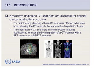
IAEA
International Atomic Energy Agency
Slide set of 190 slides based on the chapter authored by
J Gelijns
of the IAEA publication (ISBN 978-92-0-131010-1):
Diagnostic Radiology Physics:
A Handbook for Teachers and Students
Objective:
To familiarize the student with CT scanning principles, acquisition
and reconstruction, technology and image quality.
Chapter 11: Computed Tomography
Slide set prepared
by S. Edyvean
IAEA
CHAPTER 11. TABLE OF CONTENTS
11.1. Introduction
11.2. CT principles
11.3. The CT imaging system
11.4. Image reconstruction and processing
11.5. Acquisition
11.6. Computed tomography image quality
Bibliography
Diagnostic Radiology Physics: a Handbook for Teachers and Students – chapter 11, 2
IAEA
Clinical Computed Tomography (CT) was introduced in
1971 - limited to axial imaging of the brain in
neuroradiology
It developed into a versatile 3D whole body imaging
modality for a wide range of applications in for example
• oncology, vascular radiology, cardiology, traumatology and
interventional radiology.
Computed tomography can be used for
• diagnosis and follow-up studies of patients
• planning of radiotherapy treatment
• screening of healthy subpopulations with specific risk factors.
11.1 INTRODUCTION
Diagnostic Radiology Physics: a Handbook for Teachers and Students – chapter 11, 3
IAEA
Nowadays dedicated CT scanners are available for special
clinical applications, such as
• For radiotherapy planning - these CT scanners offer an extra wide
bore, allowing the CT scans to be made with a large field of view.
• The integration of CT scanners in multi modality imaging
applications, for example by integration of a CT scanner with a
PET scanner or a SPECT scanner.
11.1 INTRODUCTION
Diagnostic Radiology Physics: a Handbook for Teachers and Students – chapter 11, 4

Computed tomography notes full notes helpfull for radiological students for their exams

  • 1.
    IAEA International Atomic EnergyAgency Slide set of 190 slides based on the chapter authored by J Gelijns of the IAEA publication (ISBN 978-92-0-131010-1): Diagnostic Radiology Physics: A Handbook for Teachers and Students Objective: To familiarize the student with CT scanning principles, acquisition and reconstruction, technology and image quality. Chapter 11: Computed Tomography Slide set prepared by S. Edyvean
  • 2.
    IAEA CHAPTER 11. TABLEOF CONTENTS 11.1. Introduction 11.2. CT principles 11.3. The CT imaging system 11.4. Image reconstruction and processing 11.5. Acquisition 11.6. Computed tomography image quality Bibliography Diagnostic Radiology Physics: a Handbook for Teachers and Students – chapter 11, 2
  • 3.
    IAEA Clinical Computed Tomography(CT) was introduced in 1971 - limited to axial imaging of the brain in neuroradiology It developed into a versatile 3D whole body imaging modality for a wide range of applications in for example • oncology, vascular radiology, cardiology, traumatology and interventional radiology. Computed tomography can be used for • diagnosis and follow-up studies of patients • planning of radiotherapy treatment • screening of healthy subpopulations with specific risk factors. 11.1 INTRODUCTION Diagnostic Radiology Physics: a Handbook for Teachers and Students – chapter 11, 3
  • 4.
    IAEA Nowadays dedicated CTscanners are available for special clinical applications, such as • For radiotherapy planning - these CT scanners offer an extra wide bore, allowing the CT scans to be made with a large field of view. • The integration of CT scanners in multi modality imaging applications, for example by integration of a CT scanner with a PET scanner or a SPECT scanner. 11.1 INTRODUCTION Diagnostic Radiology Physics: a Handbook for Teachers and Students – chapter 11, 4