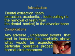
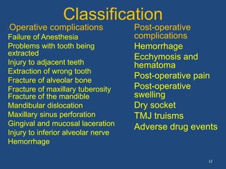
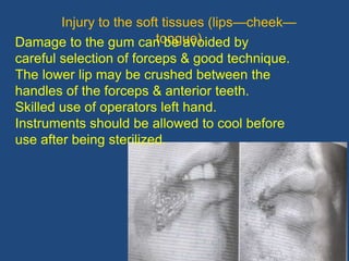
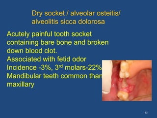
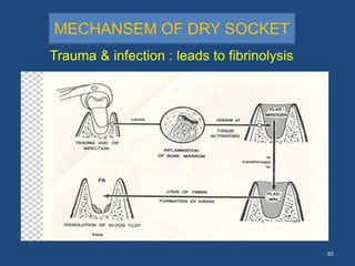
4. Introduction
Dental extraction tooth
extraction, exodontia, , tooth pulling) is
the removal of teeth from
the dental socket) in the alveolar bone
Complications
Any adverse , unplanned events that
tend to increase the morbidity above
what would be expected from a
particular operative procedure under
normal circumstances.
4
6. Surgical complications may arise from either one
or a combination of the following factors.
1-- PATIENT- Medically compromised pt. leading to
an persistent hemorrhage or delayed healing.
2-- CLINICIAN
-level of training, skills and expernice
-attitudes towards total patient care.
3-- SURGICAL PROCEDURE risks depend on :-
-complexity of the procedure.
-local anatomy of the surgical site
-proximity of important vital structures.
6
Sources of
complications
7. Possible complications
Failure to -secure anaesthesia
-remove the tooth with either
forceps or elevator
Fracture of-crown of the tooth /root
-alveolar bone
-maxillary tuberosity
-adjacent or opposing tooth
-mandible fracture
7
8. Dislocation of -adjacent tooth
-TMJ
Displacement of the root
-into the soft tissues
- maxillary antrum
-aspiration or swallowing
under G.A
8
9. Excessive hemorrhage
- During tooth removal
- on completion of the
extraction
- postoperatively
Damage to
- gums/lips/tongue/floor of
mouth
- inferior dental nerve &
branches
- lingual nerve
9
10. Postoperative pain
- damage to hard & soft tissues
- dry socket
- acute osteomyelitis of mandible
- traumatic arthritis of TMJ
Postoperative swelling due to:-
Edema
Hematomas formation
Infection
Truisms
Oro-antral communication
10
12. Classification
• Operative complications
Failure of Anesthesia
Problems with tooth being
extracted
Injury to adjacent teeth
Extraction of wrong tooth
Fracture of alveolar bone
Fracture of maxillary tuberosity
Fracture of the mandible
Mandibular dislocation
Maxillary sinus perforation
Gingival and mucosal laceration
Injury to inferior alveolar nerve
Hemorrhage
12
Post-operative
complications
Hemorrhage
Ecchymosis and
hematoma
Post-operative pain
Post-operative
swelling
Dry socket
TMJ truisms
Adverse drug events
13. 13
Failure Of Local Anesthesia
Failure of local anesthesia is usually the result of
either:
• inaccurate placement of the anesthetic solution
• too small a dosage
• not waiting long enough for the anesthesia to act before
commencing surgery
Check for subjective signs and objective signs to
confirm LA has worked.
If anesthesia cannot be secured by using conventional
techniques of infiltration or regional block
intraligamental, intraraosseous or intrapulpal injections
may be indicated, provided that the cause of the failure is
15. 15
Injuries to adjacent tooth
1- Fracture or dislodgement of adjacent
restoration
If a large restoration exists:
-Patient should be warned about possibility
of fracturing or displacing during
extraction.
Straight elevator should be inserted entirely
into the periodontal ligament space
Management
The surgeon should make sure the
restoration is removed from the mouth
injured tooth should be treated by
replacement of the displaced crown or
placement of a temporary restoration
16. 16
2-Luxation Of An Adjacent Tooth
Caused due to Inappropriate use of the extraction instruments
Prevention
Judicious use of force with elevators and forceps
• Other teeth should not be used as fulcrum for an elevator.
• Narrow forceps may be useful for the extraction of
Management s
A- If it is luxated or partially avulsed
Reposition in the tooth socket and left alone
Occlusion should be checked to ensure that the tooth has not
been displaced into a hyperocclusion and traumatic occlusion
B-If the luxated tooth is mobile
The tooth should be stabilized with semirigid fixation to maintain it
in its position
For this a simple silk suture that crosses the occlusal table and is
sutured to the adjacent gingiva is usually sufficient
17. 17
Extraction of the Wrong Tooth
Causes
-A dentist removes a tooth for another dentist
- Use of differing tooth numbering systems
-Differences in the mounting of radiographs
Prevention
Focus attention on the procedure
Check with the patient and the assistant to ensure
that the correct tooth is being removed.
Check, then recheck, images and records to confirm
the correct tooth
Management
Immediately:-The tooth should be replaced quickly
into the tooth socket Splinting is done -Endodontic
treatment after successful reattachmen
19. 19
Fracture of the tooth
Fracture of the crown of a tooth
Weakened tooth- caries or large
restoration
Improper application of the forceps
Excessive force
proper application of forceps or elevator
will deliver the tooth or Trans alveolar
method
20. 20
Root fracture
Faulty technique
Pulp less or rct treated or brittle teeth
Fracture on apical third or middle or cervical or
longitudinal fracture
Management
Trans alevolar extraction except fracture root on the
cervical third
it can be by close extraction
21. 21
Fracture of alveolar bone
Accidental inclusion of alveolar bone within forceps
blades.
Pathological changes in the bone
Shape of the alveolus
Extraction of canine is frequently complicated by fracture
of the labial plate.
Alveolar fragments which has lost one half of the
periosteal attachment should be removed. if it well
attached to periosteum, should be sutured back
22. 22
Dislocation of the mandible
It is the dislodgment of the condyloid process from its
seat in the glenoid fossa .One or both joints may be
dislocated.
--This dislocation is usually the result of using:-
1- too much pressure during extraction
2-Inadequate support of the mandible.
3-Under general anesthesia when muscles of
mastication are relaxed
Symptom
The mouth is opened and rigidly set in position with
the chin protruding .
The patient cannot close his mouth,
depression in front of the ear ,
some pain may or may not be present
25. 25
1. Dislocation of the mandible
Management
Reduction of the dislocation by the following
technique : The operator wraps his thumps with
gauze as a safeguard against injury which may
result from immediate and quick closure of the
jaw.
Then the thumbs are placed on the occlusal
surface of the mandibular teeth by which
downward and backward pressure is applied
meanwhile the free fingers support the jaw during
the application of pressure and to raise the chin
. A bandage is applied to hold the jaw in place for
48 hours. Analgesic are also indicated in the first
26. 26
Fracture of the mandible
1--Excessive or incorrectly applied force
2-Exsseve bone removal during surgical extraction
3-Pathological changes of mandible
Senile osteoporosis
Atrophy
Osteomyelitis
Previous therapeutic irradiation
Unerupted teeth, cysts, hyperparathyroidism
Or tumors may also predispose to fracture
27. 27
Fracture of Maxillary Tuberosity
This occasionally occurs when
extraction of third maxillary molars is
attempted .
It is liable to occur when such a tooth is
firmly ankylosed to the bone or isolated
or has divergent or hypercementosed
root formation.
Also liable to occur when the tuberosity
is prominent.
If accident occurs
the operator find him/herself grasping a
large segment of bone , which
31. 31
Management :
If it is a small fragment it should be surgically
excised.
A gingival incision around bone with the tooth
attached ,
the fragment is then freed and removed.
In most cases the maxillary sinus is exposed , the
bone is debrided and the flaps reposed and
sutured over the exposed sinus.
If the fragment is a large one and carrying more
than one sound tooth,
it could be repositioned and fixed with suitable
Fracture of Maxillary Tuberosity
37. 37
Diagnosis:
Bubbling through the extraction site occurs when
the nose is blocked under pressure. The patient
cannot suck through a straw.
Management:
Immediate Rx alternatives:
Replace the tooth and splint into position and
plan to extract surgically at a later date or
Cover defect with anti septic – soaked ribbon/
gauze and remove in 2-3 weeks to allow healing
by sec. intention or
Reduce bony socket edge and suture margins
together (interrupted horizontal Immediate
closure with a buccal advancement flap provided
the sinus is clear of infection.
38. 38
---Displacement the root to max sinus it can be
removed by the suction tip on perforated floor of the
sinus
if it failed it will go to Claudelwelluc operation to open
the max sinus to remove the RR,
Remove the broken roots of the lower molar sometimes
are pushed through the thin inner area of the mandible
to submandibular space below the mylohyoid muscle---
--
Push it back with finger pressure to the orifice of the
socket
Displacement of the root
- maxillary antrum
- -into the soft
tissues
47. 47
Causes
1- Uncommon occurrence in the extraction of erupted
mandibular teeth.
2- Injudicious curettage or improper use of elevators to
remove root apices
Result in paresthesia and sometimes anesthesia of half the
lower lip and chin.
Management
Most cases - the nerve regenerates within 6 weeks to 6
months.
If the nerve does not regenerate, the bony walls of the
mandibular canal may have been displaced, impinging on
it.
This condition sometimes can be remedied by a
decompression operation. • Traumatic neuroma – excised
and the nerve reanastomosed or grafted.
Injury of the inferior alveolar nerve
Intervention is required in some circumstances, as nerve
repair can improve recovery. using epineurial sutures is
more effective than entubulation and, if a damaged nerve
segment has to be excised, repair by stretching the ends
under slight tension is better than grafting
48. 48
AREA OF SENSORY DEFICIT
FOLLOWING INFERIOR ALVEOLAR
NERVE INJURY
damage can be prevented or
minimized only by pre-op
radiographic diagnosis & careful
dissection.
49. 49
Lingual nerve :
Lingual nerve is in close
proximity to roots of mandibular
third molar .
Risk of damage while taking
incision and during elevation of
lingual periosteum.
Risk of direct trauma form bur or
chisels used for removal of
bone or sectioning of the tooth
The risk associated with wisdom
tooth surgery is commonly
accepted to be 2% temporary
and 0.2% permanent.
Parasthesia half of the tongoue
Mental nerve :
Injury is caused due to surgery
in the area of mental
nerve.(premolar area)
51. 51
Injury to the soft tissues (lips—cheek—
tongue)Damage to the gum can be avoided by
careful selection of forceps & good technique.
The lower lip may be crushed between the
handles of the forceps & anterior teeth.
Skilled use of operators left hand.
Instruments should be allowed to cool before
use after being sterilized.
52. 52
Emphysema
It the swelling due to accumulation of air
into C,T of the facial muscle
Due to prolonged reflection of the large flap
together with use the drill
Swelling will take 7---10 days to be
absorbed
Antibiotic is recommended
54. 54
1--Hemorrhage
Hemorrhage is escape of blood outside the
vascular system
TYPES;-
A- internal inside the body in oral cavity
blood between the oral spaces
B-External outside the body
1- Primary ;during the procedure
2- Intermediate; after24h from the
procedure
3—Secondary;after the first day or at any
time
55. 55
Etiology of the oral hemorrhage
-Local cause;--
-Interference by the patient to the formed clot
by mechanical dislodgement of the clot
--Improper post operative instruction for
pressure packs and other instruction
--Presence of nutrient canal in the wall of the
socket
--Presence of the infection, granulation tissue,
Chronic gingival inflammation
56. 56
Systemic cause;-
--Vascular Defects;
it is structural ,functional defects of vessels affecting the
vascular contraction leads to prolonged bleeding
( Hereditary hemorrhage
telangiecutasia,Scurvy,Allergic,,)
--Platelet disorders: Thrombocytopenia antiplatelet
drugs ASA PLAVX
--Coagulation Disorder;-
It is due to absence of one or more of the clotting factor
ether congenital (: Hemophilia)
Drugs (, Anti coagulant therapy)
systemic disease
leukemia, multiple myeloma Aplastic anaemias
57. 57
Physical methods
Pressure packs
Use of LA solution with
vasoconstrictors
Socket suturing
Hemostatic forceps
Splints
Thermal measures- cautery,
hot saline packs
Firm gauze roll should be
placed upon the socket &
patient asked to bite upon it .
Horizontal mattress suture
Management
62. 62
Dry socket / alveolar osteitis/
alveolitis sicca dolorosa
Acutely painful tooth socket
containing bare bone and broken
down blood clot.
Associated with fetid odor
Incidence -3%, 3rd molars-22%
Mandibular teeth common than
maxillary
63. 63
Predisposing factors
-- infection of socket : release of plasminogen
activators
---Trauma - use of excessive force
---Vasoconstrictors (contributory factor)
---Mandibular extractions (dense & less vascular,
contaminated with food debris)
---Bacteriological origin - Treponema denticolum.
---Pt. on oral contraceptives, smokers
64. 64
Vascular injury
Activation of coag
Platelet activation
Fibrin
formation
thrombu
s
(Fibrinolytic agents)XII Plasminogen
plasmin
tissue plasminogen activator
inflammation of the tooth socket
66. 66
--granulation tissue is absent in cases of dry
socket.
--Dull, boring pain to severe throbbing pain
--Gingival margin of socket – swollen & red
--Socket filled with food debris or a brown friable
clot
---bare bone which is severely tender to touch
---Regional lymph nodes may be tender
Clinical features
67. 67
Management
--Socket irrigation with warm saline & all degenerating blood
clot removed.
---Sharp bony spurs - excised with rongeur forceps or
removed with a wheel stone
---Loose dressing – zinc oxide & oil of cloves on cotton wool is
tucked into the socket.
--Analgesic tab & hot saline mouth baths
---Recall after 3 days
68. 68
Postoperative swelling
A--EDEMA :
1-If the soft tissues are not handled carefully during an
extraction traumatic edema may be formed.
2-The use of blunt instrument, the excessive retraction of
badly designed flap, or a bur becoming entangled in the
soft tissues predispose to this condition.
3-IF sutures are tied too tightly post operative swelling due to
edema or hematoma formation may cause sloughing of
the soft tissues and breakdown of the suture line.
Usually both conditions regress if the patient uses hot saline
mouth baths frequently for 2-3 days
69. 69
b. INFECTION :-
pain and swelling
Mild- hot saline mouth baths
Severe – I & D, antibiotic & analgesics
70. 70
Truisms
Anability to open mouth due to muscle
spasm.
Caused by post op. edema, hematoma
formation or inflammation of soft tissue.
Intra oral heat by means of short wave
diathermy or use of hot saline mouth
baths.
Antibiotics
72. 72
It is extensive infection bone,(bone marrow
,periosteum, and affects a large area of
mandible bone)
Mandible tender
impairment of labial sensation
pyrexia , pain is severe
Traumatic extraction of lower molar under
LA in P/o acute gingival inflammation
predisposes to acute OML.
Patient should hospitalized for effective
treatment
73. 73
Bisphosphonates are the most widely prescribed drugs
for the treatment of osteoporosis, and are also used in
malignant bone metastases, multiple myeloma, and
Paget's disease, and provide therapeutic efficacy on
those diseases.
Adverse drug events
bisphosphonate-related
osteonecrosis of the jaws
bone in non-healing extraction
A tooth extraction during or after
bisphosphonate therapy is a
possible trigger of ONJ
Bisphosphonates remain on
hydroxapptied of the bone more than
3 month after the stop of the
treatment
Careful history from the patient