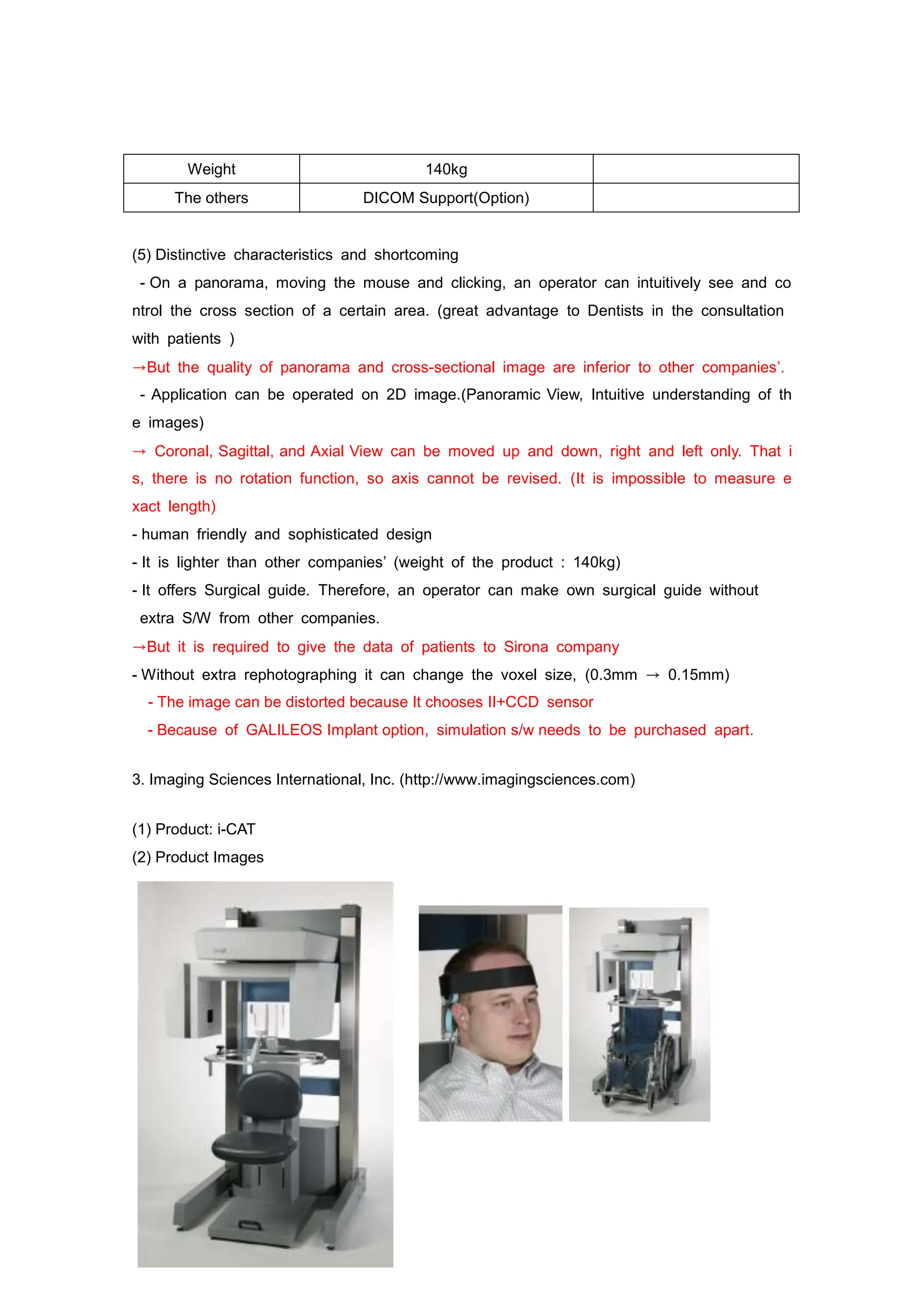
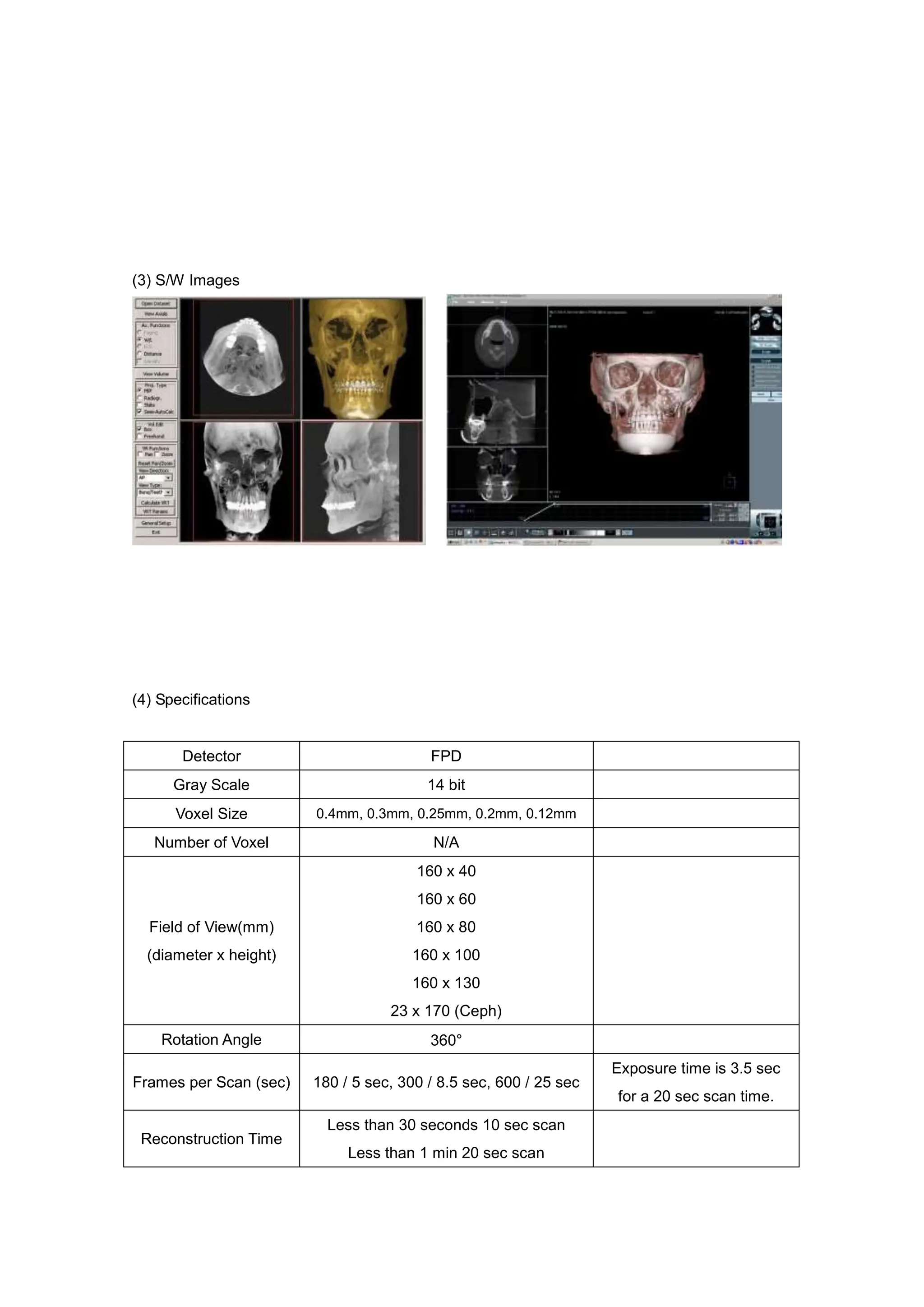
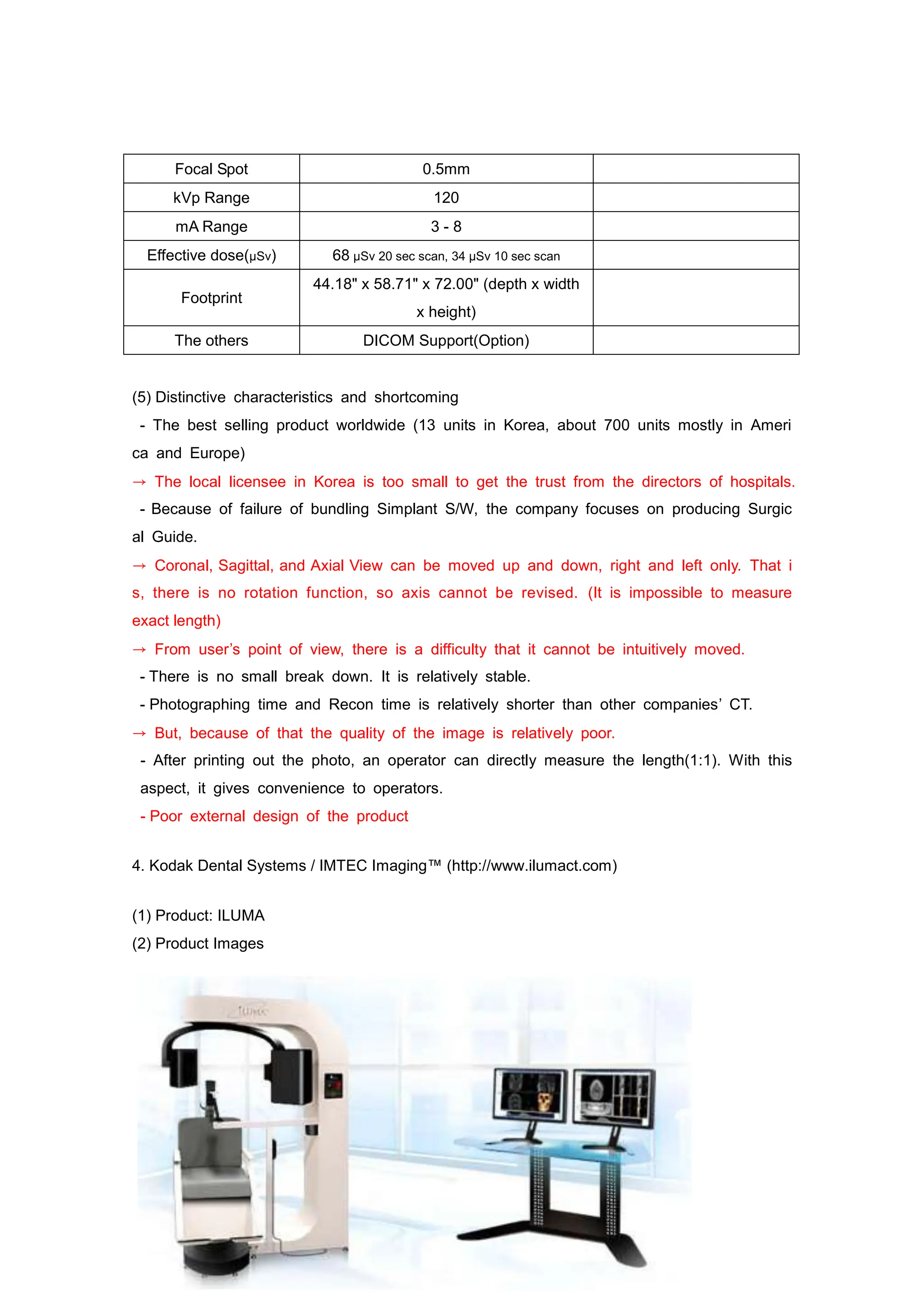
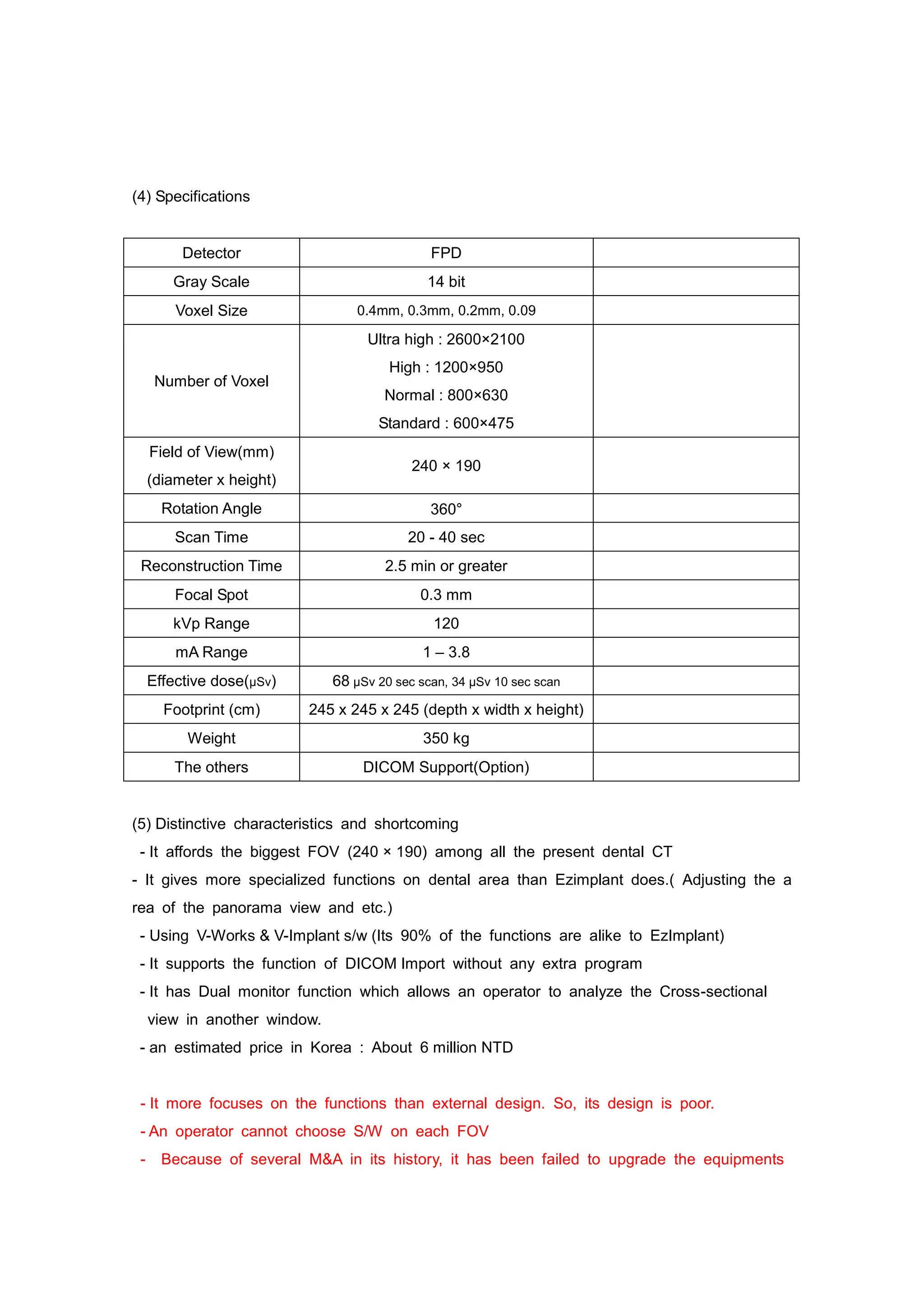
Competitor Analysis
1. J. Morita MFG. Corp (http://www.jmorita-mfg.com)
(1) Product: Veraviewpocs 3D
(2) Product Images
(3) S/W Images
(4) Specifications
Detector FPD
CSI coated CMOS FPD
Hamamatsu C9250DP
Gray Scale 13 bit
Voxel Size 0.125mm
Number of Voxel N/A
Field of View(mm)
40 x 40
40 x 80
80 x 80
Rotation Angle 180°
Exposure Time(sec)
CT ; 9.4
Pano; 7.7(Fine high), 15(Superfine)
Picasso 14sec
Reconstruction Time Less than 5 min Picasso 2.5 min
Focal Spot 0.5 mm
kVp Range 60 - 90
kVp(Peak kilovoltage): The
maximum voltage applied
across an X-ray tube, the deg
ree of intensity of X-ray is d
irectly proportional to square
of kVp
mA Range 1 - 10
mA: a unit of electric current,
or amount of electric charge
per second
Footprint
With Pano 1.35㎡
With Pano + Ceph 2.60㎡
Weight
With Pano 19kg
With Pano + Ceph 258kg
Upgradeable to Ceph 205㎏
The others DICOM Support(Option)
(5) Distinctive characteristics and shortcoming
- If necessary, Cassette is purchasable at the buyers’ option. ( In 40 x40 an operator
can take CT and Pano with only one cassette.)
→There is a possible risk when a cassette is being attached/removed
- Since its AF Auto Positioning function automatically sets up the photographing position,
an operator can easily make the best image.
- It will automatically set up the shooting(photographing) position, when an operator clicks
on the position he/she wants in Pano image.
- 80x80’s Voxel size is 0.125mm. The quality of the image is excellent
→ But Recon time takes 5 minutes
- It allows an operator to see 3D in a network with a small outlay, thanks to diversificati
on of 3D viewer
(One Data Viewer: XYZ axes move function, One Data Viewer Plus: 이미지 조정 adjustment
of image and measurement function, XYZ axes move function)
→ I-dixel program, 3D viewer S/W, doesn’t have canal coloring or implant simulation fun
ctions. It only has very simple 3D viewer functions such as length and angle measuring f
unction, profile measuring function, rotation, and etc.. In addition, since there is no rotation
function of an axis in i-dixel, an axis cannot be revised.(It is impossible to measure exa
ct length.)
- It can be installed in two ways, floor type and wall type.(The photographing can be d
one either when a patient is sitting on a chair or when a patient is standing.)
2. Sirona (http://www.sirona.com)
(1) Product: GALILEOS
(2) Product Images
(3) S/W Images
(4) Specifications
Detector II+CCD
Gray Scale 8 or 12 bit
Voxel Size
0.300mm
0.150mm
Number of Voxel
512x512x512
1024x1024x1024
Field of View(mm) 150x150x150
Rotation Angle 180°
Exposure Time(sec) 14 sec
On Brochure & homepage
Scan time; 14s,
Exposure time; 2 - 6s
Reconstruction Time 4.5 min
Focal Spot N/A
kVp Range 85
kVp(Peak kilovoltage): The
maximum voltage applied
across an X-ray tube, the deg
ree of intensity of X-ray is d
irectly proportional to square
of kVp
mA Range 5 - 7
mA: a unit of electric current,
or amount of electric charge
per second
Effective dose(µSv) 29 µSv (21mAs,85kV)
Footprint 71" x 71" x 99" (depth x width x height)
Weight 140kg
The others DICOM Support(Option)
(5) Distinctive characteristics and shortcoming
- On a panorama, moving the mouse and clicking, an operator can intuitively see and co
ntrol the cross section of a certain area. (great advantage to Dentists in the consultation
with patients )
→But the quality of panorama and cross-sectional image are inferior to other companies’.
- Application can be operated on 2D image.(Panoramic View, Intuitive understanding of th
e images)
→ Coronal, Sagittal, and Axial View can be moved up and down, right and left only. That i
s, there is no rotation function, so axis cannot be revised. (It is impossible to measure e
xact length)
- human friendly and sophisticated design
- It is lighter than other companies’ (weight of the product : 140kg)
- It offers Surgical guide. Therefore, an operator can make own surgical guide without
extra S/W from other companies.
→But it is required to give the data of patients to Sirona company
- Without extra rephotographing it can change the voxel size, (0.3mm → 0.15mm)
- The image can be distorted because It chooses II+CCD sensor
- Because of GALILEOS Implant option, simulation s/w needs to be purchased apart.
3. Imaging Sciences International, Inc. (http://www.imagingsciences.com)
(1) Product: i-CAT
(2) Product Images
(3) S/W Images
(4) Specifications
Detector FPD
Gray Scale 14 bit
Voxel Size 0.4mm, 0.3mm, 0.25mm, 0.2mm, 0.12mm
Number of Voxel N/A
Field of View(mm)
(diameter x height)
160 x 40
160 x 60
160 x 80
160 x 100
160 x 130
23 x 170 (Ceph)
Rotation Angle 360°
Frames per Scan (sec) 180 / 5 sec, 300 / 8.5 sec, 600 / 25 sec
Exposure time is 3.5 sec
for a 20 sec scan time.
Reconstruction Time
Less than 30 seconds 10 sec scan
Less than 1 min 20 sec scan
Focal Spot 0.5mm
kVp Range 120
mA Range 3 - 8
Effective dose(µSv) 68 µSv 20 sec scan, 34 µSv 10 sec scan
Footprint
44.18" x 58.71" x 72.00" (depth x width
x height)
The others DICOM Support(Option)
(5) Distinctive characteristics and shortcoming
- The best selling product worldwide (13 units in Korea, about 700 units mostly in Ameri
ca and Europe)
→ The local licensee in Korea is too small to get the trust from the directors of hospitals.
- Because of failure of bundling Simplant S/W, the company focuses on producing Surgic
al Guide.
→ Coronal, Sagittal, and Axial View can be moved up and down, right and left only. That i
s, there is no rotation function, so axis cannot be revised. (It is impossible to measure
exact length)
→ From user’s point of view, there is a difficulty that it cannot be intuitively moved.
- There is no small break down. It is relatively stable.
- Photographing time and Recon time is relatively shorter than other companies’ CT.
→ But, because of that the quality of the image is relatively poor.
- After printing out the photo, an operator can directly measure the length(1:1). With this
aspect, it gives convenience to operators.
- Poor external design of the product
4. Kodak Dental Systems / IMTEC Imaging™ (http://www.ilumact.com)
(1) Product: ILUMA
(2) Product Images
(3) S/W Images
(4) Specifications
Detector FPD
Gray Scale 14 bit
Voxel Size 0.4mm, 0.3mm, 0.2mm, 0.09
Number of Voxel
Ultra high : 2600×2100
High : 1200×950
Normal : 800×630
Standard : 600×475
Field of View(mm)
(diameter x height)
240 × 190
Rotation Angle 360°
Scan Time 20 - 40 sec
Reconstruction Time 2.5 min or greater
Focal Spot 0.3 mm
kVp Range 120
mA Range 1 – 3.8
Effective dose(µSv) 68 µSv 20 sec scan, 34 µSv 10 sec scan
Footprint (cm) 245 x 245 x 245 (depth x width x height)
Weight 350 kg
The others DICOM Support(Option)
(5) Distinctive characteristics and shortcoming
- It affords the biggest FOV (240 × 190) among all the present dental CT
- It gives more specialized functions on dental area than Ezimplant does.( Adjusting the a
rea of the panorama view and etc.)
- Using V-Works & V-Implant s/w (Its 90% of the functions are alike to EzImplant)
- It supports the function of DICOM Import without any extra program
- It has Dual monitor function which allows an operator to analyze the Cross-sectional
view in another window.
- an estimated price in Korea : About 6 million NTD
- It more focuses on the functions than external design. So, its design is poor.
- An operator cannot choose S/W on each FOV
- Because of several M&A in its history, it has been failed to upgrade the equipments
and improve the functions. (MTEC → Kodak → Onex)
There are difficulties that it has been failed to retain and support the policy of AS.
4. Planmeca (http://www.planmeca.com)
(1) Product: ProMax 3D
(2) Product Images
(3) S/W Images
(4) Specifications
Detector FPD
Gray Scale 12 bit
Voxel Size 0.16mm
Number of Voxel 501
Field of View(mm)
(diameter x height)
80 x 80
80 x 50
40 x 40
Rotation Angle 194°
Scan Time 18 sec Exposure Time: 6 - 7s
Reconstruction Time Under 3 min
Focal Spot 0.5 mm
kVp Range N/A
mA Range N/A
Effective dose(µSv) N/A
Footprint (cm) N/A
Weight N/A
The others Romexis 3D Explorer s/w
(5) Distinctive characteristics and shortcoming
- Good design
- CT sensor can easily be attached and removed
- Photographing PC and Database Server are being operated separately.
- It offers the intuitive 3D analysis program which is very necessary
- In DB server, Windows XP, Vista, 2003 Server are the supporting S/W
In client, Max Os and Linux are compatible
- The quality of the images are poor compared to the competitors’. The quality is very si
milar to tomo image, and the quality of VR is also relatively poor.)
- Implant Planning Mode option
- The maximum FOV is 8 x 8, so it cannot take the whole picture of arch at once.
- It is more expensive that Picasso Trio. ( estimated price : about 6million NTD)
- It only offers very simple functions of 3D Viewer, so it’s not good enough for consultati
on purpose.
5. QR s.r.l. (http://www.qrverona.it)
(1) Product: NewTom 3G/ NewTom VG
(2) Product Images
NewTom 3G
NewTom VG
(3) S/W Images
(4) Specifications
NewTom 3G NewTom VG
Detector N/A FPD
Gray Scale N/A 14 bit
Voxel Size 0.42mm, 0.29mm, 0.19mm 0.127mm
Number of Voxel N/A N/A
Field of View(mm)
200 x 200
150 x 150
100 x 100
160 x 140
Rotation Angle N/A 360°
Scan Time 36s 24s
Exposure Time 5.4s N/A
Reconstruction Time over 3 min 3 min
Focal Spot 0.5 mm 0.3mm
kVp Range 110 80-120
mA Range 15 N/A
Footprint(cm) 200 x 250 144 x 110 x 230
Weight 480kg 250kg
The others Print(Option), CD, DVD writer
(5) Distinctive characteristics and shortcoming
NewTom 3G
- The quality of image is good
- Restructuring images and loading are fast.
- Exposure Time is relatively short.
- It is the 3D viewer which is specialized on Dental clinic and gives a variety of the
functions more than its competitors.
- Since a patient need to lie down, it takes a lot of spaces.
NewTom VG
- It can be installed in the same area with panorama
5. PALODEX Group, SOREDEX (http://www.soredex.com)
(1) Name: SCANORA 3D
(2) Product Images
(3) S/W Images
(4) Specifications
Detector FPD(Pano-CCD, CT-CMOS)
Gray Scale N/A
Voxel Size 0.42mm, 0.29mm, 0.19mm
Number of Voxel 0.15mm, 0.35mm
Field of View(mm) 124 x 124
Rotation Angle N/A
Scan Time 10 – 24 s
Exposure Time 2 – 5 s
Reconstruction Time 1 - 3 min
Focal Spot 0.4 mm
kVp Range 65 - 85
mA Range 0.5 - 0.8
Footprint(mm) 1963 x 1541 x 1100 (H x W x D)
Weight 310kg
The others Print(Option), CD, DVD writer
(5) Distinctive characteristics and shortcoming
- When photographing Pano or CT, an operator doesn’t need to attach or remove the se
nsor. (Using Auto Switch, an operator can change it into another, and also change Pano
sensor’s position when photographing CT)
- Using Remote Control function, an operator can change and control the shooting position.
- It has a Clear Touch Control Panel which allows the operator to choose Pano or CT fun
ction right next to the product and see the preview image on the panel.
- When photographing CT, an operator can choose FOV, concerning the part of the diagn
osis.
- Its VR image has relatively better quality when using Implant Simulation. (But in 2D, th
e reality is relatively poor)
- It has Canal Coloring and Implant Simulation functions.
- Because the maximum FOV is only 75cm x 145cm, revision and MFS are limited.
- Ceph image is not available
- The quality of 2D image is poor compared to its competitors’
- Coronal, Sagittal, and Axial View can be moved up and down, right and left only. That is,
there is no rotation function, so axis cannot be revised. (It is impossible to measure
exact length)
- It only offers a very simple function of 3D Viewer

Comparison chart(English).doc

  • 1.
    Competitor Analysis 1. J.Morita MFG. Corp (http://www.jmorita-mfg.com) (1) Product: Veraviewpocs 3D (2) Product Images (3) S/W Images
  • 2.
    (4) Specifications Detector FPD CSIcoated CMOS FPD Hamamatsu C9250DP Gray Scale 13 bit Voxel Size 0.125mm Number of Voxel N/A Field of View(mm) 40 x 40 40 x 80 80 x 80 Rotation Angle 180° Exposure Time(sec) CT ; 9.4 Pano; 7.7(Fine high), 15(Superfine) Picasso 14sec Reconstruction Time Less than 5 min Picasso 2.5 min Focal Spot 0.5 mm kVp Range 60 - 90 kVp(Peak kilovoltage): The maximum voltage applied across an X-ray tube, the deg ree of intensity of X-ray is d irectly proportional to square of kVp mA Range 1 - 10 mA: a unit of electric current, or amount of electric charge per second Footprint With Pano 1.35㎡ With Pano + Ceph 2.60㎡ Weight With Pano 19kg With Pano + Ceph 258kg Upgradeable to Ceph 205㎏ The others DICOM Support(Option) (5) Distinctive characteristics and shortcoming - If necessary, Cassette is purchasable at the buyers’ option. ( In 40 x40 an operator can take CT and Pano with only one cassette.) →There is a possible risk when a cassette is being attached/removed - Since its AF Auto Positioning function automatically sets up the photographing position, an operator can easily make the best image.
  • 3.
    - It willautomatically set up the shooting(photographing) position, when an operator clicks on the position he/she wants in Pano image. - 80x80’s Voxel size is 0.125mm. The quality of the image is excellent → But Recon time takes 5 minutes - It allows an operator to see 3D in a network with a small outlay, thanks to diversificati on of 3D viewer (One Data Viewer: XYZ axes move function, One Data Viewer Plus: 이미지 조정 adjustment of image and measurement function, XYZ axes move function) → I-dixel program, 3D viewer S/W, doesn’t have canal coloring or implant simulation fun ctions. It only has very simple 3D viewer functions such as length and angle measuring f unction, profile measuring function, rotation, and etc.. In addition, since there is no rotation function of an axis in i-dixel, an axis cannot be revised.(It is impossible to measure exa ct length.) - It can be installed in two ways, floor type and wall type.(The photographing can be d one either when a patient is sitting on a chair or when a patient is standing.) 2. Sirona (http://www.sirona.com) (1) Product: GALILEOS (2) Product Images (3) S/W Images
  • 4.
    (4) Specifications Detector II+CCD GrayScale 8 or 12 bit Voxel Size 0.300mm 0.150mm Number of Voxel 512x512x512 1024x1024x1024 Field of View(mm) 150x150x150 Rotation Angle 180° Exposure Time(sec) 14 sec On Brochure & homepage Scan time; 14s, Exposure time; 2 - 6s Reconstruction Time 4.5 min Focal Spot N/A kVp Range 85 kVp(Peak kilovoltage): The maximum voltage applied across an X-ray tube, the deg ree of intensity of X-ray is d irectly proportional to square of kVp mA Range 5 - 7 mA: a unit of electric current, or amount of electric charge per second Effective dose(µSv) 29 µSv (21mAs,85kV) Footprint 71" x 71" x 99" (depth x width x height)
  • 5.
    Weight 140kg The othersDICOM Support(Option) (5) Distinctive characteristics and shortcoming - On a panorama, moving the mouse and clicking, an operator can intuitively see and co ntrol the cross section of a certain area. (great advantage to Dentists in the consultation with patients ) →But the quality of panorama and cross-sectional image are inferior to other companies’. - Application can be operated on 2D image.(Panoramic View, Intuitive understanding of th e images) → Coronal, Sagittal, and Axial View can be moved up and down, right and left only. That i s, there is no rotation function, so axis cannot be revised. (It is impossible to measure e xact length) - human friendly and sophisticated design - It is lighter than other companies’ (weight of the product : 140kg) - It offers Surgical guide. Therefore, an operator can make own surgical guide without extra S/W from other companies. →But it is required to give the data of patients to Sirona company - Without extra rephotographing it can change the voxel size, (0.3mm → 0.15mm) - The image can be distorted because It chooses II+CCD sensor - Because of GALILEOS Implant option, simulation s/w needs to be purchased apart. 3. Imaging Sciences International, Inc. (http://www.imagingsciences.com) (1) Product: i-CAT (2) Product Images
  • 6.
    (3) S/W Images (4)Specifications Detector FPD Gray Scale 14 bit Voxel Size 0.4mm, 0.3mm, 0.25mm, 0.2mm, 0.12mm Number of Voxel N/A Field of View(mm) (diameter x height) 160 x 40 160 x 60 160 x 80 160 x 100 160 x 130 23 x 170 (Ceph) Rotation Angle 360° Frames per Scan (sec) 180 / 5 sec, 300 / 8.5 sec, 600 / 25 sec Exposure time is 3.5 sec for a 20 sec scan time. Reconstruction Time Less than 30 seconds 10 sec scan Less than 1 min 20 sec scan
  • 7.
    Focal Spot 0.5mm kVpRange 120 mA Range 3 - 8 Effective dose(µSv) 68 µSv 20 sec scan, 34 µSv 10 sec scan Footprint 44.18" x 58.71" x 72.00" (depth x width x height) The others DICOM Support(Option) (5) Distinctive characteristics and shortcoming - The best selling product worldwide (13 units in Korea, about 700 units mostly in Ameri ca and Europe) → The local licensee in Korea is too small to get the trust from the directors of hospitals. - Because of failure of bundling Simplant S/W, the company focuses on producing Surgic al Guide. → Coronal, Sagittal, and Axial View can be moved up and down, right and left only. That i s, there is no rotation function, so axis cannot be revised. (It is impossible to measure exact length) → From user’s point of view, there is a difficulty that it cannot be intuitively moved. - There is no small break down. It is relatively stable. - Photographing time and Recon time is relatively shorter than other companies’ CT. → But, because of that the quality of the image is relatively poor. - After printing out the photo, an operator can directly measure the length(1:1). With this aspect, it gives convenience to operators. - Poor external design of the product 4. Kodak Dental Systems / IMTEC Imaging™ (http://www.ilumact.com) (1) Product: ILUMA (2) Product Images (3) S/W Images
  • 8.
    (4) Specifications Detector FPD GrayScale 14 bit Voxel Size 0.4mm, 0.3mm, 0.2mm, 0.09 Number of Voxel Ultra high : 2600×2100 High : 1200×950 Normal : 800×630 Standard : 600×475 Field of View(mm) (diameter x height) 240 × 190 Rotation Angle 360° Scan Time 20 - 40 sec Reconstruction Time 2.5 min or greater Focal Spot 0.3 mm kVp Range 120 mA Range 1 – 3.8 Effective dose(µSv) 68 µSv 20 sec scan, 34 µSv 10 sec scan Footprint (cm) 245 x 245 x 245 (depth x width x height) Weight 350 kg The others DICOM Support(Option) (5) Distinctive characteristics and shortcoming - It affords the biggest FOV (240 × 190) among all the present dental CT - It gives more specialized functions on dental area than Ezimplant does.( Adjusting the a rea of the panorama view and etc.) - Using V-Works & V-Implant s/w (Its 90% of the functions are alike to EzImplant) - It supports the function of DICOM Import without any extra program - It has Dual monitor function which allows an operator to analyze the Cross-sectional view in another window. - an estimated price in Korea : About 6 million NTD - It more focuses on the functions than external design. So, its design is poor. - An operator cannot choose S/W on each FOV - Because of several M&A in its history, it has been failed to upgrade the equipments
  • 9.
    and improve thefunctions. (MTEC → Kodak → Onex) There are difficulties that it has been failed to retain and support the policy of AS. 4. Planmeca (http://www.planmeca.com) (1) Product: ProMax 3D (2) Product Images (3) S/W Images
  • 10.
    (4) Specifications Detector FPD GrayScale 12 bit Voxel Size 0.16mm Number of Voxel 501 Field of View(mm) (diameter x height) 80 x 80 80 x 50 40 x 40 Rotation Angle 194° Scan Time 18 sec Exposure Time: 6 - 7s Reconstruction Time Under 3 min Focal Spot 0.5 mm kVp Range N/A
  • 11.
    mA Range N/A Effectivedose(µSv) N/A Footprint (cm) N/A Weight N/A The others Romexis 3D Explorer s/w (5) Distinctive characteristics and shortcoming - Good design - CT sensor can easily be attached and removed - Photographing PC and Database Server are being operated separately. - It offers the intuitive 3D analysis program which is very necessary - In DB server, Windows XP, Vista, 2003 Server are the supporting S/W In client, Max Os and Linux are compatible - The quality of the images are poor compared to the competitors’. The quality is very si milar to tomo image, and the quality of VR is also relatively poor.) - Implant Planning Mode option - The maximum FOV is 8 x 8, so it cannot take the whole picture of arch at once. - It is more expensive that Picasso Trio. ( estimated price : about 6million NTD) - It only offers very simple functions of 3D Viewer, so it’s not good enough for consultati on purpose. 5. QR s.r.l. (http://www.qrverona.it) (1) Product: NewTom 3G/ NewTom VG (2) Product Images NewTom 3G NewTom VG
  • 12.
    (3) S/W Images (4)Specifications NewTom 3G NewTom VG Detector N/A FPD Gray Scale N/A 14 bit Voxel Size 0.42mm, 0.29mm, 0.19mm 0.127mm Number of Voxel N/A N/A Field of View(mm) 200 x 200 150 x 150 100 x 100 160 x 140 Rotation Angle N/A 360° Scan Time 36s 24s Exposure Time 5.4s N/A Reconstruction Time over 3 min 3 min Focal Spot 0.5 mm 0.3mm kVp Range 110 80-120 mA Range 15 N/A Footprint(cm) 200 x 250 144 x 110 x 230 Weight 480kg 250kg The others Print(Option), CD, DVD writer
  • 13.
    (5) Distinctive characteristicsand shortcoming NewTom 3G - The quality of image is good - Restructuring images and loading are fast. - Exposure Time is relatively short. - It is the 3D viewer which is specialized on Dental clinic and gives a variety of the functions more than its competitors. - Since a patient need to lie down, it takes a lot of spaces. NewTom VG - It can be installed in the same area with panorama 5. PALODEX Group, SOREDEX (http://www.soredex.com) (1) Name: SCANORA 3D (2) Product Images
  • 14.
    (3) S/W Images (4)Specifications Detector FPD(Pano-CCD, CT-CMOS) Gray Scale N/A Voxel Size 0.42mm, 0.29mm, 0.19mm Number of Voxel 0.15mm, 0.35mm Field of View(mm) 124 x 124 Rotation Angle N/A Scan Time 10 – 24 s Exposure Time 2 – 5 s Reconstruction Time 1 - 3 min Focal Spot 0.4 mm kVp Range 65 - 85 mA Range 0.5 - 0.8 Footprint(mm) 1963 x 1541 x 1100 (H x W x D) Weight 310kg The others Print(Option), CD, DVD writer (5) Distinctive characteristics and shortcoming - When photographing Pano or CT, an operator doesn’t need to attach or remove the se nsor. (Using Auto Switch, an operator can change it into another, and also change Pano sensor’s position when photographing CT) - Using Remote Control function, an operator can change and control the shooting position.
  • 15.
    - It hasa Clear Touch Control Panel which allows the operator to choose Pano or CT fun ction right next to the product and see the preview image on the panel. - When photographing CT, an operator can choose FOV, concerning the part of the diagn osis. - Its VR image has relatively better quality when using Implant Simulation. (But in 2D, th e reality is relatively poor) - It has Canal Coloring and Implant Simulation functions. - Because the maximum FOV is only 75cm x 145cm, revision and MFS are limited. - Ceph image is not available - The quality of 2D image is poor compared to its competitors’ - Coronal, Sagittal, and Axial View can be moved up and down, right and left only. That is, there is no rotation function, so axis cannot be revised. (It is impossible to measure exact length) - It only offers a very simple function of 3D Viewer