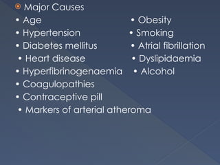
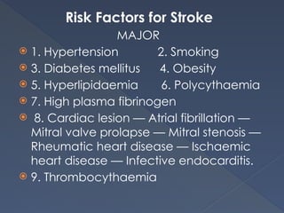
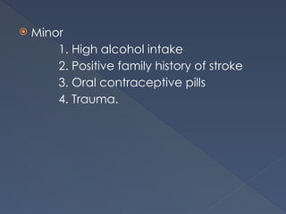
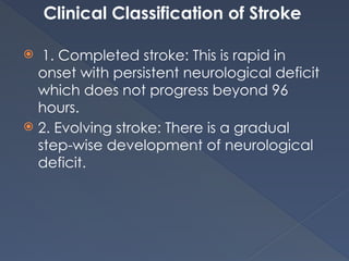
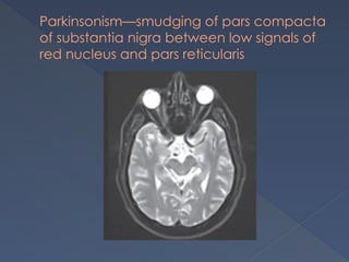
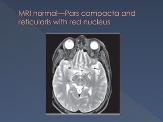
CENTRAL
NERVOUS
SYSTEM
CEREBROVASCULAR
DISORDERS
BEDSIDE ASSESSMENT
 History
 Neurological examination
HISTORY
 The time of onset of symptoms
 The nature of symptoms
Abruptly : vascular etiology
Risk factors for vascular disease,history of
seizures,migraine,insulin use or drug abude
Accompanying symptoms(severe
thunderclap headache-Sub arachnoid
hemorrhage)
 History of present illness:
Time of symptoms onset
Evolution of symptoms,recovery
Convulsion or loss of consciousness at
onset
Headache
Chestpain at onset
 Medical History:
Prior intracerebral hemorrhage,H/O
stroke,recent head trauma.myocardial
infarction
 Surgical History
Recent surgical procedures
 Medications-
Anticoagulant therapy
Record:
 Vital signs
 Blood glucose
 Level of consciousness
 Severity of deficits
NEUROLOGICAL EXAMINATION
 Identify signs of lateralised hemispheric
or brainstem dysfunction consistent with
focal cerebral ischemia.
 Level of consciusness, the presence of a
gaze deviation,Aphasia,neglect or
Hemiparesis
WHO DEFINITION OF STROKE
 A neurological deficit of sudden onset
accompanied by focal dysfunction and
symptoms lasting more than 24 hours
that are presumed to be of a non-
traumatic vascular origin.
 Major Causes
• Age • Obesity
• Hypertension • Smoking
• Diabetes mellitus • Atrial fibrillation
• Heart disease • Dyslipidaemia
• Hyperfibrinogenaemia • Alcohol
• Coagulopathies
• Contraceptive pill
• Markers of arterial atheroma
Risk Factors for Stroke
MAJOR
 1. Hypertension 2. Smoking
 3. Diabetes mellitus 4. Obesity
 5. Hyperlipidaemia 6. Polycythaemia
 7. High plasma fibrinogen
 8. Cardiac lesion — Atrial fibrillation —
Mitral valve prolapse — Mitral stenosis —
Rheumatic heart disease — Ischaemic
heart disease — Infective endocarditis.
 9. Thrombocythaemia
 Minor
1. High alcohol intake
2. Positive family history of stroke
3. Oral contraceptive pills
4. Trauma.
Clinical Classification of Stroke
 1. Completed stroke: This is rapid in
onset with persistent neurological deficit
which does not progress beyond 96
hours.
 2. Evolving stroke: There is a gradual
step-wise development of neurological
deficit.
 3. Transient ischaemic attack: The focal
neurological deficit resolves completely
within 24 hours
 4. Reversible ischaemic neurological
deficit (RIND): The neurological deficit
completely resolves within a period of 1
to 3 weeks.
Classification of Stroke
(Based on pathophysiology)
 Ischemic(85%)
 Hemorrhagic(15%)
ISCHEMIC STROKE:
 Thrombotic(55%)
 Embolic (30%)
HEMORRHAGIC STOKE:
 Subarachnoid hemorrhage
 Intracerebral and cerebellar .H
 Subdural & Extra dural.H
ISCHEMIC STROKE
THROMBOTIC
 Usually develops at night during sleep
 Symptoms perceived in morning
 Suspect in h/o of hypercoagulable
states,atherosclerosis and collagen
vascular disorders.
EMBOLIC
 Occurs at any time
 Frequently during periods of vigorous
activity
 H/O Atrial fibrillation,Valvular
vegetations,Thromboembolism from
MI,Ulcerated plaques incarotid system
 Seizures in 20% of cases
HEMORRHAGIC STROKE
 Occur during stress or exertion
 Focal deficits rapidly evolve
 Signs of raised ICT
 Confusion, coma or immediate death
Investigations
 I. Baseline investigations Full blood count,
ESR
2. Serological tests
3. Blood glucose and urea
4. Serum electrolytes and proteins
5. X-ray chest
6. ECG and ECHO
7. Carotid Doppler.
II. Special investigations (especially
young patients)
 1. Antinuclear antibodies (ANA) for SLE,
rheumatoid arthritis
 2. Antibodies to double stranded DNA
(SLE)
 3. Anticardiolipin antibodies (SLE)
 4. Lupus anticoagulant
(antiphospholipid antibodies)—SLE
 5. Serum cholesterol (familial
hyperlipidaemia).
 III.CT Scan—Indications
CT scan is mandatory for proper
initial evaluation in all cases of stroke to
categorise them into either ischaemic or
haemorrhagic origin.
1. To confirm diagnosis (haemorrhage can
be detected immediately whereas it
may take 48 hours for infarcts to be
detected).
2. To decide the line of management (to
decide on therapy with anticoagulants
or antiplatelet drugs).
3. To identify the presence of underlying
tumour, haematoma or vascular
malformation which can simulate stroke.
4. Head CT scan is diagnostic of SAH in 90%
of cases in the first 24 hours.
 IV.MRI: It is the preferred modality of
investigation in cases of posterior fossa
infarcts and for patients having TIA.
MR angiography is a useful non-
invasive test to evaluate large arteries
and veins.
 V.Cardiac Evaluation (for sources of
thromboembolism).
 VI. Angiography: It is not usually
indicated but indicated only to rule out
specific causes such as arterial
dissection.
• Definitive study for vascular
malformations
• Essential test prior to endarterectomy
• Pre-surgical evaluation of saccular
aneurysms
Infarct in anterior cerebral artery territory
CT Brain- Intracerebral bleed
with intraventricular extension
MANAGEMENT OF ISCHEMIC
STROKE
Specific Management
Medical Management
a. Blood pressure:A patients blood
pressure at presentation should not be
lowered unless it is more than 185/110
mmHg, as the ischaemic penumbral
tissue will infarct with even minor drop
in systemic blood pressure, because
cerebral autoregulation in this zone is
impaired.
 b. Anticoagulants
 c. Treatment of cerebral oedema
 d. Thrombolysis
 e. Antiplatelet drugs
2. Surgical Management
Carotid endarterectomy is useful in
patients with TIA with haemodynamically
significant (> 70%) carotid stenosis.
MANAGEMENT OF HEMORRHAGIC STROKE
 1. Maintenance of fluid and electrolyte
balance (Ryle’s tube feeding in
unconscious patients and bladder
catheterisation).
 2. Regular change of posture and care
of skin to avoid bed sores. 3. Monitoring
of blood pressure (diastolic pressure not
to be lowered below 110 mmHg in the
first 48 hours).
 4. Antioedema measures with mannitol
0.5–1.0 gm/ kg as rapid IV infusion or with
10% glycerol 500 ml IV infused over 4
hours daily for 6 days.
 5. Surgery: It is indicated in cerebellar
haemorrhage, whereby the haematoma
can be surgically removed. This is not
recommended in haemorrhage at other
sites, as this may result in permanent
neurological deficit.
II. General Management
 a. Attend to bladder, bowel, back, base
of lungs and eyes
 b. Patients should be ideally placed in
the semiprone position if unconscious to
prevent aspiration
 c. Proper nursing, feeding (if needed,
through Ryle’s tube)
 d. Intake and output chart should be
maintained
 e. Physiotherapy
i. It is started immediately to prevent
joint contractures and to promote
recovery of strength and coordination
ii. Physiotherapy of the chest is done
to prevent lung infection.
PARKINSON’S
DISEASE
PARKINSON’S DISEASE
 It is a movement disorder of unknown
aetiology due to degeneration of the
neurons in the nigrostriatal dopaminergic
system.
 There is an imbalance between
dopamine and acetylcholine
neurotransmitters (either an increase in
acetylcholine or a decrease in
dopamine level).
Causes of Parkinsonism
CLINICAL FEATURES
 A. Motor
 1. General • Expressionless face with
staring look with infrequent blinking
• Greasy skin
• Soft, rapid, indistinct
monotonous speech
• Flexed posture (universal
flexion).
 2. Gait
• Patients walk with short steps, with a
tendency to run
• Slow to start walking
• Shortened stride • Rapid small steps,
tendency to run (festination)
• Reduced arm swinging • Impaired
balance on turning
• Propulsion and retropulsion and
lateropulsion
• Kinesia paradox (patients can run fast
during emergency).
3. Tremor
 Resting tremor (4–6 Hertz)
• Usually first in fingers/thumb
• Coarse, complex movements,
flexion/extension of fingers (pill rolling
and drumbeating movements)
• Abduction/adduction of thumb
• Supination/pronation of forearm
• May affect arms, legs, feet, jaw, tongue
• Intermittent, present at rest and when
distracted
• Diminishes on action and disappears
during sleep.
 4. Rigidity
• It is seen predominantly in the limbs •
Cogwheel type, mostly appreciated in
upper limbs especially in wrist joints
(there is a phasic element to stiffness in
all directions of movement)
• Plastic (lead pipe) type, mostly
appreciated in the legs and trunk • In
the trunk, rigidity manifests itself by the
presence of a flexed, and stooped
posture.
 5. Hypokinesis
• Slowness in initiating movements
• Impaired fine movements, especially of
fingers
• Poor precision of repetitive movements
• Handwriting–micrographia.
B. NON-MOTOR
• Neuropsychiatric symptoms – anxiety,
depression, psychosis, dementia, impulse
control disorder
• Autonomic failure
• Sensory symptoms.
STAGING
Grading Clinical features
Grade I Unilateral involvement
Grade II Bilateral involvement
Grade III Bilateral with mild
postural imbalance
Grade IV Bilateral with moderate
postural imbalance and requires
assistance
Grade V Bedridden
Eye Signs in Parkinsonism
 Decreased blink rate
 Hypometric saccades
 Impaired smooth pursuit
 Reflex blepharospasm (glabellar,
Myerson’s sign)
 Blepharoclonus
 Spontaneous blepharospasm
 Oculogyric crises (postencephalitic
parkinsonism)
 Reversed Argyll Robertson pupil
(postencephalitic parkinsonism).
INVESTIGATIONS
 1. Serological tests for syphilis (all
patients)
 2. CT brain
Indications a. Patients under age 50
b. Signs entirely unilateral
c. Atypical signs (e.g.
pyramidal).
 3. Tests to exclude Wilson’s disease (in
young patients (2nd to 4th decade)
a.Serum ceruloplasmin
b. Serum copper
c. Urine copper
d. Liver function tests.
4. MRI brain
TREATMENT
 1. Treat the underlying disease
 2. Withdraw the drugs in drug-induced
parkinsonism
 3. Drug therapy.
3. Drug therapy
I. Drugs • Dopamine agonists
• Carbidopa/levodopa
• MAO-A inhibitors
• MAO-B inhibitors
• COMT inhibitors
• Amantadine
• Anticholinergics
 II. Neuroprotective agents
• L-carnitine
• Creatine monohydrate
• Dopamine agonists
• Seligiline
 III.Surgery
• Deep brain stimulation
• Substantia nigra ablation
 IV.Neurotransplantation
• Stem cell
• GDNF infusion (Glial cell derived
neurotrophic factor)
NON-MOTOR SYMPTOMS:
• Anti-depressants – Tricyclic anti-
depressants, SSRI
• Dementia – Donepezil, Rivantigmine
• Psychosis – Atypical antipsychotics
CT brain—basal ganglia calcification in
hypoparathyroidism—causing Parkinsonism
Parkinsonism—smudging of pars compacta
of substantia nigra between low signals of
red nucleus and pars reticularis
MRI normal—Pars compacta and
reticularis with red nucleus
MULTIPLE SCLEROSIS
THANK YOU

CNS.pptx stroke clinical assessment and evaluation

  • 1.
  • 2.
  • 3.
    BEDSIDE ASSESSMENT  History Neurological examination
  • 4.
    HISTORY  The timeof onset of symptoms  The nature of symptoms Abruptly : vascular etiology Risk factors for vascular disease,history of seizures,migraine,insulin use or drug abude Accompanying symptoms(severe thunderclap headache-Sub arachnoid hemorrhage)
  • 5.
     History ofpresent illness: Time of symptoms onset Evolution of symptoms,recovery Convulsion or loss of consciousness at onset Headache Chestpain at onset  Medical History: Prior intracerebral hemorrhage,H/O stroke,recent head trauma.myocardial infarction
  • 6.
     Surgical History Recentsurgical procedures  Medications- Anticoagulant therapy
  • 7.
    Record:  Vital signs Blood glucose  Level of consciousness  Severity of deficits
  • 8.
    NEUROLOGICAL EXAMINATION  Identifysigns of lateralised hemispheric or brainstem dysfunction consistent with focal cerebral ischemia.  Level of consciusness, the presence of a gaze deviation,Aphasia,neglect or Hemiparesis
  • 9.
    WHO DEFINITION OFSTROKE  A neurological deficit of sudden onset accompanied by focal dysfunction and symptoms lasting more than 24 hours that are presumed to be of a non- traumatic vascular origin.
  • 10.
     Major Causes •Age • Obesity • Hypertension • Smoking • Diabetes mellitus • Atrial fibrillation • Heart disease • Dyslipidaemia • Hyperfibrinogenaemia • Alcohol • Coagulopathies • Contraceptive pill • Markers of arterial atheroma
  • 11.
    Risk Factors forStroke MAJOR  1. Hypertension 2. Smoking  3. Diabetes mellitus 4. Obesity  5. Hyperlipidaemia 6. Polycythaemia  7. High plasma fibrinogen  8. Cardiac lesion — Atrial fibrillation — Mitral valve prolapse — Mitral stenosis — Rheumatic heart disease — Ischaemic heart disease — Infective endocarditis.  9. Thrombocythaemia
  • 12.
     Minor 1. Highalcohol intake 2. Positive family history of stroke 3. Oral contraceptive pills 4. Trauma.
  • 13.
    Clinical Classification ofStroke  1. Completed stroke: This is rapid in onset with persistent neurological deficit which does not progress beyond 96 hours.  2. Evolving stroke: There is a gradual step-wise development of neurological deficit.
  • 14.
     3. Transientischaemic attack: The focal neurological deficit resolves completely within 24 hours  4. Reversible ischaemic neurological deficit (RIND): The neurological deficit completely resolves within a period of 1 to 3 weeks.
  • 15.
    Classification of Stroke (Basedon pathophysiology)  Ischemic(85%)  Hemorrhagic(15%)
  • 16.
    ISCHEMIC STROKE:  Thrombotic(55%) Embolic (30%) HEMORRHAGIC STOKE:  Subarachnoid hemorrhage  Intracerebral and cerebellar .H  Subdural & Extra dural.H
  • 17.
    ISCHEMIC STROKE THROMBOTIC  Usuallydevelops at night during sleep  Symptoms perceived in morning  Suspect in h/o of hypercoagulable states,atherosclerosis and collagen vascular disorders.
  • 18.
    EMBOLIC  Occurs atany time  Frequently during periods of vigorous activity  H/O Atrial fibrillation,Valvular vegetations,Thromboembolism from MI,Ulcerated plaques incarotid system  Seizures in 20% of cases
  • 20.
    HEMORRHAGIC STROKE  Occurduring stress or exertion  Focal deficits rapidly evolve  Signs of raised ICT  Confusion, coma or immediate death
  • 27.
    Investigations  I. Baselineinvestigations Full blood count, ESR 2. Serological tests 3. Blood glucose and urea 4. Serum electrolytes and proteins 5. X-ray chest 6. ECG and ECHO 7. Carotid Doppler.
  • 28.
    II. Special investigations(especially young patients)  1. Antinuclear antibodies (ANA) for SLE, rheumatoid arthritis  2. Antibodies to double stranded DNA (SLE)  3. Anticardiolipin antibodies (SLE)  4. Lupus anticoagulant (antiphospholipid antibodies)—SLE  5. Serum cholesterol (familial hyperlipidaemia).
  • 29.
     III.CT Scan—Indications CTscan is mandatory for proper initial evaluation in all cases of stroke to categorise them into either ischaemic or haemorrhagic origin. 1. To confirm diagnosis (haemorrhage can be detected immediately whereas it may take 48 hours for infarcts to be detected).
  • 30.
    2. To decidethe line of management (to decide on therapy with anticoagulants or antiplatelet drugs). 3. To identify the presence of underlying tumour, haematoma or vascular malformation which can simulate stroke. 4. Head CT scan is diagnostic of SAH in 90% of cases in the first 24 hours.
  • 31.
     IV.MRI: Itis the preferred modality of investigation in cases of posterior fossa infarcts and for patients having TIA. MR angiography is a useful non- invasive test to evaluate large arteries and veins.  V.Cardiac Evaluation (for sources of thromboembolism).
  • 32.
     VI. Angiography:It is not usually indicated but indicated only to rule out specific causes such as arterial dissection. • Definitive study for vascular malformations • Essential test prior to endarterectomy • Pre-surgical evaluation of saccular aneurysms
  • 33.
    Infarct in anteriorcerebral artery territory
  • 34.
    CT Brain- Intracerebralbleed with intraventricular extension
  • 35.
    MANAGEMENT OF ISCHEMIC STROKE SpecificManagement Medical Management a. Blood pressure:A patients blood pressure at presentation should not be lowered unless it is more than 185/110 mmHg, as the ischaemic penumbral tissue will infarct with even minor drop in systemic blood pressure, because cerebral autoregulation in this zone is impaired.
  • 36.
     b. Anticoagulants c. Treatment of cerebral oedema  d. Thrombolysis  e. Antiplatelet drugs 2. Surgical Management Carotid endarterectomy is useful in patients with TIA with haemodynamically significant (> 70%) carotid stenosis.
  • 37.
    MANAGEMENT OF HEMORRHAGICSTROKE  1. Maintenance of fluid and electrolyte balance (Ryle’s tube feeding in unconscious patients and bladder catheterisation).  2. Regular change of posture and care of skin to avoid bed sores. 3. Monitoring of blood pressure (diastolic pressure not to be lowered below 110 mmHg in the first 48 hours).
  • 38.
     4. Antioedemameasures with mannitol 0.5–1.0 gm/ kg as rapid IV infusion or with 10% glycerol 500 ml IV infused over 4 hours daily for 6 days.  5. Surgery: It is indicated in cerebellar haemorrhage, whereby the haematoma can be surgically removed. This is not recommended in haemorrhage at other sites, as this may result in permanent neurological deficit.
  • 39.
    II. General Management a. Attend to bladder, bowel, back, base of lungs and eyes  b. Patients should be ideally placed in the semiprone position if unconscious to prevent aspiration  c. Proper nursing, feeding (if needed, through Ryle’s tube)  d. Intake and output chart should be maintained
  • 40.
     e. Physiotherapy i.It is started immediately to prevent joint contractures and to promote recovery of strength and coordination ii. Physiotherapy of the chest is done to prevent lung infection.
  • 41.
  • 42.
    PARKINSON’S DISEASE  Itis a movement disorder of unknown aetiology due to degeneration of the neurons in the nigrostriatal dopaminergic system.  There is an imbalance between dopamine and acetylcholine neurotransmitters (either an increase in acetylcholine or a decrease in dopamine level).
  • 43.
  • 44.
    CLINICAL FEATURES  A.Motor  1. General • Expressionless face with staring look with infrequent blinking • Greasy skin • Soft, rapid, indistinct monotonous speech • Flexed posture (universal flexion).
  • 45.
     2. Gait •Patients walk with short steps, with a tendency to run • Slow to start walking • Shortened stride • Rapid small steps, tendency to run (festination) • Reduced arm swinging • Impaired balance on turning • Propulsion and retropulsion and lateropulsion • Kinesia paradox (patients can run fast during emergency).
  • 46.
    3. Tremor  Restingtremor (4–6 Hertz) • Usually first in fingers/thumb • Coarse, complex movements, flexion/extension of fingers (pill rolling and drumbeating movements) • Abduction/adduction of thumb • Supination/pronation of forearm • May affect arms, legs, feet, jaw, tongue • Intermittent, present at rest and when distracted • Diminishes on action and disappears during sleep.
  • 47.
     4. Rigidity •It is seen predominantly in the limbs • Cogwheel type, mostly appreciated in upper limbs especially in wrist joints (there is a phasic element to stiffness in all directions of movement) • Plastic (lead pipe) type, mostly appreciated in the legs and trunk • In the trunk, rigidity manifests itself by the presence of a flexed, and stooped posture.
  • 48.
     5. Hypokinesis •Slowness in initiating movements • Impaired fine movements, especially of fingers • Poor precision of repetitive movements • Handwriting–micrographia.
  • 49.
    B. NON-MOTOR • Neuropsychiatricsymptoms – anxiety, depression, psychosis, dementia, impulse control disorder • Autonomic failure • Sensory symptoms.
  • 50.
    STAGING Grading Clinical features GradeI Unilateral involvement Grade II Bilateral involvement Grade III Bilateral with mild postural imbalance Grade IV Bilateral with moderate postural imbalance and requires assistance Grade V Bedridden
  • 51.
    Eye Signs inParkinsonism  Decreased blink rate  Hypometric saccades  Impaired smooth pursuit  Reflex blepharospasm (glabellar, Myerson’s sign)  Blepharoclonus
  • 52.
     Spontaneous blepharospasm Oculogyric crises (postencephalitic parkinsonism)  Reversed Argyll Robertson pupil (postencephalitic parkinsonism).
  • 53.
    INVESTIGATIONS  1. Serologicaltests for syphilis (all patients)  2. CT brain Indications a. Patients under age 50 b. Signs entirely unilateral c. Atypical signs (e.g. pyramidal).
  • 54.
     3. Teststo exclude Wilson’s disease (in young patients (2nd to 4th decade) a.Serum ceruloplasmin b. Serum copper c. Urine copper d. Liver function tests. 4. MRI brain
  • 55.
    TREATMENT  1. Treatthe underlying disease  2. Withdraw the drugs in drug-induced parkinsonism  3. Drug therapy.
  • 56.
    3. Drug therapy I.Drugs • Dopamine agonists • Carbidopa/levodopa • MAO-A inhibitors • MAO-B inhibitors • COMT inhibitors • Amantadine • Anticholinergics
  • 57.
     II. Neuroprotectiveagents • L-carnitine • Creatine monohydrate • Dopamine agonists • Seligiline  III.Surgery • Deep brain stimulation • Substantia nigra ablation
  • 58.
     IV.Neurotransplantation • Stemcell • GDNF infusion (Glial cell derived neurotrophic factor) NON-MOTOR SYMPTOMS: • Anti-depressants – Tricyclic anti- depressants, SSRI • Dementia – Donepezil, Rivantigmine • Psychosis – Atypical antipsychotics
  • 59.
    CT brain—basal gangliacalcification in hypoparathyroidism—causing Parkinsonism
  • 60.
    Parkinsonism—smudging of parscompacta of substantia nigra between low signals of red nucleus and pars reticularis
  • 61.
    MRI normal—Pars compactaand reticularis with red nucleus
  • 62.
  • 73.