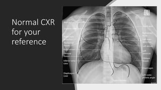
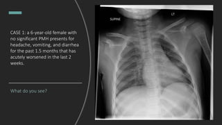
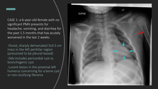
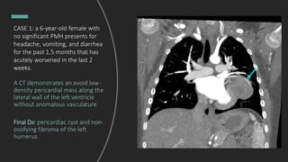
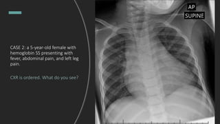
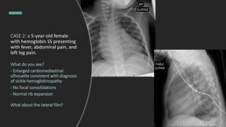
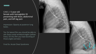
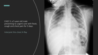
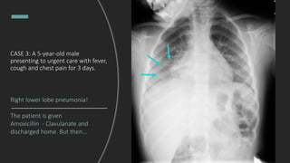
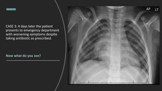
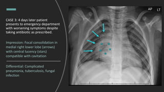
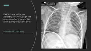
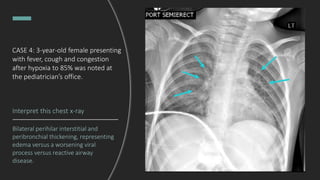
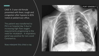
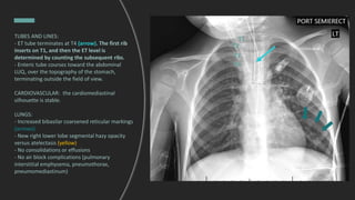
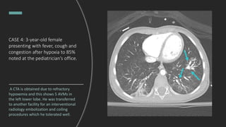
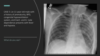
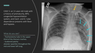
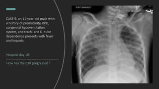
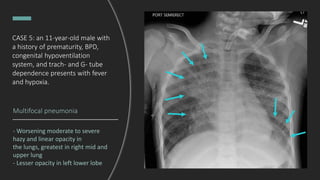
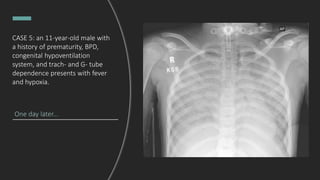
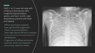
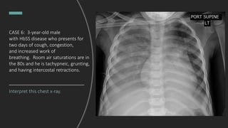
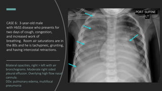
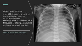
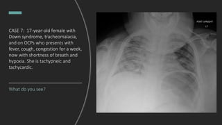
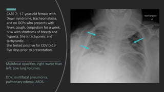
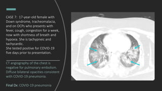
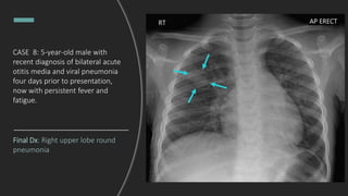
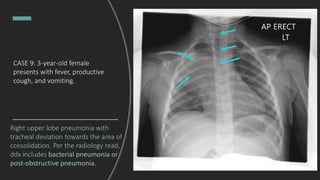
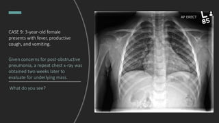
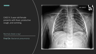
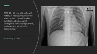
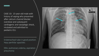
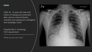
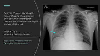
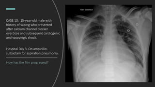
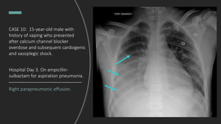
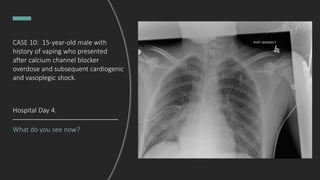
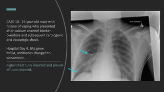
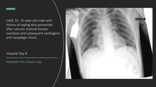
Pediatric X-Rays of the Month
Taylor Anderson, MD & Kaley El-Arab, MD
Departments of Emergency Medicine and Pediatrics
Levine Children's Hospital and Carolinas Medical Center
Nicholena Richardson, MD & Mary Grady, MD, Faculty Editors
Michael Gibbs, MD, Editor – CMC Imaging Mastery Project
January 2022
Process and Disclosures
This ongoing pediatric chest x-ray
interpretation series is proudly sponsored by
the Emergency Medicine and Pediatrics
Residency Programs and the Pediatric
Emergency Medicine Fellowship at Carolinas
Medical Center & Levine Children’s Hospital.
The goal is to promote widespread mastery of
CXR interpretation.
Cases are submitted by contributors from
many CMC departments, and now… Tanzania
and Brazil.
Ages have been changed to protect patient
confidentiality. No protected health
information (PHI) will be shared.
For more educational content, visit
EMGuidewire.com
Reading systematically…
A for airway
B for bones
C for cardiac silhouette
D for diaphragm
E for everything else
For more educational content, visit
EMGuidewire.com
Normal CXR
for your
reference
CASE 1: a 6-year-old female with
no significant PMH presents for
headache, vomiting, and diarrhea
for the past 1.5 months that has
acutely worsened in the last 2
weeks.
What do you see?
CASE 1: a 6-year-old female with no
significant PMH presents for
headache, vomiting, and diarrhea for
the past 1.5 months that has acutely
worsened in the last 2 weeks.
-Ovoid, sharply demarcated 5x3.5 cm
mass in the left perihilar region
(presumed to be pleural-based)
-Ddx includes pericardial cyst vs
bronchogenic cyst
-Lucent lesion in the proximal left
humerus concerning for a bone cyst
or non-ossifying fibroma
CASE 1: a 6-year-old female with
no significant PMH presents for
headache, vomiting, and diarrhea
for the past 1.5 months that has
acutely worsened in the last 2
weeks.
A CT demonstrates an ovoid low-
density pericardial mass along the
lateral wall of the left ventricle
without anomalous vasculature
Final Dx: pericardiac cyst and non-
ossifying fibroma of the left
humerus
CASE 2: a 5-year-old female with
hemoglobin SS presenting with
fever, abdominal pain, and left leg
pain.
CXR is ordered. What do you see?
CASE 2: a 5-year-old female
with hemoglobin SS presenting
with fever, abdominal pain, and
left leg pain.
What do you see?
- Enlarged cardiomediastinal
silhouette consistent with diagnosis
of sickle hemoglobinopathy
- No focal consolidations
- Normal rib expansion
What about the lateral film?
CASE 2: 5-year-old
female with hemoglobin SS
presenting with fever, abdominal
pain, and left leg pain
Impression: Opacity at posterior lung
bases.
Tip: On lateral film you should be able to
see sharp costal vertebral angles (yellow
lines). On this film you have blunting of
those angles (arrows).
Final Dx: Acute Chest Syndrome
CASE 3: a 5-year-old male
presenting to urgent care with fever,
cough and chest pain for 3 days.
Interpret this chest X-Ray
CASE 3: A 5-year-old male
presenting to urgent care with fever,
cough and chest pain for 3 days.
Right lower lobe pneumonia!
The patient is given
Amoxicillin - Clavulanate and
discharged home. But then...
CASE 3: 4 days later the patient
presents to emergency department
with worsening symptoms despite
taking antibiotic as prescribed.
Now what do you see?
CASE 3: 4 days later patient
presents to emergency department
with worsening symptoms despite
taking antibiotic as prescribed.
Impression: Focal consolidation in
medial right lower lobe (arrows)
with central lucency (stars)
compatible with cavitation
Differential: Complicated
pneumonia, tuberculosis, fungal
infection
CASE 4: 3-year-old female
presenting with fever, cough and
congestion after hypoxia to 85%
noted at the pediatrician’s office.
Interpret this chest x-ray
CASE 4: 3-year-old female presenting
with fever, cough and congestion
after hypoxia to 85% was noted at
the pediatrician’s office.
Interpret this chest x-ray
Bilateral perihilar interstitial and
peribronchial thickening, representing
edema versus a worsening viral
process versus reactive airway
disease.
CASE 4: 3-year-old female
presented with fever, cough and
congestion after hypoxia to 85%
noted at pediatrician office.
The patient was transferred to
PICU on hospital day 3 due to
increasing high-flow oxygen
requirements progressing to the
need for intubation. A respiratory
panel was positive for non-covid
Coronavirus.
Now interpret this chest x-ray.
TUBES AND LINES:
- ET tube terminates at T4 (arrow). The first rib
inserts on T1, and then the ET level is
determined by counting the subsequent ribs.
- Enteric tube courses toward the abdominal
LUQ, over the topography of the stomach,
terminating outside the field of view.
CARDIOVASCULAR: the cardiomediastinal
silhouette is stable.
LUNGS:
- Increased bibasilar coarsened reticular markings
(arrows)
- New right lower lobe segmental hazy opacity
versus atelectasis (yellow)
- No consolidations or effusions
- No air block complications (pulmonary
interstitial emphysema, pneumothorax,
pneumomediastinum)
T1
T2
T3
T4
CASE 4: 3-year-old female
presenting with fever, cough and
congestion after hypoxia to 85%
noted at the pediatrician’s office.
A CTA is obtained due to refractory
hypoxemia and this shows 5 AVMs in
the left lower lobe. He was transferred
to another facility for an interventional
radiology embolization and coiling
procedures which he tolerated well.
What do you see?
CASE 5: an 11-year-old male with
a history of prematurity, BPD,
congenital hypoventilation
system, and trach- and G- tube
dependence presents with fever
and hypoxia.
CASE 5: an 11-year-old male with
a history of prematurity, BPD,
congenital hypoventilation
system, and trach- and G- tube
dependence presents with fever
and hypoxia.
What do you see?
- Tracheostomy tube in the upper
third of the thoracic trachea.
- Scattered patchy
alveolar opacities throughout the
mid and lower left lung.
Hospital day 10:
How has the CXR progressed?
CASE 5: an 11-year-old male with
a history of prematurity, BPD,
congenital hypoventilation
system, and trach- and G- tube
dependence presents with fever
and hypoxia.
Multifocal pneumonia
- Worsening moderate to severe
hazy and linear opacity in
the lungs, greatest in right mid and
upper lung
- Lesser opacity in left lower lobe
CASE 5: an 11-year-old male with
a history of prematurity, BPD,
congenital hypoventilation
system, and trach- and G- tube
dependence presents with fever
and hypoxia.
One day later...
CASE 5: an 11-year-old male with
a history of prematurity, BPD,
congenital hypoventilation
system, and trach- and G- tube
dependence presents with fever
and hypoxia.
IMPRESSION:
Progression of diffuse pulmonary
opacities which may represent
pneumonia, edema or ARDS.
Diffuse pulmonary opacities
have significantly progressed.
There is no pneumothorax.
Trace right pleural effusion is present.
CASE 5: an 11-year-old male with
a history of prematurity, BPD,
congenital hypoventilation
system, and trach- and G- tube
dependence presents with fever
and hypoxia.
CASE 6: 3-year-old male
with HbSS disease who presents for
two days of cough, congestion,
and increased work of
breathing. Room air saturations are in
the 80s and he is tachypneic, grunting,
and having intercostal retractions.
Interpret this chest x-ray.
Bilateral opacities, right > left with air
bronchograms. Moderate right sided
pleural effusion. Overlying high flow nasal
cannula.
DDx: pulmonary edema, multifocal
pneumonia
CASE 6: 3-year-old male
with HbSS disease who presents for
two days of cough, congestion,
and increased work of
breathing. Room air saturations are in
the 80s and he is tachypneic, grunting,
and having intercostal retractions.
Final Dx: Acute chest syndrome
CASE 6: 3-year-old male
with HbSS disease who presents for
two days of cough, congestion,
and increased work of
breathing. Room air saturations are in
the 80s and he is tachypneic, grunting,
and having intercostal retractions.
CASE 7: 17-year-old female with
Down syndrome, tracheomalacia,
and on OCPs who presents with
fever, cough, congestion for a week,
now with shortness of breath and
hypoxia. She is tachypneic and
tachycardic.
What do you see?
Multifocal opacities, right worse than
left. Low lung volumes.
DDx: multifocal pneumonia,
pulmonary edema, ARDS.
CASE 7: 17-year-old female with
Down syndrome, tracheomalacia,
and on OCPs who presents with
fever, cough, congestion for a week,
now with shortness of breath and
hypoxia. She is tachypneic and
tachycardic.
She tested positive for COVID-19
five days prior to presentation.
CT angiography of the chest is
negative for pulmonary embolism.
Diffuse bilateral opacities consistent
with COVID-19 pneumonia.
Final Dx: COVID-19 pneumonia
CASE 7: 17-year-old female with
Down syndrome, tracheomalacia,
and on OCPs who presents with
fever, cough, congestion for a week,
now with shortness of breath and
hypoxia. She is tachypneic and
tachycardic.
She tested positive for COVID-19
five days prior to presentation.
CASE 8: 5-year-old male with
recent diagnosis of bilateral acute
otitis media and viral pneumonia
four days prior to presentation,
now with persistent fever and
fatigue.
What do you see?
Final Dx: Right upper lobe round
pneumonia
CASE 8: 5-year-old male with
recent diagnosis of bilateral acute
otitis media and viral pneumonia
four days prior to presentation,
now with persistent fever and
fatigue.
CASE 9: 3-year-old female
presents with fever, productive
cough, and vomiting.
Interpret this chest X-ray.
CASE 9: 3-year-old female
presents with fever, productive
cough, and vomiting.
Right upper lobe pneumonia with
tracheal deviation towards the area of
consolidation. Per the radiology read,
ddx includes bacterial pneumonia or
post-obstructive pneumonia.
CASE 9: 3-year-old female
presents with fever, productive
cough, and vomiting.
Given concerns for post-obstructive
pneumonia, a repeat chest x-ray was
obtained two weeks later to
evaluate for underlying mass.
What do you see?
CASE 9: 3-year-old female
presents with fever, productive
cough, and vomiting.
Final Dx: Bacterial pneumonia
Normal chest x-ray!
CASE 10: 15-year-old male with
history of vaping who presented
after calcium channel blocker
overdose and subsequent
cardiogenic and vasoplegic shock,
intubated and admitted to
pediatric ICU.
What do you see?
Endotracheal tube in good position.
Hazy perihilar opacities.
DDx: pulmonary edema, aspiration
pneumonia
CASE 10: 15-year-old male with
history of vaping who presented
after calcium channel blocker
overdose and subsequent
cardiogenic and vasoplegic shock,
intubated and admitted to
pediatric ICU.
What do you see now?
Hospital Day 2. Increasing
FiO2 requirement.
CASE 10: 15-year-old male with
history of vaping who presented
after calcium channel blocker
overdose and subsequent cardiogenic
and vasoplegic shock.
Right lower lobe consolidation.
Dx: Aspiration pneumonia
Hospital Day 2.
Increasing FiO2 Requirement.
CASE 10: 15-year-old male with
history of vaping who presented
after calcium channel blocker
overdose and subsequent cardiogenic
and vasoplegic shock.
CASE 10: 15-year-old male with
history of vaping who presented
after calcium channel blocker
overdose and subsequent cardiogenic
and vasoplegic shock.
How has the film progressed?
Hospital Day 3. On ampicillin-
sulbactam for aspiration pneumonia.
CASE 10: 15-year-old male with
history of vaping who presented
after calcium channel blocker
overdose and subsequent cardiogenic
and vasoplegic shock.
Right parapneumonic effusion.
Hospital Day 3. On ampicillin-
sulbactam for aspiration pneumonia.
What do you see now?
Hospital Day 4.
CASE 10: 15-year-old male with
history of vaping who presented
after calcium channel blocker
overdose and subsequent cardiogenic
and vasoplegic shock.
Pigtail chest tube inserted and pleural
effusion drained.
Hospital Day 4. BAL grew
MRSA, antibiotics changed to
vancomycin.
CASE 10: 15-year-old male with
history of vaping who presented
after calcium channel blocker
overdose and subsequent cardiogenic
and vasoplegic shock.
Interpret this chest x-ray.
Hospital Day 8.
CASE 10: 15-year-old male with
history of vaping who presented
after calcium channel blocker
overdose and subsequent cardiogenic
and vasoplegic shock.
Interval extubation and removal
of chest tube. No pneumothorax.
No recurrent effusion. Clear lungs.
Final Dx: MRSA pneumonia
with parapneumonic effusion.
Hospital Day 8.
CASE 10: 15-year-old male with
history of vaping who presented
after calcium channel blocker
overdose and subsequent cardiogenic
and vasoplegic shock.
Summary of This
Month’s Diagnoses
• Pericardial cyst
• Acute chest syndrome
• Cavitary pneumonia
• Non-COVID coronavirus pneumonia
with pulmonary arteriovenous
malformations
• Multifocal pneumonia with ARDS
• COVID-19 pneumonia
• Round pneumonia
• Lobar pneumonia
• Aspiration pneumonia with
parapneumonic effusion
For more educational content, visit
EMGuidewire.com

CMC Pediatric X-Ray Mastery: January Cases

  • 1.
    Pediatric X-Rays ofthe Month Taylor Anderson, MD & Kaley El-Arab, MD Departments of Emergency Medicine and Pediatrics Levine Children's Hospital and Carolinas Medical Center Nicholena Richardson, MD & Mary Grady, MD, Faculty Editors Michael Gibbs, MD, Editor – CMC Imaging Mastery Project January 2022
  • 2.
    Process and Disclosures Thisongoing pediatric chest x-ray interpretation series is proudly sponsored by the Emergency Medicine and Pediatrics Residency Programs and the Pediatric Emergency Medicine Fellowship at Carolinas Medical Center & Levine Children’s Hospital. The goal is to promote widespread mastery of CXR interpretation. Cases are submitted by contributors from many CMC departments, and now… Tanzania and Brazil. Ages have been changed to protect patient confidentiality. No protected health information (PHI) will be shared. For more educational content, visit EMGuidewire.com
  • 3.
    Reading systematically… A forairway B for bones C for cardiac silhouette D for diaphragm E for everything else For more educational content, visit EMGuidewire.com
  • 4.
  • 5.
    CASE 1: a6-year-old female with no significant PMH presents for headache, vomiting, and diarrhea for the past 1.5 months that has acutely worsened in the last 2 weeks. What do you see?
  • 6.
    CASE 1: a6-year-old female with no significant PMH presents for headache, vomiting, and diarrhea for the past 1.5 months that has acutely worsened in the last 2 weeks. -Ovoid, sharply demarcated 5x3.5 cm mass in the left perihilar region (presumed to be pleural-based) -Ddx includes pericardial cyst vs bronchogenic cyst -Lucent lesion in the proximal left humerus concerning for a bone cyst or non-ossifying fibroma
  • 7.
    CASE 1: a6-year-old female with no significant PMH presents for headache, vomiting, and diarrhea for the past 1.5 months that has acutely worsened in the last 2 weeks. A CT demonstrates an ovoid low- density pericardial mass along the lateral wall of the left ventricle without anomalous vasculature Final Dx: pericardiac cyst and non- ossifying fibroma of the left humerus
  • 8.
    CASE 2: a5-year-old female with hemoglobin SS presenting with fever, abdominal pain, and left leg pain. CXR is ordered. What do you see?
  • 9.
    CASE 2: a5-year-old female with hemoglobin SS presenting with fever, abdominal pain, and left leg pain. What do you see? - Enlarged cardiomediastinal silhouette consistent with diagnosis of sickle hemoglobinopathy - No focal consolidations - Normal rib expansion What about the lateral film?
  • 10.
    CASE 2: 5-year-old femalewith hemoglobin SS presenting with fever, abdominal pain, and left leg pain Impression: Opacity at posterior lung bases. Tip: On lateral film you should be able to see sharp costal vertebral angles (yellow lines). On this film you have blunting of those angles (arrows). Final Dx: Acute Chest Syndrome
  • 11.
    CASE 3: a5-year-old male presenting to urgent care with fever, cough and chest pain for 3 days. Interpret this chest X-Ray
  • 12.
    CASE 3: A5-year-old male presenting to urgent care with fever, cough and chest pain for 3 days. Right lower lobe pneumonia! The patient is given Amoxicillin - Clavulanate and discharged home. But then...
  • 13.
    CASE 3: 4days later the patient presents to emergency department with worsening symptoms despite taking antibiotic as prescribed. Now what do you see?
  • 14.
    CASE 3: 4days later patient presents to emergency department with worsening symptoms despite taking antibiotic as prescribed. Impression: Focal consolidation in medial right lower lobe (arrows) with central lucency (stars) compatible with cavitation Differential: Complicated pneumonia, tuberculosis, fungal infection
  • 15.
    CASE 4: 3-year-oldfemale presenting with fever, cough and congestion after hypoxia to 85% noted at the pediatrician’s office. Interpret this chest x-ray
  • 16.
    CASE 4: 3-year-oldfemale presenting with fever, cough and congestion after hypoxia to 85% was noted at the pediatrician’s office. Interpret this chest x-ray Bilateral perihilar interstitial and peribronchial thickening, representing edema versus a worsening viral process versus reactive airway disease.
  • 17.
    CASE 4: 3-year-oldfemale presented with fever, cough and congestion after hypoxia to 85% noted at pediatrician office. The patient was transferred to PICU on hospital day 3 due to increasing high-flow oxygen requirements progressing to the need for intubation. A respiratory panel was positive for non-covid Coronavirus. Now interpret this chest x-ray.
  • 18.
    TUBES AND LINES: -ET tube terminates at T4 (arrow). The first rib inserts on T1, and then the ET level is determined by counting the subsequent ribs. - Enteric tube courses toward the abdominal LUQ, over the topography of the stomach, terminating outside the field of view. CARDIOVASCULAR: the cardiomediastinal silhouette is stable. LUNGS: - Increased bibasilar coarsened reticular markings (arrows) - New right lower lobe segmental hazy opacity versus atelectasis (yellow) - No consolidations or effusions - No air block complications (pulmonary interstitial emphysema, pneumothorax, pneumomediastinum) T1 T2 T3 T4
  • 19.
    CASE 4: 3-year-oldfemale presenting with fever, cough and congestion after hypoxia to 85% noted at the pediatrician’s office. A CTA is obtained due to refractory hypoxemia and this shows 5 AVMs in the left lower lobe. He was transferred to another facility for an interventional radiology embolization and coiling procedures which he tolerated well.
  • 20.
    What do yousee? CASE 5: an 11-year-old male with a history of prematurity, BPD, congenital hypoventilation system, and trach- and G- tube dependence presents with fever and hypoxia.
  • 21.
    CASE 5: an11-year-old male with a history of prematurity, BPD, congenital hypoventilation system, and trach- and G- tube dependence presents with fever and hypoxia. What do you see? - Tracheostomy tube in the upper third of the thoracic trachea. - Scattered patchy alveolar opacities throughout the mid and lower left lung.
  • 22.
    Hospital day 10: Howhas the CXR progressed? CASE 5: an 11-year-old male with a history of prematurity, BPD, congenital hypoventilation system, and trach- and G- tube dependence presents with fever and hypoxia.
  • 23.
    Multifocal pneumonia - Worseningmoderate to severe hazy and linear opacity in the lungs, greatest in right mid and upper lung - Lesser opacity in left lower lobe CASE 5: an 11-year-old male with a history of prematurity, BPD, congenital hypoventilation system, and trach- and G- tube dependence presents with fever and hypoxia.
  • 24.
    One day later... CASE5: an 11-year-old male with a history of prematurity, BPD, congenital hypoventilation system, and trach- and G- tube dependence presents with fever and hypoxia.
  • 25.
    IMPRESSION: Progression of diffusepulmonary opacities which may represent pneumonia, edema or ARDS. Diffuse pulmonary opacities have significantly progressed. There is no pneumothorax. Trace right pleural effusion is present. CASE 5: an 11-year-old male with a history of prematurity, BPD, congenital hypoventilation system, and trach- and G- tube dependence presents with fever and hypoxia.
  • 26.
    CASE 6: 3-year-oldmale with HbSS disease who presents for two days of cough, congestion, and increased work of breathing. Room air saturations are in the 80s and he is tachypneic, grunting, and having intercostal retractions. Interpret this chest x-ray.
  • 27.
    Bilateral opacities, right> left with air bronchograms. Moderate right sided pleural effusion. Overlying high flow nasal cannula. DDx: pulmonary edema, multifocal pneumonia CASE 6: 3-year-old male with HbSS disease who presents for two days of cough, congestion, and increased work of breathing. Room air saturations are in the 80s and he is tachypneic, grunting, and having intercostal retractions.
  • 28.
    Final Dx: Acutechest syndrome CASE 6: 3-year-old male with HbSS disease who presents for two days of cough, congestion, and increased work of breathing. Room air saturations are in the 80s and he is tachypneic, grunting, and having intercostal retractions.
  • 29.
    CASE 7: 17-year-oldfemale with Down syndrome, tracheomalacia, and on OCPs who presents with fever, cough, congestion for a week, now with shortness of breath and hypoxia. She is tachypneic and tachycardic. What do you see?
  • 30.
    Multifocal opacities, rightworse than left. Low lung volumes. DDx: multifocal pneumonia, pulmonary edema, ARDS. CASE 7: 17-year-old female with Down syndrome, tracheomalacia, and on OCPs who presents with fever, cough, congestion for a week, now with shortness of breath and hypoxia. She is tachypneic and tachycardic. She tested positive for COVID-19 five days prior to presentation.
  • 31.
    CT angiography ofthe chest is negative for pulmonary embolism. Diffuse bilateral opacities consistent with COVID-19 pneumonia. Final Dx: COVID-19 pneumonia CASE 7: 17-year-old female with Down syndrome, tracheomalacia, and on OCPs who presents with fever, cough, congestion for a week, now with shortness of breath and hypoxia. She is tachypneic and tachycardic. She tested positive for COVID-19 five days prior to presentation.
  • 32.
    CASE 8: 5-year-oldmale with recent diagnosis of bilateral acute otitis media and viral pneumonia four days prior to presentation, now with persistent fever and fatigue. What do you see?
  • 33.
    Final Dx: Rightupper lobe round pneumonia CASE 8: 5-year-old male with recent diagnosis of bilateral acute otitis media and viral pneumonia four days prior to presentation, now with persistent fever and fatigue.
  • 34.
    CASE 9: 3-year-oldfemale presents with fever, productive cough, and vomiting. Interpret this chest X-ray.
  • 35.
    CASE 9: 3-year-oldfemale presents with fever, productive cough, and vomiting. Right upper lobe pneumonia with tracheal deviation towards the area of consolidation. Per the radiology read, ddx includes bacterial pneumonia or post-obstructive pneumonia.
  • 36.
    CASE 9: 3-year-oldfemale presents with fever, productive cough, and vomiting. Given concerns for post-obstructive pneumonia, a repeat chest x-ray was obtained two weeks later to evaluate for underlying mass. What do you see?
  • 37.
    CASE 9: 3-year-oldfemale presents with fever, productive cough, and vomiting. Final Dx: Bacterial pneumonia Normal chest x-ray!
  • 38.
    CASE 10: 15-year-oldmale with history of vaping who presented after calcium channel blocker overdose and subsequent cardiogenic and vasoplegic shock, intubated and admitted to pediatric ICU. What do you see?
  • 39.
    Endotracheal tube ingood position. Hazy perihilar opacities. DDx: pulmonary edema, aspiration pneumonia CASE 10: 15-year-old male with history of vaping who presented after calcium channel blocker overdose and subsequent cardiogenic and vasoplegic shock, intubated and admitted to pediatric ICU.
  • 40.
    What do yousee now? Hospital Day 2. Increasing FiO2 requirement. CASE 10: 15-year-old male with history of vaping who presented after calcium channel blocker overdose and subsequent cardiogenic and vasoplegic shock.
  • 41.
    Right lower lobeconsolidation. Dx: Aspiration pneumonia Hospital Day 2. Increasing FiO2 Requirement. CASE 10: 15-year-old male with history of vaping who presented after calcium channel blocker overdose and subsequent cardiogenic and vasoplegic shock.
  • 42.
    CASE 10: 15-year-oldmale with history of vaping who presented after calcium channel blocker overdose and subsequent cardiogenic and vasoplegic shock. How has the film progressed? Hospital Day 3. On ampicillin- sulbactam for aspiration pneumonia.
  • 43.
    CASE 10: 15-year-oldmale with history of vaping who presented after calcium channel blocker overdose and subsequent cardiogenic and vasoplegic shock. Right parapneumonic effusion. Hospital Day 3. On ampicillin- sulbactam for aspiration pneumonia.
  • 44.
    What do yousee now? Hospital Day 4. CASE 10: 15-year-old male with history of vaping who presented after calcium channel blocker overdose and subsequent cardiogenic and vasoplegic shock.
  • 45.
    Pigtail chest tubeinserted and pleural effusion drained. Hospital Day 4. BAL grew MRSA, antibiotics changed to vancomycin. CASE 10: 15-year-old male with history of vaping who presented after calcium channel blocker overdose and subsequent cardiogenic and vasoplegic shock.
  • 46.
    Interpret this chestx-ray. Hospital Day 8. CASE 10: 15-year-old male with history of vaping who presented after calcium channel blocker overdose and subsequent cardiogenic and vasoplegic shock.
  • 47.
    Interval extubation andremoval of chest tube. No pneumothorax. No recurrent effusion. Clear lungs. Final Dx: MRSA pneumonia with parapneumonic effusion. Hospital Day 8. CASE 10: 15-year-old male with history of vaping who presented after calcium channel blocker overdose and subsequent cardiogenic and vasoplegic shock.
  • 48.
    Summary of This Month’sDiagnoses • Pericardial cyst • Acute chest syndrome • Cavitary pneumonia • Non-COVID coronavirus pneumonia with pulmonary arteriovenous malformations • Multifocal pneumonia with ARDS • COVID-19 pneumonia • Round pneumonia • Lobar pneumonia • Aspiration pneumonia with parapneumonic effusion For more educational content, visit EMGuidewire.com

Editor's Notes

  • #7 The remainder of the right lung appears completely atelectatic. No left-sided effusion.
  • #8 The remainder of the right lung appears completely atelectatic. No left-sided effusion.