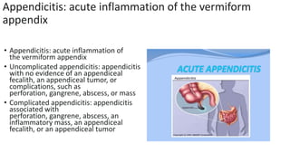
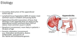
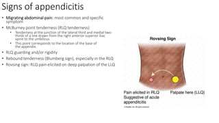
Classification of acute
Appendicitis: acute inflammation of the vermiform
appendix
• Appendicitis: acute inflammation of
the vermiform appendix
• Uncomplicated appendicitis: appendicitis
with no evidence of an appendiceal
fecalith, an appendiceal tumor, or
complications, such as
perforation, gangrene, abscess, or mass
• Complicated appendicitis: appendicitis
associated with
perforation, gangrene, abscess, an
inflammatory mass, an appendiceal
fecalith, or an appendiceal tumor
Etiology
• Caused by obstruction of the appendiceal
lumen due to:
• Lymphoid tissue hyperplasia (60% of cases): most
common cause in children and young adults
• Appendiceal fecalith (concretion of feces that
develops in the appendix that can obstruct the
appendiceal lumen) and fecal stasis (35% of
cases): most common cause in adults
• Neoplasm (uncommon): more likely in patients >
50 years of age
• Parasitic infestation (uncommon):
e.g., Enterobius vermicularis, Ascaris
lumbricoides, and species of
the Taenia and Schistosoma genera
Signs of appendicitis
• Migrating abdominal pain: most common and specific
symptom
• McBurney point tenderness (RLQ tenderness)
• Tenderness at the junction of the lateral third and medial two-
thirds of a line drawn from the right anterior superior iliac
spine to the umbilicus
• This point corresponds to the location of the base of
the appendix.
• RLQ guarding and/or rigidity
• Rebound tenderness (Blumberg sign), especially in the RLQ
• Rovsing sign: RLQ pain elicited on deep palpation of the LLQ
Laboratory studies
CBC: mild leukocytosis with left
shift
• CRP: elevated (> 10 mg/L) [10]
• BMP:
↑ creatinine, electrolyte abnorm
alities may be present in patients
with
severe vomiting and diarrhea
• Urinalysis: typically normal in
appendicitis; possible findings of
mild pyuria and/or hematuria
• Urine/serum β-hCG test: perform in
all women of reproductive age to rule
out pregnancy (including ectopic
pregnancy)
Abdominal ultrasound
• Supportive findings [10][31]
• Distended appendix (diameter > 6
mm)
• Noncompressible, aperistaltic,
distended appendix
• Target sign: concentric rings of
hypo- and hyperechogenicity in the
axial/transverse section of
the appendix
CT abdomen with IV contrast
• CT abdomen is the most accurate
initial imaging modality for
appendicitis. [10][12][28]
• Supportive findings [28]
• Distended appendix (diameter > 6
mm)
• Edematous appendix with
periappendiceal fat stranding
• Possible appendiceal fecalith:
focal hyperdensity within the
appendiceal lumen
• Evidence of complications

Classification of acute appendicitis.pptx

  • 1.
  • 5.
    Appendicitis: acute inflammationof the vermiform appendix • Appendicitis: acute inflammation of the vermiform appendix • Uncomplicated appendicitis: appendicitis with no evidence of an appendiceal fecalith, an appendiceal tumor, or complications, such as perforation, gangrene, abscess, or mass • Complicated appendicitis: appendicitis associated with perforation, gangrene, abscess, an inflammatory mass, an appendiceal fecalith, or an appendiceal tumor
  • 6.
    Etiology • Caused byobstruction of the appendiceal lumen due to: • Lymphoid tissue hyperplasia (60% of cases): most common cause in children and young adults • Appendiceal fecalith (concretion of feces that develops in the appendix that can obstruct the appendiceal lumen) and fecal stasis (35% of cases): most common cause in adults • Neoplasm (uncommon): more likely in patients > 50 years of age • Parasitic infestation (uncommon): e.g., Enterobius vermicularis, Ascaris lumbricoides, and species of the Taenia and Schistosoma genera
  • 7.
    Signs of appendicitis •Migrating abdominal pain: most common and specific symptom • McBurney point tenderness (RLQ tenderness) • Tenderness at the junction of the lateral third and medial two- thirds of a line drawn from the right anterior superior iliac spine to the umbilicus • This point corresponds to the location of the base of the appendix. • RLQ guarding and/or rigidity • Rebound tenderness (Blumberg sign), especially in the RLQ • Rovsing sign: RLQ pain elicited on deep palpation of the LLQ
  • 14.
    Laboratory studies CBC: mildleukocytosis with left shift • CRP: elevated (> 10 mg/L) [10] • BMP: ↑ creatinine, electrolyte abnorm alities may be present in patients with severe vomiting and diarrhea • Urinalysis: typically normal in appendicitis; possible findings of mild pyuria and/or hematuria • Urine/serum β-hCG test: perform in all women of reproductive age to rule out pregnancy (including ectopic pregnancy)
  • 15.
    Abdominal ultrasound • Supportivefindings [10][31] • Distended appendix (diameter > 6 mm) • Noncompressible, aperistaltic, distended appendix • Target sign: concentric rings of hypo- and hyperechogenicity in the axial/transverse section of the appendix
  • 16.
    CT abdomen withIV contrast • CT abdomen is the most accurate initial imaging modality for appendicitis. [10][12][28] • Supportive findings [28] • Distended appendix (diameter > 6 mm) • Edematous appendix with periappendiceal fat stranding • Possible appendiceal fecalith: focal hyperdensity within the appendiceal lumen • Evidence of complications