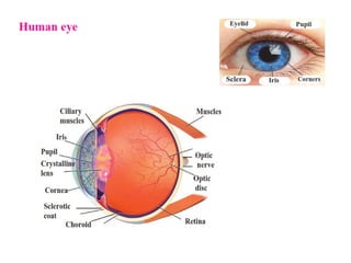
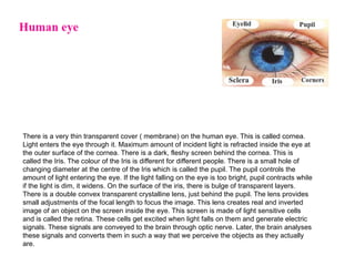
There is avery thin transparent cover ( membrane) on the human eye. This is called cornea.
Light enters the eye through it. Maximum amount of incident light is refracted inside the eye at
the outer surface of the cornea. There is a dark, fleshy screen behind the cornea. This is
called the Iris. The colour of the Iris is different for different people. There is a small hole of
changing diameter at the centre of the Iris which is called the pupil. The pupil controls the
amount of light entering the eye. If the light falling on the eye is too bright, pupil contracts while
if the light is dim, it widens. On the surface of the iris, there is bulge of transparent layers.
There is a double convex transparent crystalline lens, just behind the pupil. The lens provides
small adjustments of the focal length to focus the image. This lens creates real and inverted
image of an object on the screen inside the eye. This screen is made of light sensitive cells
and is called the retina. These cells get excited when light falls on them and generate electric
signals. These signals are conveyed to the brain through optic nerve. Later, the brain analyses
these signals and converts them in such a way that we perceive the objects as they actually
are.
15.
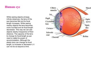
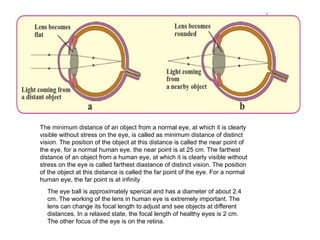
While seeing objectsat large,
infinite distances, the lens of the
eye becomes flat and its focal
length increases. While seeing
nearby objects the lens becomes
more rounded and its focal length
decreases. This way we can see
objects clearly irrespective of their
distance. The capacity of the lens
to change its focal length as per
need is called its power of
accommodation. Although the
elastic lens can change its focal
length, to increase or decrease it, it
can not do so beyond a limit.
16.
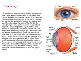
The retina inour eyes is made up of many light sensitive
cells. These cells are shaped like a rod and like a cone.
The rod like cells respond to the intensity of light and give
information about the brightness or dimness of the object
to the brain. The conical cells respond to the colour and
give information about the colour of the object to the
brain. Brain processes all the information received and
we see the actual image of the object. Rod like cells
respond to faint light also but conical cells do not. Thus
we perceive colours only in bright light. The conical cells
can respond differently to red, green and blue colours.
When red colour falls on the eyes, the cells responding to
red light get excited more than those responding to other
colours and we get the sensation of red colour. Some
people lack conical cells responding to certain colours.
These persons cannot recognize those colours or cannot
distinguish between different colours. These persons are
said to be colour blind. Apart from not being able to
distinguish between different colours, their eye sight is
normal.
17.
The minimum distanceof an object from a normal eye, at which it is clearly
visible without stress on the eye, is called as minimum distance of distinct
vision. The position of the object at this distance is called the near point of
the eye, for a normal human eye, the near point is at 25 cm. The farthest
distance of an object from a human eye, at which it is clearly visible without
stress on the eye is called farthest diastance of distinct vision. The position
of the object at this distance is called the far point of the eye. For a normal
human eye, the far point is at infinity
The eye ball is approximately sperical and has a diameter of about 2.4
cm. The working of the lens in human eye is extremely important. The
lens can change its focal length to adjust and see objects at different
distances. In a relaxed state, the focal length of healthy eyes is 2 cm.
The other focus of the eye is on the retina.
18.
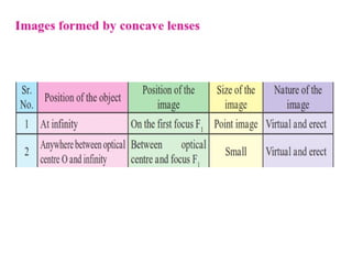
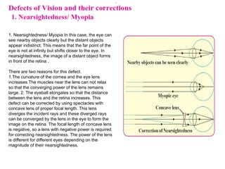
1. Nearsightedness/ MyopiaIn this case, the eye can
see nearby objects clearly but the distant objects
appear indistinct. This means that the far point of the
eye is not at infinity but shifts closer to the eye. In
nearsightedness, the image of a distant object forms
in front of the retina .
There are two reasons for this defect.
1.The curvature of the cornea and the eye lens
increases.The muscles near the lens can not relax
so that the converging power of the lens remains
large. 2. The eyeball elongates so that the distance
between the lens and the retina increases. This
defect can be corrected by using spectacles with
concave lens of proper focal length. This lens
diverges the incident rays and these diverged rays
can be converged by the lens in the eye to form the
image on the retina. The focal length of concave lens
is negative, so a lens with negative power is required
for correcting nearsightedness. The power of the lens
is different for different eyes depending on the
magnitude of their nearsightedness.
19.
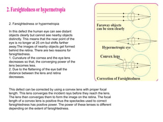
2. Farsightedness orhypermetropia
In this defect the human eye can see distant
objects clearly but cannot see nearby objects
distinctly. This means that the near point of the
eye is no longer at 25 cm but shifts farther
away.The images of nearby objects get formed
behind the retina. There are two reasons for
farsightedness.
1. Curvature of the cornea and the eye lens
decreases so that, the converging power of the
lens becomes less.
2. Due to the flattening of the eye ball the
distance between the lens and retina
decreases.
This defect can be corrected by using a convex lens with proper focal
length. This lens converges the incident rays before they reach the lens.
The lens then converges them to form the image on the retina. The focal
length of a convex lens is positive thus the spectacles used to correct
farsightedness has positive power. The power of these lenses is different
depending on the extent of farsightedness.
20.
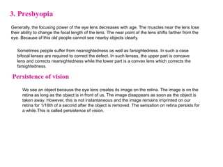
Generally, the focusingpower of the eye lens decreases with age. The muscles near the lens lose
their ability to change the focal length of the lens. The near point of the lens shifts farther from the
eye. Because of this old people cannot see nearby objects clearly.
Sometimes people suffer from nearsightedness as well as farsightedness. In such a case
bifocal lenses are required to correct the defect. In such lenses, the upper part is concave
lens and corrects nearsightedness while the lower part is a convex lens which corrects the
farsightedness.
We see an object because the eye lens creates its image on the retina. The image is on the
retina as long as the object is in front of us. The image disappears as soon as the object is
taken away. However, this is not instantaneous and the image remains imprinted on our
retina for 1/16th of a second after the object is removed. The sensation on retina persists for
a while.This is called persistence of vision.
21.
Use of concavelenses
a. Medical equipments, scanner, CD player – These
instuments use laser light. For proper working of these
equipments concave lenses are used.
b. The peep hole in door- This is a small safety device which
helps us see a large area outside the door. This uses one or
more concave lenses.
c. Spectacles- Concave lenses are used in spectacles to
correct nearsightedness. d. Torch- Concave lens is used to
spread widely the light produced by a small bulb inside a
torch. e. Camera, telescope and microscope- These
instruments mainly use convex lenses. To get good quality
images a concave lens is used in front of the eyepiece or
inside it.
22.
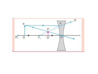
Use of convexlenses
a. Simple microscope : A convex lens with small focal length produces a virtual, erect and bigger
image of an object. Such a lens is called simple microscope or magnifying lens. One can get a 20
times larger image of an object using such microscopes. These are used for watch repair, testing
precious gems and finding their defects.
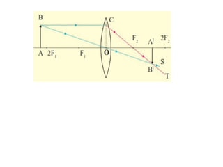
b. Compound microscope
Simple microscope is used to observe small sized objects. But minute objects like blood
cells, cells of plants and animals and minute living beings like bacteria cannot be
magnified sufficiently by simple microscope. Compound microscopes are used to study
these objects. A compound microscope is made of two convex lenses: objective and eye
piece. The objective has smaller cross-section and smaller focal length. The eye piece
has bigger crosssection, its focal length is also larger than that of the objective. Higher
magnification can be obtained by the combined effect of the two lenses. 7.16 Simple
microscope a. Object is close to the lens b. Object is at the focus 7.17 A compound
microscope As shown in the figure 7.17, the magnification occurs in two stages. The
image formed by the first lens acts as the object for the second lens. The axes of both
lenses are along the same line. The lenses are fitted inside a metallic tube in such a way
that the distance between can be changed.
23.
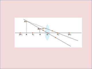
c. Telescope
Telescope isused to see distant objects clearly in their magnified form. The telescopes used to observe
astronomical sources like the stars and the planets are called astronomical telescopes. Telescopes are of
two types. 1. Refracting telescope – This uses lenses 2. Reflecting telescope – This uses mirrors and
also lenses. In both of these, the image formed by the objective acts as object for the eye piece which
forms the final image. Objective lens has large diameter and larger focal length because of which
maximum amount of light coming from the distant object can be collected.
On the other hand the size of the eyepiece is smaller and its focal length is also
less. Both the lenses are fitted inside a metallic tube in such a way that the
distance between them can be changed. The principal axes of both the lenses
are along the same straight line. Generally, using the same objective but
different eye pieces, different magnification can be obtained.
d. Optical instrument Convex lenses are used in various other optical instruments like
camera, projector, spectrograph etc.