This document is an American Heart Association scientific statement that provides an updated review of the cardiovascular risks and benefits of physical activity and exercise. It finds that while regular physical activity and higher cardiorespiratory fitness are associated with significant reductions in cardiovascular disease risk, vigorous or high-intensity physical activity can acutely increase the risk of sudden cardiac death and acute myocardial infarction in susceptible individuals. Recent studies also show that high volumes and intensities of exercise may be associated with potential cardiac maladaptations like fibrosis. The statement aims to advise healthcare professionals on preparticipation screening and informing patients of appropriate physical activity levels based on current understanding of risks.

![Franklin et al Exercise-Related Acute Cardiovascular Events
March 31, 2020 Circulation. 2020;141:e705–e736. DOI: 10.1161/CIR.0000000000000749e706
CLINICALSTATEMENTS
ANDGUIDELINES
S
ubstantial epidemiological, clinical, and basic sci-
ence evidence suggests that regular physical ac-
tivity (PA), higher cardiorespiratory fitness (CRF),
or both, delay the development of atherosclerotic car-
diovascular disease (CVD) and reduce the incidence of
coronary heart disease (CHD) events.1–3
PA is defined as
any bodily movement resulting from the contraction of
skeletal muscle that increases energy expenditure above
the basal level. A systematic review and meta-analysis
of 33 PA studies (n=883 372 participants) reported risk
reductions of 30% to 50% for cardiovascular mortality
and 20% to 50% for all-cause mortality with increas-
ing volumes of PA.4
More recently, researchers from the
Nurses’ Health Study (n=78 865) and the Health Pro-
fessionals Follow-up Study (n=44 354) estimated the
impact of 5 lifestyle factors, including ≥30 min/d of
moderate to vigorous PA, on life expectancy in the US
population.5
During up to 34 years of follow-up, the
most physically active cohorts of men and women dem-
onstrated 7- to 8-year gains in life expectancy.
Exercise training, as a subcategory of PA, is defined as
any planned and structured intervention with the objec-
tive of improving or maintaining CRF or health, achiev-
ing athletic goals, or both. Aerobic capacity or CRF can
be directly measured during cardiopulmonary exercise
testing via gas-exchange measurements or estimated
from the attained treadmill speed, percent grade, and
duration (minutes) or the cycle ergometer workload,
expressed as kilogram meters per minute. After a 12-
week exercise-based cardiac rehabilitation program in a
cohort of 5600 patients with known CVD, each 1-met-
abolic equivalent (MET; 1 MET=3.5 mL O2
·kg−1
·min−1
)
improvement in estimated CRF (CRFe) was associated
with a 13% overall reduced risk of all-cause mortality
and, in the least fit patient cohort (5 MET capacity),
a 30% reduction in mortality.6
These risk reductions
compare favorably with the survival benefit conferred
by commonly prescribed cardioprotective medications,
including statins.7
In US veterans, the incidence of major
CVD events was 16% lower for every 1-MET increase
in CRFe. Compared with the least fit veterans, the risk
of CVD events was ≈70% lower for individuals in the
highest fitness category.8
A prospective cohort study
of 120 000 consecutive patients who underwent
maximal treadmill testing and 1.1 million person-years
of observation reported no upper limit for the protec-
tive effect of higher CRFe on all-cause mortality. Com-
pared with the lowest performers (25th percentile),
elite CRFe (97.7th percentile) was associated with an
80% reduction in mortality risk. Remarkably, the effect
of low CRFe on all-cause mortality was comparable to
or greater than that of traditional risk factors such as
smoking and diabetes mellitus for coronary artery dis-
ease (CAD).9
A study from the UK Biobank (n=502 635)
showed inverse associations between CRFe and CHD
(hazard ratio [HR], 0.51 [95% CI, 0.38–0.69]) and CRFe
and atrial fibrillation (AF; HR, 0.40 [95% CI, 0.30–0.55])
among individuals at high genetic risk for these diseas-
es.10
Others have reported that highly fit individuals, re-
gardless of their risk factor profile, have an ≈50% lower
30-year CVD mortality than their counterparts with low
fitness.11
Similarly, men with subclinical CAD (coronary
artery calcium [CAC] score ≥100) whose CRFe was ≥10
METs have an age-adjusted HR for CHD events of 0.26
(95% CI, 0.15–0.45) compared with men whose CRFe
is 10 METs.12
More recently, compared with the least
fit men, CVD events were shown to be progressively re-
duced with increasing fitness levels, and the effect was
more prominent in individuals with the highest levels
of CAC.13
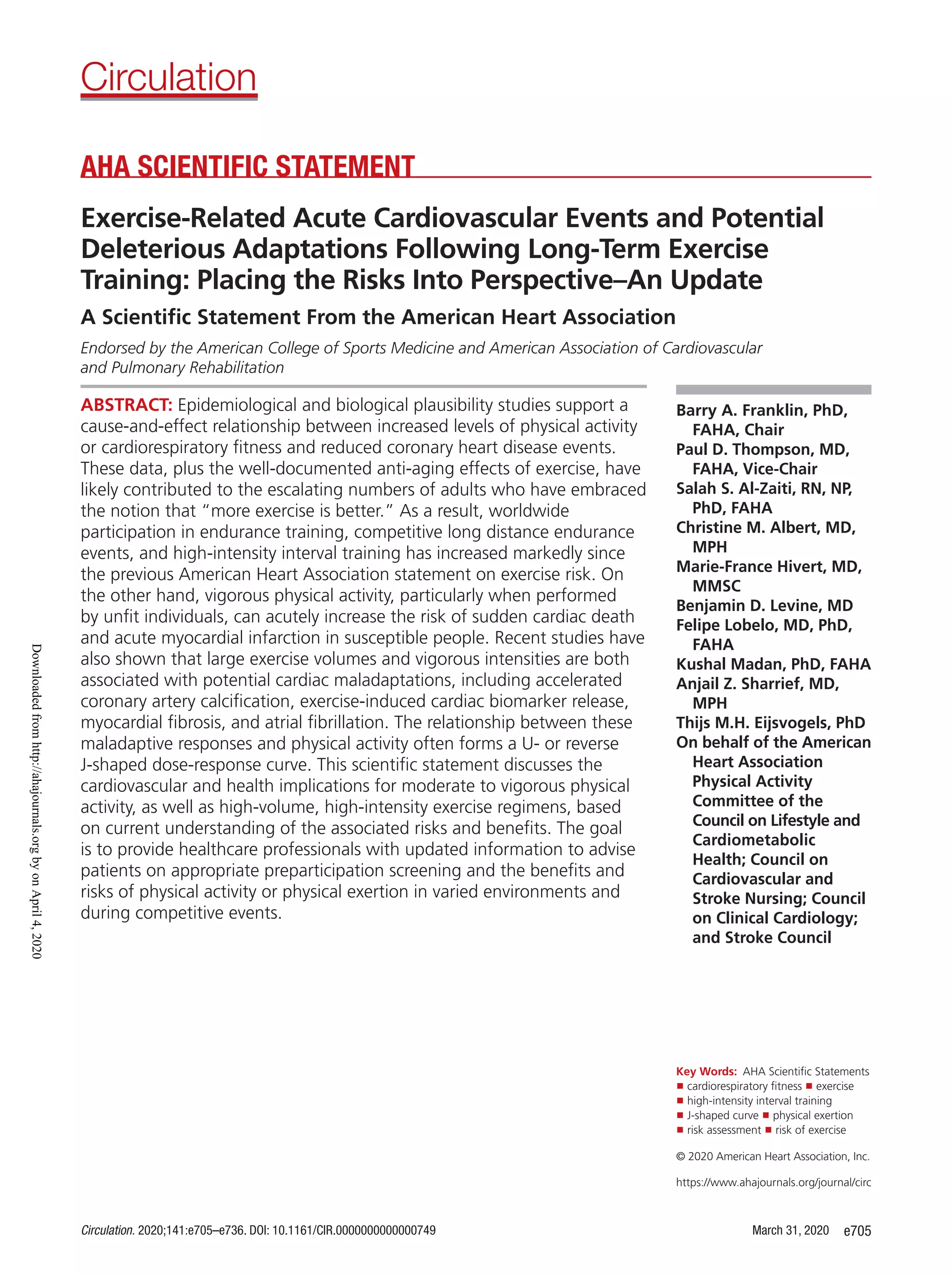
Collectively, these epidemiological analyses,
combined with evidence of biological plausibility (Fig-
ure 1),14,15
support a cause-and-effect relationship be-
tween increased levels of PA and CRF and reduced CVD
mortality16–18
and suggest that being unfit is an indepen-
dent risk factor for CHD (Figure 2).1,19
Higher levels of PA or CRF (5 METs) before hospi-
talization for acute coronary syndromes (ACS) and sur-
gical procedures are associated with better short-term
outcomes,20,21
possibly because of exercise-induced
ischemic preconditioning.22
An investigation of 2172
patients hospitalized for ACS evaluated the effect of
preadmission PA on in-hospital and 1-month post-
discharge CVD health outcomes.23
Patients were cat-
egorized as physically inactive (reference category),
minimally active, and highly active. Multivariate analysis
revealed that compared with the inactive cohort, mini-
mal or high activity was associated with a 44% (95%
CI, 10%–68%) reduction of in-hospital mortality and
a 20% (95% CI, 1%–50%) lower risk of a CVD event
within the first month of hospital discharge.23
Compli-
cations after elective or emergent surgical procedures,
including bariatric surgery24
and coronary artery bypass
grafting,25
are also increased in those with reduced pre-
operative levels of PA or CRF (Figure 3).26
The favorable risk factor profiles and superior car-
diac performance of long-distance runners and the ob-
servation that vigorous PA and high levels of CRF are
associated with reduced use of diabetic, hypertensive,
and hypercholesterolemia medications27
suggest that
high-volume and high-intensity endurance training reg-
imens, including high-intensity interval training (HIIT),28
are cardioprotective in individuals with and without
CHD. These data have likely contributed to increasing
numbers of middle-aged and older adults concluding
that “more exercise is better.”29
As a result, worldwide
participation in half- and full-marathon races, triath-
lon events, and HIIT has increased markedly since the
American Heart Association’s (AHA’s) previous state-
ment on exercise risk.30
Nevertheless, prolonged exer-
cise increases cardiac biomarkers31–33
and postexercise
transient myocardial dysfunction, and endurance ath-
letes 35 years of age have increased myocardial late
Downloadedfromhttp://ahajournals.orgbyonApril4,2020](https://image.slidesharecdn.com/circulation-200407184256/85/Circulation-2-320.jpg)
![Franklin et al Exercise-Related Acute Cardiovascular Events
Circulation. 2020;141:e705–e736. DOI: 10.1161/CIR.0000000000000749 March 31, 2020 e709
CLINICALSTATEMENTS
ANDGUIDELINES
risk of SCD during vigorous exercise (RR=5068
to 7448
)
compared with those who exercised regularly (RR=268
to 1148
).43
However, in 2 of these studies, the risk of
SCD remained elevated during exertion even among
the most habitually active men.43,48
There are limited data regarding whether more mod-
erate levels of exercise might trigger SCD. One prospec-
tive case-crossover study among middle-aged nurses
reported the RR of SCD associated with combined expo-
sure to both moderate and vigorous levels of exertion.45
Because of the small number of exercise-associated
SCDs in women, the risk of moderate versus vigorous
exercise could not be analyzed separately. The RR of
SCD was modestly but significantly elevated during and
30 minutes after moderate and vigorous exercise, with
an estimated RR of 2.4 (95% CI, 1.2–4.6; P=0.01). This
transient increase in risk was again reduced by regular
exercise participation, with the highest risks observed
among women who reported 2 hours per week of
moderate to vigorous exertion (RR, 9.0 [95% CI, 3.3–
24.3]). In contrast to prior studies that examined vigor-
ous exercise in men,43,48
this transient elevation in risk
was no longer significant among the most habitually
active women. Women who reported participating in
moderate to vigorous exercise ≥2 hours per week had
an RR of 1.5 (95% CI, 0.61–3.61).
Myocardial Infarction
Several studies using differing designs have also dem-
onstrated significant 2- to 10-fold elevations in the like-
lihood of experiencing an AMI within 1 hour of par-
ticipating in vigorous exertion,41,60–63,70,71
with 1 study
demonstrating risk elevations persisting to 2 hours.62
When the available data from 7 studies (n=5503 pa-
tients) were combined using meta-analysis, the summa-
ry RR of MI associated with bouts of physical exertion
was 3.45 (95% CI, 2.33–5.13), with substantial be-
tween-study heterogeneity that was not explained by
study size or the year the study was conducted.72
Effect
sizes for the association of episodic PA did not appear
to change significantly over time. Again, as is the case
for SCD, the magnitude of association between PA as
a trigger of MI was greater in individuals with the low-
est habitual activity. In these 7 studies, estimates of the
RR associated with a bout of vigorous exertion in the
least habitually active individuals ranged from 4.571
to
10741
for MI. In comparison, RR estimates of MI in the
setting of exertion were much more modest (0.88–3.3)
in those with the highest levels of habitual PA.72
In a
meta-regression analysis, the RR of MI associated with
episodic PA was decreased by ≈45% for each additional
time per week a person habitually exercised.72
Since this meta-analysis was published in 2011, the
INTERHEART Study has reported results from a retro-
spective case-crossover design performed in the larg-
est population to date, including 12 461 patients with
first MI in 262 centers across 52 countries.63
Participa-
tion in heavy physical exertion was reported by 13.6%
of participants within the 1 hour before MI compared
with 9.1% at the same hour on a previous day (control
period). Compared with the control period, the adjust-
ed RR of AMI within 1 hour of physical exertion was
estimated to be 2.3 (95% CI, 2.0−2.7), correspond-
ing to a population attributable risk of 7.7% (95% CI,
6.3%−8.8%). Unlike prior studies, this study did not
demonstrate evidence of effect modification by base-
line PA level. However, the latter analysis only stratified
by overall participation in categories of PA by intensity
(sedentary, mild, moderate, or strenuous) and not by
frequency of participation.
Compared with vigorous exercise, the data on
whether moderate exercise also acutely elevates the
risk of MI are much more limited. One case-crossover
study within the Myocardial Infarction Registry in Augs-
burg, Germany, reported a modest increase in the risk
of MI within 2 hours of participation in moderate exer-
tion (METs=5; RR, 1.6 [95% CI, 1.2−2.1]).71
However,
a prior study from the same region in Germany found
no elevation in the risk of MI associated with moder-
ate physical exertion.60
Both studies demonstrated no
association between lighter levels of exertion and AMI.
Absolute Risks
Although there is a heightened RR of acute cardiac
events with unaccustomed vigorous PA, it is important
to recognize that the absolute risk of experiencing SCD
or MI during physical exertion is very small. For example,
in the Physicians’ Health Study, marked elevations in the
RR of SCD during and after a bout of vigorous physical
exertion translated into an absolute risk of only 1 SCD for
every 1.51 million episodes.48
This absolute risk is even
lower in middle-aged women, among whom 1 SCD oc-
curred for every 36.5 million person-hours of participa-
tion in moderate to vigorous exercise.45
Retrospective
multicenter data from fitness facilities also report low
absolute rates of adverse CVD events: 1 per 1 124 200
and 1 per 887 526 person-hours for nonfatal and fatal
cardiovascular events, respectively.73
In patients with es-
tablished heart disease in supervised cardiac rehabilita-
tion facilities, the absolute CVD event rate is higher (1
event per 58 000 patient-hours), but still low.74
Given that even the most avid exercisers do not
spend the majority of their time exercising, these low
absolute risks translate into extremely low incident rates
of exertion-related SCD in the general population. Re-
ported annual incidence rates of exertion-related SCD
in the population at large and across various subgroups
are shown in Table 1. Population-based estimates of the
frequency of exercise-related out-of-hospital cardiac ar-
rest and SCD range from 2.1 per 100 000 person-years
in the Netherlands,49
to 0.46 per 100 000 person-years
Downloadedfromhttp://ahajournals.orgbyonApril4,2020](https://image.slidesharecdn.com/circulation-200407184256/85/Circulation-5-320.jpg)
![Franklin et al Exercise-Related Acute Cardiovascular Events
Circulation. 2020;141:e705–e736. DOI: 10.1161/CIR.0000000000000749 March 31, 2020 e717
CLINICALSTATEMENTS
ANDGUIDELINES
12-lead resting ECG.49,51,118,148–151
Adding a resting ECG
to screening can enhance the detection of disorders as-
sociated with a heightened risk of SCD but increases
false-positive results, with costly follow-up noninvasive/
invasive studies and possible associated downstream psy-
chological harm.152
Consequently, a relevant AHA/Ameri-
can College of Cardiology scientific statement concluded
that the available data do not support a significant public
health benefit from using the 12-lead ECG as a universal
screening tool for athletes.153
This contrasts with recom-
mendations from the European Society of Cardiology.154
There are multiple issues with routine cardiovascular
screening in asymptomatic athletes. The true incidence
of sports-related SCA is unknown. This has been gener-
ally estimated at ≈1 per 200 000 annually,155
but studies
in accomplished athletes, including National Collegiate
Athletic Association basketball players119
and English
youth soccer players,156
have reported annualized rates
of 1 per 5200 and 1 per 14 794, respectively. It is also
not clear how truly asymptomatic athletes with a car-
diac condition should be managed. Others emphasize
that preparticipation screening of large groups of as-
ymptomatic athletes without known CVD will inevitably
result in many false-positive responses, unnecessarily
restricting PA among athletes who are free from CVD,
while simultaneously providing a false sense of security
to those with unremarkable findings.151
Other options are available to reduce exercise-re-
lated cardiac events. Coaches and athletes should be
educated about possible warning signs/symptoms (eg,
syncope, lightheadedness, perceived palpitations or ar-
rhythmias) that could be harbingers of acute cardiac
events. The Sudden Unexplained Death Study reported
that of 3775 SCAs in all age groups in the Portland,
OR, metropolitan area between 2002 and 2015, 186
(5%) occurred in the young (mean±SD age 25.9±6.8
years; 67% male), and the prevalence of warning signs
before SCA was 29%. Moreover, 26 of the 186 (14%)
were associated with sports as a trigger.53
Such symp-
toms mandate the immediate cessation of training/
competition and medical review. Regular emergency
drills, bystander cardiopulmonary resuscitation, and the
use of automated external defibrillators have increased
survival rates in the general157
and athletic158
popula-
tion. This writing group maintains that if cardiovascular
preparticipation screening is offered to young athletes,
it should be voluntary and conducted by highly expe-
rienced clinicians, but we recognize that the ability to
alter survival by removal from sports participation or by
other interventions has not been proven.156,159
CAN EXTREME EXERCISE HARM THE
HEART?
Long-term exercise training alters cardiac structure and
function. The athlete’s heart is characterized by (1)
enlargement of all cardiac chambers,160
(2) improve-
ment of cardiac function161
and compliance,162
and (3)
electrical remodeling, such as sinus bradycardia, sinus
arrhythmia, and first-degree atrioventricular block.163
These exercise training–induced adaptations are be-
lieved to be benign. Emerging evidence, however, sug-
gests that over time, high-volume, high-intensity exer-
cise training can induce cardiac maladaptations such as
an increased risk for AF, coronary artery calcification,
and myocardial fibrosis.164
Hence, there is debate as to
whether intensive exercise can be harmful to the heart,
especially in some individuals.165–167
Atrial Fibrillation
AF is characterized by chaotic electrical activity that re-
places normal sinus rhythm and eliminates the contri-
bution of atrial contraction to left ventricular filling. AF
is the most common arrhythmia in the general popula-
tion,168
and the risk of AF depends on subject charac-
teristics (age, race, height), health status (weight, blood
pressure, [para]sympathetic tone, diabetes mellitus, a
history of MI or heart failure), lifestyle factors (alcohol
use, smoking, PA), obstructive sleep apnea,169
and car-
diac characteristics (left atrial size and pressure).170,171
AF is associated with an increased risk for stroke,172
MI,173
heart failure,174
and a multitude of adverse clini-
cal consequences.175
The relation between exercise and incident AF is
complicated. Low levels of CRFe (6 METs) are asso-
ciated with a higher risk for AF, and individuals with
higher levels of CRFe (7.9±1.0 and 9.3±1.2 METs) have
a dose-dependent decrease in AF risk.176,177
Similarly, fit
AF patients have a lower risk for AF recurrences during
follow-up than their unfit counterparts.178
AF burden
and symptom severity decreased significantly in patients
with AF who increased their fitness during an exercise
training program versus those who failed to improve178
and among patients with AF randomized to aerobic in-
terval training in a small clinical trial.179
Although these
observations suggest that fitter individuals have the
lowest AF risk, there is substantial evidence that the
risk for AF is higher in athletes than in control subjects.
High-intensity exercise training180
and faster finishing
times181
were associated with an increase of AF in physi-
cally active older adults and long-distance cross-country
skiers, respectively. In the US Physician’s Health Study,
men who jogged 5 to 7 times per week had a 50%
higher risk of AF than men who did not exercise vigor-
ously, even after adjustment for multiple cardiovascular
risk factors.182
Three meta-analyses found that AF risk
was 2- to 10-fold higher in endurance athletes than in
control participants.170,183,184
Furthermore, long-term vol-
ume of vigorous endurance exercise (ie, ≥2000 hours
of training169
or ≥20 years of training185
) was strongly
associated with an increased risk for lone AF. These data
Downloadedfromhttp://ahajournals.orgbyonApril4,2020](https://image.slidesharecdn.com/circulation-200407184256/85/Circulation-13-320.jpg)
![Franklin et al Exercise-Related Acute Cardiovascular Events
Circulation. 2020;141:e705–e736. DOI: 10.1161/CIR.0000000000000749 March 31, 2020 e719
CLINICALSTATEMENTS
ANDGUIDELINES
coronary artery atherosclerosis among veteran athletes.
Möhlenkamp et al203
were the first to demonstrate that
German marathon runners had a higher prevalence of
CAC scores ≥100 Agatston units compared with an
age- and risk factor–matched control group from the
general population (36% versus 22%, P=0.02). Aen-
gevaeren et al34
reported an increased CAC prevalence
across progressive tertiles of PA volumes, 43%, 50%,
and 68% (P=0.005), respectively, among Dutch male
amateur athletes. More importantly, the most active
athletes had a lower prevalence of unstable mixed
plaques (48% versus 69%; OR, 0.35 [95% CI, 0.15–
0.85]) and more often had only stable calcified plaques
(38% versus 16%; OR, 3.57 [95% CI, 1.28–9.97])
compared with the least active athletes.34
Merghani
et al35
reported similar findings in a cohort of British
male master endurance athletes and sedentary control
participants. The athletes had a higher prevalence of
atherosclerotic stable plaques with any degree of lumi-
nal stenosis compared with control participants (44.3%
versus 22.2%, P0.001). Athletes demonstrated pre-
dominantly stable calcified plaques (73% versus 31%,
P0.001) and fewer vulnerable mixed plaques (23%
versus 62%).35
These observations demonstrated an
increased prevalence of stable plaques among highly
active middle-aged endurance athletes.
Only 2 small studies (n=4635
and n=26204
) assessed
atherosclerotic characteristics among female athletes.
There were no differences in CAC and plaque preva-
lence between female athletes and control participants
in a British cohort.35
In contrast, among American
women undergoing computed tomography angiog-
raphy to evaluate CAD, athletes had lower coronary
plaque prevalence and calcific plaque volumes than
comparison women.204
Because the reference group
had a significantly higher prevalence of hypertension,
hyperlipidemia, smoking, and family history of CAD,
the decreased plaque prevalence in the athletes could
be more related to their low atherosclerotic risk profile
than their exercise behavior.
The clinical relevance of accelerated coronary ar-
tery atherosclerosis in athletes performing very high-
volume, high-intensity exercise is unknown. In clinical
populations, any CAC or plaque presence represents
a higher cardiovascular risk.205,206
On the other hand,
coronary artery size and dilating capacity are increased
among athletes compared with control subjects,207
which could offset the apparently negative adapta-
tion of a higher CAC. Elevated CAC scores in athletes
might also indicate increased cardiovascular risk, but
definite data to evaluate this hypothesis are lacking.208
Traditional CAC risk predictions might not be applicable
to athletes, because physically active individuals have
a lower risk for adverse cardiovascular outcomes than
less active individuals with similar CAC scores.209
The
hypothesis that athletes are at reduced risk despite their
higher CAC scores is supported by the observation that
the most active athletes had fewer unstable “mixed”
plaques and more stable “calcified” plaques.34,35
Mixed
plaques are associated with a high risk of cardiovascular
events, whereas calcified plaques are associated with a
lower risk.199
Very high volumes of exercise training can
therefore produce calcified stabilized plaques, similar to
the observation that statins can increase CAC scores
although they reduce atheroma volume and acute
cardiovascular events.13,210
In a large cohort of men
(n=21 758) with more than a decade of follow-up in the
Cooper Clinic Longitudinal study, adjusted risk of CAC
score ≥100 was greater among individuals with very
high levels of PA (n=432; ≥3000 MET-min/wk) com-
pared with those with lower activity levels. Neverthe-
less, their risk for all-cause and cardiovascular mortality
was not higher than that for those exercising at more
moderate levels, and it was lower than the least active
cohort with similar CAC scores. These findings refute
the notion that high-volume endurance activity (1 h/d)
increases mortality risk, regardless of CAC level.211
Several mechanisms have been proposed to explain
increased CAC after long-term, high-intensity exercise
training. Exercise-related changes in calcium metabo-
lism might contribute to the higher CAC score in ath-
letes. Vitamin D levels are lower in athletes,212
and low
vitamin D levels are associated with increased CAC in
the general population.213
In contrast, exercise acutely
increases parathyroid hormone,214
which is associated
with increased arterial calcification.215
Alternatively,
vigorous-intensity exercise can increase mechanical
stress on the coronary wall, causing disrupted flow pat-
terns,216
elevate blood pressure, and cause shear stress,
each of which is associated with accelerated athero-
sclerosis.217,218
Regular high-intensity exercise training
also transiently increases other nontraditional athero-
sclerotic risk factors, including oxidative free radicals,219
dicarbonyl stress,220
and systemic inflammation.221
Myocardial Fibrosis
Myocardial fibrosis is characterized by collagen infiltra-
tion in the myocardial extracellular matrix and can de-
velop after injury from myocardial ischemia (hypoxia),
inflammation, hypertensive overload, or combinations
thereof.222
It is associated with increased myocardial
stiffness,223
a higher incidence of ventricular arrhyth-
mias,224,225
and adverse cardiac outcomes.226
Myocardial
fibrosis is determined noninvasively using cardiac mag-
netic resonance imaging with gadolinium to determine
LGE, a marker for myocardial fibrosis. The development
of myocardial fibrosis generally occurs with cardiac re-
modeling secondary to diseases such as heart failure,
hypertension, and valvular dysfunction,227
but sev-
eral studies have found evidence of LGE in apparently
healthy athletes.228
The prevalence of LGE in endurance
Downloadedfromhttp://ahajournals.orgbyonApril4,2020](https://image.slidesharecdn.com/circulation-200407184256/85/Circulation-15-320.jpg)
![March 31, 2020 Circulation. 2020;141:e705–e736. DOI: 10.1161/CIR.0000000000000749e720
CLINICALSTATEMENTS
ANDGUIDELINES Franklin et al Exercise-Related Acute Cardiovascular Events
athletes has ranged from 0%229
to 50%.230
A system-
atic review of 19 studies reported LGE in 5.9% of the
509 athletes.228
Only 6 studies35,230–234
directly compared
LGE prevalence between athletes and control subjects
(Table 3). Five found a higher LGE prevalence in ath-
letes, and 1 study found no difference between athletes
and control subjects, but the prevalence of LGE varied
widely across studies, probably because of sample size
differences. Overall, competitive endurance athletes
had a substantially higher LGE prevalence than control
subjects performing PA at or below World Health Orga-
nization recommendations (12% versus 1.5%).
Several studies have attempted to identify factors as-
sociated with the presence of LGE. Long-term exercise
dose quantified as years of training230,235
and number
of completed foot races ≥42 km203,230
was consistently
found as a strong predictor for LGE, which suggests that
high-volume, high-intensity exercise training may in-
crease the risk of myocardial fibrosis. Alternatively, LGE
may also represent edema,236
inflammation,237
or myo-
cardial disarray238
rather than scar tissue. LGE in athletes
is often located where the right ventricle inserts into
the ventricular septum. Indeed, 48% of athletes with
LGE had LGE in the septum or right ventricular insertion
points.228
Short-term and prolonged exposure to high
cardiac strain can also cause edema-induced LGE.236
Vigorous exercise elicits a greater relative increase in
pulmonic than aortic systolic pressure, estimated as an
increase in wall stress of 125% versus 4% for the right
and left ventricles, respectively.124
This greater relative
increase in wall stress imposed on the thinner right
ventricular wall may contribute to edema and cardio-
myocyte damage in the septum and right ventricular
septal insertion points. An animal study found higher
levels of myocardial fibrosis in the right but not left ven-
tricle in rats running 60 min/d for 16 weeks compared
with sedentary controls.237
The amount of fibrosis was
time dependent and accompanied by myofiber disarray,
leukocyte infiltration, and expression of proinflamma-
tion factors (IL-1β [interleukin-1β] and MCP-1 [mono-
cyte chemoattractant protein-1]), which suggests that
exercise can induce deleterious remodeling.237
It is un-
known how these findings translate to humans, and
future studies are needed to determine the nature and
cause of LGE in athletes and whether edema-induced
LGE will regress, remain unchanged, or progress to scar
tissue over time.
There is little information about the clinical conse-
quences of LGE in athletes. Coronary revascularization
was more common in one study of athletes with versus
without LGE (25% versus 1%, P0.001) during 21±3
months of follow-up, but it is unclear how knowledge
of the fibrosis influenced clinical decisions.233
Also,
nonsustained ventricular arrhythmias, symptomatic
ventricular tachycardia, and progressive left ventricular
dysfunction were found in a case series of athletes with
subepicardial fibrosis.239
Although overall systolic and
diastolic cardiac function is usually normal in athletes
with LGE, one study found wall motion abnormalities
in myocardial segments with LGE.240
However, mas-
ters athletes have more compliant hearts and blood
vessels than matched healthy, sedentary individuals or
Table 3. Overview of Studies Comparing Prevalence of LGE Between Male Athletes and Control Subjects
Study Athlete Cohort Control Group
LGE Prevalence
in Athletes,
n (%)
LGE Prevalence in
Control Subjects,
n (%)
Abdullah et al231
21 competitive master athletes (68 [66–70]
y of age), performing 6 or 7 exercise
sessions per week for 25 y
25 healthy seniors (69 [65–71] y of age)
performing 1 exercise session per week
for 25 y
0/21 (0) 0/25 (0)
Bohm et al232
33 competitive elite male master endurance
athletes (47±8 y of age) with a training
history of 29±8 y
33 individuals exercising ≤3 h/wk 1/33 (3) 0/33 (0)
Breuckmann et al233
102 marathon runners (57±6 y of age) with
a history of ≥5 marathons in ≤3 y
102 individuals (57±6 y of age) without
exceptional endurance sports activity
12/102 (12) 4/102 (4)
Merghani et al35
106 masters athletes (55±9 y of age) with a
training history 31±13 y
54 individuals (53±8 y of age) engaged
in habitual physical activity (ie, walking,
jogging, or swimming) according to WHO
recommendations (150 min/wk)
15/106 (14) 0/54 (0)
Tahir et al234
54 male competitive triathletes (44±10 y of
age), training 10 h/wk
29 female competitive triathletes (42±10 y
of age), training 10 h/wk
22 sedentary controls (40±12 y of age),
exercising ≤3 h/wk
14 sedentary controls (45±12 y of age),
exercising ≤3 h/wk
9/54 (17)
0/29 (0)
0/22 (0)
0/14 (0)
Wilson et al230
12 veteran endurance athletes (57±6 y of
age) with a training history of 43±6 y
20 sedentary controls (60±5 y of age) 6/12 (50) 0/20 (0)
Total 357 competitive endurance athletes 270 controls performing physical activity at
or below WHO recommendations*
43/357 (12.0) 4/270 (1.5)
LGE indicates late gadolinium enhancement; and WHO, World Health Organization.
*WHO recommendation: 150 minutes of moderate intensity physical activity, 75 minutes of vigorous, or an equivalent combination per week.
Downloadedfromhttp://ahajournals.orgbyonApril4,2020](https://image.slidesharecdn.com/circulation-200407184256/85/Circulation-16-320.jpg)
![Franklin et al Exercise-Related Acute Cardiovascular Events
Circulation. 2020;141:e705–e736. DOI: 10.1161/CIR.0000000000000749 March 31, 2020 e721
CLINICALSTATEMENTS
ANDGUIDELINES
individuals who perform lower amounts of regular exer-
cise.162,241
Because the clinical course and consequences
of LGE in endurance athletes are unclear, these athletes
require additional follow-up to determine the signifi-
cance of this finding.
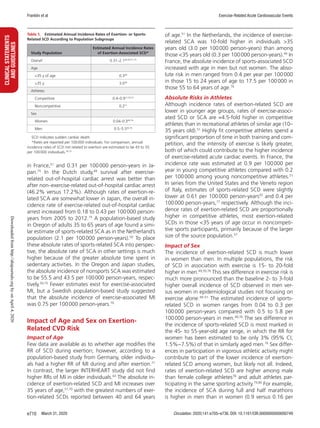
Curvilinear Versus U-Shaped Associations
There is debate regarding the dose-response relation-
ship between the volume of PA or exercise training and
cardiovascular health outcomes. The prevailing dogma
suggests a curvilinear relationship between exercise vol-
ume and cardiovascular health risks (Figure 8A), which
indicates that individuals performing none to very low
volumes of exercise training have the highest risk for
adverse outcomes, whereas individuals who exercise
the most have the lowest risk. Epidemiological evidence
strongly supports this relationship.38,242,243
Wen et al38
found that 92 minutes per week of moderate- to vigor-
ous-intensity PA was associated with a 19% risk reduc-
tion for CVD mortality (HR, 0.81 [95% CI, 0.71−0.93])
and a 14% risk reduction for all-cause mortality (HR,
0.86 [95% CI, 0.81−0.91]). Larger exercise volumes
yielded greater health benefits, with a maximal risk
reduction of 45% for CVD mortality (HR, 0.55 [95%
CI, 0.46−0.66]) and 35% for all-cause mortality (HR,
0.65 [95% CI, 0.60−0.70]) in individuals performing
523 minutes of exercise per week. This exercise volume
equals 3.5 to 4 times the amount of PA recommended
in the 2018 Federal Physical Activity Guidelines,244
but
an increasing number of athletes are exceeding this ex-
ercise volume during their training. Arem et al242
investi-
gated the health benefits of a physically active lifestyle in
individuals performing up to 10 times the World Health
Organization–recommended exercise dose. Maximal
mortality risk reduction (HR, 0.61 [95% CI, 0.59−0.62])
occurred at an exercise volume equaling 3 to 5 times
the recommendations. Although the most active indi-
viduals (≥10 times the recommendations) had a lower
mortality risk (HR, 0.69 [95% CI, 0.59−0.78]) compared
with inactive control subjects, their risk reduction was
not as large as the group with optimal exercise volume.
Similar findings with large CIs for risk estimates among
the most active exercisers were observed in other stud-
ies38,82,242,243,245–247
(Table 4) and may be explained by
the relatively small sample sizes of the most active sub-
groups (typically 5% of the total cohort).
The observation that very high volumes of PA may
yield lower risk reductions than moderate- to high-
activity volumes resulted in the extreme exercise hy-
pothesis,37,248
which postulates a U-shaped relationship
between PA volumes and health outcomes (Figure 8B)
and is characterized by partial loss of exercise-induced
health benefits among the most active individuals.
However, only limited data are available to support this
hypothesis. Schnohr et al246
found an increased mor-
tality risk for strenuous versus light joggers (Table 4),
but there were few individuals in the most active group
(n=36) and only 2 deaths (no cause of death identi-
fied), resulting in an unusually large CI (0.48–8.14) and
discussion about its interpretation.249
Similarly, Lear et
al245
reported less mortality reduction from recreational
PA in the most active exercisers (Table 4), but this effect
disappeared when recreational and nonrecreational
physical activities were combined. Armstrong et al82
showed a higher risk ratio of cerebrovascular disease
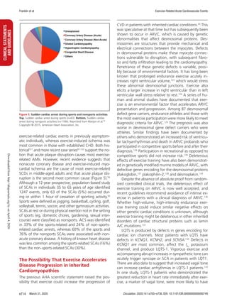
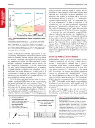
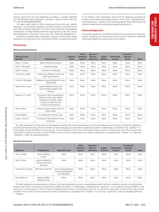
Figure 8. Conceptual overview of dose-response association between physical activity volume and cardiovascular health outcomes.
Conceptual overview of dose-response association between physical activity volume and cardiovascular health outcomes in line with (A) the current dogma and
(B) an alternative hypothesis. There is currently no compelling evidence to reject the curvilinear association (A) between exercise volumes and cardiovascular health
outcomes, with atrial fibrillation as a possible exception. Adapted from Eijsvogels et al.37
Copyright © 2018, The Authors. This article is distributed under the terms
of the Creative Commons Attribution 4.0 International License (http://creativecommons.org/licenses/by/4.0/), which permits unrestricted use, distribution, and
reproduction in any medium, provided you give appropriate credit to the original author(s) and the source, provide a link to the Creative Commons license, and
indicate if changes were made.
Downloadedfromhttp://ahajournals.orgbyonApril4,2020](https://image.slidesharecdn.com/circulation-200407184256/85/Circulation-17-320.jpg)
![March 31, 2020 Circulation. 2020;141:e705–e736. DOI: 10.1161/CIR.0000000000000749e722
CLINICALSTATEMENTS
ANDGUIDELINES Franklin et al Exercise-Related Acute Cardiovascular Events
and venous thromboembolism in women performing
strenuous activities daily (RR, 0.96 [95% CI, 0.89−1.04]
and RR, 1.08 [95% CI, 0.99−1.17], respectively) com-
pared with women performing 2 to 3 sessions of stren-
uous activities per week (RR, 0.81 [95% CI, 0.78−0.84]
and RR, 0.83 [95% CI, 0.79−0.87], respectively). The
higher prevalence of smoking (25.6% versus 13.3%)
and lower socioeconomic status (20.1% versus 13.1%)
among daily strenuous exercisers compared with con-
ventional exercisers might explain this finding in part
and suggests that daily exercisers had a different life-
style than the typical endurance athlete. In aggregate,
there is currently no evidence to reject the curvilinear
association (Figure 8A) between exercise volumes and
cardiovascular health outcomes, with AF as a possible
exception.
The health benefits of long-term, high-volume,
high-intensity exercise training are supported by data
comparing life expectancy in elite athletes with con-
trol populations. Kettunen et al250
reported that endur-
ance athletes (n=437; HR, 0.70 [95% CI, 0.61−0.79])
and team sports athletes (n=1046; HR, 0.80 [95% CI,
0.72−0.89]) had a lower all-cause mortality rate than
control subjects (n=1712), whereas no difference was
found in life expectancy between power sports ath-
letes (n=941; HR, 0.93 [95% CI, 0.85−1.03]), includ-
ing power lifters, short-distance runners, and jumpers,
and control subjects, in part because of a high number
of non-natural causes of death (ie, suicide/homicide)
in the former. Similar data were found in a meta-
analysis of cohort studies among elite athletes. The
standardized mortality ratio was significantly lower in
elite athletes (n=42 807) for all-cause mortality (stan-
dardized mortality ratio, 0.67 [95% CI, 0.55−0.81]),
cardiovascular mortality (standardized mortality ratio,
0.73 [95% CI, 0.65−0.82]), and cancer mortality (stan-
dardized mortality ratio, 0.60 [95% CI, 0.38−0.94]).251
On average, elite athletes lived 3 to 6 years longer
than the general population.250,252,253
An important
limitation of these observational data is the inability
to differentiate cause and effect, because some traits
of elite athletes (eg, genetics, socioeconomic status,
other lifestyle habits) unrelated to PA can also affect
longevity. A Finnish study sought to minimize biologi-
cal and behavioral confounders by comparing life ex-
pectancy between Finnish elite athletes (n=900) and
their brothers (n=900).254
Elite athletes had a more ac-
tive lifestyle, smoked less, and lived ≈3 years longer
than their brothers. These data suggest that both exer-
cise training and a healthy lifestyle contributed to the
athletes’ increased longevity.
Strenuous Exercise in Cardiac Patients
Supervised exercise training and habitual PA are a class
1 recommendation for cardiac patients255–257
because
there is strong evidence that higher CRF258
and high-
er PA volumes259
are associated with a lower risk for
adverse outcomes. Some have suggested using HIIT
with cardiac patients,260
because HIIT elicits greater
increases in fitness than moderate-intensity continu-
ous training (MICT) in healthy young to middle-aged
adults.261
There are conflicting reports on the benefits
of HIIT versus MICT in cardiac rehabilitation. A me-
ta-analysis including CHD patients engaging in HIIT
(n=218) or MICT (n=254) found a 1.78 mL·kg−1
·min−1
(95% CI, 0.45–3.1 mL·kg−1
·min−1
) greater increase
in CRF with HIIT.262
Other meta-analyses including
Table 4. All-Cause Mortality (Unshaded Columns) and Incident Cardiovascular Diseases (Shaded Columns) Between the Optimal and Most Active
Groups From Epidemiological Studies
Study
Arem et al242
Lear et al245
Lee et al243
Schnohr et al246
Wen et al38
Armstrong et al82
Maessen et al247
Sample size
Total cohort 661 137 (100%) 130 843 (100%) 55 137 (100%) 1252 (100%) 416 175 (100%) 1 094 327 (100%) 12 440 (100%)
Reference group 52 848 (8%) N/A* 42 121 (76%) 394 (31%) 226 493 (54%) 516 035 (47%) 417 (4%)
Optimal group 124 446 (18.8%) 94 893 (72%) 2584 (5%) 570 (46%) 20 390 (5%)† 34 967 (3%) 2127 (19%)
Most active group 4077 (0.6%) 3597 (3%) 2570 (5%) 36 (3%) 20 390 (5%)† 34 947 (3%) 2130 (19%)
Exercise type and volume MVPA Recreational PA Running time Jogging MVPA Strenuous activity MVPA
Reference group 0 MET-h/wk N/A* 0 min/wk Nonjoggers 0 MET-h/wk 0 times/wk 0 MET-h/wk
Optimal group 22.5–40 MET-h/wk 0–10 MET-h/wk 51–80 min/wk Light jogger 41±14 MET-h/wk† 4–6 times/wk 12.8–18.2 MET-h/wk
Most active group ≥75 MET-h/wk ≥50 MET-h/wk ≥176 min/wk Strenuous jogger 41±14 MET-h/wk† 7 times/wk 29.5 MET-h/wk
Health risks HR (95% CI) HR (95% CI) HR (95% CI) HR (95% CI) HR (95% CI) RR (95% CI) OR (95% CI)
Optimal group 0.61 (0.59−0.62) 0.89 (0.82–0.95) 0.67 (0.55–0.80) 0.22 (0.10−0.47) 0.65 (0.60−0.70)† 0.80 (0.76−0.85) 0.31 (0.20−0.48)
Most active group 0.69 (0.59−0.78) 0.98 (0.91–1.06) 0.77 (0.63–0.92) 1.97 (0.48−8.14) 0.65 (0.60−0.70)† 0.89 (0.84−0.93) 0.43 (0.28−0.65)
HR indicates hazard ratio; MET, metabolic equivalents; MVPA, moderate to vigorous intensity physical activity; N/A, not applicable; PA, physical activity; OR, odds
ratio; and RR, relative risk.
*A cubic spline regression analyses was performed; thus, no dedicated reference group was created.
†The most active group was the optimal group in the study of Wen et al38
; thus, presented data are from the same subgroup.
Downloadedfromhttp://ahajournals.orgbyonApril4,2020](https://image.slidesharecdn.com/circulation-200407184256/85/Circulation-18-320.jpg)
![Franklin et al Exercise-Related Acute Cardiovascular Events
Circulation. 2020;141:e705–e736. DOI: 10.1161/CIR.0000000000000749 March 31, 2020 e723
CLINICALSTATEMENTS
ANDGUIDELINES
594 CHD patients263
and 411 patients with heart
failure and reduced ejection fraction264
found a 1.3
mL·kg−1
·min−1
(95% CI, 0.6–1.9 mL·kg−1
·min−1
) and
a 1.4 mL·kg−1
·min−1
(95% CI, 0.1–2.6 mL·kg−1
·min−1
)
greater increase in CRF with HIIT versus MICT for
both groups, respectively, but these effects disap-
peared after HIIT and MICT were matched for energy
expenditure.263,264
Furthermore, MICT was associated
with greater reductions in body mass (−0.48 kg [95%
CI, 0.15–0.81 kg]) and resting heart rate (−1.8 bpm
[95% CI, 0.71−2.89 bpm) compared with HIIT,261
and
there were no differences in quality of life outcomes
between the training modalities.263,264
These findings
suggest that the exercise-induced health benefits of
HIIT and MICT in cardiac patients are comparable.
A major concern in using HIIT in cardiac patients is
the increased risk for acute coronary events.265
Data
on the safety of HIIT in clinical populations are scarce.
A systematic review reported an adverse event rate of
8% using HIIT in patients with cardiometabolic diseas-
es.266
Adverse responses included vasovagal reaction,
nausea, ventricular bigeminy, atrial tachycardia, tran-
sient cerebral ischemia, and myocardial ischemia. An-
other study compared the cardiovascular event rates
during HIIT and MICT in 4846 coronary patients par-
ticipating in cardiac rehabilitation.267
The absolute risk
was low for HIIT (1 event per 23 182 patient-hours)
and MICT (1 event per 129 456 patient-hours) but
5.6 times higher in the HIIT group. Although HIIT is a
purported time-saving alternative to MICT, additional
long-term studies assessing the safety, compliance,
and morbidity and mortality after HIIT are needed be-
fore it can be widely adopted in patients with known
or suspected CAD, especially in unsupervised, non-
medical settings.268
Several studies have explored the effects of high-
intensity or high-volume PA on all-cause and cardio-
vascular mortality in cardiac patients outside of formal
cardiac rehabilitation programs. Williams and Thomp-
son269
reported a gradual risk reduction (−15% per
MET-h/d) of cardiovascular mortality in heart attack sur-
vivors (n=2377) running or walking up to 7.2 MET-h/d.
However, patients running ≥7.2 MET-h/d had a mortal-
ity risk similar to inactive patients (HR, 0.88 [95% CI,
0.45−1.58]). Other studies have also reported a partial
loss of the health benefits of PA among the most ac-
tive cardiac patients. Keteyian et al270
found the larg-
est risk reduction for major adverse cardiovascular
outcomes among patients with heart failure (n=959)
exercising 3 to 5 MET-h/wk (−37%), but no risk re-
duction in patients performing ≥7 MET-h/wk. Wan-
namethee et al271
reported a lower mortality risk for
patients with CHD (n=772) performing light (RR, 0.42
[95% CI, 0.25−0.71]) or moderate (RR, 0.47 [95% CI,
0.24−0.92]) activities but a nonsignificant reduction in
events in patients performing vigorous activities (RR,
0.63 [95% CI, 0.39−1.03]). Mons et al272
demonstrated
that patients with CHD (n=1038) who performed vig-
orous exercise 2 to 4 times weekly had the lowest all-
cause mortality (7.6 per 1000 person-years), but that
patients performing either no exercise (HR, 3.81 [95%
CI, 2.17−6.70]) or 7 sessions/week (HR, 1.77 [95%
CI, 0.90−3.47]) had an increased mortality risk. These
findings suggest a U-shaped relationship between exer-
cise volumes and health outcomes in cardiac patients;
however, other studies suggest a curvilinear relation-
ship. Stewart et al83
demonstrated a graded risk reduc-
tion for all-cause mortality with increasing PA volumes
among a large sample of CHD patients (n=15 486). The
largest risk reductions were observed in patients per-
forming any vigorous-intensity PA, irrespective of the
PA volume. Similarly, Moholdt et al273
found the lowest
mortality risk in the most active (≥4 sessions/wk) CHD
patients (n=3504; HR, 0.77 [95% CI, 0.66−0.89]). The
conflicting outcomes on the association between high-
intensity, high-volume PA and all-cause and cardiovas-
cular mortality warrant further exploration. Neverthe-
less, all studies demonstrated that inactive patients
have the highest mortality risk. Such results emphasize
the importance of regular PA for cardiac patients, ideal-
ly via traditional medically supervised center- or home-
based cardiac rehabilitation programs,274
as well as the
importance of prescribing exercise volumes according
to current AHA/American College of Cardiology recom-
mendations (Table 5).
Population Attributable Risk of Excessive
Exercise/PA
The population attributable fraction is the proportion
(fraction) of all cases in the population that can be at-
tributed to an exposure, whereas the population at-
tributable risk is the proportion of the incidence of a
disease-related event in the population (exposed and
unexposed) that is attributable to exposure. Nawrot et
al278
identified studies of specific triggers of nonfatal MI,
including vigorous physical exertion (≥6 METs; 6 reports,
n=5208), to calculate the corresponding population at-
tributable fractions. Physical exertion was associated
with increased odds of AMI (OR, 4.25 [95% CI, 3.17–
5.68]), with a population attributable fraction of 6.16%
(95% CI, 4.20%–8.64%). In INTERHEART (n=12 461), a
case-control study of first AMI in 52 countries, investi-
gators used a case-crossover approach to estimate ORs
for AMI occurring within 1 hour of triggers.63
Popula-
tion attributable risk was calculated from the propor-
tion of participants with exposure to physical exertion in
the case and control periods. Any PA in the case period
was associated with an OR of 2.31 (99% CI, 1.96–2.72),
with a population attributable risk of 7.7% (99% CI,
6.3%–8.8%). However, moderate to strenuous exercise
was associated with a population attributable risk of
Downloadedfromhttp://ahajournals.orgbyonApril4,2020](https://image.slidesharecdn.com/circulation-200407184256/85/Circulation-19-320.jpg)
![Franklin et al Exercise-Related Acute Cardiovascular Events
Circulation. 2020;141:e705–e736. DOI: 10.1161/CIR.0000000000000749 March 31, 2020 e725
CLINICALSTATEMENTS
ANDGUIDELINES
regimens. Because the least active, least fit individu-
als are at greatest risk for exercise-related acute car-
diac events, novice exercisers should follow a gradual
increase in exercise intensity to increase CRF without
greatly increasing their cardiovascular risk by starting
with vigorous exercise (Figure 4).41,94
Although most
cardiac patients initiate exercise-based rehabilitation
programs at ≈2 to 3 METs, uptitration of training in-
tensities over time is often suboptimal, especially when
direct early outpatient medical supervision and continu-
ous electrocardiographic monitoring have ceased.279
Such patients should be counseled to gradually increase
walking speed or grade over time to a moderate inten-
sity (40%–59% functional capacity or 3.0–5.9 METs),
provided they remain asymptomatic136
This approach is
prudent because these intensities are below the vigor-
ous PA level (≥60% functional capacity or ≥6 METs) that
is commonly associated with the triggering of exercise-
related acute cardiovascular events.41
Patients should be counseled to include a warm-
up and cool-down period during exercise training
sessions to reduce the likelihood of inducing cardiac
ischemia with sudden, intense physical effort280,281
and
to avoid the decrease in central blood volume that can
occur with abrupt cessation of PA. Previously inac-
tive patients with or without known CVD should also
be advised to avoid unaccustomed, vigorous physical
exertion and highly strenuous activities (eg, racquet
sports,95
snow removal106
), to recognize potential
exertion-related warning signs and symptoms (eg,
chest pain or pressure, lightheadedness, palpitations
or arrhythmias) that require cessation of exercise and
medical evaluation, and to adapt the exercise to the
environment. Because exercise in hot, humid environ-
ments requires an increased heart rate response to
handle the associated thermal load,282
patients should
be advised to reduce the intensity of exercise under
hot, humid conditions. Increased altitude reduces
oxygen availability and further augments the cardio-
respiratory and hemodynamic responses to a given
submaximal work rate, thereby increasing cardiac de-
mands. Accordingly, individuals exercising at altitudes
of 1500 m should limit the intensity of their exercise
until acclimated.101,137,283
Patients with CVD who are
interested in participating in competitive sports should
be evaluated and advised in accordance with the AHA/
American College of Cardiology eligibility and disqual-
ification recommendations for competitive athletes
with cardiovascular abnormalities.284–286
Prophylactic Use of Cardioprotective
Medications Before Exercise
Although some authors have suggested that individu-
als at risk for CAD may benefit from taking aspirin
or β-blockers shortly before competitive exercise,287–289
there are no definitive data that these medications
prevent exercise-related acute cardiac events. The
rationale for low-dose, uncoated aspirin to reduce
the elevated, transient risk for exertion-related ath-
erothrombosis is based on aspirin’s ability to inhibit
epinephrine-induced platelet aggregation, the obser-
vation that distance runners often show elevated in-
flammatory and hemostatic markers during races,290–292
the finding that aspirin discontinuation is associated
with an increase in acute cardiovascular events,293
and
the fact that aspirin use would preemptively provide
athletes with the only medication with a class 1A rec-
ommendation for prehospital administration to treat
ACS.287,288
Nevertheless, in the TIMI II study (Throm-
bolysis in Myocardial Infarction phase II), aspirin use
did not significantly reduce the likelihood of AMI dur-
ing physical exertion.289
Similarly, in the Myocardial In-
farction Onset Study, aspirin use did not alter the RR
of triggering AMI by heavy physical exertion.41
In the
RACER study, Kim et al80
also concluded that taking
aspirin before endurance running events would have
limited efficacy, because acute plaque rupture and
thrombosis were not found in any of the 5 runners
who were discovered to have died with CHD.
Taking short-acting β-blockers before strenuous ex-
ercise has also been suggested,294
despite their potential
to reduce exercise tolerance.295–297
Tofler et al294
report-
ed that peak and average heart rates during standard-
ized bouts of physical exertion were significantly lower
30 minutes after ingestion of a β-blocker and aspirin
than during a control period. Similarly, Kokkinos et al298
concluded that β-blocker therapy could protect against
excessive and repetitive elevations in blood pressure
in hypertensive patients engaged in vigorous activities
such as basketball, tennis, and racquetball. Analysis of
the TIMI II database suggested that β-blockers provide
the most compelling evidence for protection against
physical exertion.289
In contrast, the Myocardial Infarc-
tion Onset Study found that β-blockers did not signifi-
cantly reduce the likelihood of AMI with heavy physical
exertion, although the RR was slightly lower in users
(RR, 4.2 [95% CI, 1.8–9.9]) than nonusers (RR, 6.2
[95% CI, 4.7–8.2]).41
Collectively, these data and more recent reports63
suggest there is insufficient evidence to recommend the
routine use of either aspirin or β-blockers before strenu-
ous PA or sports participation. Decisions to recommend
these prophylactic agents requires assessment of the in-
dividual patient’s risk and a discussion with the patient
of potential risks and benefits.
CONCLUSIONS AND PERSPECTIVES
There is abundant evidence that the amount of ha-
bitual PA and the level of CRF are inversely related to
the risk of cardiovascular morbidity and mortality.299
Downloadedfromhttp://ahajournals.orgbyonApril4,2020](https://image.slidesharecdn.com/circulation-200407184256/85/Circulation-21-320.jpg)

![Franklin et al Exercise-Related Acute Cardiovascular Events
Circulation. 2020;141:e705–e736. DOI: 10.1161/CIR.0000000000000749 March 31, 2020 e729
CLINICALSTATEMENTS
ANDGUIDELINES
mechanisms, and implications. J Am Coll Cardiol. 2010;56:169–176. doi:
10.1016/j.jacc.2010.03.037
34. Aengevaeren VL, Mosterd A, Braber TL, Prakken NHJ, Doevendans PA,
Grobbee DE, Thompson PD, Eijsvogels TMH, Velthuis BK. Relationship be-
tween lifelong exercise volume and coronary atherosclerosis in athletes. Circu-
lation. 2017;136:138–148. doi: 10.1161/CIRCULATIONAHA.117.027834
35. Merghani A, Maestrini V, Rosmini S, Cox AT, Dhutia H,
Bastiaenan R, David S, Yeo TJ, Narain R, Malhotra A, et al. Prevalence
of subclinical coronary artery disease in masters endurance athletes with
a low atherosclerotic risk profile. Circulation. 2017;136:126–137. doi:
10.1161/CIRCULATIONAHA.116.026964
36. O’Keefe JH, Franklin B, Lavie CJ. Exercising for health and longevity vs
peak performance: different regimens for different goals. Mayo Clin Proc.
2014;89:1171–1175. doi: 10.1016/j.mayocp.2014.07.007
37. Eijsvogels TMH, Thompson PD, Franklin BA. The “extreme exercise hypoth-
esis”: recent findings and cardiovascular health implications. Curr Treat
Options Cardiovasc Med. 2018;20:84. doi: 10.1007/s11936-018-0674-3
38. Wen CP, Wai JP, Tsai MK, Yang YC, Cheng TY, Lee MC, Chan HT, Tsao CK,
Tsai SP, Wu X. Minimum amount of physical activity for reduced mor-
tality and extended life expectancy: a prospective cohort study. Lancet.
2011;378:1244–1253. doi: 10.1016/S0140-6736(11)60749-6
39. Swain DP. Moderate or vigorous intensity exercise: which is bet-
ter for improving aerobic fitness? Prev Cardiol. 2005;8:55–58. doi:
10.1111/j.1520-037x.2005.02791.x
40. Swain DP, Franklin BA. Comparison of cardioprotective benefits of
vigorous versus moderate intensity aerobic exercise. Am J Cardiol.
2006;97:141–147. doi: 10.1016/j.amjcard.2005.07.130
41. Mittleman MA, Maclure M, Tofler GH, Sherwood JB, Goldberg RJ, Muller JE;
Determinants of Myocardial Infarction Onset Study Investigators. Trigger-
ing of acute myocardial infarction by heavy physical exertion: protection
against triggering by regular exertion. N Engl J Med. 1993;329:1677–
1683. doi: 10.1056/NEJM199312023292301
42. Franklin BA, Billecke S. Putting the benefits and risks of aerobic ex-
ercise in perspective. Curr Sports Med Rep. 2012;11:201–208. doi:
10.1249/JSR.0b013e31825dabd4
43. Siscovick DS, Weiss NS, Fletcher RH, Lasky T. The incidence of primary
cardiac arrest during vigorous exercise. N Engl J Med. 1984;311:874–877.
doi: 10.1056/NEJM198410043111402
44. Lemaitre RN, Siscovick DS, Raghunathan TE, Weinmann S, Arbogast P,
Lin DY. Leisure-time physical activity and the risk of primary cardiac arrest.
Arch Intern Med. 1999;159:686–690. doi: 10.1001/archinte.159.7.686
45. Whang W, Manson JE, Hu FB, Chae CU, Rexrode KM,
Willett WC, Stampfer MJ, Albert CM. Physical exertion, exercise, and
sudden cardiac death in women. JAMA. 2006;295:1399–1403. doi:
10.1001/jama.295.12.1399
46. Manson JE, Greenland P, LaCroix AZ, Stefanick ML, Mouton CP, Oberman A,
Perri MG, Sheps DS, Pettinger MB, Siscovick DS. Walking compared with
vigorous exercise for the prevention of cardiovascular events in women. N
Engl J Med. 2002;347:716–725. doi: 10.1056/NEJMoa021067
47. Kannel WB. Exercise and sudden death. JAMA. 1982;248:3143–3144.
48. Albert CM, Mittleman MA, Chae CU, Lee IM, Hennekens CH, Manson JE.
Triggering of sudden death from cardiac causes by vigorous exertion. N
Engl J Med. 2000;343:1355–1361. doi: 10.1056/NEJM200011093431902
49. Berdowski J, de Beus MF, Blom M, Bardai A, Bots ML, Doevendans PA,
Grobbee DE, Tan HL, Tijssen JG, Koster RW, et al. Exercise-related out-of-
hospital cardiac arrest in the general population: incidence and prognosis.
Eur Heart J. 2013;34:3616–3623. doi: 10.1093/eurheartj/eht401
50. Marijon E, Uy-Evanado A, Reinier K, Teodorescu C, Narayanan K,
Jouven X, Gunson K, Jui J, Chugh SS. Sudden cardiac arrest during
sports activity in middle age. Circulation. 2015;131:1384–1391. doi:
10.1161/CIRCULATIONAHA.114.011988
51. Marijon E, Tafflet M, Celermajer DS, Dumas F, Perier MC, Mustafic H,
Toussaint JF, Desnos M, Rieu M, Benameur N, et al. Sports-related sud-
den death in the general population. Circulation. 2011;124:672–681. doi:
10.1161/CIRCULATIONAHA.110.008979
52. Toukola T, Junttila MJ, Holmström LTA, Haukilahti MA, Tikkanen JT,
Terho H, Kenttä TV, Aro AL, Anttonen O, Kerola T, et al. Fragmented QRS
complex as a predictor of exercise-related sudden cardiac death. J Cardio-
vasc Electrophysiol. 2018;29:55–60. doi: 10.1111/jce.13341
53. Jayaraman R, Reinier K, Nair S, Aro AL, Uy-Evanado A, Rusinaru C,
Stecker EC, Gunson K, Jui J, Chugh SS. Risk factors of sudden cardiac death
in the young: multiple-year community-wide assessment. Circulation.
2018;137:1561–1570. doi: 10.1161/CIRCULATIONAHA.117.031262
54. Pilmer CM, Porter B, Kirsh JA, Hicks AL, Gledhill N, Jamnik V, Faught BE,
Hildebrandt D, McCartney N, Gow RM, et al. Scope and nature of sudden
cardiac death before age 40 in Ontario: a report from the cardiac death
advisory committee of the office of the chief coroner. Heart Rhythm.
2013;10:517–523. doi: 10.1016/j.hrthm.2012.12.003
55. Maron BJ, Doerer JJ, Haas TS, Tierney DM, Mueller FO. Sudden
deaths in young competitive athletes: analysis of 1866 deaths in the
United States, 1980-2006. Circulation. 2009;119:1085–1092. doi:
10.1161/CIRCULATIONAHA.108.804617
56. Finocchiaro G, Papadakis M, Robertus JL, Dhutia H, Steriotis AK, Tome M,
Mellor G, Merghani A, Malhotra A, Behr E, et al. Etiology of sudden death
in sports: insights from a United Kingdom regional registry. J Am Coll Car-
diol. 2016;67:2108–2115. doi: 10.1016/j.jacc.2016.02.062
57. Maron BJ, Haas TS, Duncanson ER, Garberich RF, Baker AM,
Mackey-Bojack S. Comparison of the frequency of sudden cardiovascu-
lar deaths in young competitive athletes versus nonathletes: should we
really screen only athletes? Am J Cardiol. 2016;117:1339–1341. doi:
10.1016/j.amjcard.2016.01.026
58. Asatryan B, Vital C, Kellerhals C, Medeiros-Domingo A, Gräni C,
Trachsel LD, Schmied CM, Saguner AM, Eser P, Herzig D, et al. Sports-
related sudden cardiac deaths in the young population of Switzerland.
PLoS One. 2017;12:e0174434. doi: 10.1371/journal.pone.0174434
59. Burke AP, Farb A, Malcom GT, Liang Y, Smialek JE, Virmani R. Plaque rup-
ture and sudden death related to exertion in men with coronary artery
disease. JAMA. 1999;281:921–926. doi: 10.1001/jama.281.10.921
60. Willich SN, Lewis M, Löwel H, Arntz HR, Schubert F, Schröder R; Triggers
and Mechanisms of Myocardial Infarction Study Group. Physical exertion
as a trigger of acute myocardial infarction. N Engl J Med. 1993;329:1684–
1690. doi: 10.1056/NEJM199312023292302
61. Giri S, Thompson PD, Kiernan FJ, Clive J, Fram DB, Mitchel JF,
Hirst JA, McKay RG, Waters DD. Clinical and angiographic characteris-
tics of exertion-related acute myocardial infarction [published correction
appears in JAMA. 1999;282:2124]. JAMA. 1999;282:1731–1736. doi:
10.1001/jama.282.18.1731
62. Baylin A, Hernandez-Diaz S, Siles X, Kabagambe EK, Campos H. Triggers
of nonfatal myocardial infarction in Costa Rica: heavy physical exertion,
sexual activity, and infection. Ann Epidemiol. 2007;17:112–118. doi:
10.1016/j.annepidem.2006.05.004
63. Smyth A, O’Donnell M, Lamelas P, Teo K, Rangarajan S,
Yusuf S; on behalf of the INTERHEART Investigators. Physical activity
and anger or emotional upset as triggers of acute myocardial infarc-
tion: the INTERHEART study. Circulation. 2016;134:1059–1067. doi:
10.1161/CIRCULATIONAHA.116.023142
64. Schachner T, Fischler N, Dumfarth J, Bonaros N, Krapf C,
Schobersberger W, Grimm M. Aortic dissection type A in alpine skiers.
Biomed Res Int. 2013;2013:192459. doi: 10.1155/2013/192459
65. Hatzaras I, Tranquilli M, Coady M, Barrett PM, Bible J, Elefteriades JA.
Weight lifting and aortic dissection: more evidence for a connection. Car-
diology. 2007;107:103–106. doi: 10.1159/000094530
66. Elefteriades JA, Hatzaras I, Tranquilli MA, Elefteriades AJ, Stout R,
Shaw RK, Silverman D, Barash P. Weight lifting and rupture of silent aortic
aneurysms. JAMA. 2003;290:2803. doi: 10.1001/jama.290.21.2803
67. Maron BJ, Haas TS, Ahluwalia A, Murphy CJ, Garberich RF. Demographics
and epidemiology of sudden deaths in young competitive athletes: from
the United States National Registry. Am J Med. 2016;129:1170–1177.
doi: 10.1016/j.amjmed.2016.02.031
68. Selb Semerl J, Kenda MF. Out of hospital sudden cardiac death among
physically active and inactive married persons younger than 65 years in
Slovenia. J Clin Basic Cardiol. 2003;6:63−67.
69. Thompson PD, Funk EJ, Carleton RA, Sturner WQ. Incidence of death
during jogging in Rhode Island from 1975 through 1980. JAMA.
1982;247:2535–2538.
70. Hallqvist J, Möller J, Ahlbom A, Diderichsen F, Reuterwall C, de Faire U.
Does heavy physical exertion trigger myocardial infarction? A case-cross-
over analysis nested in a population-based case-referent study. Am J Epi-
demiol. 2000;151:459–467. doi: 10.1093/oxfordjournals.aje.a010231
71. von Klot S, Mittleman MA, Dockery DW, Heier M, Meisinger C,
Hörmann A, Wichmann HE, Peters A. Intensity of physical exertion and
triggering of myocardial infarction: a case-crossover study. Eur Heart J.
2008;29:1881–1888. doi: 10.1093/eurheartj/ehn235
72. Dahabreh IJ, Paulus JK. Association of episodic physical and sexual activ-
ity with triggering of acute cardiac events: systematic review and meta-
analysis. JAMA. 2011;305:1225–1233. doi: 10.1001/jama.2011.336
73. Goodman JM, Burr JF, Banks L, Thomas SG. The acute risks of ex-
ercise in apparently healthy adults and relevance for prevention
of cardiovascular events. Can J Cardiol. 2016;32:523–532. doi:
10.1016/j.cjca.2016.01.019
Downloadedfromhttp://ahajournals.orgbyonApril4,2020](https://image.slidesharecdn.com/circulation-200407184256/85/Circulation-25-320.jpg)
![Franklin et al Exercise-Related Acute Cardiovascular Events
Circulation. 2020;141:e705–e736. DOI: 10.1161/CIR.0000000000000749 March 31, 2020 e731
CLINICALSTATEMENTS
ANDGUIDELINES
116. Ullal AJ, Abdelfattah RS, Ashley EA, Froelicher VF. Hypertrophic cardiomy-
opathy as a cause of sudden cardiac death in the young: a meta-analysis.
Am J Med. 2016;129:486–496.e2. doi: 10.1016/j.amjmed.2015.12.027
117. Bagnall RD, Weintraub RG, Ingles J, Duflou J, Yeates L, Lam L, Davis AM,
Thompson T, Connell V, Wallace J, et al. A prospective study of sud-
den cardiac death among children and young adults. N Engl J Med.
2016;374:2441–2452. doi: 10.1056/NEJMoa1510687
118. Landry CH, Allan KS, Connelly KA, Cunningham K, Morrison LJ,
Dorian P; Rescu Investigators. Sudden cardiac arrest during participa-
tion in competitive sports. N Engl J Med. 2017;377:1943–1953. doi:
10.1056/NEJMoa1615710
119. Harmon KG, Asif IM, Maleszewski JJ, Owens DS, Prutkin JM, Salerno JC,
Zigman ML, Ellenbogen R, Rao AL, Ackerman MJ, et al. Incidence, cause,
and comparative frequency of sudden cardiac death in National Col-
legiate Athletic Association athletes: a decade in review. Circulation.
2015;132:10–19. doi: 10.1161/CIRCULATIONAHA.115.015431
120. Maron BJ. Clinical course and management of hypertrophic cardiomyop-
athy. N Engl J Med. 2018;379:655–668. doi: 10.1056/NEJMra1710575
121. Black A, Black MM, Gensini G. Exertion and acute coronary artery injury.
Angiology. 1975;26:759–783. doi: 10.1177/000331977502601101
122. Albano AJ, Thompson PD, Kapur NK. Acute coronary thrombosis in
Boston marathon runners. N Engl J Med. 2012;366:184–185. doi:
10.1056/NEJMc1111015
123. Douglas PS, O’Toole ML, Hiller WD, Reichek N. Different effects of
prolonged exercise on the right and left ventricles. J Am Coll Cardiol.
1990;15:64–69. doi: 10.1016/0735-1097(90)90176-p
124. La Gerche A, Heidbüchel H, Burns AT, Mooney DJ, Taylor AJ,
Pfluger HB, Inder WJ, Macisaac AI, Prior DL. Disproportionate exercise
load and remodeling of the athlete’s right ventricle. Med Sci Sports Exerc.
2011;43:974–981. doi: 10.1249/MSS.0b013e31820607a3
125. James CA, Bhonsale A, Tichnell C, Murray B, Russell SD, Tandri H,
Tedford RJ, Judge DP, Calkins H. Exercise increases age-related pene-
trance and arrhythmic risk in arrhythmogenic right ventricular dysplasia/
cardiomyopathy-associated desmosomal mutation carriers. J Am Coll
Cardiol. 2013;62:1290–1297. doi: 10.1016/j.jacc.2013.06.033
126. RuwaldAC,MarcusF,EstesNA3rd,LinkM,McNittS,PolonskyB,CalkinsH,
Towbin JA, Moss AJ, Zareba W. Association of competitive and recreation-
al sport participation with cardiac events in patients with arrhythmogenic
right ventricular cardiomyopathy: results from the North American mul-
tidisciplinary study of arrhythmogenic right ventricular cardiomyopathy.
Eur Heart J. 2015;36:1735–1743. doi: 10.1093/eurheartj/ehv110
127. Kirchhof P, Fabritz L, Zwiener M, Witt H, Schäfers M, Zellerhoff S, Paul M,
Athai T, Hiller KH, Baba HA, et al. Age- and training-dependent develop-
ment of arrhythmogenic right ventricular cardiomyopathy in heterozy-
gous plakoglobin-deficient mice. Circulation. 2006;114:1799–1806. doi:
10.1161/CIRCULATIONAHA.106.624502
128. Cruz FM, Sanz-Rosa D, Roche-Molina M, García-Prieto J, García-Ruiz JM,
Pizarro G, Jiménez-Borreguero LJ, Torres M, Bernad A, Ruíz-Cabello J,
et al. Exercise triggers ARVC phenotype in mice expressing a disease-
causing mutated version of human plakophilin-2. J Am Coll Cardiol.
2015;65:1438–1450. doi: 10.1016/j.jacc.2015.01.045
129. Martherus R, Jain R, Takagi K, Mendsaikhan U, Turdi S, Osinska H,
James JF, Kramer K, Purevjav E, Towbin JA. Accelerated cardiac remodel-
ing in desmoplakin transgenic mice in response to endurance exercise is
associated with perturbed Wnt/β-catenin signaling. Am J Physiol Heart
Circ Physiol. 2016;310:H174–H187. doi: 10.1152/ajpheart.00295.2015
130. Al-Khatib SM, Stevenson WG, Ackerman MJ, Bryant WJ, Callans DJ,
Curtis AB, Deal BJ, Dickfeld T, Field ME, Fonarow GC, et al. 2017 AHA/
ACC/HRS guideline for management of patients with ventricular ar-
rhythmias and the prevention of sudden cardiac death: a report of the
American College of Cardiology Foundation/American Heart Association
Task Force on Clinical Practice Guidelines and the Heart Rhythm Society
[published correction appears in Circulation. 2018;138:e419–e420]. Cir-
culation. 2018;138:e272–e391. doi: 10.1161/CIR.0000000000000549
131. Pasotti M, Klersy C, Pilotto A, Marziliano N, Rapezzi C, Serio A,
Mannarino S, Gambarin F, Favalli V, Grasso M, et al. Long-term outcome
and risk stratification in dilated cardiolaminopathies. J Am Coll Cardiol.
2008;52:1250–1260. doi: 10.1016/j.jacc.2008.06.044
132. Schwartz PJ, Crotti L, Insolia R. Long-QT syndrome: from genetics to man-
agement [published correction appears in Circ Arrhythm Electrophysiol.
2012;5:e119–e120]. Circ Arrhythm Electrophysiol. 2012;5:868–877. doi:
10.1161/CIRCEP.111.962019
133. Schwartz PJ, Vanoli E, Crotti L, Spazzolini C, Ferrandi C, Goosen A,
Hedley P, Heradien M, Bacchini S, Turco A, et al. Neural control of heart
rate is an arrhythmia risk modifier in long QT syndrome. J Am Coll Car-
diol. 2008;51:920–929. doi: 10.1016/j.jacc.2007.09.069
134. Crotti L, Spazzolini C, Porretta AP, Dagradi F, Taravelli E, Petracci B,
Vicentini A, Pedrazzini M, La Rovere MT, Vanoli E, et al. Vagal reflexes fol-
lowing an exercise stress test: a simple clinical tool for gene-specific risk
stratification in the long QT syndrome. J Am Coll Cardiol. 2012;60:2515–
2524. doi: 10.1016/j.jacc.2012.08.1009
135. Whitfield GP, Pettee Gabriel KK, Rahbar MH, Kohl HW 3rd. Application
of the American Heart Association/American College of Sports Medicine
Adult Preparticipation Screening Checklist to a nationally representative
sample of US adults aged ≥40 years from the National Health and Nu-
trition Examination Survey 2001 to 2004. Circulation. 2014;129:1113–
1120. doi: 10.1161/CIRCULATIONAHA.113.004160
136. Riebe D, Franklin BA, Thompson PD, Garber CE, Whitfield GP, Magal M,
Pescatello LS. Updating ACSM’s recommendations for exercise prepartici-
pation health screening [published correction appears in Med Sci Sports
Exerc. 2016;48:579]. Med Sci Sports Exerc. 2015;47:2473–2479. doi:
10.1249/MSS.0000000000000664
137. Riebe D, Ehrman JK, Liguori G, Magal M, eds. ACSM’s Guidelines for
Exercise Testing and Prescription. 10th ed. Philadelphia, PA: Lippincott
Williams Wilkins; 2017.
138. Armstrong M, Paternostro-Bayles M, Conroy MB, Franklin BA,
Richardson C, Kriska A. Preparticipation screening prior to physical ac-
tivity in community lifestyle interventions. Transl J Am Coll Sports Med.
2018;3:176–180. doi: 10.1249/TJX.0000000000000073
139. de Barros e Silva PG, Califf RM, Sun JL, McMurray JJ, Holman R,
Haffner S, Thomas L, Lopes RD. Chronic obstructive pulmonary disease
and cardiovascular risk: insights from the NAVIGATOR trial. Int J Cardiol.
2014;176:1126–1128.
140. Curry SJ, Krist AH, Owens DK, Barry MJ, Caughey AB, Davidson KW,
Doubeni CA, Epling JW Jr, Kemper AR, Kubik M, et al; US Preventive
Services Task Force. Screening for cardiovascular disease risk with electro-
cardiography: US Preventive Services Task Force recommendation state-
ment. JAMA. 2018;319:2308–2314. doi: 10.1001/jama.2018.6848
141. Jonas DE, Reddy S, Middleton JC, Barclay C, Green J, Baker C,
Asher GN. Screening for cardiovascular disease risk with resting or ex-
ercise electrocardiography: evidence report and systematic review for
the US Preventive Services Task Force. JAMA. 2018;319:2315–2328. doi:
10.1001/jama.2018.6897
142. American College of Physicians. Choosing wisely initiative. http://www.
choosingwisely.org/. Accessed March 1, 2019.
143. Gill TM, DiPietro L, Krumholz HM. Role of exercise stress testing and
safety monitoring for older persons starting an exercise program. JAMA.
2000;284:342–349. doi: 10.1001/jama.284.3.342
144. Nasir K, Rubin J, Blaha MJ, Shaw LJ, Blankstein R, Rivera JJ, Khan AN,
Berman D, Raggi P, Callister T, et al. Interplay of coronary artery calcifica-
tion and traditional risk factors for the prediction of all-cause mortality
in asymptomatic individuals. Circ Cardiovasc Imaging. 2012;5:467–473.
doi: 10.1161/CIRCIMAGING.111.964528
145. Shaw LJ, Mieres JH, Hendel RH, Boden WE, Gulati M, Veledar E,
Hachamovitch R, Arrighi JA, Merz CN, Gibbons RJ, et al; for the WOMEN
Trial Investigators. Comparative effectiveness of exercise electrocardi-
ography with or without myocardial perfusion single photon emission
computed tomography in women with suspected coronary artery dis-
ease: results from the What Is the Optimal Method for Ischemia Evalu-
ation in Women (WOMEN) trial. Circulation. 2011;124:1239–1249. doi:
10.1161/CIRCULATIONAHA.111.029660
146. Kannankeril PJ, Moore JP, Cerrone M, Priori SG, Kertesz NJ, Ro PS,
Batra AS, Kaufman ES, Fairbrother DL, Saarel EV, et al. Efficacy of fle-
cainide in the treatment of catecholaminergic polymorphic ventricular
tachycardia: a randomized clinical trial. JAMA Cardiol. 2017;2:759–766.
doi: 10.1001/jamacardio.2017.1320
147. Mascia G, Hernandez-Ojeda J, Solimene F, Brugada R, Brugada J. Bru-
gada syndrome and exercise practice: current knowledge, shortcomings
and open questions [published correction appears in Int J Sports Med.
2017;38:e2]. Int J Sports Med. 2017;38:e2. doi: 10.1055/s-0043-120577
148. Steinvil A, Chundadze T, Zeltser D, Rogowski O, Halkin A, Galily Y,
Perluk H, Viskin S. Mandatory electrocardiographic screening of athletes
to reduce their risk for sudden death: proven fact or wishful thinking? J
Am Coll Cardiol. 2011;57:1291–1296. doi: 10.1016/j.jacc.2010.10.037
149. Maron BJ, Levine BD, Washington RL, Baggish AL, Kovacs RJ, Maron MS;
on behalf of the American Heart Association Electrocardiography and
Arrhythmias Committee of the Council on Clinical Cardiology, Council
on Cardiovascular Disease in the Young, Council on Cardiovascular and
Downloadedfromhttp://ahajournals.orgbyonApril4,2020](https://image.slidesharecdn.com/circulation-200407184256/85/Circulation-27-320.jpg)
![Franklin et al Exercise-Related Acute Cardiovascular Events
March 31, 2020 Circulation. 2020;141:e705–e736. DOI: 10.1161/CIR.0000000000000749e732
CLINICALSTATEMENTS
ANDGUIDELINES
Stroke Nursing, Council on Functional Genomics and Translational Bi-
ology, and the American College of Cardiology. Eligibility and disquali-
fication recommendations for competitive athletes with cardiovascular
abnormalities: Task Force 2: preparticipation screening for cardiovascular
disease in competitive athletes: a scientific statement from the Ameri-
can Heart Association and American College of Cardiology. Circulation.
2015;132:e267–e272. doi: 10.1161/CIR.0000000000000238
150. Van Brabandt H, Desomer A, Gerkens S, Neyt M. Harms and benefits
of screening young people to prevent sudden cardiac death. BMJ.
2016;353:i1156. doi: 10.1136/bmj.i1156
151. Mosterd A. Pre-participation screening of asymptomatic athletes:
“Don’t do stupid stuff.” Neth Heart J. 2018;26:123–126. doi:
10.1007/s12471-018-1075-7
152. Drezner JA, O’Connor FG, Harmon KG, Fields KB, Asplund CA, Asif IM,
Price DE, Dimeff RJ, Bernhardt DT, Roberts WO. AMSSM position state-
ment on cardiovascular preparticipation screening in athletes: current
evidence, knowledge gaps, recommendations and future directions. Br
J Sports Med. 2017;51:153–167. doi: 10.1136/bjsports-2016-096781
153. Maron BJ, Friedman RA, Kligfield P, Levine BD, Viskin S, Chaitman BR,
Okin PM, Saul JP, Salberg L, Van Hare GF, et al; on behalf of the Ameri-
can Heart Association Council on Clinical Cardiology, Advocacy Coor-
dinating Committee, Council on Cardiovascular Disease in the Young,
Council on Cardiovascular Surgery and Anesthesia, Council on Epide-
miology and Prevention, Council on Functional Genomics and Trans-
lational Biology, Council on Quality of Care and Outcomes Research,
and American College of Cardiology. Assessment of the 12-lead ECG
as a screening test for detection of cardiovascular disease in healthy
general populations of young people (12–25 years of age): a scien-
tific statement from the American Heart Association and the Ameri-
can College of Cardiology. Circulation. 2014;130:1303–1334. doi:
10.1161/CIR.0000000000000025
154. Mont L, Pelliccia A, Sharma S, Biffi A, Borjesson M, Brugada Terradellas J,
Carré F, Guasch E, Heidbuchel H, et al. Pre-participation cardiovascular
evaluation for athletic participants to prevent sudden death: position
paper from the EHRA and the EACPR, branches of the ESC. Endorsed
by APHRS, HRS, and SOLAECE. Eur J Prev Cardiol. 2017;24:41–69. doi:
10.1177/2047487316676042
155. Lawless CE. Minnesota high school athletes 1993–2012: evidence that
American screening strategies and sideline preparedness are associ-
ated with very low rates of sudden cardiac deaths. J Am Coll Cardiol.
2013;62:1302–1303. doi: 10.1016/j.jacc.2013.06.036
156. Malhotra A, Dhutia H, Finocchiaro G, Gati S, Beasley I, Clift P, Cowie C,
Kenny A, Mayet J, Oxborough D, et al. Outcomes of cardiac screening
in adolescent soccer players. N Engl J Med. 2018;379:524–534. doi:
10.1056/NEJMoa1714719
157. BlomMT,BeesemsSG,HommaPC,ZijlstraJA,HullemanM,vanHoeijenDA,
Bardai A, Tijssen JG, Tan HL, Koster RW. Improved survival after out-of-
hospital cardiac arrest and use of automated external defibrillators [pub-
lished correction appears in Circulation. 2014;130:e436]. Circulation.
2014;130:1868–1875. doi: 10.1161/CIRCULATIONAHA.114.010905
158. Kinoshi T, Tanaka S, Sagisaka R, Hara T, Shirakawa T, Sone E, Takahashi H,
Sakurai M, Maki A, Takyu H, et al. Mobile automated external defibril-
lator response system during road races. N Engl J Med. 2018;379:488–
489. doi: 10.1056/NEJMc1803218
159. Sharma S, Merghani A, Gati S. Cardiac screening of young athletes pri-
or to participation in sports: difficulties in detecting the fatally flawed
among the fabulously fit. JAMA Intern Med. 2015;175:125–127. doi:
10.1001/jamainternmed.2014.6023
160. Pluim BM, Zwinderman AH, van der Laarse A, van der Wall EE. The ath-
lete’s heart: a meta-analysis of cardiac structure and function. Circula-
tion. 2000;101:336–344. doi: 10.1161/01.cir.101.3.336
161. Utomi V, Oxborough D, Whyte GP, Somauroo J, Sharma S, Shave R,
Atkinson G, George K. Systematic review and meta-analysis of training
mode, imaging modality and body size influences on the morphology
and function of the male athlete’s heart. Heart. 2013;99:1727–1733.
doi: 10.1136/heartjnl-2012-303465
162. Bhella PS, Hastings JL, Fujimoto N, Shibata S, Carrick-Ranson G,
Palmer MD, Boyd KN, Adams-Huet B, Levine BD. Impact of lifelong ex-
ercise “dose” on left ventricular compliance and distensibility. J Am Coll
Cardiol. 2014;64:1257–1266. doi: 10.1016/j.jacc.2014.03.062
163. Sharma S, Drezner JA, Baggish A, Papadakis M, Wilson MG, Prutkin JM,
La Gerche A, Ackerman MJ, Borjesson M, Salerno JC, et al. International
recommendations for electrocardiographic interpretation in athletes. J
Am Coll Cardiol. 2017;69:1057–1075. doi: 10.1016/j.jacc.2017.01.015
164. Eijsvogels TM, Molossi S, Lee DC, Emery MS, Thompson PD. Exercise at
the extremes: the amount of exercise to reduce cardiovascular events. J
Am Coll Cardiol. 2016;67:316–329. doi: 10.1016/j.jacc.2015.11.034
165. Eijsvogels TM, Fernandez AB, Thompson PD. Are there deleterious
cardiac effects of acute and chronic endurance exercise? Physiol Rev.
2016;96:99–125. doi: 10.1152/physrev.00029.2014
166. Levine BD. Can intensive exercise harm the heart? The benefits of competi-
tive endurance training for cardiovascular structure and function. Circula-
tion. 2014;130:987–991. doi: 10.1161/CIRCULATIONAHA.114.008142
167. La Gerche A, Heidbuchel H. Can intensive exercise harm the heart? You
can get too much of a good thing. Circulation. 2014;130:992–1002. doi:
10.1161/CIRCULATIONAHA.114.008141
168. Miyasaka Y, Barnes ME, Gersh BJ, Cha SS, Bailey KR, Abhayaratna WP,
Seward JB, Tsang TS. Secular trends in incidence of atrial fibrillation
in Olmsted County, Minnesota, 1980 to 2000, and implications on
the projections for future prevalence [published correction appears
in Circulation. 2006;114:e498]. Circulation. 2006;114:119–125. doi:
10.1161/CIRCULATIONAHA.105.595140
169. Calvo N, Ramos P, Montserrat S, Guasch E, Coll-Vinent B, Domenech M,
Bisbal F, Hevia S, Vidorreta S, Borras R, et al. Emerging risk factors and
the dose-response relationship between physical activity and lone atrial
fibrillation: a prospective case-control study. Europace. 2016;18:57–63.
doi: 10.1093/europace/euv216
170. Sorokin AV, Araujo CG, Zweibel S, Thompson PD. Atrial fibrillation in
endurance-trained athletes. Br J Sports Med. 2011;45:185–188. doi:
10.1136/bjsm.2009.057885
171. Alonso A, Krijthe BP, Aspelund T, Stepas KA, Pencina MJ, Moser CB,
Sinner MF, Sotoodehnia N, Fontes JD, Janssens AC, et al. Simple risk
model predicts incidence of atrial fibrillation in a racially and geographi-
cally diverse population: the CHARGE-AF consortium. J Am Heart Assoc.
2013;2:e000102. doi: 10.1161/JAHA.112.000102
172. Reiffel JA. Atrial fibrillation and stroke: epidemiology. Am J Med.
2014;127:e15–e16. doi: 10.1016/j.amjmed.2013.06.002
173. Ruddox V, Sandven I, Munkhaugen J, Skattebu J, Edvardsen T, Otterstad JE.
Atrial fibrillation and the risk for myocardial infarction, all-cause mortal-
ity and heart failure: a systematic review and meta-analysis. Eur J Prev
Cardiol. 2017;24:1555–1566. doi: 10.1177/2047487317715769
174. Chatterjee NA, Chae CU, Kim E, Moorthy MV, Conen D, Sandhu RK,
Cook NR, Lee IM, Albert CM. Modifiable risk factors for incident heart
failure in atrial fibrillation. JACC Heart Fail. 2017;5:552–560. doi:
10.1016/j.jchf.2017.04.004
175. Odutayo A, Wong CX, Hsiao AJ, Hopewell S, Altman DG, Emdin CA.
Atrial fibrillation and risks of cardiovascular disease, renal disease, and
death: systematic review and meta-analysis. BMJ. 2016;354:i4482. doi:
10.1136/bmj.i4482
176. Qureshi WT, Alirhayim Z, Blaha MJ, Juraschek SP, Keteyian SJ,
Brawner CA, Al-Mallah MH. Cardiorespiratory fitness and risk of incident
atrial fibrillation: results from the Henry Ford Exercise Testing (FIT) Project.
Circulation. 2015;131:1827–1834. doi: 10.1161/CIRCULATIONAHA.
114.014833
177. Faselis C, Kokkinos P, Tsimploulis A, Pittaras A, Myers J, Lavie CJ, Kyritsi F,
Lovic D, Karasik P, Moore H. Exercise capacity and atrial fibrillation risk
in veterans: a cohort study. Mayo Clin Proc. 2016;91:558–566. doi:
10.1016/j.mayocp.2016.03.002
178. Pathak RK, Elliott A, Middeldorp ME, Meredith M, Mehta AB, Mahajan R,
Hendriks JM, Twomey D, Kalman JM, Abhayaratna WP, et al. Impact
of CARDIOrespiratory FITness on arrhythmia recurrence in obese indi-
viduals with atrial fibrillation: the CARDIO-FIT study. J Am Coll Cardiol.
2015;66:985–996. doi: 10.1016/j.jacc.2015.06.488
179. Malmo V, Nes BM, Amundsen BH, Tjonna AE, Stoylen A, Rossvoll O,
Wisloff U, Loennechen JP. Aerobic interval training reduces the burden
of atrial fibrillation in the short term: a randomized trial. Circulation.
2016;133:466–473. doi: 10.1161/CIRCULATIONAHA.115.018220
180. Mozaffarian D, Furberg CD, Psaty BM, Siscovick D. Physical ac-
tivity and incidence of atrial fibrillation in older adults: the Car-
diovascular Health Study. Circulation. 2008;118:800–807. doi:
10.1161/CIRCULATIONAHA.108.785626
181. Andersen K, Farahmand B, Ahlbom A, Held C, Ljunghall S,
Michaëlsson K, Sundström J. Risk of arrhythmias in 52 755 long-distance
cross-country skiers: a cohort study. Eur Heart J. 2013;34:3624–3631.
doi: 10.1093/eurheartj/eht188
182. Aizer A, Gaziano JM, Cook NR, Manson JE, Buring JE, Albert CM. Re-
lation of vigorous exercise to risk of atrial fibrillation. Am J Cardiol.
2009;103:1572–1577. doi: 10.1016/j.amjcard.2009.01.374
Downloadedfromhttp://ahajournals.orgbyonApril4,2020](https://image.slidesharecdn.com/circulation-200407184256/85/Circulation-28-320.jpg)
![Franklin et al Exercise-Related Acute Cardiovascular Events
Circulation. 2020;141:e705–e736. DOI: 10.1161/CIR.0000000000000749 March 31, 2020 e733
CLINICALSTATEMENTS
ANDGUIDELINES
183. Abdulla J, Nielsen JR. Is the risk of atrial fibrillation higher in athletes
than in the general population? A systematic review and meta-analysis.
Europace. 2009;11:1156–1159. doi: 10.1093/europace/eup197
184. Mont L, Elosua R, Brugada J. Endurance sport practice as a risk factor
for atrial fibrillation and atrial flutter. Europace. 2009;11:11–17. doi:
10.1093/europace/eun289
185. Myrstad M, Nystad W, Graff-Iversen S, Thelle DS, Stigum H,
Aarønæs M, Ranhoff AH. Effect of years of endurance exercise on risk of
atrial fibrillation and atrial flutter. Am J Cardiol. 2014;114:1229–1233.
doi: 10.1016/j.amjcard.2014.07.047
186. Ricci C, Gervasi F, Gaeta M, Smuts CM, Schutte AE, Leitzmann MF. Physi-
cal activity volume in relation to risk of atrial fibrillation: a non-linear
meta-regression analysis. Eur J Prev Cardiol. 2018;25:857–866. doi:
10.1177/2047487318768026
187. McNamara DA, Aiad N, Howden E, Hieda M, Link MS, Palmer D,
Samels M, Everding B, Ng J, Adams-Huet B, et al. Left atrial electrome-
chanical remodeling following 2 years of high-intensity exercise training
in sedentary middle-aged adults. Circulation. 2019;139:1507–1516. doi:
10.1161/CIRCULATIONAHA.118.037615
188. Flannery MD, Kalman JM, Sanders P, La Gerche A. State of the art re-
view: atrial fibrillation in athletes. Heart Lung Circ. 2017;26:983–989.
doi: 10.1016/j.hic.2017.05.132
189. Campuzano O, Perez-Serra A, Iglesias A, Brugada R. Genetic basis of atrial
fibrillation. Genes Dis. 2016;3:257–262. doi: 10.1016/j.gendis.2016.09.003
190. Otway R, Vandenberg JI, Guo G, Varghese A, Castro ML, Liu J, Zhao J,
Bursill JA, Wyse KR, Crotty H, et al. Stretch-sensitive KCNQ1 mutation:
a link between genetic and environmental factors in the pathogen-
esis of atrial fibrillation? J Am Coll Cardiol. 2007;49:578–586. doi:
10.1016/j.jacc.2006.09.044
191. Mills JD, Moore GE, Thompson PD. The athlete’s heart. Clin Sports Med.
1997;16:725–737. doi: 10.1016/s0278-5919(05)70050-8
192. Pelliccia A, Maron BJ, Di Paolo FM, Biffi A, Quattrini FM, Pisicchio C,
Roselli A, Caselli S, Culasso F. Prevalence and clinical significance of left
atrial remodeling in competitive athletes. J Am Coll Cardiol. 2005;46:690–
696. doi: 10.1016/j.jacc.2005.04.052
193. Elosua R, Arquer A, Mont L, Sambola A, Molina L, García-Morán E,
Brugada J, Marrugat J. Sport practice and the risk of lone atrial fi-
brillation: a case-control study [published correction appears in
Int J Cardiol. 2007;123:74]. Int J Cardiol. 2006;108:332–337. doi:
10.1016/j.ijcard.2005.05.020
194. La Gerche A, Claessen G. Increased flow, dam walls, and upstream
pressure: the physiological challenges and atrial consequences of in-
tense exercise. JACC Cardiovasc Imaging. 2016;9:1389–1391. doi:
10.1016/j.jcmg.2016.06.008
195. Swanson DR. Atrial fibrillation in athletes: implicit literature-based con-
nections suggest that overtraining and subsequent inflammation may be
a contributory mechanism. Med Hypotheses. 2006;66:1085–1092. doi:
10.1016/j.mehy.2006.01.006
196. Elliott AD, Linz D, Verdicchio CV, Sanders P. Exercise and atrial fibrillation:
prevention or causation? Heart Lung Circ. 2018;27:1078–1085. doi:
10.1016/j.hlc.2018.04.296
197. Wong ND. Epidemiological studies of CHD and the evolution of
preventive cardiology. Nat Rev Cardiol. 2014;11:276–289. doi:
10.1038/nrcardio.2014.26
198. Finn AV, Nakano M, Narula J, Kolodgie FD, Virmani R. Concept of vul-
nerable/unstable plaque. Arterioscler Thromb Vasc Biol. 2010;30:1282–
1292. doi: 10.1161/ATVBAHA.108.179739
199. Hou ZH, Lu B, Gao Y, Jiang SL, Wang Y, Li W, Budoff MJ. Prognostic value
of coronary CT angiography and calcium score for major adverse cardiac
events in outpatients. JACC Cardiovasc Imaging. 2012;5:990–999. doi:
10.1016/j.jcmg.2012.06.006
200. Libby P. Mechanisms of acute coronary syndromes and their im-
plications for therapy. N Engl J Med. 2013;368:2004–2013. doi:
10.1056/NEJMra1216063
201. Sattelmair J, Pertman J, Ding EL, Kohl HW 3rd, Haskell W, Lee IM.
Dose response between physical activity and risk of coronary heart dis-
ease: a meta-analysis. Circulation. 2011;124:789–795. doi: 10.1161/
CIRCULATIONAHA.110.010710
202. Mora S, Cook N, Buring JE, Ridker PM, Lee IM. Physical activity and reduced
riskofcardiovascularevents:potentialmediatingmechanisms.Circulation.
2007;116:2110–2118. doi: 10.1161/CIRCULATIONAHA.107.729939
203. Möhlenkamp S, Lehmann N, Breuckmann F, Bröcker-Preuss M,
Nassenstein K, Halle M, Budde T, Mann K, Barkhausen J, Heusch G, et al;
Marathon Study Investigators; Heinz Nixdorf Recall Study Investigators.
Running: the risk of coronary events: prevalence and prognostic rel-
evance of coronary atherosclerosis in marathon runners. Eur Heart J.
2008;29:1903–1910. doi: 10.1093/eurheartj/ehn163
204. Roberts WO, Schwartz RS, Kraus SM, Schwartz JG, Peichel G,
Garberich RF, Lesser JR, Oesterle SN, Wickstrom KK, Knickelbine T, et
al. Long-term marathon running is associated with low coronary plaque
formation in women. Med Sci Sports Exerc. 2017;49:641–645. doi:
10.1249/MSS.0000000000001154
205. Detrano R, Guerci AD, Carr JJ, Bild DE, Burke G, Folsom AR, Liu K, Shea S,
Szklo M, Bluemke DA, et al. Coronary calcium as a predictor of coronary
events in four racial or ethnic groups. N Engl J Med. 2008;358:1336–
1345. doi: 10.1056/NEJMoa072100
206. Budoff MJ, Shaw LJ, Liu ST, Weinstein SR, Mosler TP, Tseng PH,
Flores FR, Callister TQ, Raggi P, Berman DS. Long-term prognosis
associated with coronary calcification: observations from a registry
of 25,253 patients. J Am Coll Cardiol. 2007;49:1860–1870. doi:
10.1016/j.jacc.2006.10.079
207. Haskell WL, Sims C, Myll J, Bortz WM, St Goar FG, Alderman EL. Coro-
nary artery size and dilating capacity in ultradistance runners. Circulation.
1993;87:1076–1082. doi: 10.1161/01.cir.87.4.1076
208. Baggish AL, Levine BD. Coronary artery calcification among endur-
ance athletes: “hearts of stone.” Circulation. 2017;136:149–151. doi:
10.1161/CIRCULATIONAHA.117.028750
209. Arnson Y, Rozanski A, Gransar H, Hayes SW, Friedman JD, Thomson LEJ,
Berman DS. Impact of exercise on the relationship between CAC scores
and all-cause mortality. JACC Cardiovasc Imaging. 2017;10:1461–1468.
doi: 10.1016/j.jcmg.2016.12.030
210. Puri R, Nicholls SJ, Shao M, Kataoka Y, Uno K, Kapadia SR, Tuzcu EM,
Nissen SE. Impact of statins on serial coronary calcification during ather-
oma progression and regression. J Am Coll Cardiol. 2015;65:1273–1282.
doi: 10.1016/j.jacc.2015.01.036
211. DeFina LF, Radford NB, Barlow CE, Willis BL, Leonard D, Haskell WL,
Farrell SW, Pavlovic A, Abel K, Berry JD, et al. Association of all-cause and
cardiovascular mortality with high levels of physical activity and concur-
rent coronary artery calcification. JAMA Cardiol. 2019;4:174–181. doi:
10.1001/jamacardio.2018.4628
212. Farrokhyar F, Tabasinejad R, Dao D, Peterson D, Ayeni OR,
Hadioonzadeh R, Bhandari M. Prevalence of vitamin D inadequacy in ath-
letes: a systematic-review and meta-analysis. Sports Med. 2015;45:365–
378. doi: 10.1007/s40279-014-0267-6
213. Watson KE, Abrolat ML, Malone LL, Hoeg JM, Doherty T,
Detrano R, Demer LL. Active serum vitamin D levels are inversely corre-
lated with coronary calcification. Circulation. 1997;96:1755–1760. doi:
10.1161/01.cir.96.6.1755
214. Bouassida A, Latiri I, Bouassida S, Zalleg D, Zaouali M, Feki Y, Gharbi N,
Zbidi A, Tabka Z. Parathyroid hormone and physical exercise: a brief re-
view. J Sports Sci Med. 2006;5:367–374.
215. Hagström E, Michaëlsson K, Melhus H, Hansen T, Ahlström H,
Johansson L, Ingelsson E, Sundström J, Lind L, Arnlöv J. Plasma-parathy-
roid hormone is associated with subclinical and clinical atherosclerotic
disease in 2 community-based cohorts. Arterioscler Thromb Vasc Biol.
2014;34:1567–1573. doi: 10.1161./ATVBAHA.113.303062
216. Ding Z, Zhu H, Friedman MH. Coronary artery dynamics in vivo. Ann
Biomed Eng. 2002;30:419–429. doi: 10.1114/1.1467925
217. Chiu JJ, Chien S. Effects of disturbed flow on vascular endothe-
lium: pathophysiological basis and clinical perspectives. Physiol Rev.
2011;91:327–387. doi: 10.1152/physrev.00047.2009
218. Kronmal RA, McClelland RL, Detrano R, Shea S, Lima JA, Cushman M,
Bild DE, Burke GL. Risk factors for the progression of coronary artery
calcification in asymptomatic subjects: results from the Multi-Ethnic
Study of Atherosclerosis (MESA). Circulation. 2007;115:2722–2730. doi:
10.1161/CIRCULATIONAHA.106.674143
219. Kojda G, Hambrecht R. Molecular mechanisms of vascular adaptations to
exercise: physical activity as an effective antioxidant therapy? Cardiovasc
Res. 2005;67:187–197. doi: 10.1016/j.cardiores.2005.04.032
220. Maessen MFH, Schalkwijk CG, Verheggen RJHM, Aengevaeren VL,
Hopman MTE, Eijsvogels TMH. A comparison of dicarbonyl stress
and advanced glycation endproducts in lifelong endurance ath-
letes vs. sedentary controls. J Sci Med Sport. 2017;20:921–926. doi:
10.1016/j.jsams.2017.03.011
221. Suzuki K, Nakaji S, Yamada M, Liu Q, Kurakake S, Okamura N,
Kumae T, Umeda T, Sugawara K. Impact of a competitive marathon race
on systemic cytokine and neutrophil responses. Med Sci Sports Exerc.
2003;35:348–355. doi: 10.1249/01.MSS.0000048861.57899.04
Downloadedfromhttp://ahajournals.orgbyonApril4,2020](https://image.slidesharecdn.com/circulation-200407184256/85/Circulation-29-320.jpg)
![Franklin et al Exercise-Related Acute Cardiovascular Events
March 31, 2020 Circulation. 2020;141:e705–e736. DOI: 10.1161/CIR.0000000000000749e734
CLINICALSTATEMENTS
ANDGUIDELINES
222. Zannad F, Radauceanu A. Effect of MR blockade on collagen formation
and cardiovascular disease with a specific emphasis on heart failure.
Heart Fail Rev. 2005;10:71–78. doi: 10.1007/s10741-005-2351-3
223. Sugihara N, Genda A, Shimizu M, Suematsu T, Kita Y, Minamoto M,
Kawagoshi H, Umeda K, Chin S, Takeda R. Diastolic dysfunction and its
relation to myocardial fibrosis in essential hypertension [in Japanese]. J
Cardiol. 1988;18:353–361.
224. Díez J. Mechanisms of cardiac fibrosis in hypertension. J Clin Hypertens
(Greenwich). 2007;9:546–550. doi: 10.1111/j.1524-6175.2007.06626.x
225. McLenachan JM, Dargie HJ. Ventricular arrhythmias in hypertensive
left ventricular hypertrophy: relationship to coronary artery disease,
left ventricular dysfunction, and myocardial fibrosis. Am J Hypertens.
1990;3:735–740. doi: 10.1093/ajh/3.10.735
226. Kwong RY, Sattar H, Wu H, Vorobiof G, Gandla V, Steel K, Siu S,
Brown KA. Incidence and prognostic implication of unrecognized myo-
cardial scar characterized by cardiac magnetic resonance in diabetic
patients without clinical evidence of myocardial infarction. Circulation.
2008;118:1011–1020. doi: 10.1161/CIRCULATIONAHA.107.727826
227. Hill JA, Olson EN. Cardiac plasticity. N Engl J Med. 2008;358:1370–1380.
doi: 10.1056/NEJMra072139
228. vandeSchoorFR,AengevaerenVL,HopmanMT,OxboroughDL,GeorgeKP,
Thompson PD, Eijsvogels TM. Myocardial fibrosis in athletes. Mayo Clin
Proc. 2016;91:1617–1631. doi: 10.1016/j.mayocp.2016.07.012
229. Hanssen H, Keithahn A, Hertel G, Drexel V, Stern H, Schuster T, Lorang D,
Beer AJ, Schmidt-Trucksäss A, Nickel T, et al. Magnetic resonance imag-
ing of myocardial injury and ventricular torsion after marathon running.
Clin Sci (Lond). 2011;120:143–152. doi: 10.1042/CS20100206
230. Wilson M, O’Hanlon R, Prasad S, Deighan A, Macmillan P, Oxborough D,
Godfrey R, Smith G, Maceira A, Sharma S, et al. Diverse patterns of
myocardial fibrosis in lifelong, veteran endurance athletes. J Appl Physiol
(1985). 2011;110:1622–1626. doi: 10.1152/japplphysiol.01280.2010
231. Abdullah SM, Barkley KW, Bhella PS, Hastings JL, Matulevicius S,
Fujimoto N, Shibata S, Carrick-Ranson G, Palmer MD, Gandhi N, et
al. Lifelong physical activity regardless of dose is not associated with
myocardial fibrosis. Circ Cardiovasc Imaging. 2016;9:e005511. doi:
10.1161/CIRCIMAGING.116.005511
232. Bohm P, Schneider G, Linneweber L, Rentzsch A, Krämer N,
Abdul-Khaliq H, Kindermann W, Meyer T, Scharhag J. Right and left
ventricular function and mass in male elite master athletes: a controlled
contrast-enhanced cardiovascular magnetic resonance study. Circulation.
2016;133:1927–1935. doi: 10.1161/CIRCULATIONAHA.115.020975
233. Breuckmann F, Möhlenkamp S, Nassenstein K, Lehmann N, Ladd S,
Schmermund A, Sievers B, Schlosser T, Jöckel KH, Heusch G, et al. Myo-
cardial late gadolinium enhancement: prevalence, pattern, and prog-
nostic relevance in marathon runners. Radiology. 2009;251:50–57. doi:
10.1148/radiol.2511081118
234. Tahir E, Starekova J, Muellerleile K, von Stritzky A, Münch J,
Avanesov M, Weinrich JM, Stehning C, Bohnen S, Radunski UK, et al.
Myocardial fibrosis in competitive triathletes detected by contrast-en-
hanced CMR correlates with exercise-induced hypertension and com-
petition history. JACC Cardiovasc Imaging. 2018;11:1260–1270. doi:
10.1016/j.jcmg.2017.09.016
235. La Gerche A, Burns AT, Mooney DJ, Inder WJ, Taylor AJ, Bogaert J,
Macisaac AI, Heidbüchel H, Prior DL. Exercise-induced right ventricular
dysfunction and structural remodelling in endurance athletes. Eur Heart
J. 2012;33:998–1006. doi: 10.1093/eurheartj/ehr397
236. Sato T, Tsujino I, Ohira H, Oyama-Manabe N, Ito YM, Noguchi T,
Yamada A, Ikeda D, Watanabe T, Nishimura M. Paradoxical interventricu-
lar septal motion as a major determinant of late gadolinium enhance-
ment in ventricular insertion points in pulmonary hypertension. PLoS
One. 2013;8:e66724. doi: 10.1371/journal.pone.0066724
237. Rao Z, Wang S, Bunner WP, Chang Y, Shi R. Exercise induced right ven-
tricular fibrosis is associated with myocardial damage and inflammation.
Korean Circ J. 2018;48:1014–1024. doi: 10.4070/kcj.2018.0084
238. Bradlow WM, Assomull R, Kilner PJ, Gibbs JS, Sheppard MN,
Mohiaddin RH. Understanding late gadolinium enhancement in pul-
monary hypertension. Circ Cardiovasc Imaging. 2010;3:501–503. doi:
10.1161/CIRCIMAGING.109.919779
239. Schnell F, Claessen G, La Gerche A, Bogaert J, Lentz PA, Claus P, Mabo P,
Carré F, Heidbuchel H. Subepicardial delayed gadolinium enhancement
in asymptomatic athletes: let sleeping dogs lie? Br J Sports Med.
2016;50:111–117. doi: 10.1136/bjsports-2014-094546
240. Eijsvogels TMH, Oxborough DL, O’Hanlon R, Sharma S, Prasad S,
Whyte G, George KP, Wilson MG. Global and regional cardiac function
in lifelong endurance athletes with and without myocardial fibrosis. Eur J
Sport Sci. 2017;17:1297–1303. doi: 10.1080/17461391.2017.1373864
241. Arbab-Zadeh A, Dijk E, Prasad A, Fu Q, Torres P, Zhang R,
Thomas JD, Palmer D, Levine BD. Effect of aging and physical activity
on left ventricular compliance. Circulation. 2004;110:1799–1805. doi:
10.1161/01.CIR.0000142863.71285.74
242. Arem H, Moore SC, Patel A, Hartge P, Berrington de Gonzalez A,
Visvanathan K, Campbell PT, Freedman M, Weiderpass E, Adami HO, et
al. Leisure time physical activity and mortality: a detailed pooled analysis
of the dose-response relationship. JAMA Intern Med. 2015;175:959–
967. doi: 10.1001/jamainternmed.2015.0533
243. Lee DC, Pate RR, Lavie CJ, Sui X, Church TS, Blair SN. Leisure-time run-
ning reduces all-cause and cardiovascular mortality risk. J Am Coll Car-
diol. 2014;64:472–481. doi: 10.1016/j.jacc.2014.04.058
244. Piercy KL, Troiano RP, Ballard RM, Carlson SA, Fulton JE, Galuska DA,
George SM, Olson RD. The Physical Activity Guidelines for Americans.
JAMA. 2018;320:2020–2028. doi: 10.1001/jama.2018.14854
245. Lear SA, Hu W, Rangarajan S, Gasevic D, Leong D, Iqbal R, Casanova A,
Swaminathan S, Anjana RM, Kumar R, et al. The effect of physical activ-
ity on mortality and cardiovascular disease in 130 000 people from 17
high-income, middle-income, and low-income countries: the PURE study.
Lancet. 2017;390:2643–2654. doi: 10.1016/S0140-6736(17)31634-3
246. Schnohr P, O’Keefe JH, Marott JL, Lange P, Jensen GB. Dose of jogging
and long-term mortality: the Copenhagen City Heart Study. J Am Coll
Cardiol. 2015;65:411–419. doi: 10.1016/j.jacc.2014.11.023
247. Maessen MF, Verbeek AL, Bakker EA, Thompson PD, Hopman MT,
Eijsvogels TM. Lifelong exercise patterns and cardiovascular health. Mayo
Clin Proc. 2016;91:745–754. doi: 10.1016/j.mayocp.2016.02.028
248. Eijsvogels TM, Thompson PD. Exercise is medicine: at any dose? JAMA.
2015;314:1915–1916. doi: 10.1001/jama.2015.10858
249. Maessen MFH, Hopman MTE, Verbeek ALM, Eijsvogels TMH. Dose of
jogging: mortality versus longevity. J Am Coll Cardiol. 2015;65:2672–
2673. doi: 10.1016/j.jacc.2015.02.080
250. Kettunen JA, Kujala UM, Kaprio J, Bäckmand H, Peltonen M, Eriksson JG,
Sarna S. All-cause and disease-specific mortality among male, former elite
athletes: an average 50-year follow-up. Br J Sports Med. 2015;49:893–
897. doi: 10.1136/bjsports-2013-093347
251. Garatachea N, Santos-Lozano A, Sanchis-Gomar F, Fiuza-Luces C,
Pareja-Galeano H, Emanuele E, Lucia A. Elite athletes live longer than
the general population: a meta-analysis. Mayo Clin Proc. 2014;89:1195–
1200. doi: 10.1016/j.mayocp.2014.06.004
252. Clarke PM, Walter SJ, Hayen A, Mallon WJ, Heijmans J, Studdert DM.
Survival of the fittest: retrospective cohort study of the longevity of Olym-
pic medallists in the modern era. Br J Sports Med. 2015;49:898–902. doi:
10.1136/bjsports-2015-e8308rep
253. Marijon E, Tafflet M, Antero-Jacquemin J, El Helou N, Berthelot G,
Celermajer DS, Bougouin W, Combes N, Hermine O, Empana JP, et al.
Mortality of French participants in the Tour de France (1947-2012). Eur
Heart J. 2013;34:3145–3150. doi: 10.1093/eurheartj/eht347
254. Kontro TK, Sarna S, Kaprio J, Kujala UM. Mortality and health-related
habits in 900 Finnish former elite athletes and their brothers. Br J Sports
Med. 2018;52:89–95. doi: 10.1136/bjsports-2017-098206
255. Amsterdam EA, Wenger NK, Brindis RG, Casey DE Jr, Ganiats TG,
Holmes DR Jr, Jaffe AS, Jneid H, Kelly RF, Kontos MC, et al. 2014 AHA/
ACC guideline for the management of patients with non–ST-eleva-
tion acute coronary syndromes: executive summary: a report of the
American College of Cardiology/American Heart Association Task
Force on Practice Guidelines [published correction appears in Circula-
tion. 2014;130:e431–e432]. Circulation. 2014;130:2354–2394. doi:
10.1161/CIR.0000000000000133
256. O’Gara PT, Kushner FG, Ascheim DD, Casey DE Jr, Chung MK,
de Lemos JA, Ettinger SM, Fang JC, Fesmire FM, Franklin BA, et al. 2013
ACCF/AHA guideline for the management of ST-elevation myocardial
infarction: a report of the American College of Cardiology Foundation/
American Heart Association Task Force on Practice Guidelines. Circula-
tion. 2013;127:e362–e425. doi: 10.1161/CIR.0b013e3182742cf6
257. Yancy CW, Jessup M, Bozkurt B, Butler J, Casey DE Jr, Drazner MH,
Fonarow GC, Geraci SA, Horwich T, Januzzi JL, et al. 2013 ACCF/AHA
guideline for the management of heart failure: executive summary:
a report of the American College of Cardiology Foundation/Ameri-
can Heart Association Task Force on practice guidelines. Circulation.
2013;128:1810–1852. doi: 10.1161/CIR.0b013e31829e8807
258. Keteyian SJ, Patel M, Kraus WE, Brawner CA, McConnell TR,
Piña IL, Leifer ES, Fleg JL, Blackburn G, Fonarow GC, et al; HF-ACTION
Downloadedfromhttp://ahajournals.orgbyonApril4,2020](https://image.slidesharecdn.com/circulation-200407184256/85/Circulation-30-320.jpg)
![Franklin et al Exercise-Related Acute Cardiovascular Events
Circulation. 2020;141:e705–e736. DOI: 10.1161/CIR.0000000000000749 March 31, 2020 e735
CLINICALSTATEMENTS
ANDGUIDELINES
Investigators. Variables measured during cardiopulmonary exercise test-
ing as predictors of mortality in chronic systolic heart failure. J Am Coll
Cardiol. 2016;67:780–789. doi: 10.1016/j.jacc.2015.11.050
259. Moholdt T, Lavie CJ, Nauman J. Sustained physical activity, not weight
loss, associated with improved survival in coronary heart disease [pub-
lished correction appears in J Am Coll Cardiol. 2018;71:1499]. J Am Coll
Cardiol. 2018;71:1094–1101. doi: 10.1016/j.jacc.2018.01.011
260. Gayda M, Ribeiro PA, Juneau M, Nigam A. Comparison of different
forms of exercise training in patients with cardiac disease: where does
high-intensity interval training fit? Can J Cardiol. 2016;32:485–494. doi:
10.1016/j.cjca.2016.01.017
261. Milanović Z, Sporiš G, Weston M. Effectiveness of high-intensity interval
training (HIT) and continuous endurance training for VO2max
improve-
ments: a systematic review and meta-analysis of controlled trials. Sports
Med. 2015;45:1469–1481. doi: 10.1007/s40279-015-0365-0
262. Liou K, Ho S, Fildes J, Ooi SY. High intensity interval versus moderate
intensity continuous training in patients with coronary artery disease: a
meta-analysis of physiological and clinical parameters. Heart Lung Circ.
2016;25:166–174. doi: 10.1016/j.hlc.2015.06.828
263. Gomes-Neto M, Durães AR, Reis HFCD, Neves VR, Martinez BP,
Carvalho VO. High-intensity interval training versus moderate-intensity
continuous training on exercise capacity and quality of life in patients
with coronary artery disease: a systematic review and meta-analysis. Eur
J Prev Cardiol. 2017;24:1696–1707. doi: 10.1177/2047487317728370
264. Gomes Neto M, Durães AR, Conceição LSR, Saquetto MB,
Ellingsen Ø, Carvalho VO. High intensity interval training versus mod-
erate intensity continuous training on exercise capacity and quality of
life in patients with heart failure with reduced ejection fraction: a sys-
tematic review and meta-analysis. Int J Cardiol. 2018;261:134–141. doi:
10.1016/j.ijcard.2018.02.076
265. Cobb LA, Weaver WD. Exercise: a risk for sudden death in patients
with coronary heart disease. J Am Coll Cardiol. 1986;7:215–219. doi:
10.1016/s0735-1097(86)80284-4
266. Levinger I, Shaw CS, Stepto NK, Cassar S, McAinch AJ, Cheetham C,
Maiorana AJ. What doesn’t kill you makes you fitter: a systematic re-
view of high-intensity interval exercise for patients with cardiovascular
and metabolic diseases. Clin Med Insights Cardiol. 2015;9:53–63. doi:
10.4137/CMC.S26230
267. Rognmo Ø, Moholdt T, Bakken H, Hole T, Mølstad P, Myhr NE, Grimsmo J,
Wisløff U. Cardiovascular risk of high- versus moderate-intensity aerobic
exercise in coronary heart disease patients. Circulation. 2012;126:1436–
1440. doi: 10.1161/CIRCULATIONAHA.112.123117
268. Quindry JC, Franklin BA, Chapman M, Humphrey R,
Mathis S. Benefits and risks of high-intensity interval training in patients
with coronary artery disease. Am J Cardiol. 2019;123:1370–1377. doi:
10.1016/j.amjcard.2019.01.008
269. Williams PT, Thompson PD. Increased cardiovascular disease mortality as-
sociated with excessive exercise in heart attack survivors. Mayo Clin Proc.
2014;89:1187–1194. doi: 10.1016/j.mayocp.2014.05.006
270. Keteyian SJ, Leifer ES, Houston-Miller N, Kraus WE, Brawner CA,
O’Connor CM, Whellan DJ, Cooper LS, Fleg JL, Kitzman DW, et al; HF-
ACTION Investigators. Relation between volume of exercise and clinical
outcomes in patients with heart failure. J Am Coll Cardiol. 2012;60:1899–
1905. doi: 10.1016/j.jacc.2012.08.958
271. Wannamethee SG, Shaper AG, Walker M. Physical activity and mortal-
ity in older men with diagnosed coronary heart disease. Circulation.
2000;102:1358–1363. doi: 10.1161/01.cir.102.12.1358
272. Mons U, Hahmann H, Brenner H. A reverse J-shaped association of lei-
sure time physical activity with prognosis in patients with stable coronary
heart disease: evidence from a large cohort with repeated measurements.
Heart. 2014;100:1043–1049. doi: 10.1136/heartjnl-2013-305242
273. Moholdt T, Wisløff U, Nilsen TI, Slørdahl SA. Physical activity and mortal-
ity in men and women with coronary heart disease: a prospective popu-
lation-based cohort study in Norway (the HUNT study). Eur J Cardiovasc
Prev Rehabil. 2008;15:639–645. doi: 10.1097/HJR.0b013e3283101671
274. Thomas RJ, Beatty AL, Beckie TM, Brewer LC, Brown TM, Forman DE,
Franklin BA, Keteyian SJ, Kitzman DW, Regensteiner JG, et al. Home-
based cardiac rehabilitation: a scientific statement from the American As-
sociation of Cardiovascular and Pulmonary Rehabilitation, the American
Heart Association, and the American College of Cardiology. Circulation.
2019;140:e69–e89. doi: 10.1161/CIR.0000000000000663
275. Stout KK, Daniels CJ, Aboulhosn JA, Bozkurt B, Broberg CS, Colman JM,
Crumb SR, Dearani JA, Fuller S, Gurvitz M, et al. 2018 AHA/ACC guide-
line for the management of adults with congenital heart disease:
executive summary: a report of the American College of Cardiology/
American Heart Association Task Force on Clinical Practice Guidelines
[published correction appears in Circulation. 2019;139:e831–e832]. Cir-
culation. 2019;139:e637–e697. doi: 10.1161/CIR.0000000000000602
276. Smith SC Jr, Benjamin EJ, Bonow RO, Braun LT, Creager MA, Franklin BA,
Gibbons RJ, Grundy SM, Hiratzka LF, Jones DW, et al; on behalf of the
World Heart Federation and the Preventive Cardiovascular Nurses Asso-
ciation. AHA/ACCF secondary prevention and risk reduction therapy for
patients with coronary and other atherosclerotic vascular disease: 2011
update: a guideline from the American Heart Association and Ameri-
can College of Cardiology Foundation [published correction appears in
Circulation. 2015;131:e408]. Circulation. 2011;124:2458–2473. doi:
10.1161/CIR.0b013e318235eb4d
277. Fraker TD Jr, Fihn SD, Gibbons RJ, Abrams J, Chatterjee K, Daley J,
Deedwania PC, Douglas JS, Ferguson TB Jr, Fihn SD, et al; on behalf
of the 2002 Chronic Stable Angina Writing Committee. 2007 Chronic
angina focused update of the ACC/AHA 2002 guidelines for the man-
agement of patients with chronic stable angina: a report of the American
College of Cardiology/American Heart Association Task Force on Practice
Guidelines Writing Group to Develop the Focused Update of the 2002
guidelines for the management of patients with chronic stable angina
[published correction appears in Circulation. 2007;116:e558]. Circulation.
2007;116:2762–2772. doi: 10.1161/CIRCULATIONAHA.107.187930
278. Nawrot TS, Perez L, Künzli N, Munters E, Nemery B. Public health impor-
tance of triggers of myocardial infarction: a comparative risk assessment.
Lancet. 2011;377:732–740. doi: 10.1016/S0140-6736(10)62296-9
279. Squires RW, Kaminsky LA, Porcari JP, Ruff JE, Savage PD, Williams MA.
Progression of exercise training in early outpatient cardiac rehabilitation:
an official statement from the American Association of Cardiovascular
and Pulmonary Rehabilitation. J Cardiopulm Rehabil Prev. 2018;38:139–
146. doi: 10.1097/HCR.0000000000000337
280. Barnard RJ, MacAlpin R, Kattus AA, Buckberg GD. Ischemic response
to sudden strenuous exercise in healthy men. Circulation. 1973;48:936–
942. doi: 10.1161/01.cir.48.5.936
281. Barnard RJ, Gardner GW, Diaco NV, MacAlpin RN, Kattus AA. Car-
diovascular responses to sudden strenuous exercise: heart rate,
blood pressure, and ECG. J Appl Physiol. 1973;34:833–837. doi:
10.1152/jappl.1973.34.6.833
282. Pandolf KB, Cafarelli E, Noble BJ, Metz KF. Hyperthermia: effect on exer-
cise prescription. Arch Phys Med Rehabil. 1975;56:524–526.
283. Levine BD, Zuckerman JH, deFilippi CR. Effect of high-altitude expo-
sure in the elderly: the Tenth Mountain Division study. Circulation.
1997;96:1224–1232. doi: 10.1161/01.cir.96.4.1224
284. Maron BJ, Zipes DP, Kovacs RJ; on behalf of the American Heart Asso-
ciation Electrocardiography and Arrhythmias Committee of the Council
on Clinical Cardiology, Council on Cardiovascular Disease in the Young,
Council on Cardiovascular and Stroke Nursing, Council on Functional
Genomics and Translational Biology, and the American College of Car-
diology. Eligibility and disqualification recommendations for competi-
tive athletes with cardiovascular abnormalities: preamble, principles,
and general considerations: a scientific statement from the American
Heart Association and American College of Cardiology. Circulation.
2015;132:e256–e261. doi: 10.1161/CIR.0000000000000236
285. Levine BD. Going high with heart disease: the effect of high altitude
exposure in older individuals and patients with coronary artery disease.
High Alt Med Biol. 2015;16:89–96. doi: 10.1089/ham.2015.0043
286. Parati G, Agostoni P, Basnyat B, Bilo G, Brugger H, Coca A, Festi L,
Giardini G, Lironcurti A, Luks AM, et al. Clinical recommendations for
high altitude exposure of individuals with pre-existing cardiovascular
conditions: a joint statement by the European Society of Cardiology,
the Council on Hypertension of the European Society of Cardiology, the
European Society of Hypertension, the International Society of Moun-
tain Medicine, the Italian Society of Hypertension and the Italian So-
ciety of Mountain Medicine. Eur Heart J. 2018;39:1546–1554. doi:
10.1093/eurheartj/ehx720
287. Siegel AJ. Pheidippides redux: reducing risk for acute cardiac events
during marathon running. Am J Med. 2012;125:630–635. doi:
10.1016/j.amjmed.2011.11.008
288. Siegel AJ, Noakes TD. Can pre-race aspirin prevent sudden cardiac
death during marathons? Br J Sports Med. 2017;51:1579–1581. doi:
10.1136/bjsports-2016-096917
289. Tofler GH, Muller JE, Stone PH, Forman S, Solomon RE, Knatterud GL,
Braunwald E. Modifiers of timing and possible triggers of acute myo-
cardial infarction in the Thrombolysis in Myocardial Infarction Phase II
Downloadedfromhttp://ahajournals.orgbyonApril4,2020](https://image.slidesharecdn.com/circulation-200407184256/85/Circulation-31-320.jpg)
![Franklin et al Exercise-Related Acute Cardiovascular Events
March 31, 2020 Circulation. 2020;141:e705–e736. DOI: 10.1161/CIR.0000000000000749e736
CLINICALSTATEMENTS
ANDGUIDELINES
(TIMI II) Study Group. J Am Coll Cardiol. 1992;20:1049–1055. doi:
10.1016/0735-1097(92)90356-r
290. Siegel AJ, Stec JJ, Lipinska I, Van Cott EM, Lewandrowski KB,
Ridker PM, Tofler GH. Effect of marathon running on inflammatory
and hemostatic markers. Am J Cardiol. 2001;88:918–920, A9. doi:
10.1016/s0002-9149(01)01909-9
291. Huskens D, Roest M, Remijn JA, Konings J, Kremers RM, Bloemen S,
Schurgers E, Selmeczi A, Kelchtermans H, van Meel R, et al. Strenuous
exercise induces a hyperreactive rebalanced haemostatic state that is
more pronounced in men. Thromb Haemost. 2016;115:1109–1119. doi:
10.1160/TH15-10-0821
292. Veltmeijer MTW, Eijsvogels TMH, Barteling W, Verbeek-Knobbe K,
van Heerde WL, Hopman MTE. The impact of exercise-induced core
body temperature elevations on coagulation responses. J Sci Med Sport.
2017;20:202–207. doi: 10.1016/j.jsams.2016.01.007
293. Sundström J, Hedberg J, Thuresson M, Aarskog P, Johannesen KM,
Oldgren J. Low-dose aspirin discontinuation and risk of cardiovascular
events: a Swedish nationwide, population-based cohort study. Circulation.
2017;136:1183–1192. doi: 10.1161/CIRCULATIONAHA.117.028321
294. Tofler GH, Spinaze M, Shaw E, Buckley T. Therapy for triggered acute
risk prevention in subjects at increased cardiovascular risk. Am J Cardiol.
2013;111:1755–1758. doi: 10.1016/j.amjcard.2013.02.030
295. Folgering H, van Bussel M. Maximal exercise power after a single dose
of metoprolol and of slow-release metoprolol. Eur J Clin Pharmacol.
1980;18:225–229. doi: 10.1007/bf00563003
296. Wilmore JH, Freund BJ, Joyner MJ, Hetrick GA, Hartzell AA, Strother RT,
Ewy GA, Faris WE. Acute response to submaximal and maximal exercise
consequent to beta-adrenergic blockade: implications for the prescrip-
tion of exercise. Am J Cardiol. 1985;55:135D–141D.
297. Anderson RL, Wilmore JH, Joyner MJ, Freund BJ, Hartzell AA, Todd CA,
Ewy GA. Effects of cardioselective and nonselective beta-adrenergic
blockade on the performance of highly trained runners. Am J Cardiol.
1985;55:149D–154D. doi: 10.1016/0002-9149(85)91072-0
298. Kokkinos P, Chrysohoou C, Panagiotakos D, Narayan P, Greenberg M,
Singh S. Beta-blockade mitigates exercise blood pressure in hyper-
tensive male patients. J Am Coll Cardiol. 2006;47:794–798. doi:
10.1016/j.jacc.2005.09.057
299. Thompson PD, Eijsvogels TMH. New physical activity guidelines: a call
to activity for clinicians and patients. JAMA. 2018;320:1983–1984. doi:
10.1001/jama.2018.16070
300. Leon AS, Franklin BA, Costa F, Balady GJ, Berra KA, Stewart KJ,
Thompson PD, Williams MA, Lauer MS. Cardiac rehabilitation and sec-
ondary prevention of coronary heart disease: an American Heart As-
sociation scientific statement from the Council on Clinical Cardiology
(Subcommittee on Exercise, Cardiac Rehabilitation, and Prevention) and
the Council on Nutrition, Physical Activity, and Metabolism (Subcommit-
tee on Physical Activity), in collaboration with the American association
of Cardiovascular and Pulmonary Rehabilitation [published correction ap-
pears in Circulation. 2005;111:1717]. Circulation. 2005;111:369–376.
doi: 10.1161/01.CIR.0000151788.08740.5C
301. Haskell WL, Lee IM, Pate RR, Powell KE, Blair SN, Franklin BA, Macera CA,
Heath GW, Thompson PD, Bauman A. Physical activity and public health:
updated recommendation for adults from the American College of
Sports Medicine and the American Heart Association. Circulation.
2007;116:1081–1093. doi: 10.1161/CIRCULATIONAHA.107.185649
302. Keteyian SJ, Ehrman JK, Fuller B, Pack QR. Exercise testing and exercise
rehabilitation for patients with atrial fibrillation. J Cardiopulm Rehabil
Prev. 2019;39:65–72. doi: 10.1097/HCR.0000000000000423
Downloadedfromhttp://ahajournals.orgbyonApril4,2020](https://image.slidesharecdn.com/circulation-200407184256/85/Circulation-32-320.jpg)


![CTEV [ clubfoot] DR ARUN LAL ,DR MOHAMED ASHRAF travancore medical college k...](https://cdn.slidesharecdn.com/ss_thumbnails/ctevclubfootdrarunlaldrmohamedashraftravancoremedicalcollegekollamkeralaindia-260208063247-18fc466c-thumbnail.jpg?width=640&height=640&fit=bounds)


![PERI-PROSTHETIC FRACTURE NAIL-PLATE CONSTRUCT [NPC].pptx](https://cdn.slidesharecdn.com/ss_thumbnails/drarunkumardrmohamedashrafperiprostheticfrasturenail-plateconstructnpc-260209164459-7e9d15a1-thumbnail.jpg?width=640&height=640&fit=bounds)


