SlideShare a Scribd company logo
1 of 51
Comparison of Breathing
Circuits
of
Modern Anesthesia Machines:
A transitional graphic presentation
Created by:
Michael A. Olympio, MD
Chairman, Committee on Technology
Anesthesia Patient Safety Foundation
Professor of Anesthesiology
Vice Chairman for Education
Department of Anesthesiology
Wake Forest University School of Medicine
December 2003, Version I
Disclaimer
This educational tool is the sole creation of the author, who takes full responsibility for its accuracy and
representation. Efforts have been taken to ensure this accuracy, even though the schematics are not
intended to duplicate official drawings from the manufacturer. Indeed, the variation in manufacturer’
schematics makes it very difficult to compare one circuit to another. It has previously been impossible
to overlay one schematic onto another to compare their differences.
These circuits represent functional interpretations of the author, particularly with regard to the
adjustable pressure limiting valve (A), and its analogous relationship to the ventilator relief valve (VR).
Since these two valves serve analogous functions, they are drawn as one and the same, in context with
the manual or mechanical circuit; The actual machines depicted do not share these valves. Similarly,
the names that I have ascribed to these components do not always match the names given by the
manufacturers, although there is general consistency. The drawings and components are not drawn to
scale, but are rather sized to conveniently fit on the page.
The author regrets that not all manufacturers and not all machines are represented, though they may
share many similarities. The author invites those manufacturers to send me their schematic diagrams
for potential future inclusion. Only USA FDA-approved machines are considered in this version.
Since this original work has not been published elsewhere, the author retains all copyright privileges.
This work may not be replicated or copied in any form, nor taken from this website without the express
written permission of the author. It is a personally-funded, and unsupported contribution to the APSF
web site, for the benefit of its readership, and to promote patient safety through the understanding of
complex new breathing circuits.
Michael A. Olympio, MD
Professor of Anesthesiology
Wake Forest University School of Medicine molympio@wfubmc.edu
Acknowledgement
The author would like to thank the Datex-Ohmeda Corporation, Madison, WI, and the Dräger
Medical Corporation, Telford, PA, and its many educators and engineers who have graciously
helped to educate me over the past 20 years. These individuals have been most influential in my
career. They have supplied educational materials in both hard and electronic form, have welcomed
me and/or supported my travel to their educational courses and factories, have voluntarily joined
me at Wake Forest University, in the conduct of our anesthesia machine workshops, and they have
consistently answered my questions, challenges, and criticisms regarding their products.
The clarity, precision, and thoroughness of their two educational books, Explore!, from Datex-
Ohmeda, Inc., and the ‘green book’, “Operating Principles of Narkomed Anesthesia Systems” from
Dräger Medical, Inc., have profoundly structured my own educational foundation. The creators of
these two books have accomplished much more than they will ever know.
While providing such an education, these companies have allowed me to remain objective and
have encouraged comparisons between equipment. I am very appreciative of these rare
opportunities, and hope that others may learn from my experience.
I am also indebted to my physician mentors, Drs. James Eisenkraft, Sem Lampotang, Jan
Ehrenwerth, Al Grogono, Jeff Andrews and Mike Good, who’ve given me a roadmap to learn, while
placing mountains in my path. Thank you.
Instructions
• You will view a series of 13 different circuits, beginning with the
conventional, and transitioning to other modern circuits.
• Be sure to view the full-screen, slide show images.
• Each is preceded by an explanation of what you will see, followed by
its image.
• The comprehensive circuit will have a photograph of the machine from
which it is derived.
• Upcoming transitions will be explained. The reader is encouraged to
page up and down between images, in order to appreciate the
transition. PowerPoint should already be set to slide transition/fade
smoothly/slow
• Subsequent slides may repeat a previous circuit, followed by the
transition to a new circuit, so that you might appreciate other changes
in configuration.
Conventional Manual Circuit: Fig. 1
There are specific reasons for the location of the various components of the
breathing circuit within a gas machine. The circuit contains two unidirectional
valves, I (inspiratory) and E (expiratory), which allow the inspired and expired gas to flow
in one direction, thus preventing the rebreathing of carbon dioxide. Fig. 1 demonstrates
inspiration during manual ventilation, by squeezing the reservoir bag, B.
Note how the location of the fresh gas inflow, F, just proximal to I, allows the patient to
first receive fresh gas during inspiration. The location of the absorber (CO2) is such that
it does not absorb carbon dioxide during exhalation, but rather when the expired gas is
“pushed” through it during inspiration.
When F is higher than inspiratory flow, it will create a reversal of flow through the
absorber during inspiration, and a redirection of the expired gas within the reservoir bag
towards the APL (adjustable pressure-limiting), A, valve. In this manner of high F, the
absorber will be spared from absorption of carbon dioxide, but it might become
dehydrated; the patient will only receive the desired fresh gas components.
Note that B is not placed between the absorber and the inspiratory valve, because use
of the absorber would be less efficient during controlled ventilation. Some of the
patient’s gas that is “scrubbed” of carbon dioxide during exhalation would then be
scavenged, S, during flow reversal as the bag is squeezed for inspiration.
(Read: Dorsch. Understanding Anesthesia Equipment 4th Ed. 1999: pp244-5. Williams and Wilkins)
Conventional manual breathing circuit.
Conventional Mechanical Ventilation:
Fig. 2
Conventional anesthesia machines provide mechanical ventilators to “squeeze the bag”
for us. The ventilator bellows, VB, should be placed in the same relative location as the
previous reservoir bag, to provide efficiency of absorbent use.
Conventional ventilators also contain their own “APL” valve, which serves an analogous
function. It may be called a ventilator relief valve, VR, as it relieves excess gas from the
patient breathing circuit at the end of exhalation, contrary to manual ventilation, which
relieves excess patient gas during inspiration. The VR also seals the circuit during
inspiration, as shown diagrammatically. Note how I used the same APL valve for the
ventilator, to emphasize functional similarities, even though conventional ventilators
depicted in this series have their own separate valve.
For either manual or mechanical ventilation, the APL or VR should be located between
the expiratory valve and the absorber, such that exhaled gas with carbon dioxide is
preferentially vented to the scavenger, as it is “chased” by fresh gas coming from the
other direction.
Next, you will review the preceding Fig. 1, and then transition into a conventional
mechanical ventilator circuit, Fig. 2, representing the Dräger Narkomed AV2+.
Conventional manual breathing circuit.
Conventional mechanical ventilation.
(Representing Dräger Narkomed AV2+)
Representing Narkomed AV-2+: Fig.2
The hanging reservoir bag was replaced by a standing bellows, VB, which makes it easy to
detect a disconnection or leak in the circuit, through collapse of the bellows. The bellows is
“squeezed” (all the way down!) by pressure-regulated ventilator drive gas, VD, entraining room
air through the venturi, VV. The ventilator exhaust valve, VE, must close during inspiration, to
allow compression of VB. To prevent escape of patient gas during inspiration, a pilot line must
transmit the drive gas pressure to the ventilator relief valve, VR, holding it closed.
Ventilator drive gas is released directly into the room, via VE, during end-inspiratory pause and
during exhalation.
Note the location of the PEEP valve, P, which would serve to pressurize all of the breathing
circuit except the bellows, VB, at the end of exhalation. The amount of PEEP may reduce
delivered tidal volume according to its location, circuit design, and total compliance of the
patient and circuit. (Elliott. J. Clin. Mon. 1989;5:100-4)
Carefully note how the fresh gas, F, continues to flow into the patient during the inspiratory
phase, contributing to tidal volume and airway pressure.
Not shown is a mechanical pressure limiter on top of the ventilator bellows canister.
Representing the Datex-Ohmeda 7900
Smart Vent: Fig. 3
Next, you will repeat Fig. 2, and then transition into another type of ventilator circuit, the
Datex-Ohmeda 7900 Smart Vent.
Watch for the re-routing of the ventilator drive gas, with connections distal to the ventilator
relief valve, VR. Here, the ventilator exhaust, VE, and PEEP valves, P, are combined as
one, and are in-line with the patient gas that is routed to the scavenger. This changes a
number of things:
• there is a higher demand (flow) on the scavenger system
• there is no venturi to release extra ventilator drive gas, VD, because drive gas in this
system is metered according to desired tidal volume, Vt. VB starts at the top
and only travels down to the metered distance.
• the entire circuit is subjected to PEEP, which could have caused significant reductions in
delivered tidal volume (Elliott. J. Clin. Mon. 1989;5:100-4), were it not for:
• a servo-feedback loop that measures delivered tidal volume at the inspiratory valve and
gradually adjusts the ventilator drive gas accordingly
• PEEP is now electronically controlled
• the servo mechanism can also adjust VD according to F, in order to deliver the desired
Vt. This is one form of fresh gas decoupling.
Conventional mechanical ventilation.
(Representing Dräger Narkomed AV2+)
Servo-controlled volume ventilation, electronic
PEEP. (Representing Datex-Ohmeda 7900)
Relocation of Fresh Gas Flow: Fig. 4
Now let’s explore some new (probably old!) circuit designs. What would happen if we
relocate the fresh gas flow, F, distal to the inspiratory, I, valve?
First of all, F would still flow continuously, but now it would continuously flow forward,
instead of reversing during exhalation. Combined with a conventional mechanical
ventilation system (as in Fig. 2), the patient would still receive extra tidal volume during I,
but a respirometer on the exhalation limb would inappropriately measure exhaled gas
combined with continuously flowing F.
Also during exhalation, this location of F would create inefficient mixing of fresh gas with
exhaled gas, such that gas exiting the scavenger would also contain fresh gas. Any gas
going through the absorber would always contain carbon dioxide. In other words, the
concentration of carbon dioxide exiting the scavenger would be reduced, thereby creating
higher demand on the absorbent. (Read: Dorsch. Understanding Anesthesia Equipment
4th Ed. 1999: pp244-5. Williams and Wilkins)
Watch what happens as we transition, first, from a conventional manual circuit during
inspiration, Fig. 1, to a circuit with F distal to the I-valve, Fig. 4, during exhalation.
Conventional manual breathing circuit.
Relocation of fresh gas, F, distal to I valve.
FIGURE 4 Representing the Datex-Ohmeda S/5 ADU
Datex-Ohmeda S/5 ADU: Fig. 4
The “new” location of fresh gas inflow in the ADU provides and/or demands several
advantages:
1. It purges and primes the inspiratory limb of the breathing circuit with the desired
fresh gas prior to onset of the inspiratory phase, and then continues to deliver the
fresh gas flow during the remainder of the I-phase. This may cause faster
equilibration of inspired gas with fresh gas, and may be an advantage during low
flow anesthesia. Notice in a conventional circuit that at end-inspiration, the
inspiratory limb of the breathing circuit contains the least amount of fresh gas
(as appreciated in Fig. 1).
2. The respirometer (Vt) must now be placed at the endotracheal tube for accurate
measurement. This allows both inspired and expired volume measurements, and
can therefore measure an endotracheal tube cuff leak, partial disconnection at
the tube, or broncho-pleural fistula, for example.
3. The relocation of the respirometer also allows airway pressure monitoring at the
patient, where it is most representative of actual airway pressure.
4. The combination of volume and pressure measurement allows for the creation
and analysis of spirometry.
ADU A-EV1 Ventilator: Fig. 5
Having explored the ADU manual circuit, we can now compare its mechanical
ventilator circuit to Fig. 2, the Dräger Narkomed AV2+. The next frame will repeat
the AV2+ and then transition into the ADU mechanical ventilation circuit (A-EV1),
shown during inspiration. Unlike the AV2+, there is no venturi, because VD is a
metered amount of gas. There is now, however, a PEEP valve in a similar location.
You will again see the relocation of the fresh gas inflow, as it was in Fig. 4.
Notice in Fig. 5 how the fresh gas, F, continues to flow into the patient during
inspiration. However, F is measured electronically and sent to the ventilator
computer, which in turn regulates the output of VD, the drive gas, in order to
maintain the desired tidal volume, Vt. Vt is measured and reported to the clinician
from the spirometry sensor, but is not used in any way by the ventilator control, as it
was in Fig. 3 for the Aestiva/5 7900.
Another new feature is compliance compensation of Vt, as shown by the electronic
arrow. The measured compliance of the system and hoses will subsequently adjust
the VD to maintain desired Vt. This is also a feature of the Dräger 6400, Fabius,
and Julian.
Conventional mechanical ventilation.
(Representing Dräger Narkomed AV2+)
Compliance
compensation
Representing the Datex-Ohmeda S/5 ADU with A-EV1 ventilator.
FIGURE 5
Relocation of the reservoir bag: Fig. 6
What if we started with the conventional manual circuit, Fig. 1 in the next slide, and then
transitioned to Fig. 6, which shows the reservoir bag now distal to the absorber, and
proximal to the fresh gas inflow? A number of things would change:
• During inspiration, as shown, a combination of reservoir gas and fresh gas would still
go to the patient, as in Fig. 1.
• Now, however, some of the gas from the reservoir bag will go backwards across the
absorbent and out the APL valve. This would mean that some of the exhaled gas that
has already been scrubbed of carbon dioxide, would then be dumped to the scavenger.
This is inefficient use of the absorbent.
• Since the forced inspiration of gas now goes backwards across the absorbent, it could
be an advantage in preventing any absorbent dust from getting blown into the patient.
• During exhalation (not shown), it provides a preferential route for fresh gas flow into
the reservoir bag. At higher fresh gas flow, however, the bag will fill unmixed with
exhaled gas. In Fig. 1 you can imagine how fresh gas flows through the absorber and
always combines with exhaled gas before it reaches the bag.
Conventional manual breathing circuit.
Relocation of the reservoir bag.
This transition does NOT
represent any USA
machine circuit.
FIGURE 6
Piston Ventilator: Fig 7
For reasons mentioned, the previous circuit (Fig. 6) is not suitable for manual
ventilation, but could it be adapted to mechanical ventilation?
Considering this new reservoir of fresh gas, and the manner in which it fills during
exhalation, several new concepts merged into the creation of another modern
anesthesia machine circuit:
First, instead of isolating the reservoir bag from the circuit during use of the
mechanical ventilator, as in all the previous circuits, why not leave it there as a
collection reservoir of fresh gas during inspiration? In this manner, the fresh gas
could be re-routed to the bag, and away from the patient during inspiration. This
would represent another form of (true) fresh gas decoupling whereby the gas does
not flow to the patient at all during inspiration.
Second, instead of using the conventional bellows-in-a-bottle ventilator, controlled
by electronic-pneumatic circuits, why not switch to an electronic-mechanical piston
ventilator reminiscent of those used in ICU’s some 40 years ago?
Watch how the next transition from Fig. 6 to Fig. 7 incorporates these two changes.
Relocation of the reservoir bag.
This transition does NOT
represent any USA
machine circuit.
FIGURE 6
Addition of a piston ventilator.
This transition does NOT
represent any USA
machine circuit.
FIGURE 7
Piston Ventilator: Fig 7
Now there might appear to be some major problems with this new circuit:
First, as the ventilator piston rises on the upstroke (inspiration, as shown), the gas
is going to go straight into the reservoir bag instead of into the patient.
Second, at high fresh gas flow (not shown), the bag could be distended, fresh gas
would reverse flow upwards and then again, contribute to tidal volume as it joined
the gas coming from the piston. This would defeat the goal of fresh gas
decoupling, and would be unlikely to inflate the lungs because of the bag
compliance.
Third, the circuit may share some of the disadvantages of Fig. 6, in terms of
economy of absorbent use.
Fourth, the APL valve must be converted into an automated ventilator relief valve,
to prevent the escape of gas during inspiration and:
Fifth, the piston has to know when and how to retract, since it is mechanically
driven and electronically controlled.
Control Valves: Fig. 8
The modern age of computer circuits and electronically controlled, pneumatically
operated valves has allowed us to solve most of these problems.
Watch the transition to Fig. 8, after repeating Fig. 7, and look for the appearance
of three new valves:
1. A decoupling valve, DV, at the fresh gas inlet to the reservoir bag
2. A manual/spontaneous-exhaust, M/S-E, valve at the inlet of the APL valve
3. Another ventilator relief valve, VR, that provides a direct route to a new
scavenger valve, SV.
Addition of a piston ventilator.
This transition does NOT
represent any USA
machine circuit.
FIGURE 7
Almost a representation
of the Dräger Narkomed
6400.
A
M/S-E
Insertion of control valves.
FIGURE 8
Piston Ventilation Possible: Fig. 8
Now we can solve the problems listed in the previous slide, because we have
added 4 valves to this circuit. Let’s see how they function.
First, the decoupling valve closes during inspiration, such that the gas from the
piston will be directed into the patient and not into the reservoir bag.
Second, the decoupling valve forces fresh gas to flow only into the reservoir bag,
even at high flow, and not contribute to tidal volume during inspiration.
Third, the economy of absorbent was not improved because exhaled gas will still
traverse the absorbent as it flows to the reservoir bag during exhalation (not
shown).
Fourth, during mechanical ventilation, the manual/spontaneous - exhaust valve,
M/S-E, remains closed in order to cyclicly route all gas through the ventilator relief
valve, VR, on to the scavenger valve, SV. The VR closes during inspiration, and
opens at the end of exhalation, after the DV has opened, and then after the piston
has retracted. Note that during manual ventilation (not shown), the VR remains
closed, with M/S-E open. During spontaneous ventilation, functionally, M/S would
be closed while VR and A are opened.
Fifth, the basic piston movement has been described, but its operation and control
is complex, and determined by the mode of ventilation.
Dräger 6400 Divan Ventilator: Fig. 9
Watch next as we repeat the previous image of Figure 8 and transition into the
Dräger 6400 Divan Ventilator circuit in Figure 9.
Figure 9 demonstrates the status of the circuit during exhalation, whereas Figure 8
was drawn in inspiration. We have also added a 5th valve, for PEEP, and it is
located in the expiratory limb of the breathing circuit, proximal to the expiratory
valve.
Next, a word about the M/S-E valve. Although the circuit is drawn functionally
equivalent to the Narkomed 6400, there is a manually operated lever, physically
located on top of the APL valve, A, which Dräger calls a “Man/Spont” valve. The
actual M/S-E valve, represented in these figures, is hidden within the valve block
assembly of the breathing circuit, and it is electronically regulated to close during
mechanical ventilation (E-function). Obviously, the APL must be isolated from the
mechanical circuit during mechanical ventilation, and that is how it is accomplished.
Almost a representation
of the Dräger Narkomed
6400.
A
M/S-E
Insertion of control valves.
FIGURE 8
Representation of the Dräger Narkomed 6400
with Divan ventilator. A
M/S-E
FIGURE 9
Dräger Narkomed 6400: Fig. 9
In addition to that described above, realize the following points about this circuit:
• With the initial opening of the decoupling valve, exhaled gas is initially
scrubbed of carbon dioxide, and dilutes the fresh gas that has accumulated in
the reservoir bag.
• Regulation of the resistor valve, P, provides PEEP during exhalation.
• Then, as the piston retracts, it draws the previously scrubbed gas
backwards over the absorbent and into the piston cylinder.
• Next, the ventilator relief valve opens, and:
• If fresh gas flow is high enough, excess gas, under slight pressure, will
passively open the scavenger valve and vent to the scavenger suction
canister (not shown).
Time to Re-think Everything
Wow, that seemed like it was getting complicated. Four electronically regulated
valves, a passive scavenger valve and a newly designed APL valve that allows
quick-opening to atmosphere without unscrewing the APL.
And what about all that new gas mixing? How readily can I change the
concentration of the inspired mixture? We’ll have to wait on some more bench
research before the mixing effects are clear.
Can we make this piston circuit any simpler? Well, lets go back to Fig. 6, when
all we did was to place the reservoir bag next to the fresh gas flow.
Then, we are going to maintain the same concept of fresh gas decoupling into
the bag, while another piston is used to mechanically ventilate the patient. This
time, however, let’s place the piston in the same area, distal to the fresh gas
flow, instead of the conventional location in the Narkomed 6400.
Watch this transition from Figure 6 to Figure 10.
Relocation of the reservoir bag.
This transition does NOT
represent any USA
machine circuit.
FIGURE 6
This figure does NOT
represent any USA-
FDA circuit.
New location of a ventilator piston.
FIGURE 10
New Location for Ventilator Piston:
Fig. 10
With the piston in this location, as shown during inspiration, we can see that the
ventilator gas will follow the path of least resistance, and probably end up in the
reservoir bag.
This system, therefore, would not function during mechanical ventilation.
An ingenious mind, however, might suddenly see a simple solution to the
problem. Let’s watch what happens as we transition from the previous Fig. 10
to the creation of another new machine, the Dräger Fabius GS, in Fig. 11.
This figure does NOT
represent any USA-
FDA circuit.
New location of a ventilator piston.
FIGURE 10
Representing the Dräger Fabius GS
in mechanical ventilation mode.
FIGURE 11
Dräger Fabius GS: Fig. 11
Did you notice the simple solution to the problem of backflow? Dräger simply added a
mechanical one-way check valve, the decoupling valve, DV, which closes automatically
as the piston travels upwards during inspiration. Now the gas will flow forward to the
patient, but there will not be any back pressure on the expiratory valve, E, to close it.
Therefore, the engineers had to add the exhalation valve, EV/P, in the exhalation limb,
just proximal to the E valve. This valve is electronically regulated and pneumatically
controlled, and is shown in the closed EV position during inspiration. (Referred to as
PEEP/PMAX valve by Dräger.)
Notice how the M/S valve must be automatically opened by the ventilator computer
during mechanical ventilation. This bypasses the APL (Dräger calls it the “APL bypass
valve”) and allows excess gas to escape from the system, after lifting the low pressure
scavenger valve, SV. If this valve did not open, and the APL valve was closed, pressure
would build in the system, dependent upon fresh gas flow.
In spontaneous ventilation mode, the M/S valve in this diagram is functionally similar to
the “spont” setting within the APL, because, when open, it routes the excess gas directly
to the SV. The M/S valve in the diagram is seen in the photo as the silver dome beneath
the grey APL on top of the black breathing circuit manifold.
Realize that during exhalation (not shown), the EV/P functions as a PEEP valve.
Isn’t there a simpler way?
Fresh Gas Flow Interruption: Fig. 12
Since we have computer controlled ventilators, why not have computer controlled
fresh gas flow? Everything to this point has had needle-valve control of fresh gas,
even though your ADU and Fabius GS measure and report the flow digitally. If we
can control, measure, and report flow electronically, then we can also interrupt the
flow during inspiration and provide another form of fresh gas decoupling.
Note how the next transition from the conventional circuit, Fig. 1, to this new circuit,
Fig. 12, also re-routes the fresh gas into the center of the absorber. This was done
to provide a convenient entry port for the return of sampled gas from the gas
analyzer, should that design become FDA approved.
Note the obvious addition of the decoupling valve, DV, as described.
Conventional manual breathing circuit.
Fresh gas flow interruption during inspiration.
This will become the
Dräger Julian.
FIGURE 12
The Dräger Julian: Fig. 13
The previous circuit, as shown during manual inspiration, would not require a decoupling
valve, nor does it use one during inspiration, because we rely upon the APL valve to
adjust the amount of gas in the circuit for a given tidal volume.
What we will see next, is the transition from this manual circuit to the Julian mechanical
ventilation circuit. You will see the reservoir bag replaced by the bellows, but notice
something different:
• The bellows is now ‘hanging’ instead of ‘standing’
• The APL, as depicted, functionally ‘becomes’ a ventilator relief valve, as before
• We still need a ventilator exhaust valve, VE, as in Figs. 2 and 3, and:
• The VE can also serve as a PEEP valve as it does with the Aestiva 7900 smart vent.
Fresh gas flow interruption during inspiration.
This will become the
Dräger Julian.
FIGURE 12
The Dräger Julian
FIGURE 13
The Dräger Julian: Fig. 13
Here we have the Dräger Julian, with its hanging bellows, VB. How do we
prevent an undetected disconnection of the breathing circuit? Recall that
hanging ventilators of the 1970’s would continue to cycle because they
entrained room air as the bellows descended, and they delivered an
‘inspiratory’ volume as the bellows cycled during inspiration.
Notice how the electric eye ‘looks for’ the descent of the bellows. With a partial
disconnect, and at low flow, and with a light-weight bellows, one could imagine
a slow bellows descent to the bottom of the canister during exhalation. This
might trigger an alarm.
If one added PEEP to the canister, and surrounded the entire bellows with end-
expiratory pressure, then the bellows would not fall, but might actually stay at
the top during exhalation. Thus, the placement and use of PEEP could
facilitate detection of a disconnect. As always, however, we rely on multiple
other monitors of ventilation.
Summary of New Safety Features
• More accurate and/or corrected tidal volume through compliance
and fresh gas compensation
• Potential return of sampled gas to facilitate low-flow
• Fresh gas decoupling may prevent hyperinflation of the lung
• Some forms of decoupling will even reroute the high flow of oxygen
flush if it is depressed during mechanical inspiration
• Incorporation of electronic PEEP may prevent inaccurate, improper,
or unintended PEEP
• Electronic selection of ventilation parameters might prevent
improper setup of a mechanical ventilator
• Automated checkout procedures (presumably) will detect a problem
that clinicians may not, particularly in these complex systems
• New disconnect alarm system for hanging bellows might reduce the
incidence of inadequate ventilation
Conclusion
I hope this transitional graphic presentation of modern breathing circuits has
helped you to understand the function of modern anesthesia machines.
To look at them, they are quite ominous and frightening, because we can no
longer see the ‘pipes’ nor even imagine the direction of gas flow within the
system. They are complicated, indeed, and require a multitude of sensors that
are also used in conjunction with automated electronic checkout procedures.
The computer must sequentially and intelligently test the valves with and
without gas flow, and during synchronized compression of the ventilator.
We must try to understand these systems, so that we will understand the
importance, significance, and safety of auto-testing, and the meaning of various
warnings and alarms. Manufacturers are developing safer and smarter
equipment, and have provided machines that do what we want, and exactly
what we expect them to do.
Good luck and enjoy the new technology!
Please send feedback to molympio@wfubmc.edu

More Related Content

Similar to circuit.ppt

Breathing systems open circuit- shoeib
Breathing systems  open circuit- shoeibBreathing systems  open circuit- shoeib
Breathing systems open circuit- shoeibhavalprit
 
Breathing circuit's
Breathing circuit'sBreathing circuit's
Breathing circuit'sImran Sheikh
 
Basic hydraulic systems
Basic hydraulic systemsBasic hydraulic systems
Basic hydraulic systemsHong Giang
 
Overview of Mechanical Ventilation - Critical Care Medicine - Merck Manuals P...
Overview of Mechanical Ventilation - Critical Care Medicine - Merck Manuals P...Overview of Mechanical Ventilation - Critical Care Medicine - Merck Manuals P...
Overview of Mechanical Ventilation - Critical Care Medicine - Merck Manuals P...Fernando Velez Varela
 
mechanical ventilation.pptx
mechanical ventilation.pptxmechanical ventilation.pptx
mechanical ventilation.pptxGETAYE5
 
mechanical ventilation.ppt
mechanical ventilation.pptmechanical ventilation.ppt
mechanical ventilation.pptankur551312
 
Retrofitting of Centrifugal Blower for ME Lab.
Retrofitting of Centrifugal Blower for ME Lab.Retrofitting of Centrifugal Blower for ME Lab.
Retrofitting of Centrifugal Blower for ME Lab.Christian Allana
 
Mechanical ventilators- Applications and Usage
Mechanical ventilators- Applications and UsageMechanical ventilators- Applications and Usage
Mechanical ventilators- Applications and Usageshashi sinha
 
TestChest: an introduction to the physiological patient simulator
TestChest: an introduction to the physiological patient simulatorTestChest: an introduction to the physiological patient simulator
TestChest: an introduction to the physiological patient simulatorJosef X. Brunner
 
ventilator2-160425163023.pdf
ventilator2-160425163023.pdfventilator2-160425163023.pdf
ventilator2-160425163023.pdfAtiqurRahman7153
 
breathing system anaesthesia by Dr Prakriti Maiti
breathing system anaesthesia by Dr Prakriti Maitibreathing system anaesthesia by Dr Prakriti Maiti
breathing system anaesthesia by Dr Prakriti MaitiPrakriti88
 

Similar to circuit.ppt (20)

Breathing systems open circuit- shoeib
Breathing systems  open circuit- shoeibBreathing systems  open circuit- shoeib
Breathing systems open circuit- shoeib
 
Breathing circuit's
Breathing circuit'sBreathing circuit's
Breathing circuit's
 
Basic hydraulic systems
Basic hydraulic systemsBasic hydraulic systems
Basic hydraulic systems
 
Overview of Mechanical Ventilation - Critical Care Medicine - Merck Manuals P...
Overview of Mechanical Ventilation - Critical Care Medicine - Merck Manuals P...Overview of Mechanical Ventilation - Critical Care Medicine - Merck Manuals P...
Overview of Mechanical Ventilation - Critical Care Medicine - Merck Manuals P...
 
mechanical ventilation.pptx
mechanical ventilation.pptxmechanical ventilation.pptx
mechanical ventilation.pptx
 
Breathing Circuits.pptx
Breathing Circuits.pptxBreathing Circuits.pptx
Breathing Circuits.pptx
 
#Reservoir bag
#Reservoir bag#Reservoir bag
#Reservoir bag
 
c2.pdf
c2.pdfc2.pdf
c2.pdf
 
Criteria of avp
Criteria of avpCriteria of avp
Criteria of avp
 
Breathing circuit
Breathing circuitBreathing circuit
Breathing circuit
 
mechanical ventilation.ppt
mechanical ventilation.pptmechanical ventilation.ppt
mechanical ventilation.ppt
 
Retrofitting of Centrifugal Blower for ME Lab.
Retrofitting of Centrifugal Blower for ME Lab.Retrofitting of Centrifugal Blower for ME Lab.
Retrofitting of Centrifugal Blower for ME Lab.
 
Mechanical ventilators- Applications and Usage
Mechanical ventilators- Applications and UsageMechanical ventilators- Applications and Usage
Mechanical ventilators- Applications and Usage
 
TestChest: an introduction to the physiological patient simulator
TestChest: an introduction to the physiological patient simulatorTestChest: an introduction to the physiological patient simulator
TestChest: an introduction to the physiological patient simulator
 
C1.docx
C1.docxC1.docx
C1.docx
 
ventilator2-160425163023.pdf
ventilator2-160425163023.pdfventilator2-160425163023.pdf
ventilator2-160425163023.pdf
 
Ventilator
VentilatorVentilator
Ventilator
 
Mechanical ventilation
Mechanical ventilationMechanical ventilation
Mechanical ventilation
 
C1.pdf
C1.pdfC1.pdf
C1.pdf
 
breathing system anaesthesia by Dr Prakriti Maiti
breathing system anaesthesia by Dr Prakriti Maitibreathing system anaesthesia by Dr Prakriti Maiti
breathing system anaesthesia by Dr Prakriti Maiti
 

More from AbdelrahmanBAbuAmro

X-Ray_Photoelectron_Spectroscopy_(XPS)[1].pptx
X-Ray_Photoelectron_Spectroscopy_(XPS)[1].pptxX-Ray_Photoelectron_Spectroscopy_(XPS)[1].pptx
X-Ray_Photoelectron_Spectroscopy_(XPS)[1].pptxAbdelrahmanBAbuAmro
 
X-Ray Photoelectron Spectroscopy (XPS).pptx
X-Ray Photoelectron Spectroscopy (XPS).pptxX-Ray Photoelectron Spectroscopy (XPS).pptx
X-Ray Photoelectron Spectroscopy (XPS).pptxAbdelrahmanBAbuAmro
 
LukeHodgeDealing_with_Difficult_Physicians.ppt
LukeHodgeDealing_with_Difficult_Physicians.pptLukeHodgeDealing_with_Difficult_Physicians.ppt
LukeHodgeDealing_with_Difficult_Physicians.pptAbdelrahmanBAbuAmro
 
LukeHodgeDealing_with_Difficult_Physicians.ppt
LukeHodgeDealing_with_Difficult_Physicians.pptLukeHodgeDealing_with_Difficult_Physicians.ppt
LukeHodgeDealing_with_Difficult_Physicians.pptAbdelrahmanBAbuAmro
 
cprd8a7d984d8acd987d8a7d8b2-d8a7d984d8aad986d981d8b3d989-d988.ppt
cprd8a7d984d8acd987d8a7d8b2-d8a7d984d8aad986d981d8b3d989-d988.pptcprd8a7d984d8acd987d8a7d8b2-d8a7d984d8aad986d981d8b3d989-d988.ppt
cprd8a7d984d8acd987d8a7d8b2-d8a7d984d8aad986d981d8b3d989-d988.pptAbdelrahmanBAbuAmro
 

More from AbdelrahmanBAbuAmro (7)

X-Ray_Photoelectron_Spectroscopy_(XPS)[1].pptx
X-Ray_Photoelectron_Spectroscopy_(XPS)[1].pptxX-Ray_Photoelectron_Spectroscopy_(XPS)[1].pptx
X-Ray_Photoelectron_Spectroscopy_(XPS)[1].pptx
 
X-Ray Photoelectron Spectroscopy (XPS).pptx
X-Ray Photoelectron Spectroscopy (XPS).pptxX-Ray Photoelectron Spectroscopy (XPS).pptx
X-Ray Photoelectron Spectroscopy (XPS).pptx
 
XPS.pptx
XPS.pptxXPS.pptx
XPS.pptx
 
LukeHodgeDealing_with_Difficult_Physicians.ppt
LukeHodgeDealing_with_Difficult_Physicians.pptLukeHodgeDealing_with_Difficult_Physicians.ppt
LukeHodgeDealing_with_Difficult_Physicians.ppt
 
DCShock.pptx
DCShock.pptxDCShock.pptx
DCShock.pptx
 
LukeHodgeDealing_with_Difficult_Physicians.ppt
LukeHodgeDealing_with_Difficult_Physicians.pptLukeHodgeDealing_with_Difficult_Physicians.ppt
LukeHodgeDealing_with_Difficult_Physicians.ppt
 
cprd8a7d984d8acd987d8a7d8b2-d8a7d984d8aad986d981d8b3d989-d988.ppt
cprd8a7d984d8acd987d8a7d8b2-d8a7d984d8aad986d981d8b3d989-d988.pptcprd8a7d984d8acd987d8a7d8b2-d8a7d984d8aad986d981d8b3d989-d988.ppt
cprd8a7d984d8acd987d8a7d8b2-d8a7d984d8aad986d981d8b3d989-d988.ppt
 

Recently uploaded

Extrusion Processes and Their Limitations
Extrusion Processes and Their LimitationsExtrusion Processes and Their Limitations
Extrusion Processes and Their Limitations120cr0395
 
The Most Attractive Pune Call Girls Budhwar Peth 8250192130 Will You Miss Thi...
The Most Attractive Pune Call Girls Budhwar Peth 8250192130 Will You Miss Thi...The Most Attractive Pune Call Girls Budhwar Peth 8250192130 Will You Miss Thi...
The Most Attractive Pune Call Girls Budhwar Peth 8250192130 Will You Miss Thi...ranjana rawat
 
SPICE PARK APR2024 ( 6,793 SPICE Models )
SPICE PARK APR2024 ( 6,793 SPICE Models )SPICE PARK APR2024 ( 6,793 SPICE Models )
SPICE PARK APR2024 ( 6,793 SPICE Models )Tsuyoshi Horigome
 
MANUFACTURING PROCESS-II UNIT-2 LATHE MACHINE
MANUFACTURING PROCESS-II UNIT-2 LATHE MACHINEMANUFACTURING PROCESS-II UNIT-2 LATHE MACHINE
MANUFACTURING PROCESS-II UNIT-2 LATHE MACHINESIVASHANKAR N
 
Processing & Properties of Floor and Wall Tiles.pptx
Processing & Properties of Floor and Wall Tiles.pptxProcessing & Properties of Floor and Wall Tiles.pptx
Processing & Properties of Floor and Wall Tiles.pptxpranjaldaimarysona
 
Booking open Available Pune Call Girls Koregaon Park 6297143586 Call Hot Ind...
Booking open Available Pune Call Girls Koregaon Park  6297143586 Call Hot Ind...Booking open Available Pune Call Girls Koregaon Park  6297143586 Call Hot Ind...
Booking open Available Pune Call Girls Koregaon Park 6297143586 Call Hot Ind...Call Girls in Nagpur High Profile
 
Call Girls Service Nagpur Tanvi Call 7001035870 Meet With Nagpur Escorts
Call Girls Service Nagpur Tanvi Call 7001035870 Meet With Nagpur EscortsCall Girls Service Nagpur Tanvi Call 7001035870 Meet With Nagpur Escorts
Call Girls Service Nagpur Tanvi Call 7001035870 Meet With Nagpur EscortsCall Girls in Nagpur High Profile
 
MANUFACTURING PROCESS-II UNIT-5 NC MACHINE TOOLS
MANUFACTURING PROCESS-II UNIT-5 NC MACHINE TOOLSMANUFACTURING PROCESS-II UNIT-5 NC MACHINE TOOLS
MANUFACTURING PROCESS-II UNIT-5 NC MACHINE TOOLSSIVASHANKAR N
 
Software Development Life Cycle By Team Orange (Dept. of Pharmacy)
Software Development Life Cycle By  Team Orange (Dept. of Pharmacy)Software Development Life Cycle By  Team Orange (Dept. of Pharmacy)
Software Development Life Cycle By Team Orange (Dept. of Pharmacy)Suman Mia
 
Coefficient of Thermal Expansion and their Importance.pptx
Coefficient of Thermal Expansion and their Importance.pptxCoefficient of Thermal Expansion and their Importance.pptx
Coefficient of Thermal Expansion and their Importance.pptxAsutosh Ranjan
 
(RIA) Call Girls Bhosari ( 7001035870 ) HI-Fi Pune Escorts Service
(RIA) Call Girls Bhosari ( 7001035870 ) HI-Fi Pune Escorts Service(RIA) Call Girls Bhosari ( 7001035870 ) HI-Fi Pune Escorts Service
(RIA) Call Girls Bhosari ( 7001035870 ) HI-Fi Pune Escorts Serviceranjana rawat
 
Top Rated Pune Call Girls Budhwar Peth ⟟ 6297143586 ⟟ Call Me For Genuine Se...
Top Rated  Pune Call Girls Budhwar Peth ⟟ 6297143586 ⟟ Call Me For Genuine Se...Top Rated  Pune Call Girls Budhwar Peth ⟟ 6297143586 ⟟ Call Me For Genuine Se...
Top Rated Pune Call Girls Budhwar Peth ⟟ 6297143586 ⟟ Call Me For Genuine Se...Call Girls in Nagpur High Profile
 
the ladakh protest in leh ladakh 2024 sonam wangchuk.pptx
the ladakh protest in leh ladakh 2024 sonam wangchuk.pptxthe ladakh protest in leh ladakh 2024 sonam wangchuk.pptx
the ladakh protest in leh ladakh 2024 sonam wangchuk.pptxhumanexperienceaaa
 
(TARA) Talegaon Dabhade Call Girls Just Call 7001035870 [ Cash on Delivery ] ...
(TARA) Talegaon Dabhade Call Girls Just Call 7001035870 [ Cash on Delivery ] ...(TARA) Talegaon Dabhade Call Girls Just Call 7001035870 [ Cash on Delivery ] ...
(TARA) Talegaon Dabhade Call Girls Just Call 7001035870 [ Cash on Delivery ] ...ranjana rawat
 
UNIT-III FMM. DIMENSIONAL ANALYSIS
UNIT-III FMM.        DIMENSIONAL ANALYSISUNIT-III FMM.        DIMENSIONAL ANALYSIS
UNIT-III FMM. DIMENSIONAL ANALYSISrknatarajan
 
College Call Girls Nashik Nehal 7001305949 Independent Escort Service Nashik
College Call Girls Nashik Nehal 7001305949 Independent Escort Service NashikCollege Call Girls Nashik Nehal 7001305949 Independent Escort Service Nashik
College Call Girls Nashik Nehal 7001305949 Independent Escort Service NashikCall Girls in Nagpur High Profile
 
Model Call Girl in Narela Delhi reach out to us at 🔝8264348440🔝
Model Call Girl in Narela Delhi reach out to us at 🔝8264348440🔝Model Call Girl in Narela Delhi reach out to us at 🔝8264348440🔝
Model Call Girl in Narela Delhi reach out to us at 🔝8264348440🔝soniya singh
 
APPLICATIONS-AC/DC DRIVES-OPERATING CHARACTERISTICS
APPLICATIONS-AC/DC DRIVES-OPERATING CHARACTERISTICSAPPLICATIONS-AC/DC DRIVES-OPERATING CHARACTERISTICS
APPLICATIONS-AC/DC DRIVES-OPERATING CHARACTERISTICSKurinjimalarL3
 
(MEERA) Dapodi Call Girls Just Call 7001035870 [ Cash on Delivery ] Pune Escorts
(MEERA) Dapodi Call Girls Just Call 7001035870 [ Cash on Delivery ] Pune Escorts(MEERA) Dapodi Call Girls Just Call 7001035870 [ Cash on Delivery ] Pune Escorts
(MEERA) Dapodi Call Girls Just Call 7001035870 [ Cash on Delivery ] Pune Escortsranjana rawat
 

Recently uploaded (20)

Extrusion Processes and Their Limitations
Extrusion Processes and Their LimitationsExtrusion Processes and Their Limitations
Extrusion Processes and Their Limitations
 
The Most Attractive Pune Call Girls Budhwar Peth 8250192130 Will You Miss Thi...
The Most Attractive Pune Call Girls Budhwar Peth 8250192130 Will You Miss Thi...The Most Attractive Pune Call Girls Budhwar Peth 8250192130 Will You Miss Thi...
The Most Attractive Pune Call Girls Budhwar Peth 8250192130 Will You Miss Thi...
 
SPICE PARK APR2024 ( 6,793 SPICE Models )
SPICE PARK APR2024 ( 6,793 SPICE Models )SPICE PARK APR2024 ( 6,793 SPICE Models )
SPICE PARK APR2024 ( 6,793 SPICE Models )
 
MANUFACTURING PROCESS-II UNIT-2 LATHE MACHINE
MANUFACTURING PROCESS-II UNIT-2 LATHE MACHINEMANUFACTURING PROCESS-II UNIT-2 LATHE MACHINE
MANUFACTURING PROCESS-II UNIT-2 LATHE MACHINE
 
Processing & Properties of Floor and Wall Tiles.pptx
Processing & Properties of Floor and Wall Tiles.pptxProcessing & Properties of Floor and Wall Tiles.pptx
Processing & Properties of Floor and Wall Tiles.pptx
 
Booking open Available Pune Call Girls Koregaon Park 6297143586 Call Hot Ind...
Booking open Available Pune Call Girls Koregaon Park  6297143586 Call Hot Ind...Booking open Available Pune Call Girls Koregaon Park  6297143586 Call Hot Ind...
Booking open Available Pune Call Girls Koregaon Park 6297143586 Call Hot Ind...
 
Call Girls Service Nagpur Tanvi Call 7001035870 Meet With Nagpur Escorts
Call Girls Service Nagpur Tanvi Call 7001035870 Meet With Nagpur EscortsCall Girls Service Nagpur Tanvi Call 7001035870 Meet With Nagpur Escorts
Call Girls Service Nagpur Tanvi Call 7001035870 Meet With Nagpur Escorts
 
MANUFACTURING PROCESS-II UNIT-5 NC MACHINE TOOLS
MANUFACTURING PROCESS-II UNIT-5 NC MACHINE TOOLSMANUFACTURING PROCESS-II UNIT-5 NC MACHINE TOOLS
MANUFACTURING PROCESS-II UNIT-5 NC MACHINE TOOLS
 
Software Development Life Cycle By Team Orange (Dept. of Pharmacy)
Software Development Life Cycle By  Team Orange (Dept. of Pharmacy)Software Development Life Cycle By  Team Orange (Dept. of Pharmacy)
Software Development Life Cycle By Team Orange (Dept. of Pharmacy)
 
Coefficient of Thermal Expansion and their Importance.pptx
Coefficient of Thermal Expansion and their Importance.pptxCoefficient of Thermal Expansion and their Importance.pptx
Coefficient of Thermal Expansion and their Importance.pptx
 
(RIA) Call Girls Bhosari ( 7001035870 ) HI-Fi Pune Escorts Service
(RIA) Call Girls Bhosari ( 7001035870 ) HI-Fi Pune Escorts Service(RIA) Call Girls Bhosari ( 7001035870 ) HI-Fi Pune Escorts Service
(RIA) Call Girls Bhosari ( 7001035870 ) HI-Fi Pune Escorts Service
 
9953056974 Call Girls In South Ex, Escorts (Delhi) NCR.pdf
9953056974 Call Girls In South Ex, Escorts (Delhi) NCR.pdf9953056974 Call Girls In South Ex, Escorts (Delhi) NCR.pdf
9953056974 Call Girls In South Ex, Escorts (Delhi) NCR.pdf
 
Top Rated Pune Call Girls Budhwar Peth ⟟ 6297143586 ⟟ Call Me For Genuine Se...
Top Rated  Pune Call Girls Budhwar Peth ⟟ 6297143586 ⟟ Call Me For Genuine Se...Top Rated  Pune Call Girls Budhwar Peth ⟟ 6297143586 ⟟ Call Me For Genuine Se...
Top Rated Pune Call Girls Budhwar Peth ⟟ 6297143586 ⟟ Call Me For Genuine Se...
 
the ladakh protest in leh ladakh 2024 sonam wangchuk.pptx
the ladakh protest in leh ladakh 2024 sonam wangchuk.pptxthe ladakh protest in leh ladakh 2024 sonam wangchuk.pptx
the ladakh protest in leh ladakh 2024 sonam wangchuk.pptx
 
(TARA) Talegaon Dabhade Call Girls Just Call 7001035870 [ Cash on Delivery ] ...
(TARA) Talegaon Dabhade Call Girls Just Call 7001035870 [ Cash on Delivery ] ...(TARA) Talegaon Dabhade Call Girls Just Call 7001035870 [ Cash on Delivery ] ...
(TARA) Talegaon Dabhade Call Girls Just Call 7001035870 [ Cash on Delivery ] ...
 
UNIT-III FMM. DIMENSIONAL ANALYSIS
UNIT-III FMM.        DIMENSIONAL ANALYSISUNIT-III FMM.        DIMENSIONAL ANALYSIS
UNIT-III FMM. DIMENSIONAL ANALYSIS
 
College Call Girls Nashik Nehal 7001305949 Independent Escort Service Nashik
College Call Girls Nashik Nehal 7001305949 Independent Escort Service NashikCollege Call Girls Nashik Nehal 7001305949 Independent Escort Service Nashik
College Call Girls Nashik Nehal 7001305949 Independent Escort Service Nashik
 
Model Call Girl in Narela Delhi reach out to us at 🔝8264348440🔝
Model Call Girl in Narela Delhi reach out to us at 🔝8264348440🔝Model Call Girl in Narela Delhi reach out to us at 🔝8264348440🔝
Model Call Girl in Narela Delhi reach out to us at 🔝8264348440🔝
 
APPLICATIONS-AC/DC DRIVES-OPERATING CHARACTERISTICS
APPLICATIONS-AC/DC DRIVES-OPERATING CHARACTERISTICSAPPLICATIONS-AC/DC DRIVES-OPERATING CHARACTERISTICS
APPLICATIONS-AC/DC DRIVES-OPERATING CHARACTERISTICS
 
(MEERA) Dapodi Call Girls Just Call 7001035870 [ Cash on Delivery ] Pune Escorts
(MEERA) Dapodi Call Girls Just Call 7001035870 [ Cash on Delivery ] Pune Escorts(MEERA) Dapodi Call Girls Just Call 7001035870 [ Cash on Delivery ] Pune Escorts
(MEERA) Dapodi Call Girls Just Call 7001035870 [ Cash on Delivery ] Pune Escorts
 

circuit.ppt

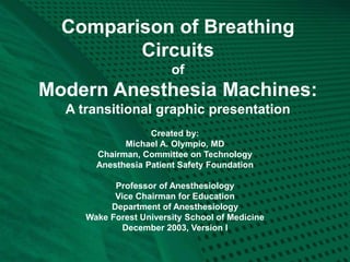
  • 1. Comparison of Breathing Circuits of Modern Anesthesia Machines: A transitional graphic presentation Created by: Michael A. Olympio, MD Chairman, Committee on Technology Anesthesia Patient Safety Foundation Professor of Anesthesiology Vice Chairman for Education Department of Anesthesiology Wake Forest University School of Medicine December 2003, Version I
  • 2. Disclaimer This educational tool is the sole creation of the author, who takes full responsibility for its accuracy and representation. Efforts have been taken to ensure this accuracy, even though the schematics are not intended to duplicate official drawings from the manufacturer. Indeed, the variation in manufacturer’ schematics makes it very difficult to compare one circuit to another. It has previously been impossible to overlay one schematic onto another to compare their differences. These circuits represent functional interpretations of the author, particularly with regard to the adjustable pressure limiting valve (A), and its analogous relationship to the ventilator relief valve (VR). Since these two valves serve analogous functions, they are drawn as one and the same, in context with the manual or mechanical circuit; The actual machines depicted do not share these valves. Similarly, the names that I have ascribed to these components do not always match the names given by the manufacturers, although there is general consistency. The drawings and components are not drawn to scale, but are rather sized to conveniently fit on the page. The author regrets that not all manufacturers and not all machines are represented, though they may share many similarities. The author invites those manufacturers to send me their schematic diagrams for potential future inclusion. Only USA FDA-approved machines are considered in this version. Since this original work has not been published elsewhere, the author retains all copyright privileges. This work may not be replicated or copied in any form, nor taken from this website without the express written permission of the author. It is a personally-funded, and unsupported contribution to the APSF web site, for the benefit of its readership, and to promote patient safety through the understanding of complex new breathing circuits. Michael A. Olympio, MD Professor of Anesthesiology Wake Forest University School of Medicine molympio@wfubmc.edu
  • 3. Acknowledgement The author would like to thank the Datex-Ohmeda Corporation, Madison, WI, and the Dräger Medical Corporation, Telford, PA, and its many educators and engineers who have graciously helped to educate me over the past 20 years. These individuals have been most influential in my career. They have supplied educational materials in both hard and electronic form, have welcomed me and/or supported my travel to their educational courses and factories, have voluntarily joined me at Wake Forest University, in the conduct of our anesthesia machine workshops, and they have consistently answered my questions, challenges, and criticisms regarding their products. The clarity, precision, and thoroughness of their two educational books, Explore!, from Datex- Ohmeda, Inc., and the ‘green book’, “Operating Principles of Narkomed Anesthesia Systems” from Dräger Medical, Inc., have profoundly structured my own educational foundation. The creators of these two books have accomplished much more than they will ever know. While providing such an education, these companies have allowed me to remain objective and have encouraged comparisons between equipment. I am very appreciative of these rare opportunities, and hope that others may learn from my experience. I am also indebted to my physician mentors, Drs. James Eisenkraft, Sem Lampotang, Jan Ehrenwerth, Al Grogono, Jeff Andrews and Mike Good, who’ve given me a roadmap to learn, while placing mountains in my path. Thank you.
  • 4. Instructions • You will view a series of 13 different circuits, beginning with the conventional, and transitioning to other modern circuits. • Be sure to view the full-screen, slide show images. • Each is preceded by an explanation of what you will see, followed by its image. • The comprehensive circuit will have a photograph of the machine from which it is derived. • Upcoming transitions will be explained. The reader is encouraged to page up and down between images, in order to appreciate the transition. PowerPoint should already be set to slide transition/fade smoothly/slow • Subsequent slides may repeat a previous circuit, followed by the transition to a new circuit, so that you might appreciate other changes in configuration.
  • 5. Conventional Manual Circuit: Fig. 1 There are specific reasons for the location of the various components of the breathing circuit within a gas machine. The circuit contains two unidirectional valves, I (inspiratory) and E (expiratory), which allow the inspired and expired gas to flow in one direction, thus preventing the rebreathing of carbon dioxide. Fig. 1 demonstrates inspiration during manual ventilation, by squeezing the reservoir bag, B. Note how the location of the fresh gas inflow, F, just proximal to I, allows the patient to first receive fresh gas during inspiration. The location of the absorber (CO2) is such that it does not absorb carbon dioxide during exhalation, but rather when the expired gas is “pushed” through it during inspiration. When F is higher than inspiratory flow, it will create a reversal of flow through the absorber during inspiration, and a redirection of the expired gas within the reservoir bag towards the APL (adjustable pressure-limiting), A, valve. In this manner of high F, the absorber will be spared from absorption of carbon dioxide, but it might become dehydrated; the patient will only receive the desired fresh gas components. Note that B is not placed between the absorber and the inspiratory valve, because use of the absorber would be less efficient during controlled ventilation. Some of the patient’s gas that is “scrubbed” of carbon dioxide during exhalation would then be scavenged, S, during flow reversal as the bag is squeezed for inspiration. (Read: Dorsch. Understanding Anesthesia Equipment 4th Ed. 1999: pp244-5. Williams and Wilkins)
  • 7. Conventional Mechanical Ventilation: Fig. 2 Conventional anesthesia machines provide mechanical ventilators to “squeeze the bag” for us. The ventilator bellows, VB, should be placed in the same relative location as the previous reservoir bag, to provide efficiency of absorbent use. Conventional ventilators also contain their own “APL” valve, which serves an analogous function. It may be called a ventilator relief valve, VR, as it relieves excess gas from the patient breathing circuit at the end of exhalation, contrary to manual ventilation, which relieves excess patient gas during inspiration. The VR also seals the circuit during inspiration, as shown diagrammatically. Note how I used the same APL valve for the ventilator, to emphasize functional similarities, even though conventional ventilators depicted in this series have their own separate valve. For either manual or mechanical ventilation, the APL or VR should be located between the expiratory valve and the absorber, such that exhaled gas with carbon dioxide is preferentially vented to the scavenger, as it is “chased” by fresh gas coming from the other direction. Next, you will review the preceding Fig. 1, and then transition into a conventional mechanical ventilator circuit, Fig. 2, representing the Dräger Narkomed AV2+.
  • 10. Representing Narkomed AV-2+: Fig.2 The hanging reservoir bag was replaced by a standing bellows, VB, which makes it easy to detect a disconnection or leak in the circuit, through collapse of the bellows. The bellows is “squeezed” (all the way down!) by pressure-regulated ventilator drive gas, VD, entraining room air through the venturi, VV. The ventilator exhaust valve, VE, must close during inspiration, to allow compression of VB. To prevent escape of patient gas during inspiration, a pilot line must transmit the drive gas pressure to the ventilator relief valve, VR, holding it closed. Ventilator drive gas is released directly into the room, via VE, during end-inspiratory pause and during exhalation. Note the location of the PEEP valve, P, which would serve to pressurize all of the breathing circuit except the bellows, VB, at the end of exhalation. The amount of PEEP may reduce delivered tidal volume according to its location, circuit design, and total compliance of the patient and circuit. (Elliott. J. Clin. Mon. 1989;5:100-4) Carefully note how the fresh gas, F, continues to flow into the patient during the inspiratory phase, contributing to tidal volume and airway pressure. Not shown is a mechanical pressure limiter on top of the ventilator bellows canister.
  • 11. Representing the Datex-Ohmeda 7900 Smart Vent: Fig. 3 Next, you will repeat Fig. 2, and then transition into another type of ventilator circuit, the Datex-Ohmeda 7900 Smart Vent. Watch for the re-routing of the ventilator drive gas, with connections distal to the ventilator relief valve, VR. Here, the ventilator exhaust, VE, and PEEP valves, P, are combined as one, and are in-line with the patient gas that is routed to the scavenger. This changes a number of things: • there is a higher demand (flow) on the scavenger system • there is no venturi to release extra ventilator drive gas, VD, because drive gas in this system is metered according to desired tidal volume, Vt. VB starts at the top and only travels down to the metered distance. • the entire circuit is subjected to PEEP, which could have caused significant reductions in delivered tidal volume (Elliott. J. Clin. Mon. 1989;5:100-4), were it not for: • a servo-feedback loop that measures delivered tidal volume at the inspiratory valve and gradually adjusts the ventilator drive gas accordingly • PEEP is now electronically controlled • the servo mechanism can also adjust VD according to F, in order to deliver the desired Vt. This is one form of fresh gas decoupling.
  • 13. Servo-controlled volume ventilation, electronic PEEP. (Representing Datex-Ohmeda 7900)
  • 14. Relocation of Fresh Gas Flow: Fig. 4 Now let’s explore some new (probably old!) circuit designs. What would happen if we relocate the fresh gas flow, F, distal to the inspiratory, I, valve? First of all, F would still flow continuously, but now it would continuously flow forward, instead of reversing during exhalation. Combined with a conventional mechanical ventilation system (as in Fig. 2), the patient would still receive extra tidal volume during I, but a respirometer on the exhalation limb would inappropriately measure exhaled gas combined with continuously flowing F. Also during exhalation, this location of F would create inefficient mixing of fresh gas with exhaled gas, such that gas exiting the scavenger would also contain fresh gas. Any gas going through the absorber would always contain carbon dioxide. In other words, the concentration of carbon dioxide exiting the scavenger would be reduced, thereby creating higher demand on the absorbent. (Read: Dorsch. Understanding Anesthesia Equipment 4th Ed. 1999: pp244-5. Williams and Wilkins) Watch what happens as we transition, first, from a conventional manual circuit during inspiration, Fig. 1, to a circuit with F distal to the I-valve, Fig. 4, during exhalation.
  • 16. Relocation of fresh gas, F, distal to I valve. FIGURE 4 Representing the Datex-Ohmeda S/5 ADU
  • 17. Datex-Ohmeda S/5 ADU: Fig. 4 The “new” location of fresh gas inflow in the ADU provides and/or demands several advantages: 1. It purges and primes the inspiratory limb of the breathing circuit with the desired fresh gas prior to onset of the inspiratory phase, and then continues to deliver the fresh gas flow during the remainder of the I-phase. This may cause faster equilibration of inspired gas with fresh gas, and may be an advantage during low flow anesthesia. Notice in a conventional circuit that at end-inspiration, the inspiratory limb of the breathing circuit contains the least amount of fresh gas (as appreciated in Fig. 1). 2. The respirometer (Vt) must now be placed at the endotracheal tube for accurate measurement. This allows both inspired and expired volume measurements, and can therefore measure an endotracheal tube cuff leak, partial disconnection at the tube, or broncho-pleural fistula, for example. 3. The relocation of the respirometer also allows airway pressure monitoring at the patient, where it is most representative of actual airway pressure. 4. The combination of volume and pressure measurement allows for the creation and analysis of spirometry.
  • 18. ADU A-EV1 Ventilator: Fig. 5 Having explored the ADU manual circuit, we can now compare its mechanical ventilator circuit to Fig. 2, the Dräger Narkomed AV2+. The next frame will repeat the AV2+ and then transition into the ADU mechanical ventilation circuit (A-EV1), shown during inspiration. Unlike the AV2+, there is no venturi, because VD is a metered amount of gas. There is now, however, a PEEP valve in a similar location. You will again see the relocation of the fresh gas inflow, as it was in Fig. 4. Notice in Fig. 5 how the fresh gas, F, continues to flow into the patient during inspiration. However, F is measured electronically and sent to the ventilator computer, which in turn regulates the output of VD, the drive gas, in order to maintain the desired tidal volume, Vt. Vt is measured and reported to the clinician from the spirometry sensor, but is not used in any way by the ventilator control, as it was in Fig. 3 for the Aestiva/5 7900. Another new feature is compliance compensation of Vt, as shown by the electronic arrow. The measured compliance of the system and hoses will subsequently adjust the VD to maintain desired Vt. This is also a feature of the Dräger 6400, Fabius, and Julian.
  • 20. Compliance compensation Representing the Datex-Ohmeda S/5 ADU with A-EV1 ventilator. FIGURE 5
  • 21. Relocation of the reservoir bag: Fig. 6 What if we started with the conventional manual circuit, Fig. 1 in the next slide, and then transitioned to Fig. 6, which shows the reservoir bag now distal to the absorber, and proximal to the fresh gas inflow? A number of things would change: • During inspiration, as shown, a combination of reservoir gas and fresh gas would still go to the patient, as in Fig. 1. • Now, however, some of the gas from the reservoir bag will go backwards across the absorbent and out the APL valve. This would mean that some of the exhaled gas that has already been scrubbed of carbon dioxide, would then be dumped to the scavenger. This is inefficient use of the absorbent. • Since the forced inspiration of gas now goes backwards across the absorbent, it could be an advantage in preventing any absorbent dust from getting blown into the patient. • During exhalation (not shown), it provides a preferential route for fresh gas flow into the reservoir bag. At higher fresh gas flow, however, the bag will fill unmixed with exhaled gas. In Fig. 1 you can imagine how fresh gas flows through the absorber and always combines with exhaled gas before it reaches the bag.
  • 23. Relocation of the reservoir bag. This transition does NOT represent any USA machine circuit. FIGURE 6
  • 24. Piston Ventilator: Fig 7 For reasons mentioned, the previous circuit (Fig. 6) is not suitable for manual ventilation, but could it be adapted to mechanical ventilation? Considering this new reservoir of fresh gas, and the manner in which it fills during exhalation, several new concepts merged into the creation of another modern anesthesia machine circuit: First, instead of isolating the reservoir bag from the circuit during use of the mechanical ventilator, as in all the previous circuits, why not leave it there as a collection reservoir of fresh gas during inspiration? In this manner, the fresh gas could be re-routed to the bag, and away from the patient during inspiration. This would represent another form of (true) fresh gas decoupling whereby the gas does not flow to the patient at all during inspiration. Second, instead of using the conventional bellows-in-a-bottle ventilator, controlled by electronic-pneumatic circuits, why not switch to an electronic-mechanical piston ventilator reminiscent of those used in ICU’s some 40 years ago? Watch how the next transition from Fig. 6 to Fig. 7 incorporates these two changes.
  • 25. Relocation of the reservoir bag. This transition does NOT represent any USA machine circuit. FIGURE 6
  • 26. Addition of a piston ventilator. This transition does NOT represent any USA machine circuit. FIGURE 7
  • 27. Piston Ventilator: Fig 7 Now there might appear to be some major problems with this new circuit: First, as the ventilator piston rises on the upstroke (inspiration, as shown), the gas is going to go straight into the reservoir bag instead of into the patient. Second, at high fresh gas flow (not shown), the bag could be distended, fresh gas would reverse flow upwards and then again, contribute to tidal volume as it joined the gas coming from the piston. This would defeat the goal of fresh gas decoupling, and would be unlikely to inflate the lungs because of the bag compliance. Third, the circuit may share some of the disadvantages of Fig. 6, in terms of economy of absorbent use. Fourth, the APL valve must be converted into an automated ventilator relief valve, to prevent the escape of gas during inspiration and: Fifth, the piston has to know when and how to retract, since it is mechanically driven and electronically controlled.
  • 28. Control Valves: Fig. 8 The modern age of computer circuits and electronically controlled, pneumatically operated valves has allowed us to solve most of these problems. Watch the transition to Fig. 8, after repeating Fig. 7, and look for the appearance of three new valves: 1. A decoupling valve, DV, at the fresh gas inlet to the reservoir bag 2. A manual/spontaneous-exhaust, M/S-E, valve at the inlet of the APL valve 3. Another ventilator relief valve, VR, that provides a direct route to a new scavenger valve, SV.
  • 29. Addition of a piston ventilator. This transition does NOT represent any USA machine circuit. FIGURE 7
  • 30. Almost a representation of the Dräger Narkomed 6400. A M/S-E Insertion of control valves. FIGURE 8
  • 31. Piston Ventilation Possible: Fig. 8 Now we can solve the problems listed in the previous slide, because we have added 4 valves to this circuit. Let’s see how they function. First, the decoupling valve closes during inspiration, such that the gas from the piston will be directed into the patient and not into the reservoir bag. Second, the decoupling valve forces fresh gas to flow only into the reservoir bag, even at high flow, and not contribute to tidal volume during inspiration. Third, the economy of absorbent was not improved because exhaled gas will still traverse the absorbent as it flows to the reservoir bag during exhalation (not shown). Fourth, during mechanical ventilation, the manual/spontaneous - exhaust valve, M/S-E, remains closed in order to cyclicly route all gas through the ventilator relief valve, VR, on to the scavenger valve, SV. The VR closes during inspiration, and opens at the end of exhalation, after the DV has opened, and then after the piston has retracted. Note that during manual ventilation (not shown), the VR remains closed, with M/S-E open. During spontaneous ventilation, functionally, M/S would be closed while VR and A are opened. Fifth, the basic piston movement has been described, but its operation and control is complex, and determined by the mode of ventilation.
  • 32. Dräger 6400 Divan Ventilator: Fig. 9 Watch next as we repeat the previous image of Figure 8 and transition into the Dräger 6400 Divan Ventilator circuit in Figure 9. Figure 9 demonstrates the status of the circuit during exhalation, whereas Figure 8 was drawn in inspiration. We have also added a 5th valve, for PEEP, and it is located in the expiratory limb of the breathing circuit, proximal to the expiratory valve. Next, a word about the M/S-E valve. Although the circuit is drawn functionally equivalent to the Narkomed 6400, there is a manually operated lever, physically located on top of the APL valve, A, which Dräger calls a “Man/Spont” valve. The actual M/S-E valve, represented in these figures, is hidden within the valve block assembly of the breathing circuit, and it is electronically regulated to close during mechanical ventilation (E-function). Obviously, the APL must be isolated from the mechanical circuit during mechanical ventilation, and that is how it is accomplished.
  • 33. Almost a representation of the Dräger Narkomed 6400. A M/S-E Insertion of control valves. FIGURE 8
  • 34. Representation of the Dräger Narkomed 6400 with Divan ventilator. A M/S-E FIGURE 9
  • 35. Dräger Narkomed 6400: Fig. 9 In addition to that described above, realize the following points about this circuit: • With the initial opening of the decoupling valve, exhaled gas is initially scrubbed of carbon dioxide, and dilutes the fresh gas that has accumulated in the reservoir bag. • Regulation of the resistor valve, P, provides PEEP during exhalation. • Then, as the piston retracts, it draws the previously scrubbed gas backwards over the absorbent and into the piston cylinder. • Next, the ventilator relief valve opens, and: • If fresh gas flow is high enough, excess gas, under slight pressure, will passively open the scavenger valve and vent to the scavenger suction canister (not shown).
  • 36. Time to Re-think Everything Wow, that seemed like it was getting complicated. Four electronically regulated valves, a passive scavenger valve and a newly designed APL valve that allows quick-opening to atmosphere without unscrewing the APL. And what about all that new gas mixing? How readily can I change the concentration of the inspired mixture? We’ll have to wait on some more bench research before the mixing effects are clear. Can we make this piston circuit any simpler? Well, lets go back to Fig. 6, when all we did was to place the reservoir bag next to the fresh gas flow. Then, we are going to maintain the same concept of fresh gas decoupling into the bag, while another piston is used to mechanically ventilate the patient. This time, however, let’s place the piston in the same area, distal to the fresh gas flow, instead of the conventional location in the Narkomed 6400. Watch this transition from Figure 6 to Figure 10.
  • 37. Relocation of the reservoir bag. This transition does NOT represent any USA machine circuit. FIGURE 6
  • 38. This figure does NOT represent any USA- FDA circuit. New location of a ventilator piston. FIGURE 10
  • 39. New Location for Ventilator Piston: Fig. 10 With the piston in this location, as shown during inspiration, we can see that the ventilator gas will follow the path of least resistance, and probably end up in the reservoir bag. This system, therefore, would not function during mechanical ventilation. An ingenious mind, however, might suddenly see a simple solution to the problem. Let’s watch what happens as we transition from the previous Fig. 10 to the creation of another new machine, the Dräger Fabius GS, in Fig. 11.
  • 40. This figure does NOT represent any USA- FDA circuit. New location of a ventilator piston. FIGURE 10
  • 41. Representing the Dräger Fabius GS in mechanical ventilation mode. FIGURE 11
  • 42. Dräger Fabius GS: Fig. 11 Did you notice the simple solution to the problem of backflow? Dräger simply added a mechanical one-way check valve, the decoupling valve, DV, which closes automatically as the piston travels upwards during inspiration. Now the gas will flow forward to the patient, but there will not be any back pressure on the expiratory valve, E, to close it. Therefore, the engineers had to add the exhalation valve, EV/P, in the exhalation limb, just proximal to the E valve. This valve is electronically regulated and pneumatically controlled, and is shown in the closed EV position during inspiration. (Referred to as PEEP/PMAX valve by Dräger.) Notice how the M/S valve must be automatically opened by the ventilator computer during mechanical ventilation. This bypasses the APL (Dräger calls it the “APL bypass valve”) and allows excess gas to escape from the system, after lifting the low pressure scavenger valve, SV. If this valve did not open, and the APL valve was closed, pressure would build in the system, dependent upon fresh gas flow. In spontaneous ventilation mode, the M/S valve in this diagram is functionally similar to the “spont” setting within the APL, because, when open, it routes the excess gas directly to the SV. The M/S valve in the diagram is seen in the photo as the silver dome beneath the grey APL on top of the black breathing circuit manifold. Realize that during exhalation (not shown), the EV/P functions as a PEEP valve.
  • 43. Isn’t there a simpler way? Fresh Gas Flow Interruption: Fig. 12 Since we have computer controlled ventilators, why not have computer controlled fresh gas flow? Everything to this point has had needle-valve control of fresh gas, even though your ADU and Fabius GS measure and report the flow digitally. If we can control, measure, and report flow electronically, then we can also interrupt the flow during inspiration and provide another form of fresh gas decoupling. Note how the next transition from the conventional circuit, Fig. 1, to this new circuit, Fig. 12, also re-routes the fresh gas into the center of the absorber. This was done to provide a convenient entry port for the return of sampled gas from the gas analyzer, should that design become FDA approved. Note the obvious addition of the decoupling valve, DV, as described.
  • 45. Fresh gas flow interruption during inspiration. This will become the Dräger Julian. FIGURE 12
  • 46. The Dräger Julian: Fig. 13 The previous circuit, as shown during manual inspiration, would not require a decoupling valve, nor does it use one during inspiration, because we rely upon the APL valve to adjust the amount of gas in the circuit for a given tidal volume. What we will see next, is the transition from this manual circuit to the Julian mechanical ventilation circuit. You will see the reservoir bag replaced by the bellows, but notice something different: • The bellows is now ‘hanging’ instead of ‘standing’ • The APL, as depicted, functionally ‘becomes’ a ventilator relief valve, as before • We still need a ventilator exhaust valve, VE, as in Figs. 2 and 3, and: • The VE can also serve as a PEEP valve as it does with the Aestiva 7900 smart vent.
  • 47. Fresh gas flow interruption during inspiration. This will become the Dräger Julian. FIGURE 12
  • 49. The Dräger Julian: Fig. 13 Here we have the Dräger Julian, with its hanging bellows, VB. How do we prevent an undetected disconnection of the breathing circuit? Recall that hanging ventilators of the 1970’s would continue to cycle because they entrained room air as the bellows descended, and they delivered an ‘inspiratory’ volume as the bellows cycled during inspiration. Notice how the electric eye ‘looks for’ the descent of the bellows. With a partial disconnect, and at low flow, and with a light-weight bellows, one could imagine a slow bellows descent to the bottom of the canister during exhalation. This might trigger an alarm. If one added PEEP to the canister, and surrounded the entire bellows with end- expiratory pressure, then the bellows would not fall, but might actually stay at the top during exhalation. Thus, the placement and use of PEEP could facilitate detection of a disconnect. As always, however, we rely on multiple other monitors of ventilation.
  • 50. Summary of New Safety Features • More accurate and/or corrected tidal volume through compliance and fresh gas compensation • Potential return of sampled gas to facilitate low-flow • Fresh gas decoupling may prevent hyperinflation of the lung • Some forms of decoupling will even reroute the high flow of oxygen flush if it is depressed during mechanical inspiration • Incorporation of electronic PEEP may prevent inaccurate, improper, or unintended PEEP • Electronic selection of ventilation parameters might prevent improper setup of a mechanical ventilator • Automated checkout procedures (presumably) will detect a problem that clinicians may not, particularly in these complex systems • New disconnect alarm system for hanging bellows might reduce the incidence of inadequate ventilation
  • 51. Conclusion I hope this transitional graphic presentation of modern breathing circuits has helped you to understand the function of modern anesthesia machines. To look at them, they are quite ominous and frightening, because we can no longer see the ‘pipes’ nor even imagine the direction of gas flow within the system. They are complicated, indeed, and require a multitude of sensors that are also used in conjunction with automated electronic checkout procedures. The computer must sequentially and intelligently test the valves with and without gas flow, and during synchronized compression of the ventilator. We must try to understand these systems, so that we will understand the importance, significance, and safety of auto-testing, and the meaning of various warnings and alarms. Manufacturers are developing safer and smarter equipment, and have provided machines that do what we want, and exactly what we expect them to do. Good luck and enjoy the new technology! Please send feedback to molympio@wfubmc.edu