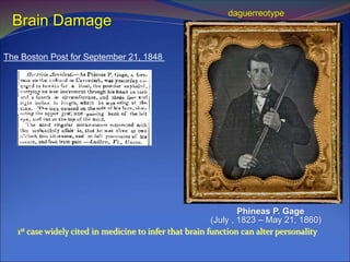
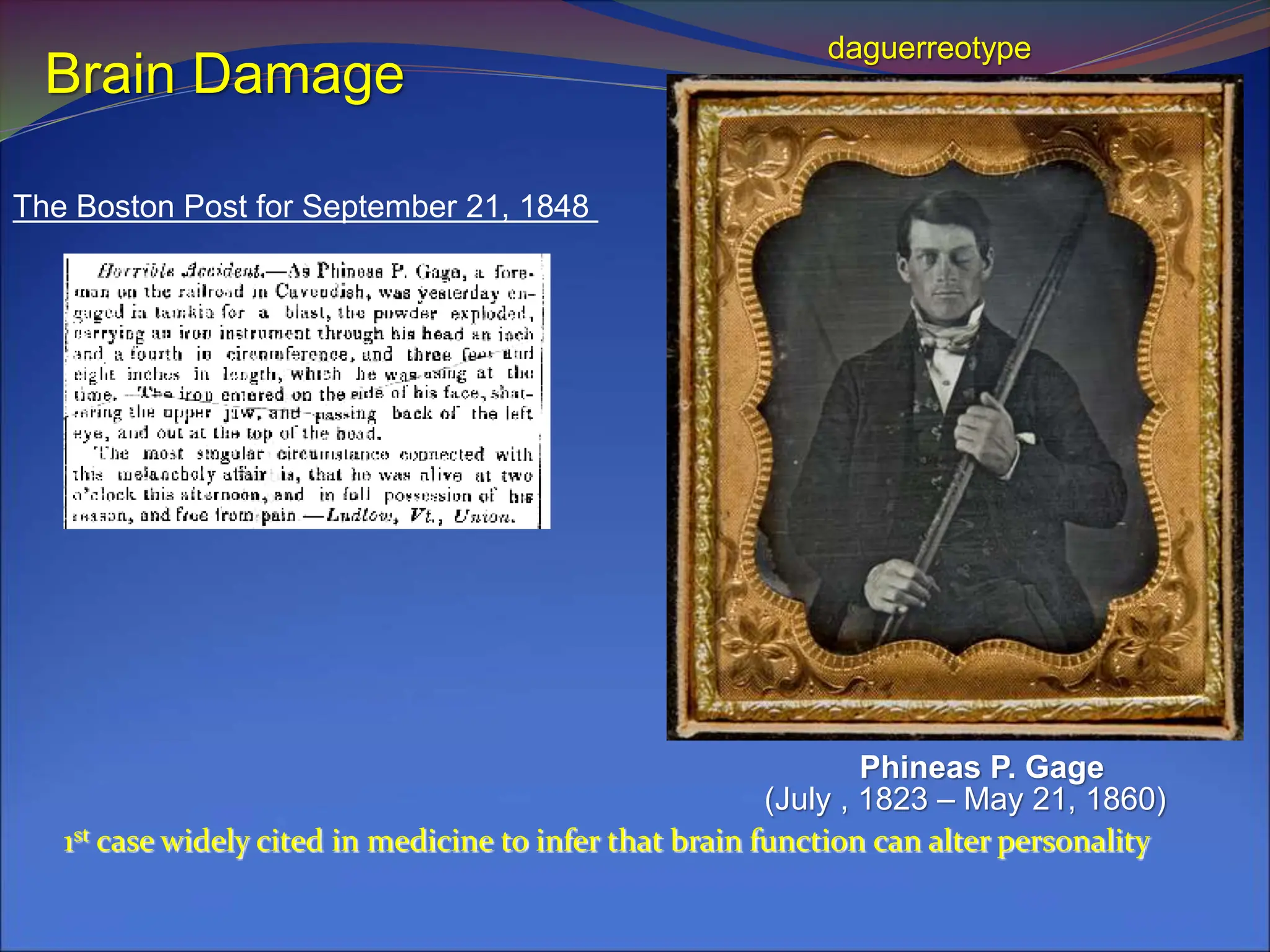
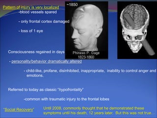
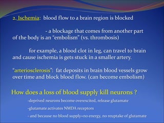
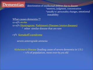
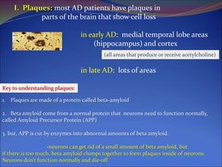
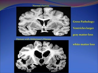
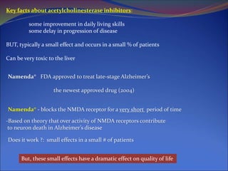
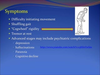
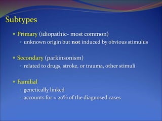
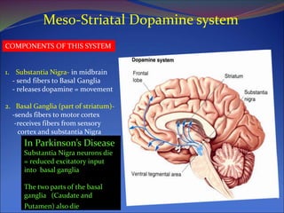
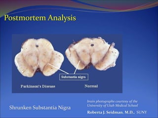
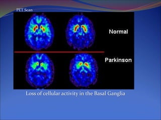
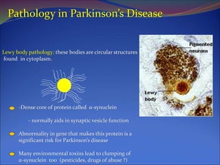
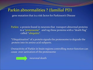
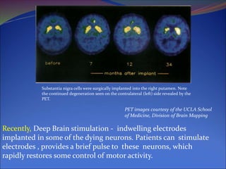
SlideShare a Scribd company logo
(July , 1823 – May 21, 1860)
Phineas P. Gage
The Boston Post for September 21, 1848
1st case widely cited in medicine to infer that brain function can alter personality
daguerreotype
Brain Damage
~1850
Pattern of injury is very localized
-blood vessels spared
- only frontal cortex damaged
- loss of 1 eye
Consciousness regained in days
- personality/behavior dramatically altered
- child-like, profane, disinhibited, inappropriate, inability to control anger and
emotions.
Referred to today as classic “hypofrontality”
-common with traumatic injury to the frontal lobes
“Social Recovery” Until 2008, commonly thought that he demonstrated these
symptoms until his death, 12 years later. But this was not true…
Brain Damage
1. Traumatic brain injury
2. Strokes
3. Alzheimer’s Disease
4. Parkinson’s Disease
Other neurological disorders discussed in the book will
NOT be subject to test questions
Traumatic Brain Injury
FACTS:
1.4 million who sustain a TBI each year in the United States (0.5% of people in U.S.)
50,000 die;
235,000 are hospitalized; and
1.1 million are treated and released from an emergency department.
What causes TBI?
Falls (28%)
Motor vehicle-traffic crashes (20%)
Struck by/against (19%)
Assaults (11%)
Who is at highest risk for TBI?
Males are about 1.5 times as likely as females to sustain a TBI.1
The two age groups at highest risk for TBI are 0 to 4 year olds and 15 to 19 year olds
Certain military duties (e.g., paratrooper) increase the risk of sustaining a TBI
Langlois JA (2006)
Types of Traumatic Brain Injury
“Closed Head Injury”
Concussion. Concussions are the most common type of TBI. A concussion is
damage to nerves or blood vessels in the brain often caused by an impact to the head.
Contusions. A contusion is a bruise or bleeding on the brain that can be caused
by an impact.
Diffuse Axonal Injury. This is tearing of nerve tissue or blood vessels when the
brain is jostled in the skull. It can result from shaking (for instance, shaken baby syndrome)
or whiplash
Coup-countrecoup Injury. impact not only injures the site of impact, but causes
the brain to impact with the skull, causing injury to the opposite side of the brain.
Symptoms:
acute: confusion, visual disturbance, unconsciousness if severe
long-term: memory problems, sensory problems .
- “dementia pugalistica”
How does traumatic brain injury develop ?
-direct, mechanical injury to neurons (axons, dendrites or soma)
- damage to blood supply deprives neurons and glia of nutrients
-Overactivity of glutamate receptors
A little bit of Ca2+ inflow is good at the right time, too much is toxic
-Activation of NMDA-type glutamate receptors
-Inflow of too much Ca2+ into neurons
- activates death enzymes that break down
neuron
-Impairs neuron ability to meet energy
requirements
-Triggers cell death
Strokes
disruption of blood flow in brain
3rd leading cause of death in adults
1. Hemorrhage: bleeding in brain, blood vessel ruptures
- can be from traumatic injury to blood vessel
- can be from “aneurysm”
weakening of blood vessel wall, balloonlike
dialation forms and can burst
caused by some infections, toxins like cigarette
Smoke. Can also be develomental abnormality.
2. Ischemia: blood flow to a brain region is blocked
- a blockage that comes from another part
of the body is an “embolism” (vs. thrombosis)
for example, a blood clot in leg, can travel to brain
and cause ischemia is gets stuck in a smaller artery.
“arteriosclerosis”: fat deposits in brain blood vessels grow
over time and block blood flow. (can become embolism)
How does a loss of blood supply kill neurons ?
-deprived neurons become overexcited, release glutamate
-glutamate activates NMDA receptors
- and because no blood supply=no energy, no reuptake of glutamate
Dementias deterioration of intellectual abilities due to disease
*memory, judgment, concentration
*usually w/ personality changes, emotional
instability
What causes dementia ??
12-15% stroke
12-15% Huntingtons, Parkinson’s Diseases (motor diseases)
* other similar disease that are rare
>5% Korsakoff’s syndrome
severe anterograde amnesia
Alzheimer’s Disease (leading cause of severe dementia in U.S.)
- +1% of population, most over 65 yrs old
The severity and type of symptoms observed is directly related to death of
neurons in several brain regions.
Causes ?
We do NOT know what causes Alzheimer’s Disease !
But, we know that…:
1. Risk of developing it is heritable
there is a “genetic” factor involved
2. Some AD patients have a specific genetic problem
That can cause early-onset and severe form (~45 yrs old)
Alzheimer’s Disease is probably just 1 term used to describe
the symptoms of lots of different diseases that cause
brain damage and similar symptoms.
3. But, many other AD patients don’t have family members
with AD and don’t have an identified genetic problem
I. Plaques: most AD patients have plaques in
parts of the brain that show cell loss
in early AD: medial temporal lobe areas
(hippocampus) and cortex
in late AD: lots of areas
(all areas that produce or receive acetylcholine)
1. Plaques are made of a protein called beta-amyloid
2. Beta amyloid come from a normal protein that neurons need to function normally,
called Amyloid Precursor Protein (APP)
3. but, APP is cut by enzymes into abnormal amounts of beta amyloid.
-neurons can get rid of a small amount of beta amyloid, but
if there is too much, beta amyloid clumps together to form plaques inside of neurons.
Neurons don’t function normally and die-off
Key to understanding plaques:
II. Tangles: Many AD patients also have “tangles” in
neurons
Microtubules are long string-like structures that transport
things from the cell body to the end of the axon and dendrites
In AD, these microtubules break apart and
collapse into a tangles mess and neurons don’t function normally,
so they die-off.
Tau proteins (like rungs of a ladder)
Normal Human
Human with Alzheimer’s Disease
Gross Pathology:
Ventricles larger
gray matter loss
white matter loss
Drug Treatment of Dementias
Drugs approved by the U.S. Food and Drug Administration
Cognex®
Aricept®
Reminyl®
Exelon®
Namenda®
All are inhibitors of acetylcholinesterase, except Namenda
1. Acetylcholine is release from “presynaptic” neuron, and then re-absorbed
lots of it in hippocampus and cortex
2. Acetylcholinesterase is an enzyme that breaks down acetylcholine
3. That means that more acetylcholine is present in synaptic cleft to stimulate
receptors on neurons in learning/memory areas
YOU DON’T NEED TO KNOW THESE
Key facts about acetylcholinesterase inhibitors:
some improvement in daily living skills
some delay in progression of disease
BUT, typically a small effect and occurs in a small % of patients
Can be very toxic to the liver
Namenda® FDA approved to treat late-stage Alzheimer’s
the newest approved drug (2004)
Namenda® - blocks the NMDA receptor for a very short period of time
-Based on theory that over activity of NMDA receptors contribute
to neuron death in Alzheimer’s disease
Does it work ?: small effects in a small # of patients
But, these small effects have a dramatic effect on quality of life
Parkinson’s Disease
"An Essay on the Shaking Palsy," published in
1817 by a London physician named James
Parkinson,
mentioned in the Ayurveda, the system of
medicine practiced in India as early 7,000 years
ago , and in the first Chinese medical text, Nei
Jing, which appeared 2500 years ago
Symptoms
 Difficulty initiating movement
 Shuffling gait
 “Cogwheel” rigidity
 Tremor at rest
 Advanced stages may include psychiatric complications
 depression
 hallucinations
 Paranoia
 Cognitive decline
http://www.youtube.com/watch?v=13ftfmYwfaw
Incidence
 Onset usually appears after age of 40 years
 but can begin at any time including childhood when
it is termed juvenile parkinsonism
 affects 1% aged 50 years and over
 10% aged 60 years and over may have undiagnosed,
early stages of the disease
 about 1½ times more common in men than in
women
Subtypes
 Primary (idiopathic- most common)
 unknown origin but not induced by obvious stimulus
 Secondary (parkinsonism)
 related to drugs, stroke, or trauma, other stimuli
 Familial
 genetically linked
 accounts for < 20% of the diagnosed cases
Progression: identified too late ?
 Considerable dopamine loss must occur before the
disease is apparent
 clinical diagnosis is usually made after  80% loss in
basal ganglia dopamine content
 symptoms may emerge after a 60% reduction in basal
ganglia dopamine content
 The disease is probably present > 20 years before
diagnosis
Meso-Striatal Dopamine system
COMPONENTS OF THIS SYSTEM
1. Substantia Nigra- in midbrain
- send fibers to Basal Ganglia
- releases dopamine = movement
2. Basal Ganglia (part of striatum)-
-sends fibers to motor cortex
-receives fibers from sensory
cortex and substantia Nigra
In Parkinson’s Disease
Substantia Nigra neurons die
= reduced excitatory input
into basal ganglia
The two parts of the basal
ganglia (Caudate and
Putamen) also die
Postmortem Analysis
brain photographs courtesy of the
University of Utah Medical School
Roberta J. Seidman, M.D., SUNY
Shrunken Substantia Nigra
Loss of cellular activity in the Basal Ganglia
PET Scan
Pathology in Parkinson’s Disease
Lewy body pathology: these bodies are circular structures
found in cytoplasm.
-Dense core of protein called α-synuclein
- normally aids in synaptic vesicle function
Abnormality in gene that makes this protein is a
significant risk for Parkinson’s disease
Many environmental toxins lead to clumping of
α-synuclein too (pesticides, drugs of abuse ?)
Parkin abnormalities ? (familial PD)
gene mutation that is a risk factor for Parkinson’s Disease
Parkin: a protein found in neurons that transport abnormal proteins
to a “proteosome” and tag these proteins with a “death flag”
called “ubiquitin”.
“Ubiquitination” of a protein signals the proteosome to degrade the
protein into its amino acid subparts.
Overactivity of Parkin in brain regions controlling motor function can
cause over-activation of the proteosome
neuronal death
Treatment
 Levodopa (L-dopa) therapy (L-dopa is a dopamine precursor)
 generally very effective for the first 2 to 5 years of
treatment after which the on-off effect develops
 Direct-acting dopamine receptor agonists
 Experimental brain surgery
 lesions of nuclei that inhibit basal ganglia
 - remove inhibition of dying neurons, they may be more active = motor
control
 neural tissue implants (e.g., fetal dopamine cells)
Recently, Deep Brain stimulation - indwelling electrodes
implanted in some of the dying neurons. Patients can stimulate
electrodes , provides a brief pulse to these neurons, which
rapidly restores some control of motor activity.
PET images courtesy of the UCLA School
of Medicine, Division of Brain Mapping
Substantia nigra cells were surgically implanted into the right putamen. Note
the continued degeneration seen on the contralateral (left) side revealed by the
PET.

More Related Content

Similar to chapter8.pptfxvdffddxxfdsfdrfsxxrggfxdffg

Alzheimers disease and its treatments
Alzheimers disease and its treatmentsAlzheimers disease and its treatments
Alzheimers disease and its treatmentsRavish Yadav
 
AlzheimersDisease-deepansha ppt.ppt
AlzheimersDisease-deepansha ppt.pptAlzheimersDisease-deepansha ppt.ppt
AlzheimersDisease-deepansha ppt.pptDeepanshaDeepansha1
 
neurodegenerative diseases
neurodegenerative diseasesneurodegenerative diseases
neurodegenerative diseasesBILAL DAR
 
Alzheimer's Disease and its pathophysiology
Alzheimer's Disease and its pathophysiologyAlzheimer's Disease and its pathophysiology
Alzheimer's Disease and its pathophysiologyAchyut Adhikari
 
Alzheimers presentation.docx
Alzheimers presentation.docxAlzheimers presentation.docx
Alzheimers presentation.docxteredeloscobos
 
Alzheimers presentation.docx
Alzheimers presentation.docxAlzheimers presentation.docx
Alzheimers presentation.docxHena Jawaid
 
Alzheimers disease by Ritika soni
Alzheimers disease by Ritika soniAlzheimers disease by Ritika soni
Alzheimers disease by Ritika soniShimla
 
Alzheimers presentation.docx (1)
Alzheimers presentation.docx (1)Alzheimers presentation.docx (1)
Alzheimers presentation.docx (1)Ashwani Kumar
 
Brain Damage and Neural Plasticity.pptx
Brain Damage and Neural Plasticity.pptxBrain Damage and Neural Plasticity.pptx
Brain Damage and Neural Plasticity.pptxPavithra L N
 
Biochemistryofalzheimers
BiochemistryofalzheimersBiochemistryofalzheimers
Biochemistryofalzheimersvince_md
 
Alzheimer’s Disease
Alzheimer’s DiseaseAlzheimer’s Disease
Alzheimer’s Diseasehulyadiels
 
Drug for Neurogenerative disorder
 Drug for Neurogenerative disorder  Drug for Neurogenerative disorder
Drug for Neurogenerative disorder kahkesha samshad
 

Similar to chapter8.pptfxvdffddxxfdsfdrfsxxrggfxdffg (20)

Pinel basics ch08
Pinel basics ch08Pinel basics ch08
Pinel basics ch08
 
Alzheimers disease and its treatments
Alzheimers disease and its treatmentsAlzheimers disease and its treatments
Alzheimers disease and its treatments
 
AlzheimersDisease-deepansha ppt.ppt
AlzheimersDisease-deepansha ppt.pptAlzheimersDisease-deepansha ppt.ppt
AlzheimersDisease-deepansha ppt.ppt
 
Dementia-final
Dementia-finalDementia-final
Dementia-final
 
Dementia
DementiaDementia
Dementia
 
neurodegenerative diseases
neurodegenerative diseasesneurodegenerative diseases
neurodegenerative diseases
 
Alzheimer's Disease and its pathophysiology
Alzheimer's Disease and its pathophysiologyAlzheimer's Disease and its pathophysiology
Alzheimer's Disease and its pathophysiology
 
QPR-CNS degenerations
QPR-CNS degenerationsQPR-CNS degenerations
QPR-CNS degenerations
 
Alzheimers presentation.docx
Alzheimers presentation.docxAlzheimers presentation.docx
Alzheimers presentation.docx
 
Alzheimers presentation.docx
Alzheimers presentation.docxAlzheimers presentation.docx
Alzheimers presentation.docx
 
Alzheimers disease by Ritika soni
Alzheimers disease by Ritika soniAlzheimers disease by Ritika soni
Alzheimers disease by Ritika soni
 
Alzheimers presentation.docx (1)
Alzheimers presentation.docx (1)Alzheimers presentation.docx (1)
Alzheimers presentation.docx (1)
 
Brain Damage and Neural Plasticity.pptx
Brain Damage and Neural Plasticity.pptxBrain Damage and Neural Plasticity.pptx
Brain Damage and Neural Plasticity.pptx
 
Alzeihmer disease
Alzeihmer diseaseAlzeihmer disease
Alzeihmer disease
 
Biochemistryofalzheimers
BiochemistryofalzheimersBiochemistryofalzheimers
Biochemistryofalzheimers
 
Alzheimer’s Disease
Alzheimer’s DiseaseAlzheimer’s Disease
Alzheimer’s Disease
 
Cate buline are buburuza
Cate buline are buburuzaCate buline are buburuza
Cate buline are buburuza
 
Basal ganglia
Basal gangliaBasal ganglia
Basal ganglia
 
Alzheimer's disease
Alzheimer's diseaseAlzheimer's disease
Alzheimer's disease
 
Drug for Neurogenerative disorder
 Drug for Neurogenerative disorder  Drug for Neurogenerative disorder
Drug for Neurogenerative disorder
 

More from nuradinman89

Communicable Disease.pptxgfgfggfffdfxfsdddf
Communicable Disease.pptxgfgfggfffdfxfsdddfCommunicable Disease.pptxgfgfggfffdfxfsdddf
Communicable Disease.pptxgfgfggfffdfxfsdddfnuradinman89
 
palliative-care-essential-facts.pdf
palliative-care-essential-facts.pdfpalliative-care-essential-facts.pdf
palliative-care-essential-facts.pdfnuradinman89
 
5. pain perception and comfort (2).pptx
5. pain perception and comfort (2).pptx5. pain perception and comfort (2).pptx
5. pain perception and comfort (2).pptxnuradinman89
 
2. Coping and stress management (2).pptx
2. Coping and stress management (2).pptx2. Coping and stress management (2).pptx
2. Coping and stress management (2).pptxnuradinman89
 
2. Family planing level3.pptx
2. Family planing level3.pptx2. Family planing level3.pptx
2. Family planing level3.pptxnuradinman89
 
2. Family planing level3.pptx
2. Family planing level3.pptx2. Family planing level3.pptx
2. Family planing level3.pptxnuradinman89
 
3. Sleep and rest pattern (2).pptx
3. Sleep and rest pattern (2).pptx3. Sleep and rest pattern (2).pptx
3. Sleep and rest pattern (2).pptxnuradinman89
 
1. Loss and grief (2).pptx
1. Loss and grief (2).pptx1. Loss and grief (2).pptx
1. Loss and grief (2).pptxnuradinman89
 

More from nuradinman89 (9)

Communicable Disease.pptxgfgfggfffdfxfsdddf
Communicable Disease.pptxgfgfggfffdfxfsdddfCommunicable Disease.pptxgfgfggfffdfxfsdddf
Communicable Disease.pptxgfgfggfffdfxfsdddf
 
palliative-care-essential-facts.pdf
palliative-care-essential-facts.pdfpalliative-care-essential-facts.pdf
palliative-care-essential-facts.pdf
 
lecture 2.ppt
lecture 2.pptlecture 2.ppt
lecture 2.ppt
 
5. pain perception and comfort (2).pptx
5. pain perception and comfort (2).pptx5. pain perception and comfort (2).pptx
5. pain perception and comfort (2).pptx
 
2. Coping and stress management (2).pptx
2. Coping and stress management (2).pptx2. Coping and stress management (2).pptx
2. Coping and stress management (2).pptx
 
2. Family planing level3.pptx
2. Family planing level3.pptx2. Family planing level3.pptx
2. Family planing level3.pptx
 
2. Family planing level3.pptx
2. Family planing level3.pptx2. Family planing level3.pptx
2. Family planing level3.pptx
 
3. Sleep and rest pattern (2).pptx
3. Sleep and rest pattern (2).pptx3. Sleep and rest pattern (2).pptx
3. Sleep and rest pattern (2).pptx
 
1. Loss and grief (2).pptx
1. Loss and grief (2).pptx1. Loss and grief (2).pptx
1. Loss and grief (2).pptx
 

Recently uploaded

ASSISTING WITH THE USE OF URINAL BY ANUSHRI SRIVASTAVA.pptx
ASSISTING WITH THE USE OF URINAL BY ANUSHRI SRIVASTAVA.pptxASSISTING WITH THE USE OF URINAL BY ANUSHRI SRIVASTAVA.pptx
ASSISTING WITH THE USE OF URINAL BY ANUSHRI SRIVASTAVA.pptxAnushriSrivastav
 
Nose-Nasal Cavity & Paranasal Sinuses BY Dr.Rabia Inam Gandapore.pptx
Nose-Nasal Cavity & Paranasal Sinuses BY Dr.Rabia Inam Gandapore.pptxNose-Nasal Cavity & Paranasal Sinuses BY Dr.Rabia Inam Gandapore.pptx
Nose-Nasal Cavity & Paranasal Sinuses BY Dr.Rabia Inam Gandapore.pptxDr. Rabia Inam Gandapore
 
Jesse Jhaj: Building Relationships with Patients as a Doctor or Healthcare Wo...
Jesse Jhaj: Building Relationships with Patients as a Doctor or Healthcare Wo...Jesse Jhaj: Building Relationships with Patients as a Doctor or Healthcare Wo...
Jesse Jhaj: Building Relationships with Patients as a Doctor or Healthcare Wo...saimasadaf14
 
About Dr Ranjit Jagtap daughter's - Aditi & Poulami Jagtap
About Dr Ranjit Jagtap daughter's - Aditi & Poulami JagtapAbout Dr Ranjit Jagtap daughter's - Aditi & Poulami Jagtap
About Dr Ranjit Jagtap daughter's - Aditi & Poulami JagtapAditi Jagtap Pune
 
Prosthesis upper limb and lower limb.pptx
Prosthesis upper limb and lower limb.pptxProsthesis upper limb and lower limb.pptx
Prosthesis upper limb and lower limb.pptxBadalverma11
 
Storage_of _Bariquin_Components_in_Storage_Boxes.pptx
Storage_of _Bariquin_Components_in_Storage_Boxes.pptxStorage_of _Bariquin_Components_in_Storage_Boxes.pptx
Storage_of _Bariquin_Components_in_Storage_Boxes.pptxBariquins
 
Breaking Down Oppositional Defiant Disorder Treatments
Breaking Down Oppositional Defiant Disorder TreatmentsBreaking Down Oppositional Defiant Disorder Treatments
Breaking Down Oppositional Defiant Disorder TreatmentsOppositional Defiant Disorder
 
Importance of Diet on Dental Health.docx
Importance of Diet on Dental Health.docxImportance of Diet on Dental Health.docx
Importance of Diet on Dental Health.docxSachin Mittal
 
Benefits of Dentulu's Salivary Testing.pptx
Benefits of Dentulu's Salivary Testing.pptxBenefits of Dentulu's Salivary Testing.pptx
Benefits of Dentulu's Salivary Testing.pptxDentulu Inc
 
Best Way 30-Days Keto Meal Plan For Diet
Best Way 30-Days Keto Meal Plan For DietBest Way 30-Days Keto Meal Plan For Diet
Best Way 30-Days Keto Meal Plan For DietGunaPerumal1
 
Management of heart failure 23.02.24.pptx
Management of heart failure 23.02.24.pptxManagement of heart failure 23.02.24.pptx
Management of heart failure 23.02.24.pptxchristina388422
 
Thoracic Vertebrae: Anatomy, Function, and Common Disorders | The Lifescience...
Thoracic Vertebrae: Anatomy, Function, and Common Disorders | The Lifescience...Thoracic Vertebrae: Anatomy, Function, and Common Disorders | The Lifescience...
Thoracic Vertebrae: Anatomy, Function, and Common Disorders | The Lifescience...The Lifesciences Magazine
 
Best Erectile Dysfunction Treatment In Narela
Best Erectile Dysfunction Treatment In NarelaBest Erectile Dysfunction Treatment In Narela
Best Erectile Dysfunction Treatment In NarelaLalClinic
 
Digital Healthcare: The Future of Medical Consultations
Digital Healthcare: The Future of Medical ConsultationsDigital Healthcare: The Future of Medical Consultations
Digital Healthcare: The Future of Medical Consultationssmartcare
 
Homeopathy Medicine for Diabetes_ Balancing Blood Sugar .pdf
Homeopathy Medicine for Diabetes_ Balancing Blood Sugar .pdfHomeopathy Medicine for Diabetes_ Balancing Blood Sugar .pdf
Homeopathy Medicine for Diabetes_ Balancing Blood Sugar .pdfDharma Homoeopathy
 
GOUT and it's Management with All the catagories like; Defination, Type, Sym...
GOUT and it's Management with All the catagories like;  Defination, Type, Sym...GOUT and it's Management with All the catagories like;  Defination, Type, Sym...
GOUT and it's Management with All the catagories like; Defination, Type, Sym...chemiology
 
PT MANAGEMENT OF URINARY INCONTINENCE.pptx
PT MANAGEMENT OF URINARY INCONTINENCE.pptxPT MANAGEMENT OF URINARY INCONTINENCE.pptx
PT MANAGEMENT OF URINARY INCONTINENCE.pptxdrtabassum4
 
Integrated Mother and Neonate Childwood Illness Health Care
Integrated Mother and Neonate Childwood Illness  Health CareIntegrated Mother and Neonate Childwood Illness  Health Care
Integrated Mother and Neonate Childwood Illness Health CareASKatoch1
 
Production.pptx\\\\\\\\\\\\\\\\\\\\\\\\\\\\\\\\\\\\\\\\\\\\\\
Production.pptx\\\\\\\\\\\\\\\\\\\\\\\\\\\\\\\\\\\\\\\\\\\\\\Production.pptx\\\\\\\\\\\\\\\\\\\\\\\\\\\\\\\\\\\\\\\\\\\\\\
Production.pptx\\\\\\\\\\\\\\\\\\\\\\\\\\\\\\\\\\\\\\\\\\\\\\DanielOliver74
 

Recently uploaded (20)

ASSISTING WITH THE USE OF URINAL BY ANUSHRI SRIVASTAVA.pptx
ASSISTING WITH THE USE OF URINAL BY ANUSHRI SRIVASTAVA.pptxASSISTING WITH THE USE OF URINAL BY ANUSHRI SRIVASTAVA.pptx
ASSISTING WITH THE USE OF URINAL BY ANUSHRI SRIVASTAVA.pptx
 
Nose-Nasal Cavity & Paranasal Sinuses BY Dr.Rabia Inam Gandapore.pptx
Nose-Nasal Cavity & Paranasal Sinuses BY Dr.Rabia Inam Gandapore.pptxNose-Nasal Cavity & Paranasal Sinuses BY Dr.Rabia Inam Gandapore.pptx
Nose-Nasal Cavity & Paranasal Sinuses BY Dr.Rabia Inam Gandapore.pptx
 
Jesse Jhaj: Building Relationships with Patients as a Doctor or Healthcare Wo...
Jesse Jhaj: Building Relationships with Patients as a Doctor or Healthcare Wo...Jesse Jhaj: Building Relationships with Patients as a Doctor or Healthcare Wo...
Jesse Jhaj: Building Relationships with Patients as a Doctor or Healthcare Wo...
 
About Dr Ranjit Jagtap daughter's - Aditi & Poulami Jagtap
About Dr Ranjit Jagtap daughter's - Aditi & Poulami JagtapAbout Dr Ranjit Jagtap daughter's - Aditi & Poulami Jagtap
About Dr Ranjit Jagtap daughter's - Aditi & Poulami Jagtap
 
Prosthesis upper limb and lower limb.pptx
Prosthesis upper limb and lower limb.pptxProsthesis upper limb and lower limb.pptx
Prosthesis upper limb and lower limb.pptx
 
Storage_of _Bariquin_Components_in_Storage_Boxes.pptx
Storage_of _Bariquin_Components_in_Storage_Boxes.pptxStorage_of _Bariquin_Components_in_Storage_Boxes.pptx
Storage_of _Bariquin_Components_in_Storage_Boxes.pptx
 
Breaking Down Oppositional Defiant Disorder Treatments
Breaking Down Oppositional Defiant Disorder TreatmentsBreaking Down Oppositional Defiant Disorder Treatments
Breaking Down Oppositional Defiant Disorder Treatments
 
Importance-of-Protein-During-Pregnancy Time
Importance-of-Protein-During-Pregnancy TimeImportance-of-Protein-During-Pregnancy Time
Importance-of-Protein-During-Pregnancy Time
 
Importance of Diet on Dental Health.docx
Importance of Diet on Dental Health.docxImportance of Diet on Dental Health.docx
Importance of Diet on Dental Health.docx
 
Benefits of Dentulu's Salivary Testing.pptx
Benefits of Dentulu's Salivary Testing.pptxBenefits of Dentulu's Salivary Testing.pptx
Benefits of Dentulu's Salivary Testing.pptx
 
Best Way 30-Days Keto Meal Plan For Diet
Best Way 30-Days Keto Meal Plan For DietBest Way 30-Days Keto Meal Plan For Diet
Best Way 30-Days Keto Meal Plan For Diet
 
Management of heart failure 23.02.24.pptx
Management of heart failure 23.02.24.pptxManagement of heart failure 23.02.24.pptx
Management of heart failure 23.02.24.pptx
 
Thoracic Vertebrae: Anatomy, Function, and Common Disorders | The Lifescience...
Thoracic Vertebrae: Anatomy, Function, and Common Disorders | The Lifescience...Thoracic Vertebrae: Anatomy, Function, and Common Disorders | The Lifescience...
Thoracic Vertebrae: Anatomy, Function, and Common Disorders | The Lifescience...
 
Best Erectile Dysfunction Treatment In Narela
Best Erectile Dysfunction Treatment In NarelaBest Erectile Dysfunction Treatment In Narela
Best Erectile Dysfunction Treatment In Narela
 
Digital Healthcare: The Future of Medical Consultations
Digital Healthcare: The Future of Medical ConsultationsDigital Healthcare: The Future of Medical Consultations
Digital Healthcare: The Future of Medical Consultations
 
Homeopathy Medicine for Diabetes_ Balancing Blood Sugar .pdf
Homeopathy Medicine for Diabetes_ Balancing Blood Sugar .pdfHomeopathy Medicine for Diabetes_ Balancing Blood Sugar .pdf
Homeopathy Medicine for Diabetes_ Balancing Blood Sugar .pdf
 
GOUT and it's Management with All the catagories like; Defination, Type, Sym...
GOUT and it's Management with All the catagories like;  Defination, Type, Sym...GOUT and it's Management with All the catagories like;  Defination, Type, Sym...
GOUT and it's Management with All the catagories like; Defination, Type, Sym...
 
PT MANAGEMENT OF URINARY INCONTINENCE.pptx
PT MANAGEMENT OF URINARY INCONTINENCE.pptxPT MANAGEMENT OF URINARY INCONTINENCE.pptx
PT MANAGEMENT OF URINARY INCONTINENCE.pptx
 
Integrated Mother and Neonate Childwood Illness Health Care
Integrated Mother and Neonate Childwood Illness  Health CareIntegrated Mother and Neonate Childwood Illness  Health Care
Integrated Mother and Neonate Childwood Illness Health Care
 
Production.pptx\\\\\\\\\\\\\\\\\\\\\\\\\\\\\\\\\\\\\\\\\\\\\\
Production.pptx\\\\\\\\\\\\\\\\\\\\\\\\\\\\\\\\\\\\\\\\\\\\\\Production.pptx\\\\\\\\\\\\\\\\\\\\\\\\\\\\\\\\\\\\\\\\\\\\\\
Production.pptx\\\\\\\\\\\\\\\\\\\\\\\\\\\\\\\\\\\\\\\\\\\\\\
 

chapter8.pptfxvdffddxxfdsfdrfsxxrggfxdffg

  • 1. (July , 1823 – May 21, 1860) Phineas P. Gage The Boston Post for September 21, 1848 1st case widely cited in medicine to infer that brain function can alter personality daguerreotype Brain Damage
  • 2. ~1850 Pattern of injury is very localized -blood vessels spared - only frontal cortex damaged - loss of 1 eye Consciousness regained in days - personality/behavior dramatically altered - child-like, profane, disinhibited, inappropriate, inability to control anger and emotions. Referred to today as classic “hypofrontality” -common with traumatic injury to the frontal lobes “Social Recovery” Until 2008, commonly thought that he demonstrated these symptoms until his death, 12 years later. But this was not true…
  • 3. Brain Damage 1. Traumatic brain injury 2. Strokes 3. Alzheimer’s Disease 4. Parkinson’s Disease Other neurological disorders discussed in the book will NOT be subject to test questions
  • 4. Traumatic Brain Injury FACTS: 1.4 million who sustain a TBI each year in the United States (0.5% of people in U.S.) 50,000 die; 235,000 are hospitalized; and 1.1 million are treated and released from an emergency department. What causes TBI? Falls (28%) Motor vehicle-traffic crashes (20%) Struck by/against (19%) Assaults (11%) Who is at highest risk for TBI? Males are about 1.5 times as likely as females to sustain a TBI.1 The two age groups at highest risk for TBI are 0 to 4 year olds and 15 to 19 year olds Certain military duties (e.g., paratrooper) increase the risk of sustaining a TBI Langlois JA (2006)
  • 5. Types of Traumatic Brain Injury “Closed Head Injury” Concussion. Concussions are the most common type of TBI. A concussion is damage to nerves or blood vessels in the brain often caused by an impact to the head. Contusions. A contusion is a bruise or bleeding on the brain that can be caused by an impact. Diffuse Axonal Injury. This is tearing of nerve tissue or blood vessels when the brain is jostled in the skull. It can result from shaking (for instance, shaken baby syndrome) or whiplash Coup-countrecoup Injury. impact not only injures the site of impact, but causes the brain to impact with the skull, causing injury to the opposite side of the brain. Symptoms: acute: confusion, visual disturbance, unconsciousness if severe long-term: memory problems, sensory problems . - “dementia pugalistica”
  • 6. How does traumatic brain injury develop ? -direct, mechanical injury to neurons (axons, dendrites or soma) - damage to blood supply deprives neurons and glia of nutrients -Overactivity of glutamate receptors A little bit of Ca2+ inflow is good at the right time, too much is toxic -Activation of NMDA-type glutamate receptors -Inflow of too much Ca2+ into neurons - activates death enzymes that break down neuron -Impairs neuron ability to meet energy requirements -Triggers cell death
  • 7. Strokes disruption of blood flow in brain 3rd leading cause of death in adults 1. Hemorrhage: bleeding in brain, blood vessel ruptures - can be from traumatic injury to blood vessel - can be from “aneurysm” weakening of blood vessel wall, balloonlike dialation forms and can burst caused by some infections, toxins like cigarette Smoke. Can also be develomental abnormality.
  • 8. 2. Ischemia: blood flow to a brain region is blocked - a blockage that comes from another part of the body is an “embolism” (vs. thrombosis) for example, a blood clot in leg, can travel to brain and cause ischemia is gets stuck in a smaller artery. “arteriosclerosis”: fat deposits in brain blood vessels grow over time and block blood flow. (can become embolism) How does a loss of blood supply kill neurons ? -deprived neurons become overexcited, release glutamate -glutamate activates NMDA receptors - and because no blood supply=no energy, no reuptake of glutamate
  • 9. Dementias deterioration of intellectual abilities due to disease *memory, judgment, concentration *usually w/ personality changes, emotional instability What causes dementia ?? 12-15% stroke 12-15% Huntingtons, Parkinson’s Diseases (motor diseases) * other similar disease that are rare >5% Korsakoff’s syndrome severe anterograde amnesia Alzheimer’s Disease (leading cause of severe dementia in U.S.) - +1% of population, most over 65 yrs old
  • 10. The severity and type of symptoms observed is directly related to death of neurons in several brain regions.
  • 11. Causes ? We do NOT know what causes Alzheimer’s Disease ! But, we know that…: 1. Risk of developing it is heritable there is a “genetic” factor involved 2. Some AD patients have a specific genetic problem That can cause early-onset and severe form (~45 yrs old) Alzheimer’s Disease is probably just 1 term used to describe the symptoms of lots of different diseases that cause brain damage and similar symptoms. 3. But, many other AD patients don’t have family members with AD and don’t have an identified genetic problem
  • 12. I. Plaques: most AD patients have plaques in parts of the brain that show cell loss in early AD: medial temporal lobe areas (hippocampus) and cortex in late AD: lots of areas (all areas that produce or receive acetylcholine) 1. Plaques are made of a protein called beta-amyloid 2. Beta amyloid come from a normal protein that neurons need to function normally, called Amyloid Precursor Protein (APP) 3. but, APP is cut by enzymes into abnormal amounts of beta amyloid. -neurons can get rid of a small amount of beta amyloid, but if there is too much, beta amyloid clumps together to form plaques inside of neurons. Neurons don’t function normally and die-off Key to understanding plaques:
  • 13. II. Tangles: Many AD patients also have “tangles” in neurons Microtubules are long string-like structures that transport things from the cell body to the end of the axon and dendrites In AD, these microtubules break apart and collapse into a tangles mess and neurons don’t function normally, so they die-off. Tau proteins (like rungs of a ladder)
  • 14. Normal Human Human with Alzheimer’s Disease Gross Pathology: Ventricles larger gray matter loss white matter loss
  • 15. Drug Treatment of Dementias Drugs approved by the U.S. Food and Drug Administration Cognex® Aricept® Reminyl® Exelon® Namenda® All are inhibitors of acetylcholinesterase, except Namenda 1. Acetylcholine is release from “presynaptic” neuron, and then re-absorbed lots of it in hippocampus and cortex 2. Acetylcholinesterase is an enzyme that breaks down acetylcholine 3. That means that more acetylcholine is present in synaptic cleft to stimulate receptors on neurons in learning/memory areas YOU DON’T NEED TO KNOW THESE
  • 16. Key facts about acetylcholinesterase inhibitors: some improvement in daily living skills some delay in progression of disease BUT, typically a small effect and occurs in a small % of patients Can be very toxic to the liver Namenda® FDA approved to treat late-stage Alzheimer’s the newest approved drug (2004) Namenda® - blocks the NMDA receptor for a very short period of time -Based on theory that over activity of NMDA receptors contribute to neuron death in Alzheimer’s disease Does it work ?: small effects in a small # of patients But, these small effects have a dramatic effect on quality of life
  • 17. Parkinson’s Disease "An Essay on the Shaking Palsy," published in 1817 by a London physician named James Parkinson, mentioned in the Ayurveda, the system of medicine practiced in India as early 7,000 years ago , and in the first Chinese medical text, Nei Jing, which appeared 2500 years ago
  • 18. Symptoms  Difficulty initiating movement  Shuffling gait  “Cogwheel” rigidity  Tremor at rest  Advanced stages may include psychiatric complications  depression  hallucinations  Paranoia  Cognitive decline http://www.youtube.com/watch?v=13ftfmYwfaw
  • 19. Incidence  Onset usually appears after age of 40 years  but can begin at any time including childhood when it is termed juvenile parkinsonism  affects 1% aged 50 years and over  10% aged 60 years and over may have undiagnosed, early stages of the disease  about 1½ times more common in men than in women
  • 20. Subtypes  Primary (idiopathic- most common)  unknown origin but not induced by obvious stimulus  Secondary (parkinsonism)  related to drugs, stroke, or trauma, other stimuli  Familial  genetically linked  accounts for < 20% of the diagnosed cases
  • 21. Progression: identified too late ?  Considerable dopamine loss must occur before the disease is apparent  clinical diagnosis is usually made after  80% loss in basal ganglia dopamine content  symptoms may emerge after a 60% reduction in basal ganglia dopamine content  The disease is probably present > 20 years before diagnosis
  • 22. Meso-Striatal Dopamine system COMPONENTS OF THIS SYSTEM 1. Substantia Nigra- in midbrain - send fibers to Basal Ganglia - releases dopamine = movement 2. Basal Ganglia (part of striatum)- -sends fibers to motor cortex -receives fibers from sensory cortex and substantia Nigra In Parkinson’s Disease Substantia Nigra neurons die = reduced excitatory input into basal ganglia The two parts of the basal ganglia (Caudate and Putamen) also die
  • 23. Postmortem Analysis brain photographs courtesy of the University of Utah Medical School Roberta J. Seidman, M.D., SUNY Shrunken Substantia Nigra
  • 24. Loss of cellular activity in the Basal Ganglia PET Scan
  • 25. Pathology in Parkinson’s Disease Lewy body pathology: these bodies are circular structures found in cytoplasm. -Dense core of protein called α-synuclein - normally aids in synaptic vesicle function Abnormality in gene that makes this protein is a significant risk for Parkinson’s disease Many environmental toxins lead to clumping of α-synuclein too (pesticides, drugs of abuse ?)
  • 26. Parkin abnormalities ? (familial PD) gene mutation that is a risk factor for Parkinson’s Disease Parkin: a protein found in neurons that transport abnormal proteins to a “proteosome” and tag these proteins with a “death flag” called “ubiquitin”. “Ubiquitination” of a protein signals the proteosome to degrade the protein into its amino acid subparts. Overactivity of Parkin in brain regions controlling motor function can cause over-activation of the proteosome neuronal death
  • 27. Treatment  Levodopa (L-dopa) therapy (L-dopa is a dopamine precursor)  generally very effective for the first 2 to 5 years of treatment after which the on-off effect develops  Direct-acting dopamine receptor agonists  Experimental brain surgery  lesions of nuclei that inhibit basal ganglia  - remove inhibition of dying neurons, they may be more active = motor control  neural tissue implants (e.g., fetal dopamine cells)
  • 28. Recently, Deep Brain stimulation - indwelling electrodes implanted in some of the dying neurons. Patients can stimulate electrodes , provides a brief pulse to these neurons, which rapidly restores some control of motor activity. PET images courtesy of the UCLA School of Medicine, Division of Brain Mapping Substantia nigra cells were surgically implanted into the right putamen. Note the continued degeneration seen on the contralateral (left) side revealed by the PET.