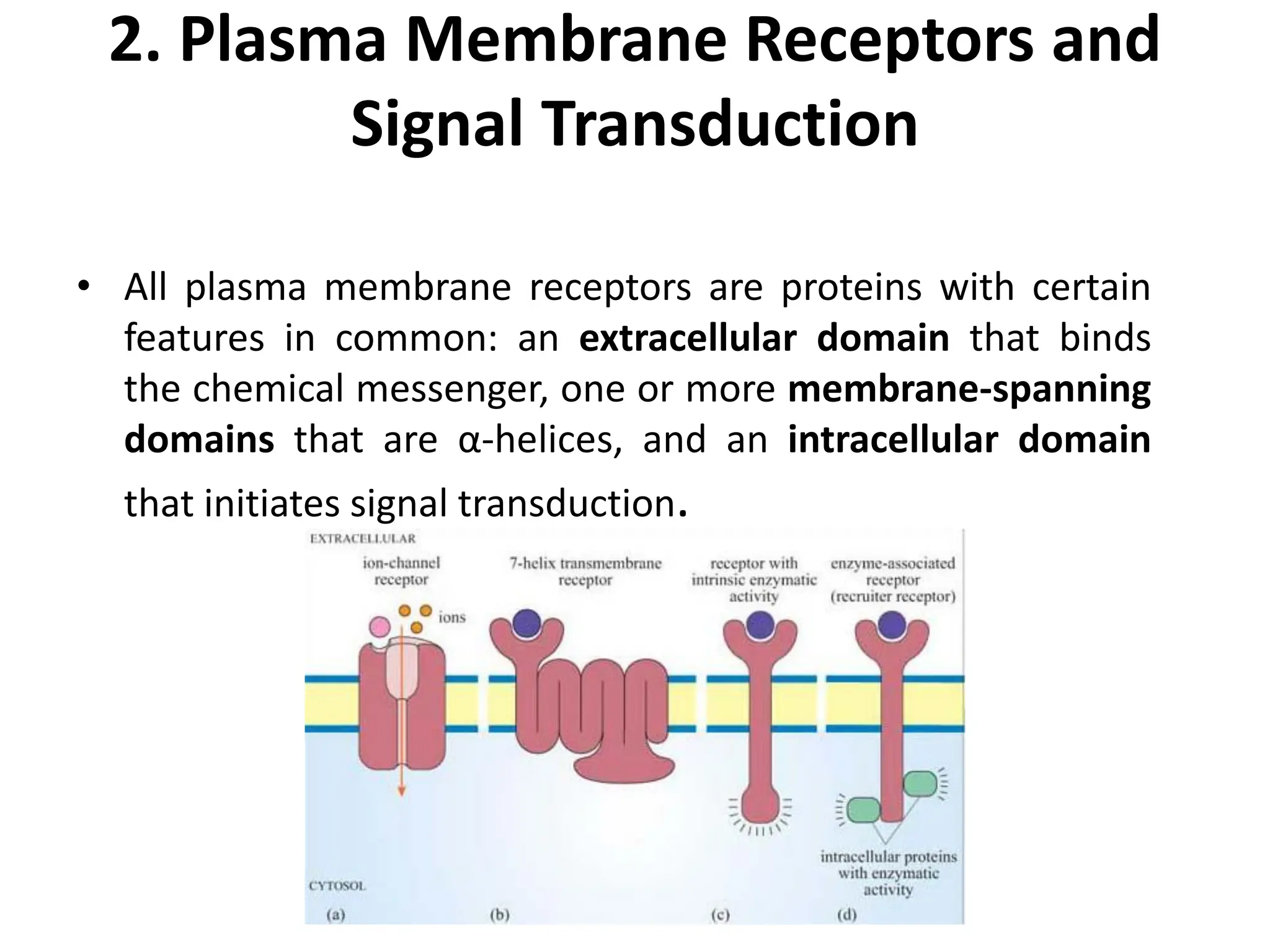
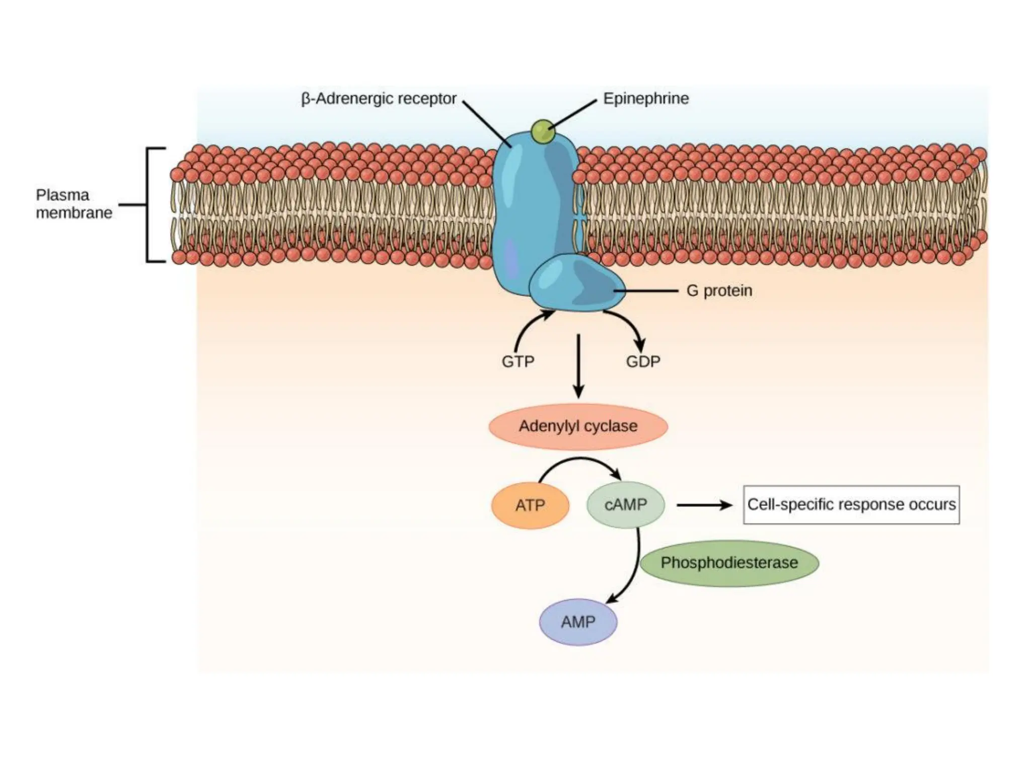
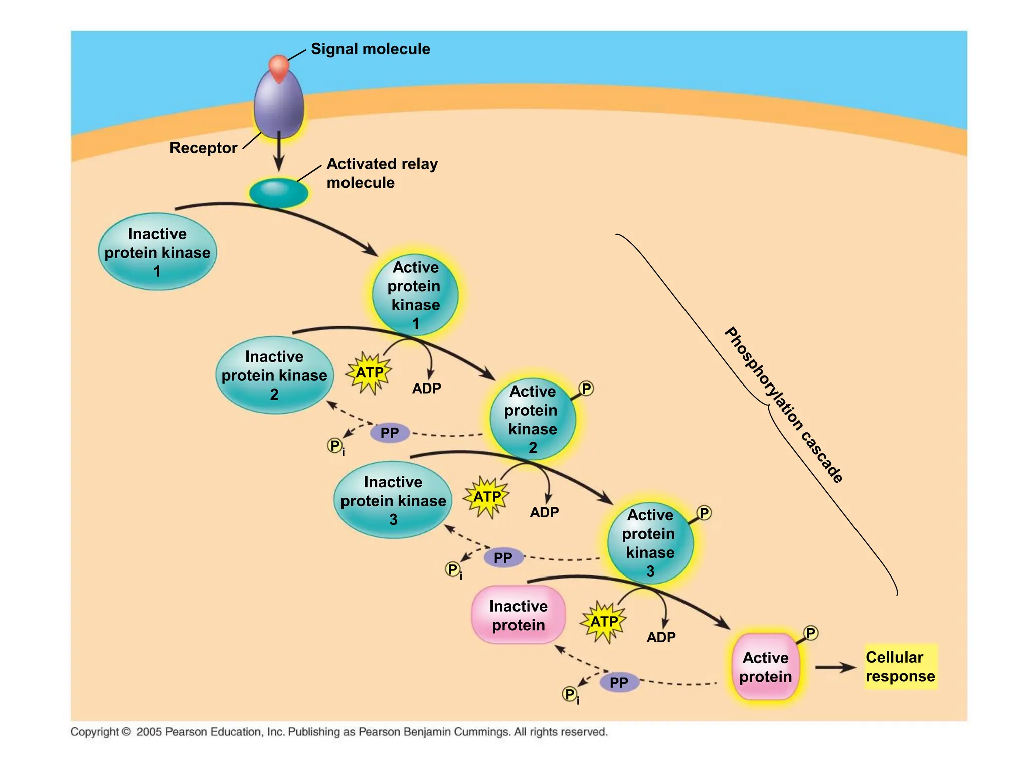
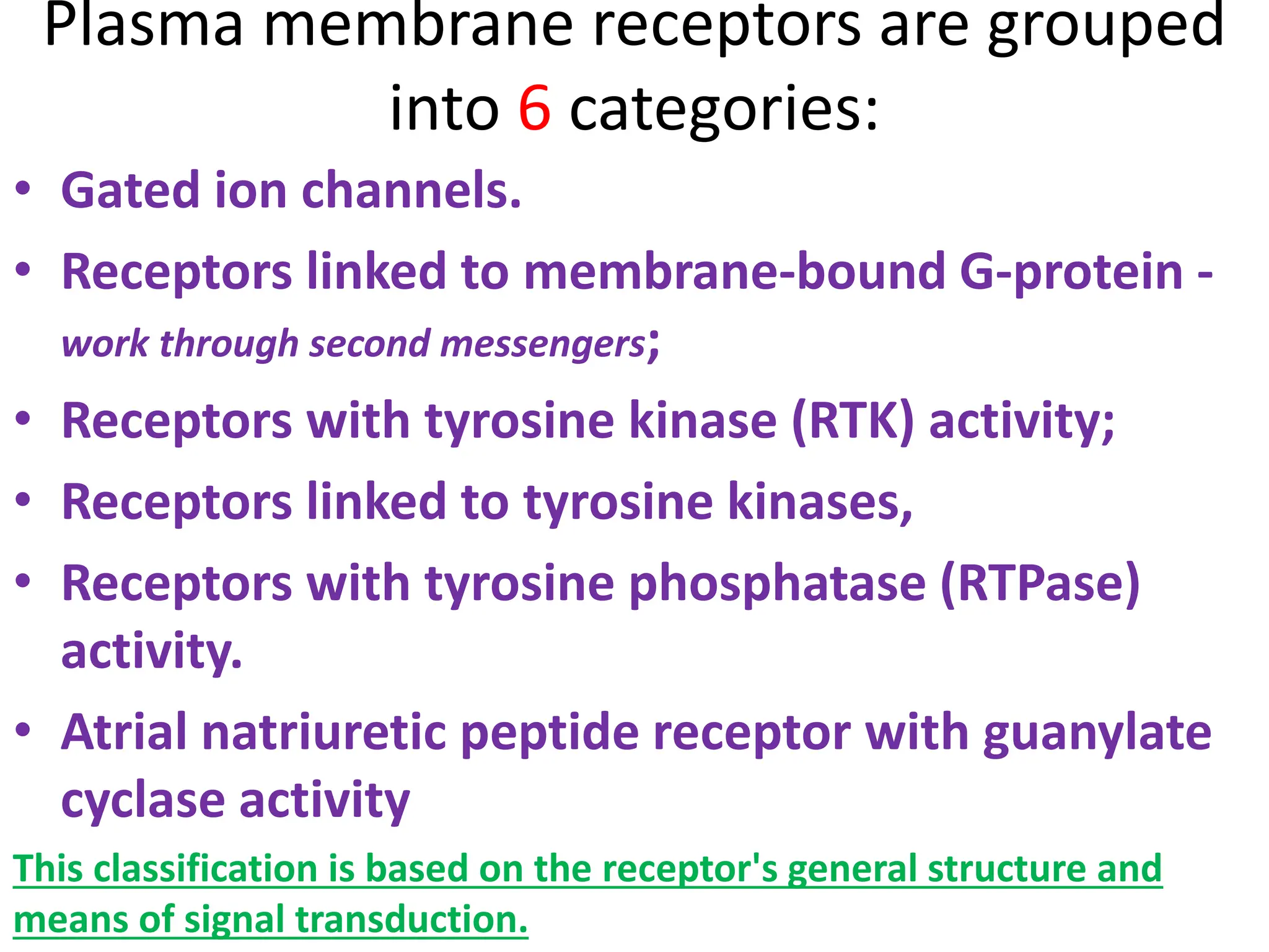
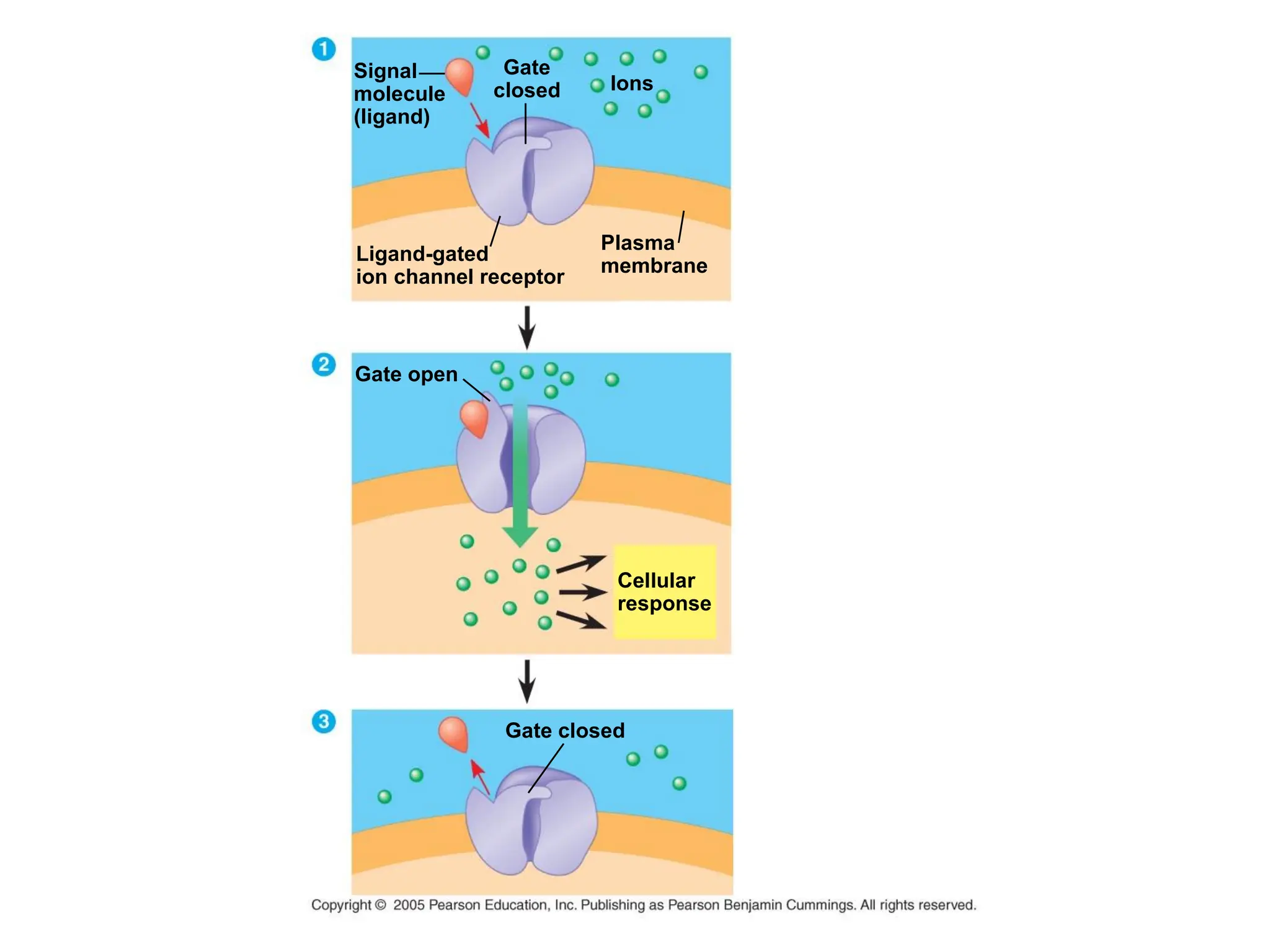
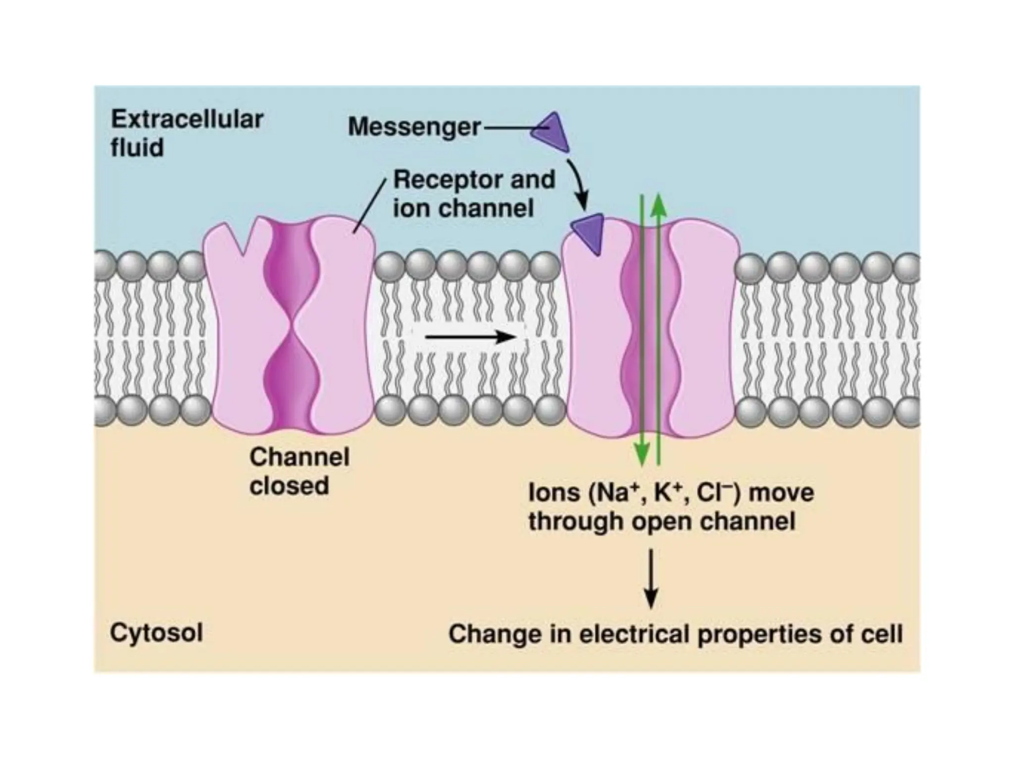
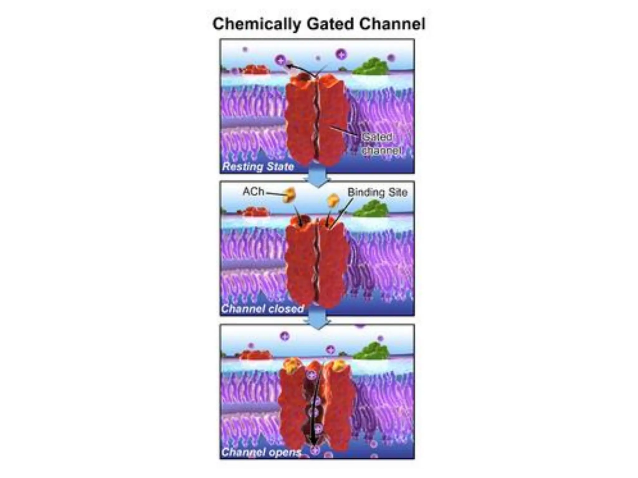
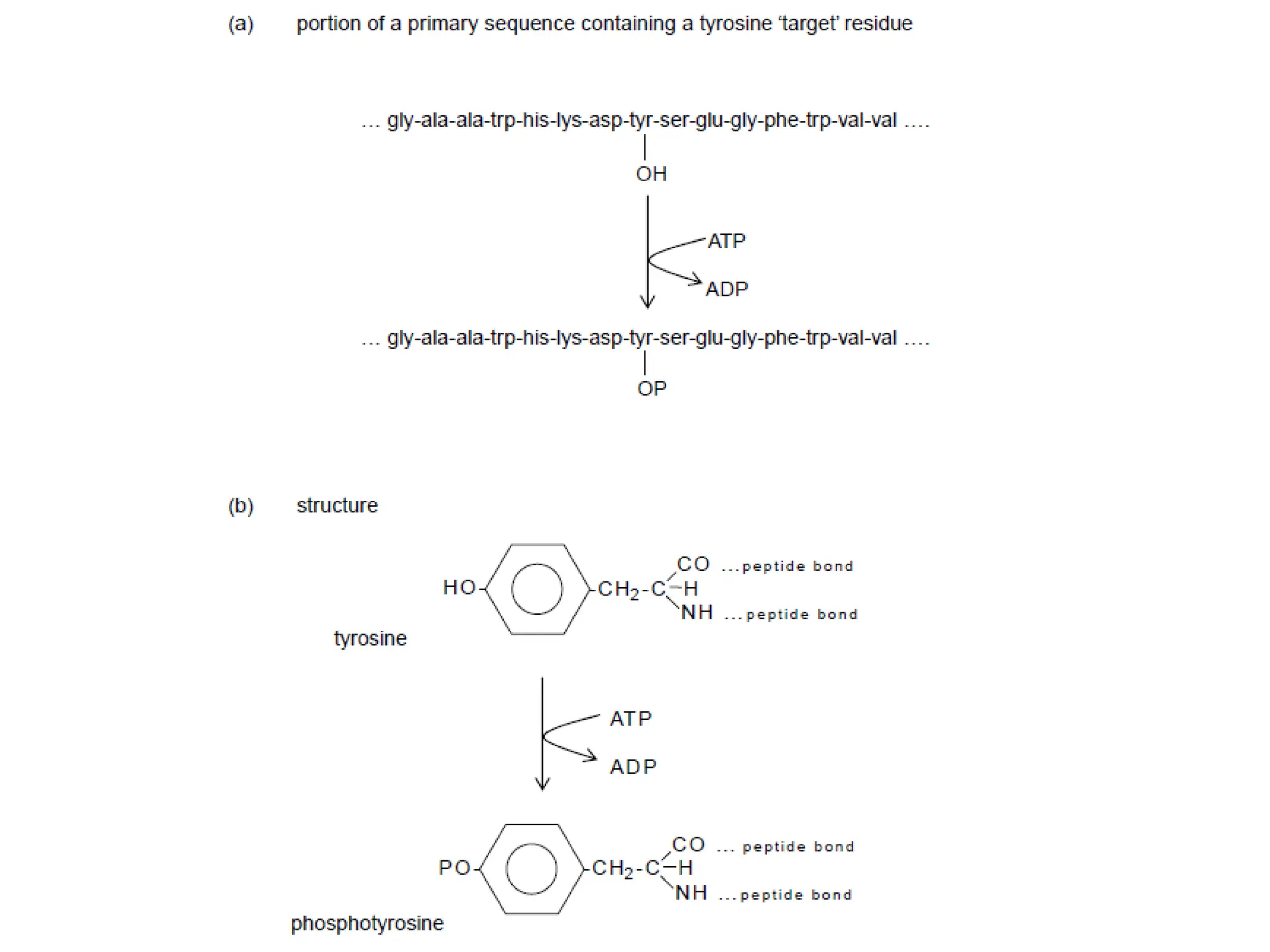
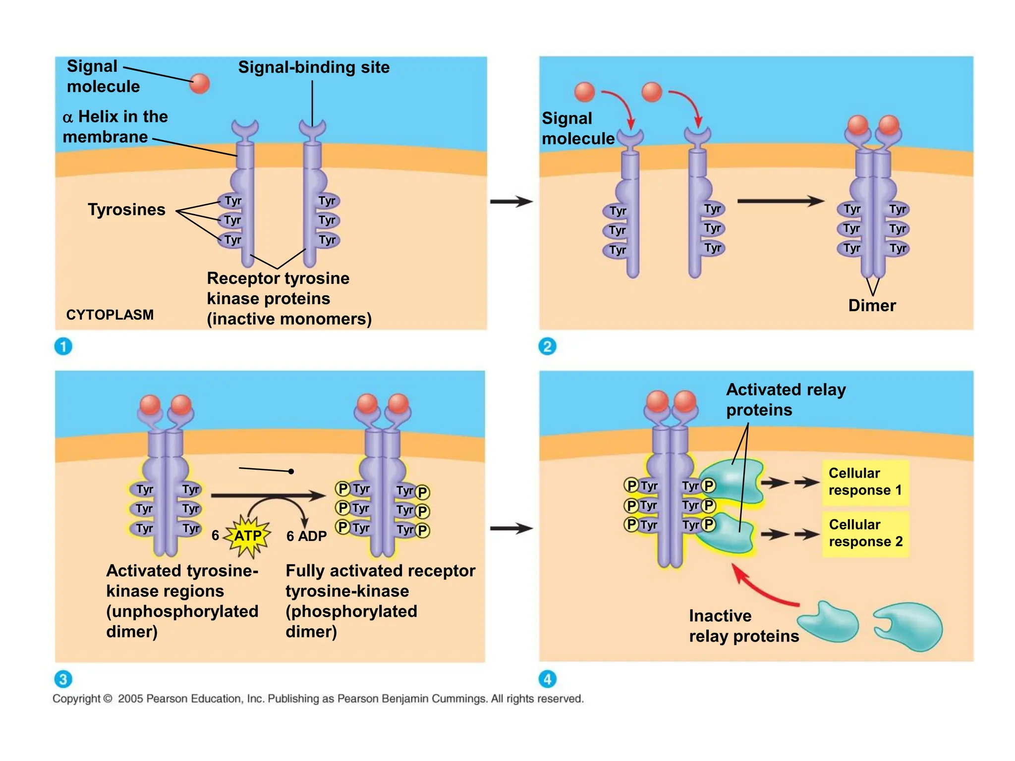
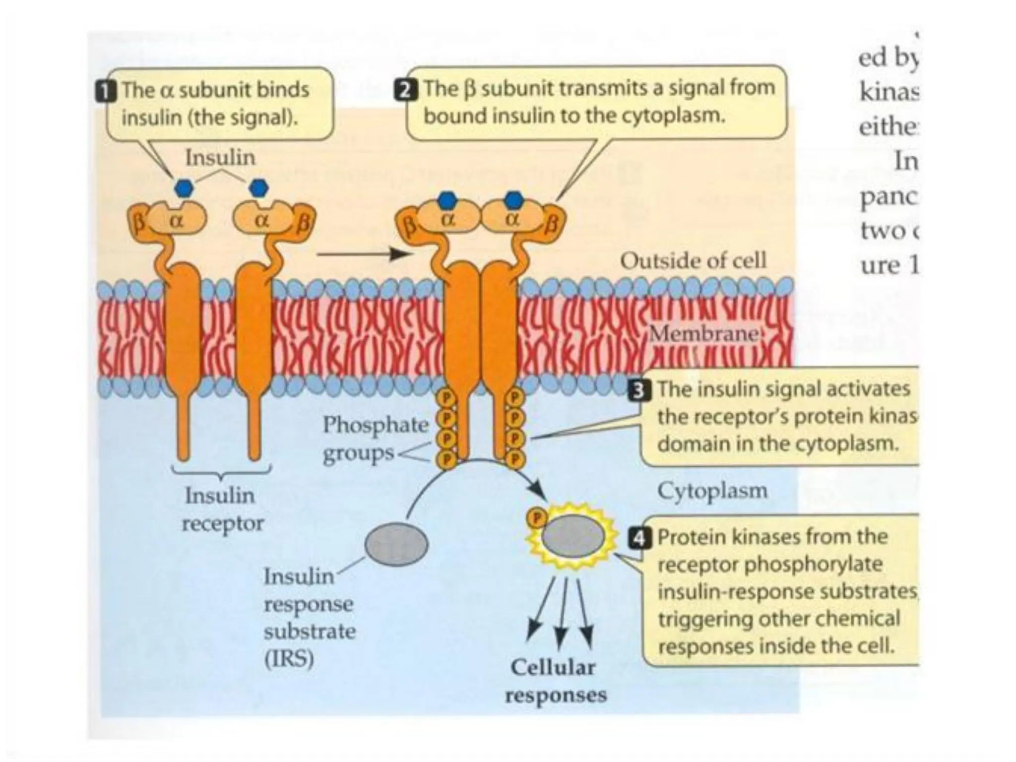
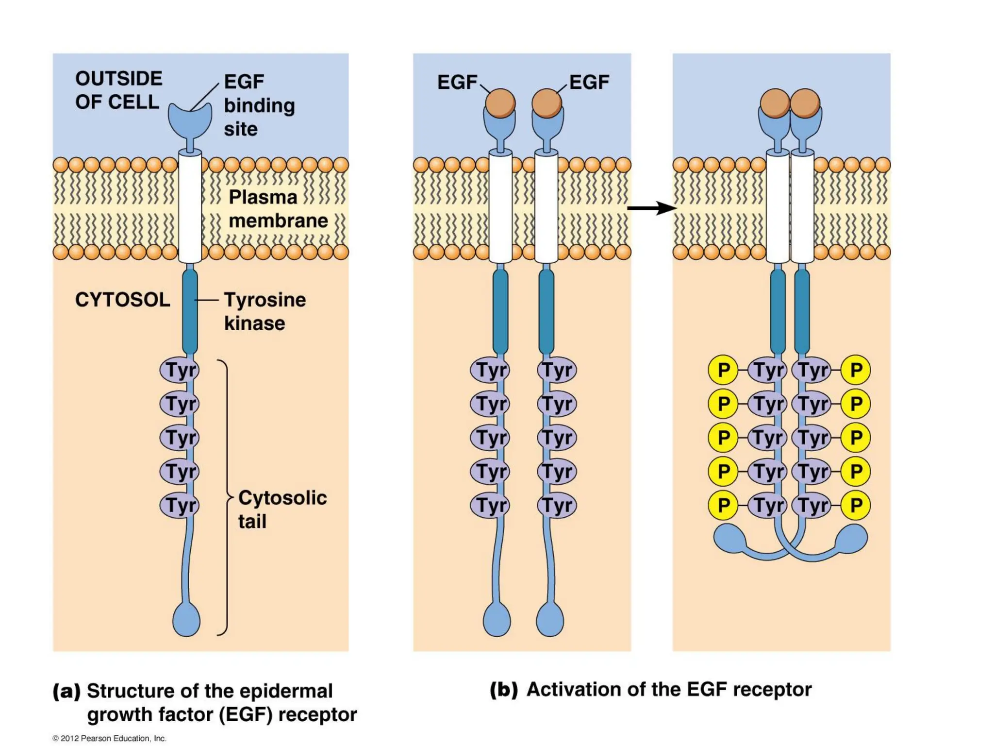
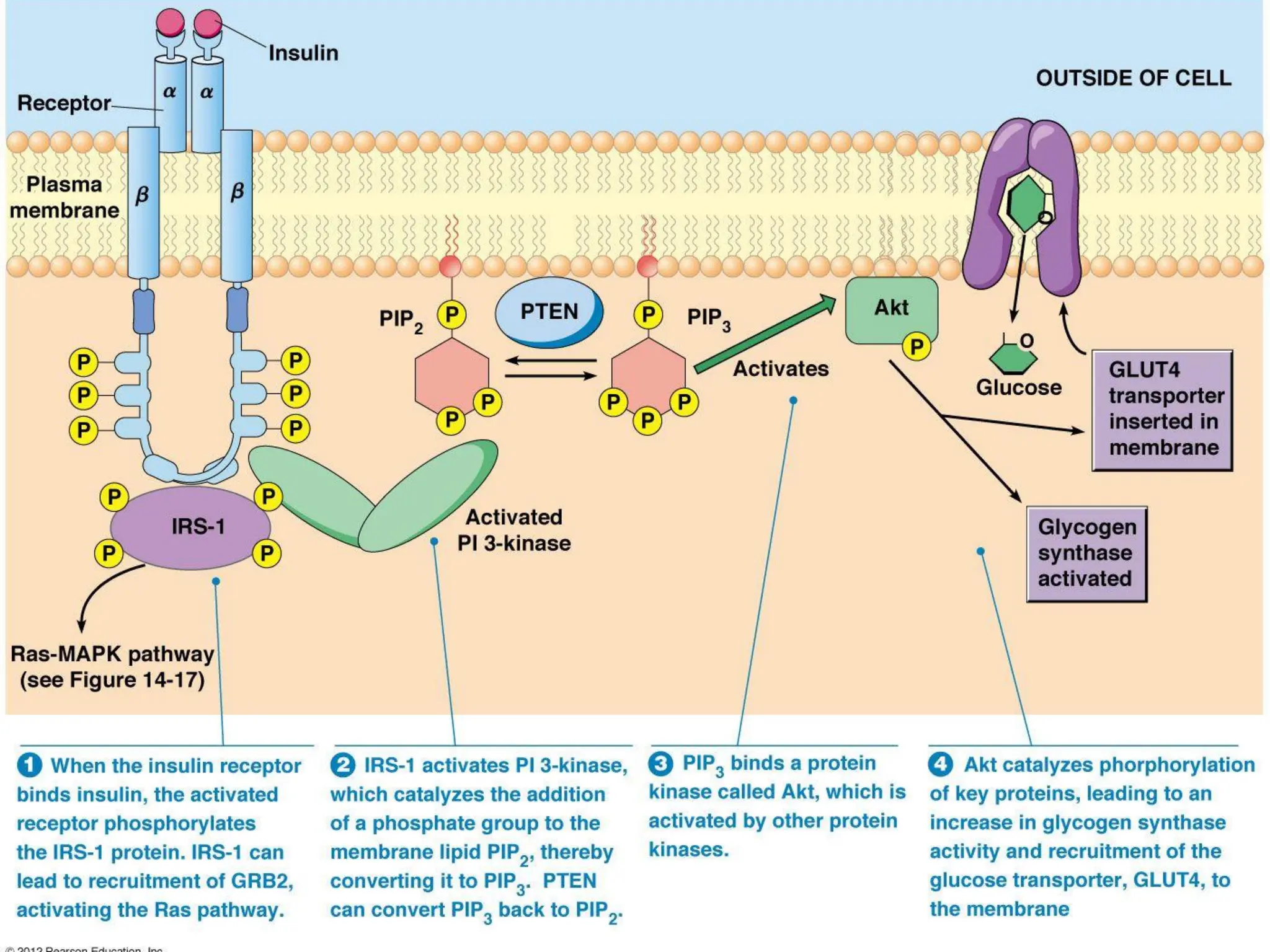
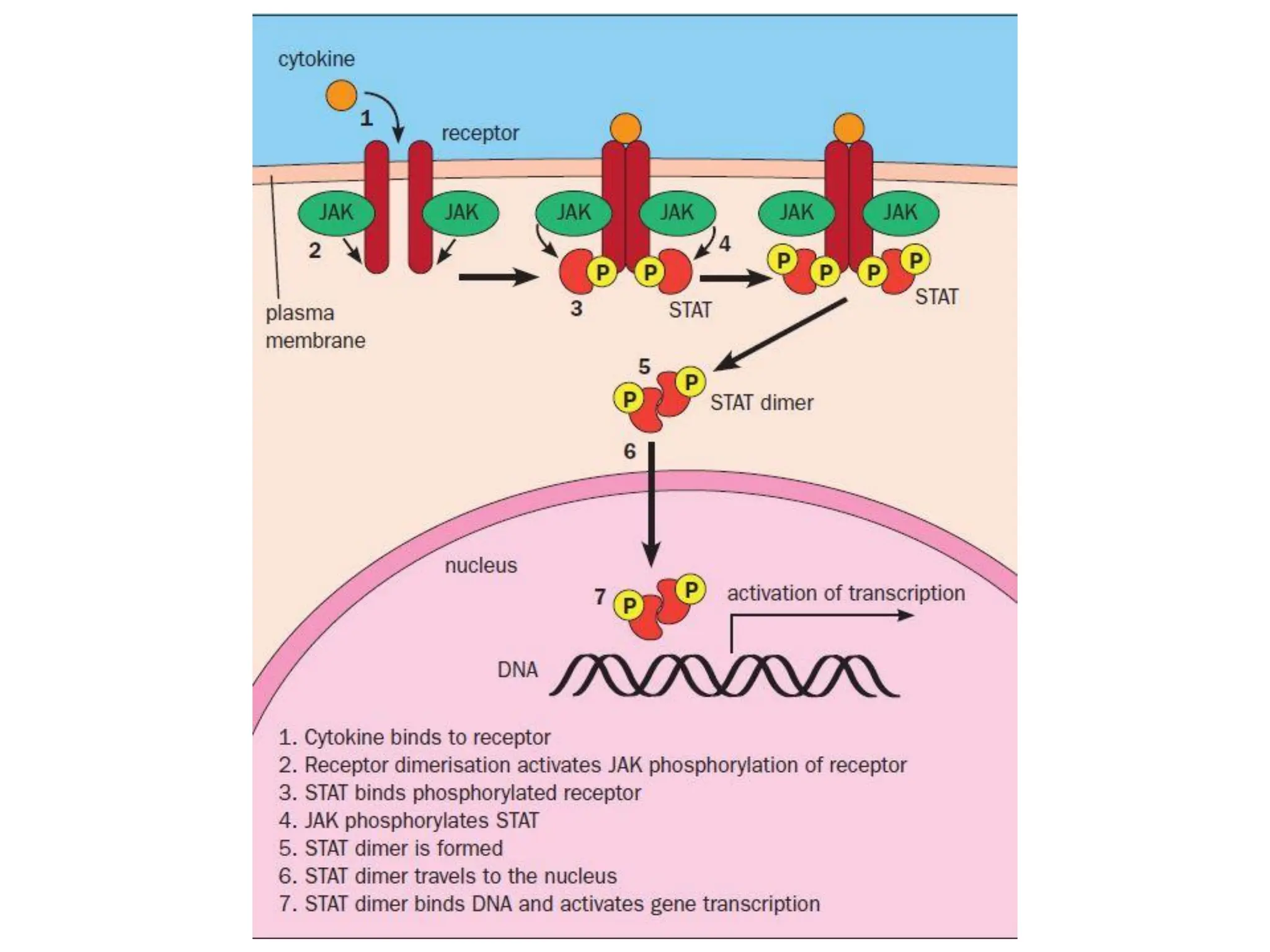
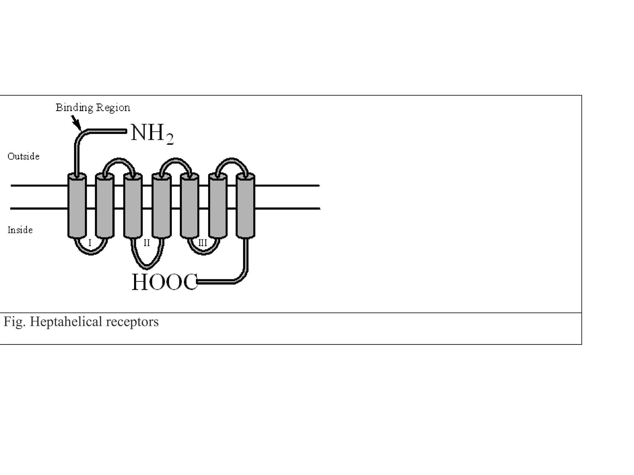
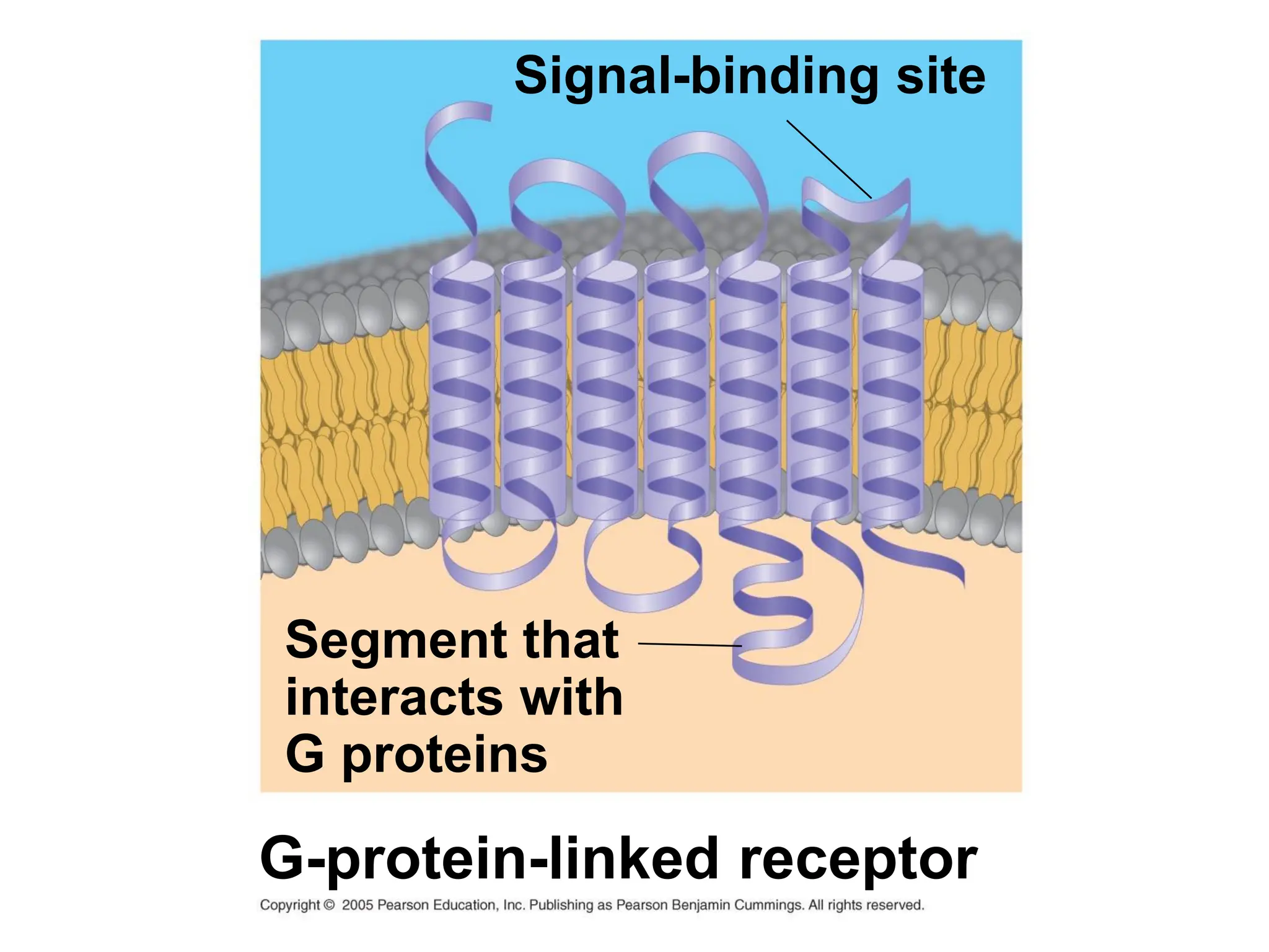
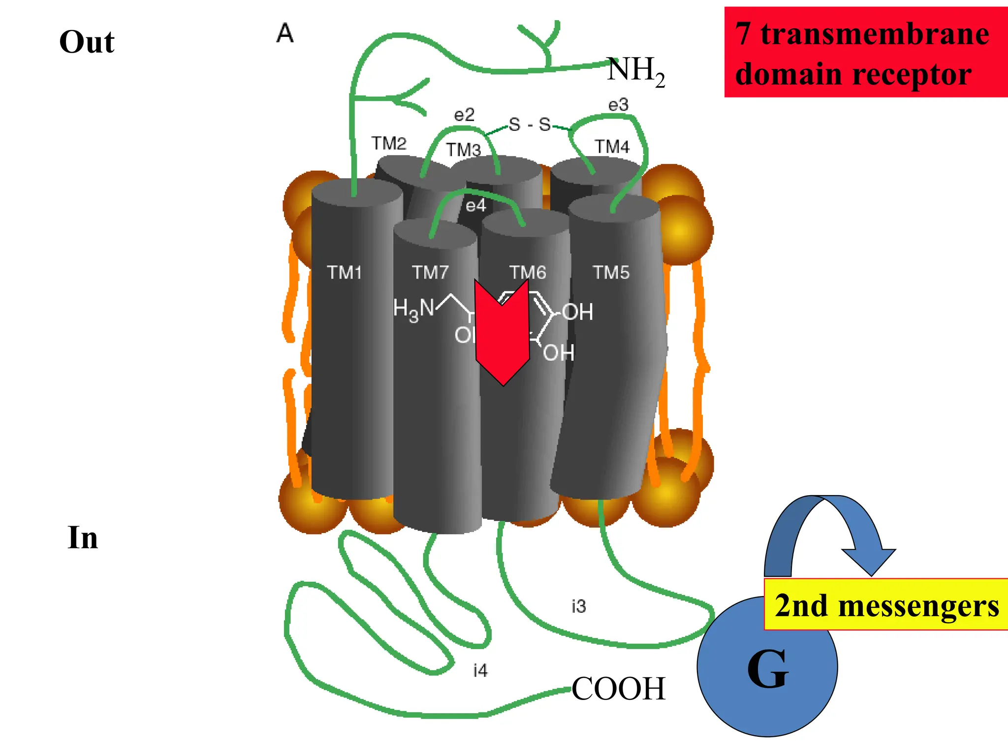
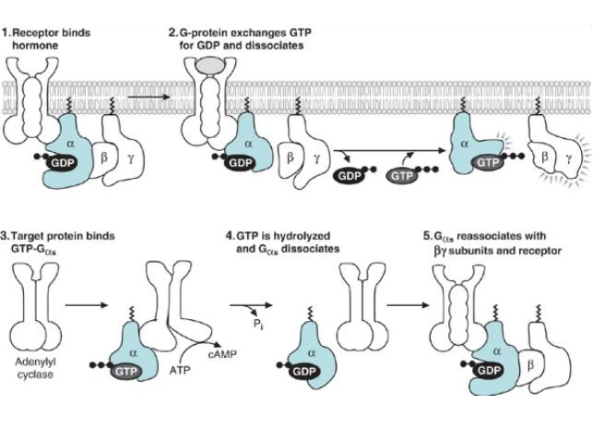
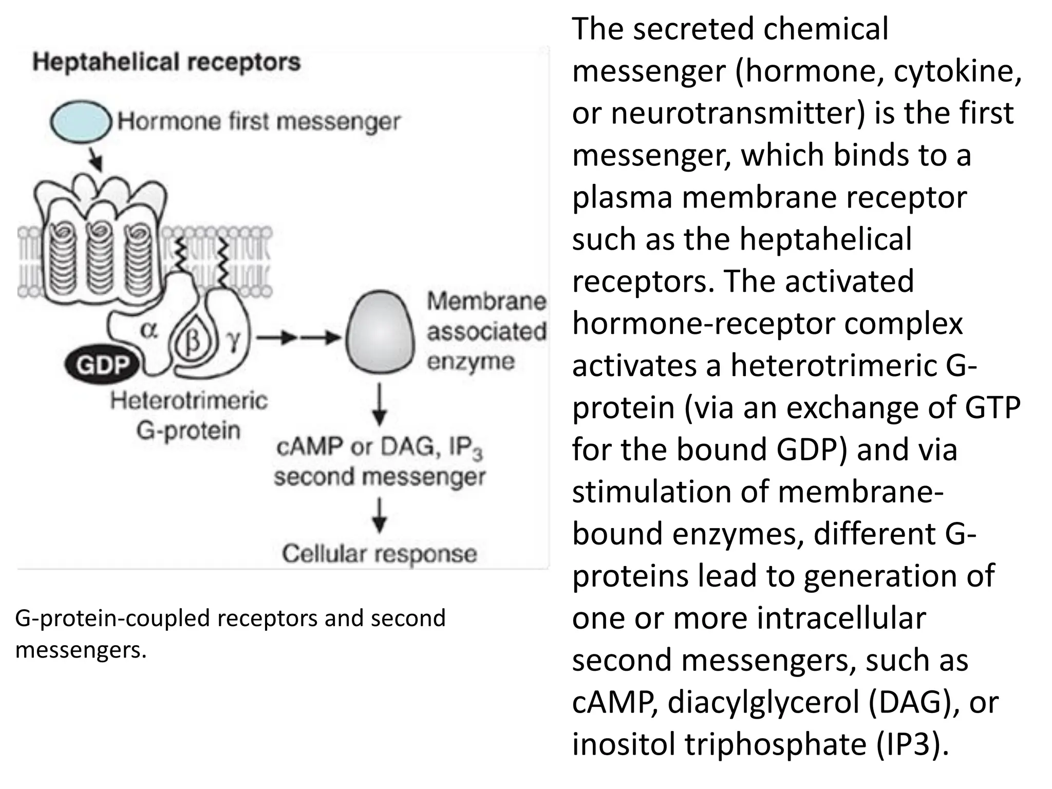
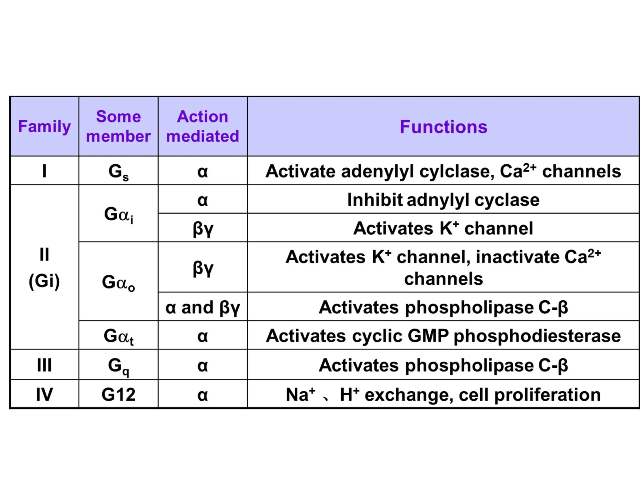
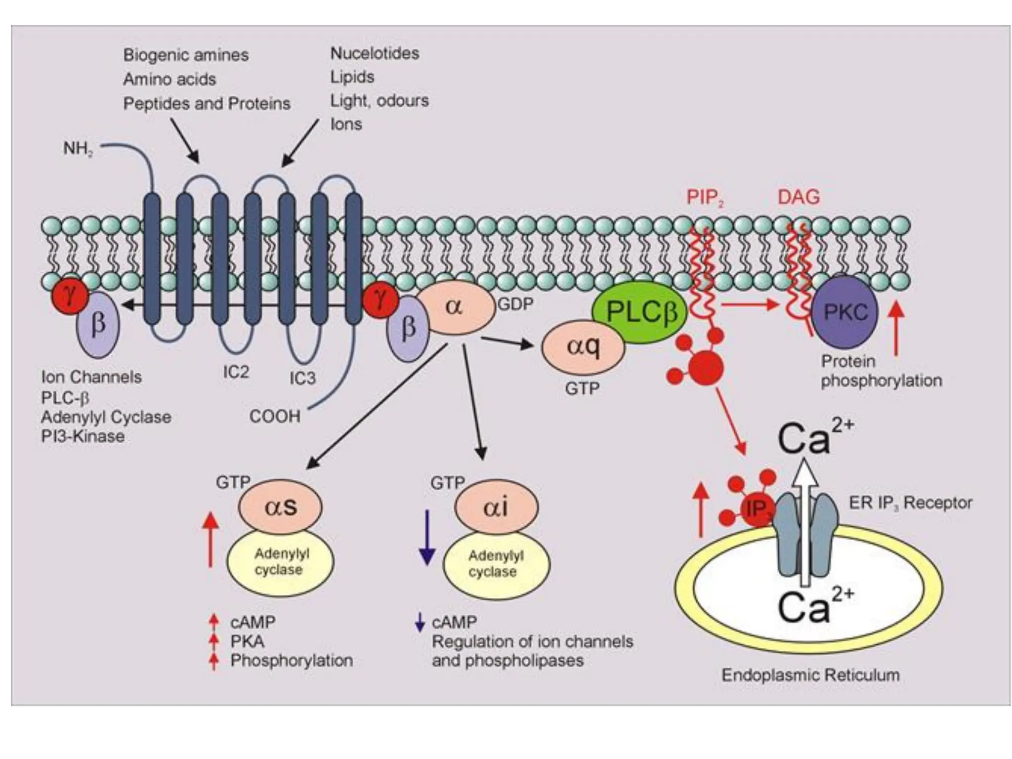
CELL SIGNALING
LESSON 2
NEUROCHEMISTRY
2. Plasma Membrane Receptors and
Signal Transduction
• All plasma membrane receptors are proteins with certain
features in common: an extracellular domain that binds
the chemical messenger, one or more membrane-spanning
domains that are α-helices, and an intracellular domain
that initiates signal transduction.
• As the ligand binds to the extracellular domain of
its receptor, it causes a conformational change
that is communicated to the intracellular domain
through the α-helix of the transmembrane domain.
• The activated intracellular domain initiates a
characteristic signal transduction pathway .
Signal transduction pathways run in one direction.
From a given point in a signal transduction pathway,
events closer to the receptor are termed “upstream”
and events closer to the response are termed
“downstream.”
• In many pathways, the signal is transmitted by a
cascade of protein phosphorylations
• This phosphorylation (kinases) and
dephosphorylation (phosphatases) system acts
as a molecular switch, turning activities on and
off
• Phosphatase - enzymes remove the phosphates
Signal molecule
Activated relay
molecule
Receptor
Inactive
protein kinase
1 Active
protein
kinase
1
Inactive
protein kinase
2 Active
protein
kinase
2
Inactive
protein kinase
3 Active
protein
kinase
3
ADP
Inactive
protein
Active
protein
Cellular
response
ATP
PP
Pi
ADP
ATP
PP
Pi
ADP
ATP
PP
Pi
P
P
P
The pathways of signal transduction
for plasma membrane receptors
The pathways of signal transduction for plasma
membrane receptors have two major types of
effects on the cell:
1. rapid and immediate effects on cellular ion
levels or activation/inhibition of enzymes
and/or
1. slower changes in the rate of gene expression
for a specific set of proteins. Often, a signal
transduction pathway will diverge to produce
both kinds of effects.
Plasma membrane receptors are grouped
into 6 categories:
• Gated ion channels.
• Receptors linked to membrane-bound G-protein -
work through second messengers;
• Receptors with tyrosine kinase (RTK) activity;
• Receptors linked to tyrosine kinases,
• Receptors with tyrosine phosphatase (RTPase)
activity.
• Atrial natriuretic peptide receptor with guanylate
cyclase activity
This classification is based on the receptor's general structure and
means of signal transduction.
1. Ion Channel Receptors or
• Ligand-gated ion channels (LGICs) (ionotropic
receptor) or channel-linked receptor.
• They are are opened or closed in response to
the binding of a chemical messenger (i.e., a
ligand), such as a neurotransmitter.
• Most small-molecule neurotransmitters and
some neuropeptides use ion channel
receptors.
NOTE
The majority of ion channels fall into two broad categories: voltage-gated ion
channels (VGIC) and ligand-gated ion channels (LGIC).
Signal
molecule
(ligand)
Gate
closed Ions
Ligand-gated
ion channel receptor
Plasma
membrane
Gate closed
Gate open
Cellular
response
• These proteins are typically composed of at least
two different domains: a transmembrane domain
which includes the ion pore, and an extracellular
domain which includes the ligand binding
domain.
• LGIC is regulated by a ligand and is usually very selective to
one or more ions like Na+, K+, Ca2+, or Cl-.
• Such receptors located at synapses convert the chemical
signal of presynaptically released neurotransmitter directly
and very quickly into a postsynaptic electrical signal.
• Many LGICs are additionally modulated by allosteric
ligands, by channel blockers, ions, or the membrane
potential.
• Ligand-gated ion channels are likely to be the major site at
which anaesthetic agents and ethanol have their effects,
halthough unequivocal evidence of this is yet to be
established
Voltage-gated ion channels
• Voltage-gated ion channels are a class of
transmembrane ion channels that are activated by
changes in electrical potential difference near the
channel; these types of ion channels are especially
critical in neurons.
• They have a crucial role in excitable neuronal and
muscle tissues, allowing a rapid and co-ordinated
depolarization in response to triggering voltage
change. Found along the axon and at the synapse,
voltage-gated ion channels directionally propagate
electrical signals.
Example: nicotinic acetylcholine receptor
• The prototypic ligand-gated ion channel is the
nicotinic acetylcholine receptor.
• The job of a neurotransmitter is to change the
membrane potential of the postsynaptic cell and
to do it quickly.
• The fastest and most direct mechanism is binding
of the neurotransmitter to a ligand-gated ion
channel in the plasma membrane.
• The nicotinic acetylcholine receptor in the
neuromuscular junction is a classic example.
• This receptor is a channel for the monovalent
cations sodium and potassium, with five
subunits that each contribute to the channel
(Fig.). The channel is closed in the resting
state, opening only when acetylcholine binds.
Opening of the channel causes a rapid influx
of sodium down its electrochemical gradient,
which depolarizes the membrane.
This receptor is a ligand-
gated channel for small
cations (Na+, K+).
Acetylcholine binds with
positive cooperativity to
the two α subunits.
Each of the five
polypeptides traverses
the membrane four
times, and one of the
transmembrane helices
in each subunit
contributes to the “gate”
in the channel.
Structure of the nicotinic acetylcholine receptor in the
neuromuscular junction.
Voltage-gated ion channels
Voltage-gated ion channels are a class of transmembrane ion channels
that are activated by changes in electrical potential difference near the
channel; these types of ion channels are especially critical in neurons.
They generally are composed
of several subunits arranged
in such a way that there is a
central pore through which
ions can travel down their
electrochemical gradients.
The channels tend to be ion-
specific, although similarly
sized and charged ions may
sometimes travel through
them.
• Examples include:
• the sodium and potassium voltage-gated
channels of nerve and muscle.
• the voltage-gated calcium channls that play a
role in neurotransmitter release in presynaptic
nerve endings.
• CLINICAL CORRELATION
• Lambert–Eaton Myasthenic Syndrome
Lambert–Eaton myasthenic syndrome (LEMS) is an autoimmune
disease in which the body raises antibodies against voltage
gated calcium channels (VGCC) located on presynaptic nerve
termini.
Upon depolarization of presynaptic neurons, calcium channels
at presynaptic nerve termini open, permitting the influx of
calcium ions. This increase in calcium ion concentration initiates
events of the synapsin cycle and leads to release of
neurotransmitters into synaptic junctions. When autoantibodies
against VGCC react with neurons at neuromuscular junctions,
calcium ions cannot enter and the amount of acetylcholine
released into synaptic junctions is diminished.
Receptor tyrosine kinases
• Kinases are the class of enzymes which
transfer the terminal phosphate group from
ATP to a substrate.
• In particular, tyrosine kinases transfer the
phosphate to a tyrosine residue.
Signal
molecule
a Helix in the
membrane
Signal-binding site
Tyr
Tyr
Tyr Tyr
Tyr
Tyr
Tyrosines
Receptor tyrosine
kinase proteins
(inactive monomers)
CYTOPLASM
Tyr
Tyr
Tyr Tyr
Tyr
Tyr Tyr
Tyr
Tyr Tyr
Tyr
Tyr
Tyr
Tyr
Tyr Tyr
Tyr
Tyr
Activated tyrosine-
kinase regions
(unphosphorylated
dimer)
Signal
molecule
Dimer
Fully activated receptor
tyrosine-kinase
(phosphorylated
dimer)
Tyr
Tyr
Tyr Tyr
Tyr
Tyr
P
P
P
P
P
P
ATP 6 ADP
Tyr
Tyr
Tyr Tyr
Tyr
Tyr
P
P
P
P
P
P
Inactive
relay proteins
Cellular
response 2
Cellular
response 1
Activated relay
proteins
6
• Signal transduction may involve two types of
tyrosine kinase:
1. receptors with inherent tyrosine kinase activity
(RTKs), for example insulin receptor; Receptors with
tyrosine kinase (RTK) activity
2. receptors, which recruit cytoplasmic tyrosine
kinases, for example growth hormone, leptin
operating through Janus kinase (JAK). Receptors
linked to tyrosine kinases,
Receptor tyrosine phosphatase
• CD45
1. It is a large(180-220 kDa)cell surface
molecule expressed by all leukocytes,
including T cell.
2. Its cytoplasmic domain has tyrosine
phosphatase activity.
G-Protein-Coupled Receptors
The G-protein-coupled receptors (also known as
heptahelical receptors) contain seven-membrane
spanning domains which are a-helices .
Although there are hundreds of hormones and
neurotransmitters that work through heptahelical
receptors, the extracellular binding domain of each
receptor is specific for just one polypeptide
hormone, catecholamine, or neurotransmitter (or its
close structural analogue).
Fig. Heptahelical receptors
Segment that
interacts with
G proteins
Signal-binding site
G-protein-linked receptor
Out
In
G
7 transmembrane
domain receptor
2nd messengers
NH2
COOH
• Heptahelical receptors initiate signal
transduction through heterotrimeric G-
proteins (proteins which are activated upon
binding GTP) composed of α-, β-, and γ-
subunits.
• However, different types of heptahelical
receptors bind different G-proteins, and
different G-proteins exert different effects on
their target proteins
Heterotrimeric G-proteins
• The function of heterotrimeric G-proteins is illustrated in Fig.
using a hormone that activates adenylyl cyclase (e.g. glucagon or
epinephrine).
• While the α-subunit contains bound GDP, it remains associated
with the β- and γ-subunits, either free in the membrane or
bound to an unoccupied receptor (see Fig, part 1).
• When the hormone binds, it causes a conformational change in
the receptor that activates GDP dissociation and GTP binding.
The exchange of GTP for bound GDP causes dissociation of the α
-subunit from the receptor and from the βγ -subunits (see Fig. ,
part 2).
• The GTP- α -subunit binds its target enzyme in the membrane,
thereby changing its activity. In this example, the α -subunit
binds and activates adenylyl cyclase, thereby increasing synthesis
of cAMP (see Fig. , part 3).
• With time, the Gα-subunit inactivates itself by
hydrolyzing its own bound GTP to GDP and Pi.
This action is unrelated to the number of cAMP
molecules formed. When this occurs, the GDP-
α -subunit dissociates from its target protein,
adenylyl cyclase (see Fig. part 4). It reforms the
trimeric G-protein complex, which may return
to bind the empty hormone receptor.
The secreted chemical
messenger (hormone, cytokine,
or neurotransmitter) is the first
messenger, which binds to a
plasma membrane receptor
such as the heptahelical
receptors. The activated
hormone-receptor complex
activates a heterotrimeric G-
protein (via an exchange of GTP
for the bound GDP) and via
stimulation of membrane-
bound enzymes, different G-
proteins lead to generation of
one or more intracellular
second messengers, such as
cAMP, diacylglycerol (DAG), or
inositol triphosphate (IP3).
G-protein-coupled receptors and second
messengers.
• There are a large number of different
heterotrimeric G-protein complexes, which are
generally categorized according to the activity of
the α-subunit. The 20 or more different isoforms of
Gα fall into four broad categories: GαS, G αi, G αq/11,
G α12/13. GαS refers to α -subunits which, like the
one in Fig., stimulate adenylyl cyclase (hence the
“s”). Gα subunits that inhibit adenylyl cyclase are
called G αi. or Gi protein -G α i/0. The βγ-subunits
likewise exist as different isoforms, which also
transmit messages. Gaqs subunits activate
phospholipase Cβ, which generates second
messengers based on phosphatidylinositol.
Cell signaling part II-1.pdfdthtrsysrysru

Cell signaling part II-1.pdfdthtrsysrysru

  • 1.
  • 2.
    2. Plasma MembraneReceptors and Signal Transduction • All plasma membrane receptors are proteins with certain features in common: an extracellular domain that binds the chemical messenger, one or more membrane-spanning domains that are α-helices, and an intracellular domain that initiates signal transduction.
  • 3.
    • As theligand binds to the extracellular domain of its receptor, it causes a conformational change that is communicated to the intracellular domain through the α-helix of the transmembrane domain. • The activated intracellular domain initiates a characteristic signal transduction pathway . Signal transduction pathways run in one direction. From a given point in a signal transduction pathway, events closer to the receptor are termed “upstream” and events closer to the response are termed “downstream.”
  • 5.
    • In manypathways, the signal is transmitted by a cascade of protein phosphorylations • This phosphorylation (kinases) and dephosphorylation (phosphatases) system acts as a molecular switch, turning activities on and off • Phosphatase - enzymes remove the phosphates
  • 6.
    Signal molecule Activated relay molecule Receptor Inactive proteinkinase 1 Active protein kinase 1 Inactive protein kinase 2 Active protein kinase 2 Inactive protein kinase 3 Active protein kinase 3 ADP Inactive protein Active protein Cellular response ATP PP Pi ADP ATP PP Pi ADP ATP PP Pi P P P
  • 7.
    The pathways ofsignal transduction for plasma membrane receptors The pathways of signal transduction for plasma membrane receptors have two major types of effects on the cell: 1. rapid and immediate effects on cellular ion levels or activation/inhibition of enzymes and/or 1. slower changes in the rate of gene expression for a specific set of proteins. Often, a signal transduction pathway will diverge to produce both kinds of effects.
  • 8.
    Plasma membrane receptorsare grouped into 6 categories: • Gated ion channels. • Receptors linked to membrane-bound G-protein - work through second messengers; • Receptors with tyrosine kinase (RTK) activity; • Receptors linked to tyrosine kinases, • Receptors with tyrosine phosphatase (RTPase) activity. • Atrial natriuretic peptide receptor with guanylate cyclase activity This classification is based on the receptor's general structure and means of signal transduction.
  • 9.
    1. Ion ChannelReceptors or • Ligand-gated ion channels (LGICs) (ionotropic receptor) or channel-linked receptor. • They are are opened or closed in response to the binding of a chemical messenger (i.e., a ligand), such as a neurotransmitter. • Most small-molecule neurotransmitters and some neuropeptides use ion channel receptors. NOTE The majority of ion channels fall into two broad categories: voltage-gated ion channels (VGIC) and ligand-gated ion channels (LGIC).
  • 10.
    Signal molecule (ligand) Gate closed Ions Ligand-gated ion channelreceptor Plasma membrane Gate closed Gate open Cellular response
  • 11.
    • These proteinsare typically composed of at least two different domains: a transmembrane domain which includes the ion pore, and an extracellular domain which includes the ligand binding domain.
  • 12.
    • LGIC isregulated by a ligand and is usually very selective to one or more ions like Na+, K+, Ca2+, or Cl-. • Such receptors located at synapses convert the chemical signal of presynaptically released neurotransmitter directly and very quickly into a postsynaptic electrical signal. • Many LGICs are additionally modulated by allosteric ligands, by channel blockers, ions, or the membrane potential. • Ligand-gated ion channels are likely to be the major site at which anaesthetic agents and ethanol have their effects, halthough unequivocal evidence of this is yet to be established
  • 14.
    Voltage-gated ion channels •Voltage-gated ion channels are a class of transmembrane ion channels that are activated by changes in electrical potential difference near the channel; these types of ion channels are especially critical in neurons. • They have a crucial role in excitable neuronal and muscle tissues, allowing a rapid and co-ordinated depolarization in response to triggering voltage change. Found along the axon and at the synapse, voltage-gated ion channels directionally propagate electrical signals.
  • 16.
    Example: nicotinic acetylcholinereceptor • The prototypic ligand-gated ion channel is the nicotinic acetylcholine receptor. • The job of a neurotransmitter is to change the membrane potential of the postsynaptic cell and to do it quickly. • The fastest and most direct mechanism is binding of the neurotransmitter to a ligand-gated ion channel in the plasma membrane. • The nicotinic acetylcholine receptor in the neuromuscular junction is a classic example.
  • 18.
    • This receptoris a channel for the monovalent cations sodium and potassium, with five subunits that each contribute to the channel (Fig.). The channel is closed in the resting state, opening only when acetylcholine binds. Opening of the channel causes a rapid influx of sodium down its electrochemical gradient, which depolarizes the membrane.
  • 19.
    This receptor isa ligand- gated channel for small cations (Na+, K+). Acetylcholine binds with positive cooperativity to the two α subunits. Each of the five polypeptides traverses the membrane four times, and one of the transmembrane helices in each subunit contributes to the “gate” in the channel. Structure of the nicotinic acetylcholine receptor in the neuromuscular junction.
  • 22.
    Voltage-gated ion channels Voltage-gatedion channels are a class of transmembrane ion channels that are activated by changes in electrical potential difference near the channel; these types of ion channels are especially critical in neurons. They generally are composed of several subunits arranged in such a way that there is a central pore through which ions can travel down their electrochemical gradients. The channels tend to be ion- specific, although similarly sized and charged ions may sometimes travel through them.
  • 23.
    • Examples include: •the sodium and potassium voltage-gated channels of nerve and muscle. • the voltage-gated calcium channls that play a role in neurotransmitter release in presynaptic nerve endings.
  • 25.
    • CLINICAL CORRELATION •Lambert–Eaton Myasthenic Syndrome Lambert–Eaton myasthenic syndrome (LEMS) is an autoimmune disease in which the body raises antibodies against voltage gated calcium channels (VGCC) located on presynaptic nerve termini. Upon depolarization of presynaptic neurons, calcium channels at presynaptic nerve termini open, permitting the influx of calcium ions. This increase in calcium ion concentration initiates events of the synapsin cycle and leads to release of neurotransmitters into synaptic junctions. When autoantibodies against VGCC react with neurons at neuromuscular junctions, calcium ions cannot enter and the amount of acetylcholine released into synaptic junctions is diminished.
  • 26.
    Receptor tyrosine kinases •Kinases are the class of enzymes which transfer the terminal phosphate group from ATP to a substrate. • In particular, tyrosine kinases transfer the phosphate to a tyrosine residue.
  • 28.
    Signal molecule a Helix inthe membrane Signal-binding site Tyr Tyr Tyr Tyr Tyr Tyr Tyrosines Receptor tyrosine kinase proteins (inactive monomers) CYTOPLASM Tyr Tyr Tyr Tyr Tyr Tyr Tyr Tyr Tyr Tyr Tyr Tyr Tyr Tyr Tyr Tyr Tyr Tyr Activated tyrosine- kinase regions (unphosphorylated dimer) Signal molecule Dimer Fully activated receptor tyrosine-kinase (phosphorylated dimer) Tyr Tyr Tyr Tyr Tyr Tyr P P P P P P ATP 6 ADP Tyr Tyr Tyr Tyr Tyr Tyr P P P P P P Inactive relay proteins Cellular response 2 Cellular response 1 Activated relay proteins 6
  • 29.
    • Signal transductionmay involve two types of tyrosine kinase: 1. receptors with inherent tyrosine kinase activity (RTKs), for example insulin receptor; Receptors with tyrosine kinase (RTK) activity 2. receptors, which recruit cytoplasmic tyrosine kinases, for example growth hormone, leptin operating through Janus kinase (JAK). Receptors linked to tyrosine kinases,
  • 34.
    Receptor tyrosine phosphatase •CD45 1. It is a large(180-220 kDa)cell surface molecule expressed by all leukocytes, including T cell. 2. Its cytoplasmic domain has tyrosine phosphatase activity.
  • 35.
    G-Protein-Coupled Receptors The G-protein-coupledreceptors (also known as heptahelical receptors) contain seven-membrane spanning domains which are a-helices . Although there are hundreds of hormones and neurotransmitters that work through heptahelical receptors, the extracellular binding domain of each receptor is specific for just one polypeptide hormone, catecholamine, or neurotransmitter (or its close structural analogue).
  • 36.
  • 37.
    Segment that interacts with Gproteins Signal-binding site G-protein-linked receptor
  • 38.
  • 39.
    • Heptahelical receptorsinitiate signal transduction through heterotrimeric G- proteins (proteins which are activated upon binding GTP) composed of α-, β-, and γ- subunits. • However, different types of heptahelical receptors bind different G-proteins, and different G-proteins exert different effects on their target proteins
  • 40.
    Heterotrimeric G-proteins • Thefunction of heterotrimeric G-proteins is illustrated in Fig. using a hormone that activates adenylyl cyclase (e.g. glucagon or epinephrine). • While the α-subunit contains bound GDP, it remains associated with the β- and γ-subunits, either free in the membrane or bound to an unoccupied receptor (see Fig, part 1). • When the hormone binds, it causes a conformational change in the receptor that activates GDP dissociation and GTP binding. The exchange of GTP for bound GDP causes dissociation of the α -subunit from the receptor and from the βγ -subunits (see Fig. , part 2). • The GTP- α -subunit binds its target enzyme in the membrane, thereby changing its activity. In this example, the α -subunit binds and activates adenylyl cyclase, thereby increasing synthesis of cAMP (see Fig. , part 3).
  • 42.
    • With time,the Gα-subunit inactivates itself by hydrolyzing its own bound GTP to GDP and Pi. This action is unrelated to the number of cAMP molecules formed. When this occurs, the GDP- α -subunit dissociates from its target protein, adenylyl cyclase (see Fig. part 4). It reforms the trimeric G-protein complex, which may return to bind the empty hormone receptor.
  • 43.
    The secreted chemical messenger(hormone, cytokine, or neurotransmitter) is the first messenger, which binds to a plasma membrane receptor such as the heptahelical receptors. The activated hormone-receptor complex activates a heterotrimeric G- protein (via an exchange of GTP for the bound GDP) and via stimulation of membrane- bound enzymes, different G- proteins lead to generation of one or more intracellular second messengers, such as cAMP, diacylglycerol (DAG), or inositol triphosphate (IP3). G-protein-coupled receptors and second messengers.
  • 44.
    • There area large number of different heterotrimeric G-protein complexes, which are generally categorized according to the activity of the α-subunit. The 20 or more different isoforms of Gα fall into four broad categories: GαS, G αi, G αq/11, G α12/13. GαS refers to α -subunits which, like the one in Fig., stimulate adenylyl cyclase (hence the “s”). Gα subunits that inhibit adenylyl cyclase are called G αi. or Gi protein -G α i/0. The βγ-subunits likewise exist as different isoforms, which also transmit messages. Gaqs subunits activate phospholipase Cβ, which generates second messengers based on phosphatidylinositol.